brain activity during traditional textbook and audiovisual
TRANSCRIPT

Brain and Behavior. 2019;00:e01427. | 1 of 13https://doi.org/10.1002/brb3.1427
wileyonlinelibrary.com/journal/brb3
Received:15May2019 | Revised:9September2019 | Accepted:10September2019DOI: 10.1002/brb3.1427
O R I G I N A L R E S E A R C H
Brain activity during traditional textbook and audiovisual‐3D learning
Jesus Pujol1,2 | Laura Blanco‐Hinojo1,2 | Gerard Martínez‐Vilavella1 | Lucila Canu‐Martín1 | Anna Pujol1 | Víctor Pérez‐Sola2,3 | Joan Deus1,4
ThisisanopenaccessarticleunderthetermsoftheCreativeCommonsAttributionLicense,whichpermitsuse,distributionandreproductioninanymedium,provided the original work is properly cited.©2019TheAuthors. Brain and BehaviorpublishedbyWileyPeriodicals,Inc.
1MRIResearchUnit,DepartmentofRadiology,HospitaldelMar,Barcelona,Spain2CentroInvestigaciónBiomédicaenReddeSaludMental,CIBERSAMG21,Barcelona,Spain3Institute of Neuropsychiatry and Addictions,HospitaldelMar,IMIM,Barcelona,Spain4Department of Psychobiology andMethodologyinHealthSciences,AutonomousUniversityofBarcelona,Barcelona,Spain
CorrespondenceJesusPujol,MRIDepartment,HospitaldelMar,PasseigMarítim25‐29,Barcelona08003,Spain.Email:[email protected]
Funding informationInstitutodeSaludCarlosIII,Grant/AwardNumber:C1589923;3DTechOmegaZeta,Barcelona
AbstractIntroduction: Audiovisualeducationaltoolshaveincreasinglybeenusedduringthepastyearstocomplementandcompetewithtraditional textbooks.However, littleis known as to how the brain processes didactic information presented in different formats. We directly assessed brain activity during learning using both traditional textbookandaudiovisual‐3Dmaterial.Methods: Ahomogeneoussampleof30youngadultswithactivestudyhabitswasassessed.EducationalmaterialonthesubjectofCardiologywasadaptedtobepre‐sented during the acquisition of functional MRI.Results: When testedafter imageacquisition,participantsobtainedsimilarexami‐nationscoresforbothformats.Evokedbrainactivitywasrobustduringbothtradi‐tionaltextbookandaudiovisual‐3Dlessons,butagreaternumberofbrainsystemswereimplicatedintheprocessingofaudiovisual‐3Dinformation,consistentwithitsmultisourcesensorynature.However,learningwasnotassociatedwithgroupmeanbrainactivations,butwasinsteadpredictedbydistinctfunctionalMRIsignalchangesinthefrontallobesandshoweddistinctcognitivecorrelates.Intheaudiovisual‐3Dversion,examinationscoreswerepositivelycorrelatedwith late‐evokedprefrontalcortexactivityandworkingmemory,andnegativelycorrelatedwithlanguage‐relatedfrontalareasandverbalmemory.Asforthetraditionaltextbookversion,thefewerresultsobtainedsuggestedtheoppositepattern,withexaminationscoresnegativelycorrelating with prefrontal cortex activity evoked during the lesson.Conclusions: Overall, theresults indicatethatasimilar levelofknowledgemaybeachievedviadifferentcognitivestrategies. Inourexperiment,audiovisual learningappeared to benefit from prefrontal executive resources (as opposed to memorizing verbal information) more than traditional textbook learning.
K E Y W O R D S
education,functionalMRI,learning,memory,prefrontalcortex
2 of 13 | PUJOL et aL.
1 | INTRODUC TION
Audiovisualeducational toolshave increasinglybeenusedduringthe past years to complement and compete with traditional text‐books(Jarvin,2015;Lewis,2003;Prakash,Muthuraman,&Anand,2017;Trelease,2016).However,littleisknownastohowthebrainprocesses didactic information presented in different formats. Someworkhasbeendone to characterize thedifferences in theevoked brain response to verbal information presented visually or aurally(e.g.,Buchweitz,Mason,Tomitch,&Just,2009;Veneziaetal.,2017),andarecentstudyhasexplicitlycomparedbrainactiv‐ity during the viewing of complex scenes (movies) and reading its screenplay text (scripts) (Tikka, Kauttonen, & Hlushchuk, 2018).Nevertheless,therearenostudiesdirectlyinvestigatingbrainac‐tivity predicting the learning achieved during both audiovisual and text lessons.
We used functional MRI (fMRI) to assess brain activity during lesson learning in a homogeneous sample of young adults with ac‐tivestudyhabits.EducationalmaterialonthesubjectofCardiologywas adapted for presentation within the constrictions of the MRI ex‐perimental environment. The traditional textbook task involved the visualpresentationoftextandhigh‐qualityillustrations.Intheaudio‐visualversion,identicalverbalinformationwaspresentedviaaudio,and visual stimuli involved a dynamic 3D illustration of the presented educational concepts.
We firstly aimed to identify differences in evoked brain ac‐tivitybetweenbothtraditionaltextbookandaudiovisual‐3Dver‐sions.Wepresumed that, as a groupeffect, the audiovisual‐3Dformat would be able to capture a greater variety of brain ele‐mentsduetoitsmultisourcesensorynature.Nevertheless,braingroup activations may not accurately reflect the neural correlates of learning (Foulkes & Blakemore, 2018; Friedman & Miyake,2017; Seghier & Price, 2018), to the extent that large individ‐ual differences typically exist in learning complex information (Jonassen&Grabowski, 1993).Therefore, a correlationanalysiswas also conducted with the aim of specifically identifying brain activitypredictingacquiredknowledgemeasuredbyamultiple‐choice examination.
According tooutstanding conceptionson theneuralbasesoflearning(Fuster,2001;Miller&Cohen,2001;Shallice&Cipolotti,2018;Wager&Smith,2003;Wood&Grafman,2003),wehypoth‐esized that knowledge acquisition would significantly depend on theparticipationofprefrontallobestructures.Thus,individualdif‐ferences in examination scores would correspond to a different in‐volvement of the prefrontal cortex controlling executive resources during learning. A selection of cognitive tests was also used tocharacterize adopted cognitive learning strategies. The assessed cognitive domains includedworkingmemory, verbalmemory, vi‐sualmemory, incidentalvisualmemory,vocabulary,andcognitiveprocessing speed.
2 | METHODS
2.1 | Study participants
Thefinalstudysampleinvolvedatotalof30universitystudents,all of whom were studying their final year of Psychology or a post‐graduate Psychology master. The selected group was therefore maximally homogeneous in terms of learning capabilities. The group included 15 men and 15 women with a mean (±SD) age of 24.2(±3.9)years,range21–39years.Participantswerenotpaidfortheir contribution.
The study was conducted in accordance with the principles ex‐pressedintheDeclarationofHelsinki.Theprotocolwasapprovedby the Autonomous University of Barcelona's Ethics CommissiononAnimalandHumanExperimentation(CEEAH:4217).Writtenin‐formed consent was obtained from all participants.
2.2 | The learning stimuli
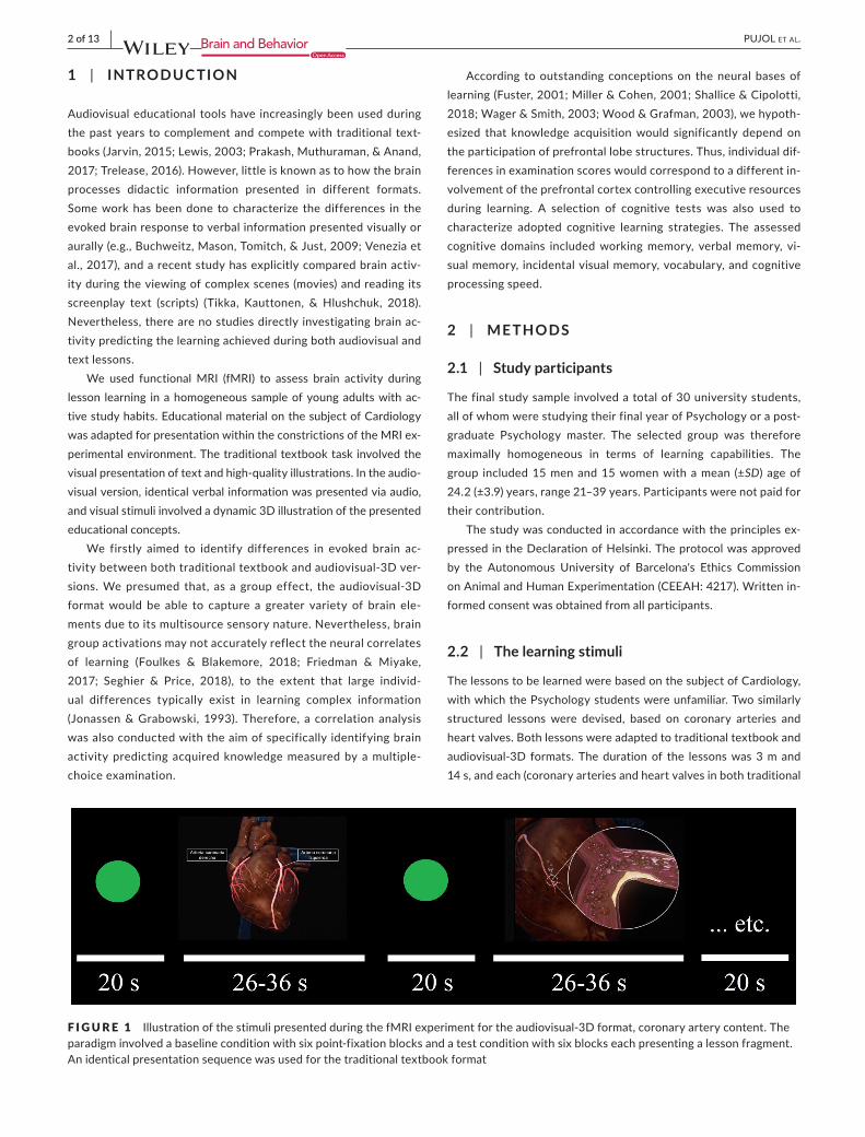
ThelessonstobelearnedwerebasedonthesubjectofCardiology,with which the Psychology students were unfamiliar. Two similarly structured lessons were devised, based on coronary arteries andheartvalves.Bothlessonswereadaptedtotraditionaltextbookandaudiovisual‐3D formats. Thedurationof the lessonswas3mand14s,andeach(coronaryarteriesandheartvalvesinbothtraditional
F I G U R E 1 IllustrationofthestimulipresentedduringthefMRIexperimentfortheaudiovisual‐3Dformat,coronaryarterycontent.Theparadigminvolvedabaselineconditionwithsixpoint‐fixationblocksandatestconditionwithsixblockseachpresentingalessonfragment.Anidenticalpresentationsequencewasusedforthetraditionaltextbookformat

| 3 of 13PUJOL et aL.
textbookandaudiovisual‐3Dformats)wassegmentedintosixfrag‐ments of 26–36 s. Lessons in traditional textbook format werepresented using PowerPoint (PPT) slides, whilst the audiovisual‐3Dversionwasdisplayed in amovie format. Ineachcase, the sixfragments were presented interleaved with a baseline condition,inwhichagreenfixationpointwaspresentedfor20s.Therefore,the experiment involved two fMRI runs of 5 m and 14 s (traditional textbookandaudiovisual‐3Dpresentedinacounterbalancedorder),eachincludingabaselineconditionwithsixpoint‐fixationblocksal‐ternatedwithsixtest‐conditionblockspresentingalessonfragment(Figure1).TheVideosS1andS2(mp4files),showafragmentofthecoronary artery lesson in both textbook and audiovisual formats. Allthedidacticmaterialwasdevelopedin3DTechOmegaZetaSL(Barcelona,Spain)bypersonnelspecializedindeveloping3Deduca‐tional platforms.
2.3 | Procedure
Participants were previously given instructions concerning fMRI testing procedures and the need to remain still during the acquisi‐tion. They were specifically requested to try and understand the di‐dacticmaterialandnotsimplymemorizethepresenteditems.Eachparticipant received one lesson (coronary arteries or heart valves) in the traditional textbook format and another (coronary arteries orheartvalves) intheaudiovisual‐3Dformat.Therefore,eachpar‐ticipantwasexposedtobothformats,butthelessonpresentedwasalways different. The order of both format (traditional textbook or audiovisual‐3D) and topic (coronary arteries or heart valves) wasfully counterbalanced across participants with a pseudorandom allocation.
FollowingthefMRIscans,knowledgeacquisitionwasassessedineachparticipantbymeansofamultiple‐choicetestincluding10questionsoneachtopic.AsaninterferencetaskbetweenthefMRIandtheexamination,theparticipantswererequiredtoreadatext(two stories from the Wechsler Memory Scale, 1987). The timeelapsed between the presentation of the Cardiology lessons and the formal examination ranged from 25 to 35 m.
Inaddition,a101‐pointnumericalratingscale(NRS)wasusedfor the participants to rate the extent to which the presented di‐dacticmaterialwasenjoyable,forbothformats.Theparticipantswere also required to express their preference for either the tradi‐tionalformatortheaudiovisualformat.Finally,theselectedcog‐nitive tests were administered in a single session the same day of the MRI experiment.
2.4 | Selected cognitive assessment
2.4.1 | Working memory
The Digit Span subtest of the Wechsler Adult Intelligence Scale(WAIS) was used as a conventional measure of working memory(Wechsler,2008). In this test, theparticipant is requiredtorepeatdigits forwardandbackward inorder, immediately after theiroral
presentationbytheexaminer.Theoverallage‐appropriatenormed(i.e.,standardizedtohaveameanof10andastandarddeviationof3)score,combiningforwardandbackwardperformance,wasusedin our analyses.
2.4.2 | Verbal memory
The classic Verbal Paired Associates test from the WechslerMemory Scale (Wechsler, 1987) was used. The test evaluatesverbalmemoryforassociatedwordpairs.After10wordpairsareread to the participant, the firstword of each pair is then readand the subject is requested to provide the corresponding associ‐ated word. The test involves three trials based on the same list presentedindifferentorder.Age‐appropriatenormedscoreswereused.
2.4.3 | Visual memory
TheVisualReproductionsubtestfromtheWechslerMemoryScale(Wechsler,1987)wasused.Inthistest,aseriesofthreegeometricpicturesof increasingcomplexity isshown,oneatatime, for10seach. Immediately after thepresentationofeachpicture, thepar‐ticipantisaskedtodrawitfrommemory.Eachpictureisscoredbyadding up the number of correctly reproduced image components. Age‐appropriatenormedscoreswereused.
2.4.4 | Processing speed and incidental visual memory
The Digit Symbol subtest of theWechsler Adult Intelligence Scale(WAIS)wasusedasaconventionalmeasureofspeedinvisuopercep‐tiveprocessing(Wechsler,2008).Theparticipantisrequiredtocopy(inthe spaces provided below the rows of numbers) the symbols matching each number on the basis of a key located at the top of the page. The participant is given a total of 120 s to copy the symbols as quickly as possible.Thenumberofcorrectsymbols,depictedwithintheallottedtime,wasmeasuredandsubsequentlyconvertedintonormedscores.
TheDigitSymboltestalsoservedtomeasureIncidentalVisualMemory,usingtheformDigitSymbol—IncidentalLearning,pairingprocedure.ImmediatelyaftertheDigitSymboltest,theparticipantisrequiredtocompletetwoseriesofDigitSymbolitemswithnotac‐cess to the code key. The participants are never informed that their memorywould be tested.Direct scores (i.e., the number of com‐pleted items) were converted into percentile scores.
2.4.5 | Vocabulary
The Vocabulary subtest of theWechsler Adult Intelligence Scale(WAIS)wasused(Wechsler,2008).Theparticipantisrequiredtode‐fine up to 30 words of increasing difficulty presented orally by the examiner. This test assesses both language and retrieval of informa‐tionfromlong‐termmemory.Directscores (from0to2pointsforeach word) were converted into normed scores.

4 of 13 | PUJOL et aL.
2.5 | Functional MRI
2.5.1 | Functional MRI acquisition
APhilips Achieva 3.0 Teslamagnet (PhilipsHealthcare), equippedwith an eight‐channel phased‐array head coil and single‐shotechoplanar imaging (EPI) software, was used for theMRI assess‐ment.StimuliwerepresentedusingMRI‐compatiblehigh‐resolutiongoggles and audio system (VisuaStim Digital System, ResonanceTechnology Inc.). Our stimuli presentation system did not include the “MR eye tracking” option.
Functional sequences consisted of gradient recalled acquisition in thesteadystate(timeofrepetition[TR],2,000ms;timeofecho[TE],35ms;pulseangle,70°)withinafieldofviewof240×240×128mm,witha64×64‐pixelmatrix,andaslicethicknessof4mm(interslicegap,0mm)andacquisitionvoxelsizeof3.75×3.75×4mm.Atotalof32interleavedsliceswereacquiredtocoverthewholebrain.Eachfunctional time series consisted of 157 consecutive image sets or volumes obtained over 5 m and 14 s. The first four (additional) image sets in each run were discarded to allow the magnetization to reach equilibrium. High‐resolution anatomical images were additionallyobtainedusingasagittalT1‐weightedthree‐dimensionalfastspoiledgradient(SPGR)sequence.Atotalof160sliceswereacquiredwithrepetitiontime=8.1ms;echotime=3.7ms;flipangle=8°,fieldofview=240×240×160mm;matrixsize256×256pixels,in‐planeresolution=0.94×0.94mm2; and slice thickness = 1 mm.
Image preprocessing
ImagingdatawereprocessedusingMATLABversion2016a(TheMathWorks Inc) and Statistical Parametric Mapping software(SPM12; The Wellcome Department of Imaging Neuroscience).Preprocessing involvedmotion correction, spatial normalization,andsmoothingbymeansofaGaussianfilter(full‐widthhalf‐maxi‐mum, 8 mm). Data were normalized to the standard SPM‐EPItemplate and resliced to 2 mm isotropic resolution in Montreal NeurologicalInstitute(MNI)space.All imagesequenceswerein‐spected for potential acquisition and normalization artifacts. Atthisstage,twoparticipantswereremovedfromaninitialbroadersampleof34subjectsasaresultofair/bone‐relatedmagneticsus‐ceptibility artifacts, a third participant due to excessive motion(meaninterscanmotionof1.9mm),andafourthsubjectshowingincidental MRI findings (white matter hypersignal intensities).
Control of potential head motion effects
Tocontrol fortheeffectsofheadmotion,weadoptedthefollow‐ing approach: (a) time series were aligned to the first image volume ineachparticipantusinga least squaresminimizationanda6‐pa‐rameter (rigid body) spatial transformation. (b) Six motion‐relatedregressorsandtheirsixderivativeswere included inthefirst‐level(single‐subject) analysis. (c) Within‐subject, censoring‐based MRIsignalartifactremoval(scrubbing)(Poweretal.,2014)wasusedtodiscardmotion‐affectedvolumes.For eachparticipant, interframemotionmeasurements(Pujoletal.,2014)servedasanindexofdata
qualitytoflagvolumesofsuspectqualityacrosstherun.Atpointswith interframemotion>0.2mm,wediscardedthecorrespondingvolume,togetherwiththeprecedingvolume,andthetwosucceed‐ingvolumes.Usingthisprocedure,ameanof2.7(1%)volumes(range0–20)was removed fromthe totalof314analyzedcombining thetwo functional MRI sequences obtained in each participant.
2.6 | Statistical analysis
2.6.1 | Behavioral data
PairedStudent'st test was used to compare mean differences within the studygroup in termsofbehavioral ratings.Achi‐squared testwas used to assess the relationships between categorical variables. Pearson's product–moment correlationswere used to test the as‐sociationbetweentheparticipants'examinationscoresandperfor‐mance in cognitive testing. Multiple regression analysis was used to predict examination scores with a combination of variables including cognitive testing and extracted measures of brain activity in regions ofinterest(i.e.,frontal loberegionsshowingsignificantcorrelationwith examination scores; see below).
2.6.2 | Functional MRI data
To obtain individual maps of brain activity evoked during lesson presentation, a boxcar regressor was generated considering thesix blocks of the baseline condition and the six blocks of the test condition and applying a hemodynamic delay of 4 s. The contrasts “baseline < test condition” (activation) and “baseline > test condition” (deactivation) were estimated for each participant. The resulting first‐levelSPMcontrastimageswerecarriedforwardtogroup‐levelrandom‐effects analyses.One‐sample t test designs were used to generate group activation maps. Paired t tests were used to compare brain activity evoked during learning using both traditional textbook andaudiovisual‐3Dformats.
In addition, voxel‐wise linear regression was used to test theassociation between examinations scores and evoked brain activity inthecorrespondingexperiment(i.e.,separatelyforthetraditionaltextbook formatand theaudiovisual‐3D format).The results fromthis correlation analysis were reported in terms of both t‐values(SPMdefault)andPearson'sr‐values(t= r∕
√
1− r2∕n−2).Aprimaryanalysis tested the correlation with brain activity evoked during the entire lesson. Further analyses tested the correlation at four dif‐ferentperiodsof6s(threeframes),usingdatafromthebeginning,middle,andendofeachlessonfragmentandimmediately(notimelap) after the lesson fragment. Data from regions showing significant correlation with examination scores were extracted for plotting pur‐posesandtocarryoutseedanalysestoidentifyco‐activatedbrainnetworks.
Seed analyses
Asinourpreviousstudies(Pujoletal.,2019,2016),theseedregionwasdefinedasa3.5‐mmradialsphere(sampling~25voxelsin2mm

| 5 of 13PUJOL et aL.
isotropic space) centered in each identified brain region. This was per‐formedusingMarsBaRregionofinterest(ROI)toolboxinMNIstereo‐taxicspace.SignalsofinterestwereextractedbycalculatingthemeanROI value at each time point across the time series. To generate the seedmaps,thesignaltimecourseofaselectedseedregionwasusedas a regressor to be correlated with the signal time course of each brain voxel to generate individual voxel‐wise statistical parametricmapsofco‐activatedregions.Ahigh‐passfiltersetat128swasusedtoremovelow‐frequencydriftsbelow~0.008Hz.Inaddition,wede‐rivedestimatesofwhitematter,CSF,globalbrainsignalfluctuations,and12motion‐related regressors tobe included in theanalyses asnuisancevariables.Thefirstlevelofourstatisticalanalysis,therefore,involved the generation of single‐subject brain maps expressed asbetaregressionestimates.Single‐subjectvoxel‐wisefunctionalcon‐nectivitymapswerethenincludedinsecond‐level(group)random‐ef‐fectsanalysestotestforgroupeffects(one‐samplet tests).
Thresholding criteria
Inwhole‐brainanalyses, clusters>1.26ml (158voxels) ataheightthreshold of p< .005wereconsidered,whichsatisfiedthefamily‐wiseerror(FWE)ratecorrectionofpFWE<.05,accordingtoMonteCarlo simulations. In analyses within the frontal lobe, the corre‐sponding cluster size considered was 0.94 ml (118 voxels).
3 | RESULTS
3.1 | Behavioral results
3.1.1 | Subjective evaluation
Theaudiovisual‐3Dformatwasconsideredbytheparticipantstobesignificantly more enjoyable than the traditional textbook format (group mean score ± SD;88.3±10.9vs.69.3±19.3; t29=5.0,andp=.00003).All30participants(100%)answered“yes”tothequestion
“Would you use the 3D video format to complement the traditional method of study?” In the event of compulsory expression of prefer‐ence,21ofthe30participantswouldchoosethevideoformatandnine the traditional textbook format for the future (χ2
1,N = 30 = 4.8; p = .028).
3.1.2 | Objective evaluation
Despite formatpreferences, theachieved learningwassimilar fol‐lowingboth lessons. In the10‐questionexaminations, thepartici‐pants’correctanswermeanwas6.1(SD,2.3andrange,1–10)whenthe didactic information was presented using the audiovisual‐3Dformat and 5.9 (SD,1.8and range3–9)whenusing the traditionaltextbook format. The difference was not statistically significant t29=0.8,andp=.752).Noteworthyaretheparticipants'wideexami‐nation score ranges denoting large individual differences in lesson learning.
Regarding the participants' poor success rate, three questionswereidentifiedashighlydifficult(i.e.,correctanswers<15%whenpresentedusingthetraditionaltextbookformat).Interestingly,whenconsideringonlysuchhighlydifficultquestions(questions1,2,and9oftheheartvalveexamination),betterexaminationoutcomeswereobtainedwiththeaudiovisual‐3Dformat (33%ofcorrectanswers)than with the traditional textbook format (12% correct answers),with t29 = 2.4 and p = .024.
3.1.3 | Cognitive testing
Table1reportstheparticipants'performanceinselectedmemoryandcognitive tests and the pattern of correlations with their scores on learningCardiologylessons.Examinationscoresforlessonspresentedwith the audiovisual‐3D format positively correlated with workingmemoryandnegativelycorrelatedwithverbalmemory.Significantly,working memory and verbal memory performance jointly explained
Performance
Correlation with examination scores
Audiovisual‐3D Traditional Textbook
Mean ± SD r (p) r (p)
Working memory (nor‐med scores)
9.8 ± 2.5 .43 (.019) −.14(.468)
Verbalmemory(normedscores)
12.3±1.6 −.42 (.020) .34(.066)
Visualmemory(normedscores)
12.1 ± 1.5 .33 (.077) −.04(.851)
Incidental visual memory (percentiles)
60.2±29.9 −.12(.514) .13 (.487)
Vocabulary(normedscores)
14.2 ± 2.3 .04 (.831) .22 (.242)
Processing speed (normed scores)
11.5 ± 2.4 .13 (.482) −.11(.561)
Note: Degrees of freedom (df),28.Significantp values are indicated in bold font.
TA B L E 1 Cognitive testing (N = 30)

6 of 13 | PUJOL et aL.
24%oftheaudiovisual‐3Dexaminationscorevariancewhenincludedin a multiple regression analysis (Multiple R=0.54,r2=0.30,adjustedr2=0.24,F2,27=5.6,p = .009). Figure 2 illustrates such an association between memory domains and audiovisual examination scores.
3.2 | Imaging results
3.2.1 | Brain activation
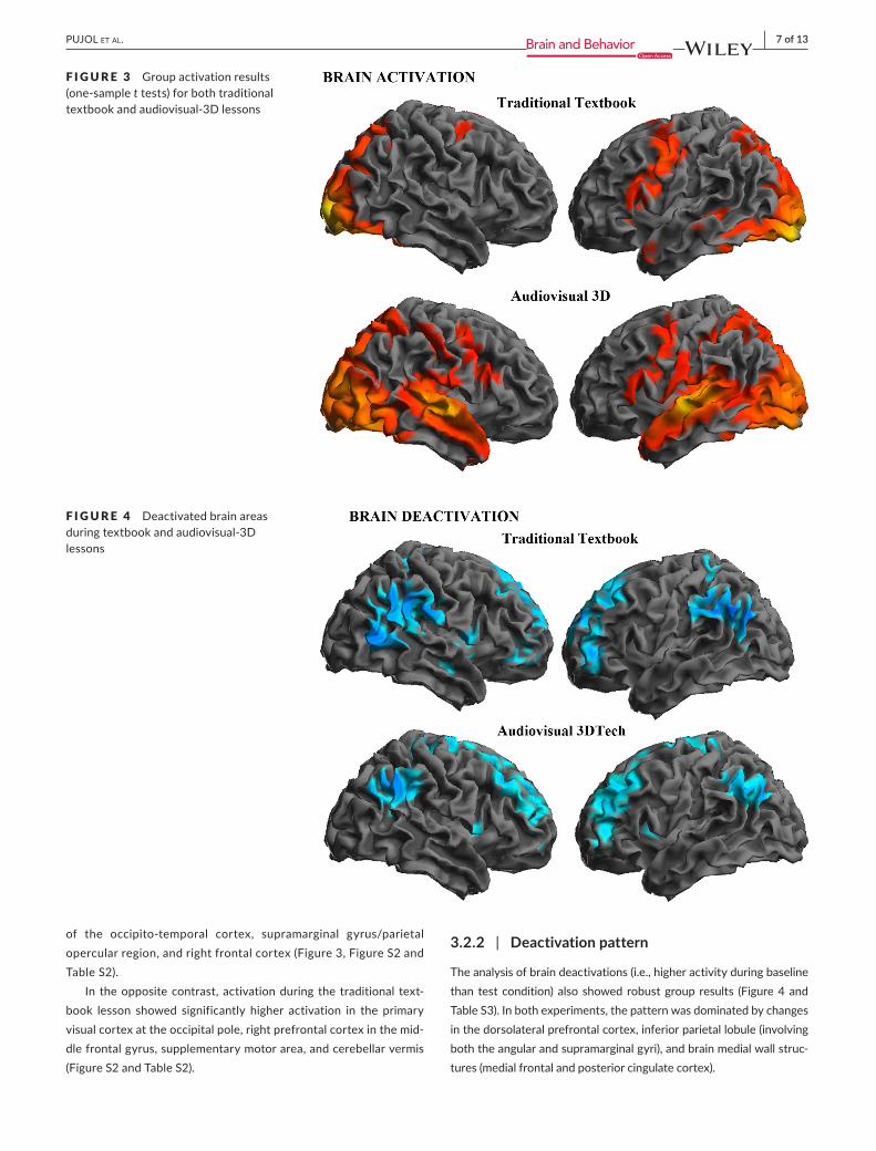
Brainactivationwasrobustinbothexperiments(Figure3andTableS1),whichotherwiseshowedhighlysignificantdifferences.During
the traditional textbook lesson, the strongest activations wereidentifiedintheventralanddorsalaspectsofthevisualcortex,hip‐pocampi,anddorsalparietalassociationcortexincludingtheintra‐parietalsulci,frontaleyefields,andleftpremotorcortexextendingtoBroca'sareaandWernicke'sarea.
Duringtheaudiovisual‐3Dlesson,allthesebrainregionswerealsoactivated(Figure3,FigureS1andTableS2),butthechangeswere more extensive and additionally involved other areas. Direct comparison between both data sets indeed showed significantly higher activation during the audiovisual‐3D lesson in the audi‐torycortexandrelatedareasofthetemporallobe,ventralaspect
F I G U R E 2 Plotsshowingtheassociationsbetweenexaminationscoresfromlessonspresentedwiththeaudiovisual‐3Dformatandmemory performance
| 7 of 13PUJOL et aL.
of the occipito‐temporal cortex, supramarginal gyrus/parietalopercularregion,andrightfrontalcortex(Figure3,FigureS2andTableS2).
In theoppositecontrast, activationduring the traditional text‐book lesson showed significantly higher activation in the primary visualcortexattheoccipitalpole,rightprefrontalcortexinthemid‐dlefrontalgyrus,supplementarymotorarea,andcerebellarvermis(FigureS2andTableS2).
3.2.2 | Deactivation pattern
Theanalysisofbraindeactivations(i.e.,higheractivityduringbaselinethan test condition) also showed robust group results (Figure 4 and TableS3).Inbothexperiments,thepatternwasdominatedbychangesinthedorsolateralprefrontalcortex,inferiorparietallobule(involvingboththeangularandsupramarginalgyri),andbrainmedialwallstruc‐tures (medial frontal and posterior cingulate cortex).
F I G U R E 3 Groupactivationresults(one‐samplet tests) for both traditional textbookandaudiovisual‐3Dlessons
F I G U R E 4 Deactivated brain areas duringtextbookandaudiovisual‐3Dlessons

8 of 13 | PUJOL et aL.
3.2.3 | Correlation analysis—Audiovisual‐3D lesson
Examinationscoresforthelessonspresentedwiththeaudiovisual‐3D format showed a dual pattern of correlations with fMRI signal changesevokedduringexposure to stimuli (Figure5). Specifically,brain activity in a left dorsal prefrontal region predicted higher ex‐aminationscores,whilstbrainactivityinaleftventralfrontalregionadjacenttoBroca'sareapredictedlowerscores(seealsoTableS4).Thatis,examinationscoresincreasesasdorsalprefrontalactivationincreases and ventral frontal activation decreases. These two op‐posite effects were independent and complementary in accounting for examination scores (Figure6). That is, in amultiple regressionanalysisbothimagingvariablesjointlyexplained57%ofaudiovisualexamination score variance (multiple R = 0.77, r2 = 0.60, adjustedr2=0.57,F2,27=20.1,p = .000005).
To illustrate the meaning of the identified associations, wemappedthenetworksofregionsco‐activatedwiththefrontalre‐gions showing positive and negative correlations with examination scores (seed analyses). Overlapping displays (Figure 5) interest‐inglyshowthattheregionsco‐activatedwiththeleftdorsalpre‐frontalregionduringtheaudiovisual‐3Dlessoncorrespondmostlytodeactivatedbrainareas.Bycontrast,regionsco‐activatedwith
the left ventral frontal region adjacent to Broca's area networkmostly overlap with activated regions. Therefore, higher exam‐ination scores are seen to be achieved by participants capable of recruiting, as opposed to deactivating, the prefrontal cortexduringthelesson.Thisisevidentintheplots(Figure6),whereallparticipantsactivatingthisregionshowedexaminationscores≥5.Also,higherscorescorrespondedtoindividualswith lessBroca'sareaactivation,asexaminationscoresnegativelycorrelatedwithactivity in this region.
To this end,we observed that the audiovisual presentation ofeducational material generated an extensive and robust pattern ofactivation thatdidnot,however,positivelypredictexaminationscores.ExaminationsuccesswasinsteadassociatedwithfMRIsignalchanges in circumscribed frontal areas.
Tocharacterizethetemporalevolutionofthelearningprocess,we further analyzed the correlation with examination scores at four differenttimes(beginning,middle,endofeachlessonfragment,andimmediatelyafterthelessonfragment).Significantcorrelationswereobserved only upon completion of and after the lesson. During the finalperiod,brainactivity inthe leftdorsalprefrontalcortexonceagainpredictedhigherexaminationscores(TableS4).However,themostextensivecorrelationfindingswereobservedafterthelesson,
F I G U R E 5 Correlationbetweenaudiovisual‐3Dexaminationscoresandbrainactivityduringaudiovisual‐3Dlessons(leftpanels).Thecentralpanelsillustratethenetworkofregions(seedanalyses)co‐activatedwiththeidentifiedfrontalregions.Therightpanelsshowhowtheregionsco‐activatedwiththeleftdorsalprefrontalregionoverlap(yellow)withdeactivatedbrainareas,andtheregionsco‐activatedwith the left ventral frontal region overlap (yellow) with activated brain areas. Numbers indicate MNI coordinates

| 9 of 13PUJOL et aL.
once theaudiovisual stimuliwereno longerpresent, and involvedventral prefrontal cortex bilaterally and the left sensorimotor cortex (Figure7andTableS4).
3.2.4 | Correlation analysis—Traditional textbook
Examinationscoresinlessonspresentedwiththetraditionaltextbookformat showed a negative correlation with fMRI signal change in a right dorsal prefrontal region (Figure 8). This correlation interestingly shows theoppositedirectiontotheassociationobservedintheaudiovisual‐3D analysis,whichwould further support the adoption of differentmechanisms for learning material presented in different formats. The temporal analysis showed a significant association between examina‐tion scores and signal changes in this right dorsal prefrontal region only inthemiddleperiod(TableS4).
4 | DISCUSSION
Educationalmedicaltopicswereadaptedtobepresentedinbothtraditional textbook and audiovisual‐3D lesson formats. On thebasisofthetestscores,studentsgainedasimilarlevelofknowl‐edge in both formats. Imaging results, however, interestinglysuggest that similar learning may be obtained via different brain resources.Evokedbrainactivitywas robustduringboth the tra‐ditionaltextbookandaudiovisual‐3Dlessons,butalargernumberofbrainsystemswereimplicatedintheprocessingofaudiovisual‐3Dinformation,consistentwithitsmultisensorynatureandhigherstimulus complexity. Significantly, learning was not associatedwithgroupmeanbrainactivations,butwas insteadpredictedbydistinct fMRI signal changes in the frontal lobes. In the audiovis‐ual‐3Dversion,examinationscorespositivelycorrelatedwithlate
F I G U R E 6 Plotsshowingtheassociationsbetweenexaminationscoresfromlessonspresentedwiththeaudiovisual‐3Dformatandbrainactivity in frontal areas

10 of 13 | PUJOL et aL.
prefrontal cortex activity and working memory, and correlatednegativelywithlanguage‐relatedfrontalareasandverbalmemory.In the traditional textbook version, the fewer results recordedsuggested the opposite pattern.
During traditional textbook learning, the identified patternof brain activation accurately reflected the expected changes fornaturalreadingandverbalinformationencoding(Choi,Desai,&Henderson, 2014; Schuster,Hawelka,Hutzler, Kronbichler, &Richlan,2016;Wandell&Le,2017),withactivationofthevisualcortex,hippocampi,frontaleyefields,intraparietalsulci,Broca'sarea, and Wernicke's area. In addition, strong activation wasidentifiedinboththeventralanddorsalvisualstreams,whichisconsistent with neural operations related to processing object features and spatial relationships in the illustrating images pre‐sentedalongwiththetext(Goodale&Milner,1992;Ungerleider&Haxby,1994).
Intheaudiovisualversion,theverbal inputwasauralandthevisual stimulation involved more complex dynamic 3D representa‐tions.Accordingly,brainactivationwasmoreextensiveandimpli‐catedthepertinentareasineachbrainlobe.Additionalactivationswere thus identified in the auditory cortex processing speech and related areas of the temporal lobe (Venezia et al., 2017),
particularly involving the superior temporal sulcus likely reflecting multisensory integration (Beauchamp, Yasar, Frye, & Ro, 2008),thelateralaspectoftheoccipito‐temporalcortexsubservingcom‐plexvisualprocessesandmotionperception(Lingnau&Downing,2015), the supramarginal gyrus/parietal opercular region criti‐cal to cross‐modal abstractions and spatial cognition (Leichnetz,2011), and right frontal cortex. Paradoxically, the larger numberof brain resources employed to process more complex audiovisual informationwasnotnecessarilyassociatedwithgreaterlearning,as,indeed,ourparticipantsactuallyobtainedsimilarexaminationscores with both formats.
The issue of stimulus complexity in encoding information has previously been discussed in the context of advertising. Somedata indicate that a higher sensory intensity of audio and visual features brings increased attention and cognitive processing,leading toagreateradvertising impact (Langlebenet al., 2009).Dynamic motion and audio features may indeed be effective in making adsmorememorable (Han et al., 2015).However, stim‐uli wealth and complexity may otherwise exceed the capacity to assimilate information and result in reduced message retention (Langleben et al., 2009; Seelig et al., 2014;Wang et al., 2013).Inouraudiovisualexperiment,somebalancemayexistbetween
F I G U R E 7 Correlation between audiovisual‐3Dexaminationscoresandbrain activity immediately after exposure toaudiovisual‐3Dmaterial
F I G U R E 8 Correlation between traditional textbook scores and brain activity during traditional textbook lessons (left panel). The central panelillustratesthenetworkofregions(seedanalysis)co‐activatedwiththeidentifiedfrontalregion.Therightpanelshowstheassociationbetween examination scores and brain activity in the frontal area. Numbers indicate MNI coordinates

| 11 of 13PUJOL et aL.
the advantages and disadvantages of providing more complex information.
The amount and complexity of the information is obviously an important factor to consider when designing audiovisual ed‐ucationalmaterial,asisthetempowithwhichtheinformationispresented. In our study, examination scores were predicted bybrain changes at the end‐of‐lesson fragments and after‐lessonfragmentswhenpresented in the audiovisual‐3D format,whichwould indicate that information perception and assimilation do not temporally coincide, thus reinforcing the notion that thetempo should be adequately adjusted when planning education. Bycontrast,inthecaseofusingthetraditionaltextbookformat,examination scores were predicted by brain changes at the mid‐dle‐of‐lessonfragmentssuggestingadifferentlearningstrategy,in which information may be more directly recorded at the per‐ception stage.
Activity in theprefrontal cortex significantlyaccounted for thenotable interindividual differences of our participants when assimi‐latingdidactic informationpresented in theaudiovisual‐3D format.Successful participants activated prefrontal cortex areas typicallyconcernedwithexecutivefunctions(Fuster,2001;Wood&Grafman,2003).Theseparticipantsalsoshowedbetterworkingmemory,whichisaprototypicalexecutivefunction(Diamond,2013;Wager&Smith,2003).Ontheotherhand,examinationscoresnegativelycorrelatedwithactivity in language‐relatedcortexandverbalmemory.Suchascenario would therefore indicate that audiovisual learning may ben‐efit fromexecutive prefrontal operations,whereas verbal process‐ing, tosomeextent, interferedwith informationassimilation inourexperiment. In other words, audiovisual learning of the presentedmaterial may be more closely related to the proper use of perceived information to integrate knowledge than to the verbal memorization ofdidacticmaterial,thatistosaylearningthroughunderstandingasopposed to memorizing.
Onthewhole,ourresultssuggestnotabledifferencesbetweentraditional textbook and audiovisual learning of an identical amount of didactic information. The limited results obtained from the tra‐ditionaltextbooktaskanalysiswereintheoppositedirection,witha negative correlation between examination scores and prefrontal cortex activity suggesting a lower involvement of executive opera‐tions.Moreover,thedorsalprefrontalcortexisoneofthefewstruc‐tures showing significantly higher activation during the traditional textbook lesson (Figure S2), as a result of being less deactivated(Figure 4). The overall pattern of results (i.e., activation/deactiva‐tion and the direction of the correlation with examination scores) may therefore support the notion that prefrontal cortex activity to some extent interferes with learning during textbook lessons. Unfortunately, we found no positive correlation to complete thepatternand,thus,theanalysisislessinformativeastohowlearningtakes place in such a situation. Further research is needed to support the proposal that traditional textbook learning is based more on lan‐guage processing and verbal memory.
A limitation in our study concerns the generalization of re‐sults.Firstly,thereisanotabledifferencebetweentheduration
of conventional college lessons and lesson duration in our exper‐iment.Therefore,ourresultscannotbegeneralizedatthislevel.Also,both textbookandaudiovisual formatsmaypotentiallybemore or less advantageous depending on the topics to be learned. We recorded the students' preference for the audiovisual ver‐sion of the lesson, but with no clear advantage in the contextof examination results. Previous studies generally coincide on reporting higher student satisfaction when using audiovisual material (Ahmad, Sleiman, Thomas, Kashani, & Ditmyer, 2016;Drapkin,Lindgren,Lopez,&Stabio,2015;Murgitroyd,Madurska,Gonzalez,&Watson,2015;Ozer,Govsa,&Bati,2017;Prakashetal.,2017;Trelease,2016),andsomestudiesalsoreportobjectiveadvantagesbasedonexaminationscores(e.g.,Ahmadetal.,2016;Drapkinetal.,2015;andrevisedinTrelease,2016).Asmightbeexpected, the topic thatmore clearly benefits from audiovisualand3Dmaterial isAnatomy (Ahmadetal.,2016;Drapkinetal.,2015; Trelease, 2016).Our study is also limited due to its rela‐tivelyreducedcognitiveevaluationandalackoflong‐termlessonlearning assessments.
Inconclusion, this isanovel functionalMRI studycharacteriz‐ing the pattern of brain activity evoked during the assimilation of educational information presented in two distinct formats. Brainactivationwas robust inboth cases,butnotabledifferenceswereidentified that may well suggest the advantages of either model. It isrelevant,however,thattheactuallearningsuccess,measuredbyamultiple‐choiceexamination,wasnotdirectlyrelatedtothemostobviousbrainactivations.Examinationscoreswereinsteadassoci‐atedwithfMRIsignalchangesinthefrontallobes.Learningduringthe audiovisual version was specifically predicted by a combination of more activity in the dorsal prefrontal cortex and less participation of the language‐relatedventral frontalcortex.Consistently,exam‐ination scores positively correlated with performance in executive functioningandnegativelywithverbalmemory.Overall,ourresultssuggest that audiovisual learning relies more on the proper use of perceived information and less on basic verbal memory processes. Further research would be necessary for a more complete charac‐terization of imaging correlates in the context of traditional textbook learning.
ACKNOWLEDG MENTS
Thisworkwassupportedby3DTechOmegaZeta,BarcelonaandtheInstituteofHealthCarlosIII‐CIBER,GovernmentofSpain,refer‐ence number 1589923.
CONFLIC T OF INTERE S T
The authors have no conflict of interest to declare.
DATA AVAIL ABILIT Y S TATEMENT
The data that support the findings of this study are available from the corresponding author upon reasonable request.

12 of 13 | PUJOL et aL.
ORCID
Jesus Pujol https://orcid.org/0000‐0002‐9946‐4547
R E FE R E N C E S
Ahmad,M.,Sleiman,N.H.,Thomas,M.,Kashani,N.,&Ditmyer,M.M.(2016).Useofhigh‐definitionaudiovisualtechnologyinagrossanat‐omy laboratory: Effect on dental students' learning outcomes andsatisfaction. Journal of Dental Education,80,128–132.
Beauchamp,M.S.,Yasar,N.E.,Frye,R.E.,&Ro,T.(2008).Touch,soundand vision in human superior temporal sulcus. NeuroImage,41,1011–1020.https://doi.org/10.1016/j.neuroimage.2008.03.015
Buchweitz,A.,Mason,R.A.,Tomitch,L.M.,&Just,M.A.(2009).Brainactivationforreadingand listeningcomprehension:AnfMRIstudyof modality effects and individual differences in language com‐prehension. Psychology & Neuroscience, 2, 111–123. https://doi.org/10.3922/j.psns.2009.2.003
Choi,W.,Desai,R.H.,&Henderson,J.M.(2014).Theneuralsubstratesofnaturalreading:Acomparisonofnormalandnonwordtextusingeyetracking and fMRI. Frontiers in Human Neuroscience,8,1024.https://doi.org/10.3389/fnhum.2014.01024
Diamond,A.(2013).Executivefunctions.Annual Review of Psychology,64,135–168.https://doi.org/10.1146/annurev‐psych‐113011‐143750
Drapkin, Z. A., Lindgren, K. A., Lopez, M. J., & Stabio, M. E. (2015).Development and assessment of a new 3D neuroanatomy teaching tool for MRI training. Anatomical Sciences Education, 8, 502–509.https ://doi.org/10.1002/ase.1509
Foulkes,L.,&Blakemore,S.J.(2018).Studyingindividualdifferencesinhuman adolescent brain development. Nature Neuroscience,21,315–323.https://doi.org/10.1038/s41593‐018‐0078‐4
Friedman,N.P.,&Miyake,A. (2017).Unity anddiversityof executivefunctions: Individual differences as a window on cognitive structure. Cortex,86,186–204.https://doi.org/10.1016/j.cortex.2016.04.023
Fuster, J. M. (2001). The prefrontal cortex–an update: Time is ofthe essence. Neuron, 30, 319–333. https://doi.org/10.1016/S0896‐6273(01)00285‐9
Goodale,M.A.,&Milner,A.D.(1992).Separatevisualpathwaysforper‐ception and action. Trends in Neurosciences,15, 20–25.https://doi.org/10.1016/0166‐2236(92)90344‐8
Han, J., Chen, C., Shao, L., Hu, X., Han, J., & Liu, T. (2015). Learningcomputational models of video memorability from fMRI brain im‐aging. IEEE Transactions on Cybernetics,45, 1692–1703.https://doi.org/10.1109/TCYB.2014.2358647
Jarvin,L.(2015).Edutainment,games,andthefutureofeducationinadigitalworld.InE.L.Grigorenko(Ed.),The global context for new di‐rections for child and adolescent development. New Directions for Child and Adolescent Development,147 (pp. 33–40).Hoboken,NJ:WileyPeriodicals,Inc.
Jonassen,D.,&Grabowski,B.(1993).Handbook of individual differences, learning, and instruction.NewYork,NY:Routledge.
Langleben,D.D.,Loughead,J.W.,Ruparel,K.,Hakun,J.G.,Busch‐Winokur,S.,Holloway,M.B.,…Lerman,C.(2009).Reducedprefrontalandtempo‐ral processing and recall of high "sensation value" ads. NeuroImage,46,219–225.https://doi.org/10.1016/j.neuroimage.2008.12.062
Leichnetz,G.R.(2011).SupramarginalGyrus.InJ.S.Kreutzer,J.DeLuca,&B.Caplan(Eds.),Encyclopedia of clinical neuropsychology(pp.180–203).NewYork,NY:Springer.
Lewis,M. J. (2003). Computer‐assisted learning for teaching anatomyand physiology in subjects allied to medicine. Medical Teacher,25,204–206.https://doi.org/10.1080/0000000000000000000a
Lingnau,A.,&Downing,P.E. (2015).The lateraloccipitotemporalcor‐tex in action. Trends in Cognitive Sciences,19, 268–277.https://doi.org/10.1016/j.tics.2015.03.006
Miller,E.K.,&Cohen,J.D. (2001).An integrativetheoryofprefrontalcortex function. Annual Review of Neuroscience,24,167–202.
Murgitroyd,E.,Madurska,M.,Gonzalez,J.,&Watson,A.(2015).3Ddig‐italanatomymodelling–Practicalorpretty?Surgeon,13,177–180.https://doi.org/10.1016/j.surge.2014.10.007
Ozer,M.A.,Govsa,F.,&Bati,A.H. (2017).Web‐basedteachingvideopackages on anatomical education. Surgical and Radiologic Anatomy,39(11),1253–1261.
Power,J.D.,Mitra,A.,Laumann,T.O.,Snyder,A.Z.,Schlaggar,B.L.,&Petersen,S.E.(2014).Methodstodetect,characterize,andremovemotion artifact in resting state fMRI. NeuroImage,84,320–341.https://doi.org/10.1016/j.neuroimage.2013.08.048
Prakash, S. S., Muthuraman, N., & Anand, R. (2017). Short‐durationpodcasts as a supplementary learning tool: Perceptions of medi‐cal students and impact on assessment performance. BMC Medical Education,17,167.https://doi.org/10.1186/s12909‐017‐1001‐5
Pujol,J.,Blanco‐Hinojo,L.,Macia,D.,Alonso,P.,Harrison,B.J.,Martínez‐Vilavella,G.,…Soriano‐Mas,C. (2019).Mappingalterationsof thefunctionalstructureofthecerebralcortexinobsessive‐compulsivedisorder. Cerebral Cortex,https://doi.org/10.1093/cercor/bhz008
Pujol,J.,Macià,D.,Blanco‐Hinojo,L.,Martínez‐Vilavella,G.,Sunyer,J.,de laTorre,R.,…Harrison,B. J. (2014).Doesmotion‐relatedbrainfunctional connectivity reflect both artifacts and genuine neural activity? NeuroImage, 101, 87–95. https://doi.org/10.1016/j.neuroimage.2014.06.065
Pujol, J.,Martínez‐Vilavella,G.,Macià,D., Fenoll,R.,Alvarez‐Pedrerol,M.,Rivas,I.,…Sunyer,J.(2016).Trafficpollutionexposureisassoci‐ated with altered brain connectivity in school children. NeuroImage,129,175–184.
Schuster,S.,Hawelka,S.,Hutzler,F.,Kronbichler,M.,&Richlan,F.(2016).Wordsincontext:Theeffectsoflength,frequency,andpredictabil‐ity on brain responses during natural reading. Cerebral Cortex,26,3889–3904.https://doi.org/10.1093/cercor/bhw184
Seelig, D., Wang, A.‐L., Jaganathan, K., Loughead, J. W., Blady, S. J.,Childress, A. R., … Langleben, D. D. (2014). Low message sensa‐tion health promotion videos are better remembered and activate areas of the brain associated with memory encoding. PLoS ONE,9,e113256.https://doi.org/10.1371/journal.pone.0113256
Seghier,M.L.,&Price,C.J.(2018).Interpretingandutilisingintersubjectvariability in brain function. Trends in Cognitive Sciences,22,517–530.https://doi.org/10.1016/j.tics.2018.03.003
Shallice,T.,&Cipolotti,L. (2018).Theprefrontalcortexandneurologi‐cal impairments of active thought. Annual Review of Psychology,69,157–180.https://doi.org/10.1146/annurev‐psych‐010416‐044123
Tikka,P.,Kauttonen,J.,&Hlushchuk,Y.(2018).Narrativecomprehensionbeyond language: Common brain networks activated by a movie and its script. PLoS ONE, 13, e0200134. https://doi.org/10.1371/journal.pone.0200134
Trelease,R.B.(2016).Fromchalkboard,slides,andpapertoe‐learning:Howcomputingtechnologieshavetransformedanatomicalscienceseducation. Anatomical Sciences Education, 9, 583–602. https://doi.org/10.1002/ase.1620
Ungerleider,L.G.,&Haxby,J.V.(1994).'What'and'where'inthehumanbrain. Current Opinion in Neurobiology, 4, 157–165. https://doi.org/10.1016/0959‐4388(94)90066‐3
Venezia,J.H.,Vaden,K.I.Jr,Rong,F.,Maddox,D.,Saberi,K.,&Hickok,G.(2017).Auditory,visualandaudiovisualspeechprocessingstreamsinsuperior temporal sulcus. Frontiers in Human Neuroscience,11,174.https ://doi.org/10.3389/fnhum.2017.00174
Wager, T. D., & Smith, E. E. (2003). Neuroimaging studies of work‐ing memory: A meta‐analysis. Cognitive, Affective, & Behavioural Neuroscience,3,255–274.https://doi.org/10.3758/CABN.3.4.255
Wandell, B. A., & Le, R. K. (2017). Diagnosing the neural circuitryof reading. Neuron, 96, 298–311. https://doi.org/10.1016/j.neuron.2017.08.007

| 13 of 13PUJOL et aL.
Wang, A. L., Ruparel, K., Loughead, J.W., Strasser, A. A., Blady, S.J., Lynch, K. G., … Langleben, D. D. (2013). Content matters:Neuroimaging investigation of brain and behavioral impact of televised anti‐tobacco public service announcements. Journal of Neuroscience, 33, 7420–7427. https://doi.org/10.1523/JNEUROSCI.3840‐12.2013
Wechsler,D.(1987).Wechsler memory scale‐revised manual.SanAntonio,TX:PsychologicalCorporation.
Wechsler,D.(2008).Wechsler adult intelligence scale,4thed.SanAntonio,TX:PearsonAssessment.
Wood,J.N.,&Grafman,J.(2003).Humanprefrontalcortex:Processingand representational perspectives. Nature Reviews Neuroscience,4,139–147.https://doi.org/10.1038/nrn1033
SUPPORTING INFORMATION
Additional supporting information may be found online in theSupportingInformationsectionattheendofthearticle.
How to cite this article:PujolJ,Blanco‐HinojoL,Martínez‐VilavellaG,etal.Brainactivityduringtraditionaltextbookandaudiovisual‐3Dlearning.Brain Behav. 2019;00:e01427. https ://doi.org/10.1002/brb3.1427