brain damage & recovery causes of brain damage some examples of brain damage effects of brain...
TRANSCRIPT
Brain Damage & Recovery
• Causes of brain damage
• Some examples of brain damage
• Effects of brain damage
• Recovery from brain damage
• Myelination disorders
Causes of Brain Damage
• Genetic (not necessarily hereditary!)
• Congenital
• Environmental (toxins)
• Neoplasms
• Apoptosis (programmed cell death)
• Cerebrovascular problems
• Head impact injuries (closed head)
• Infections
Hereditary Brain Damage
• Passed from parent to child through DNA.• Several types:
– Faulty chromosome duplication• Rare
– Dominant gene disorders• Rare, tend to be self-limiting.
– Recessive gene disorders• Not all children express the defect.
– Polygenetic disorders• By far the most common.
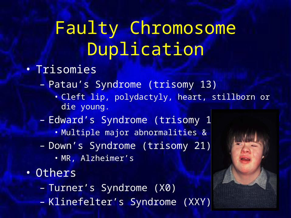
Faulty Chromosome Duplication
• Trisomies– Patau’s Syndrome (trisomy 13)
• Cleft lip, polydactyly, heart, stillborn or die young.
– Edward’s Syndrome (trisomy 18)• Multiple major abnormalities & MR
– Down’s Syndrome (trisomy 21)• MR, Alzheimer’s
• Others– Turner’s Syndrome (X0)
– Klinefelter’s Syndrome (XXY)
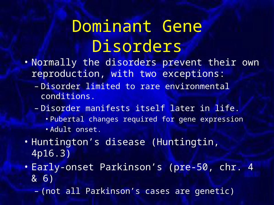
Dominant Gene Disorders
• Normally the disorders prevent their own reproduction, with two exceptions:– Disorder limited to rare environmental conditions.– Disorder manifests itself later in life.
• Pubertal changes required for gene expression
• Adult onset.
• Huntington’s disease (Huntingtin, 4p16.3)
• Early-onset Parkinson’s (pre-50, chr. 4 & 6)– (not all Parkinson’s cases are genetic)
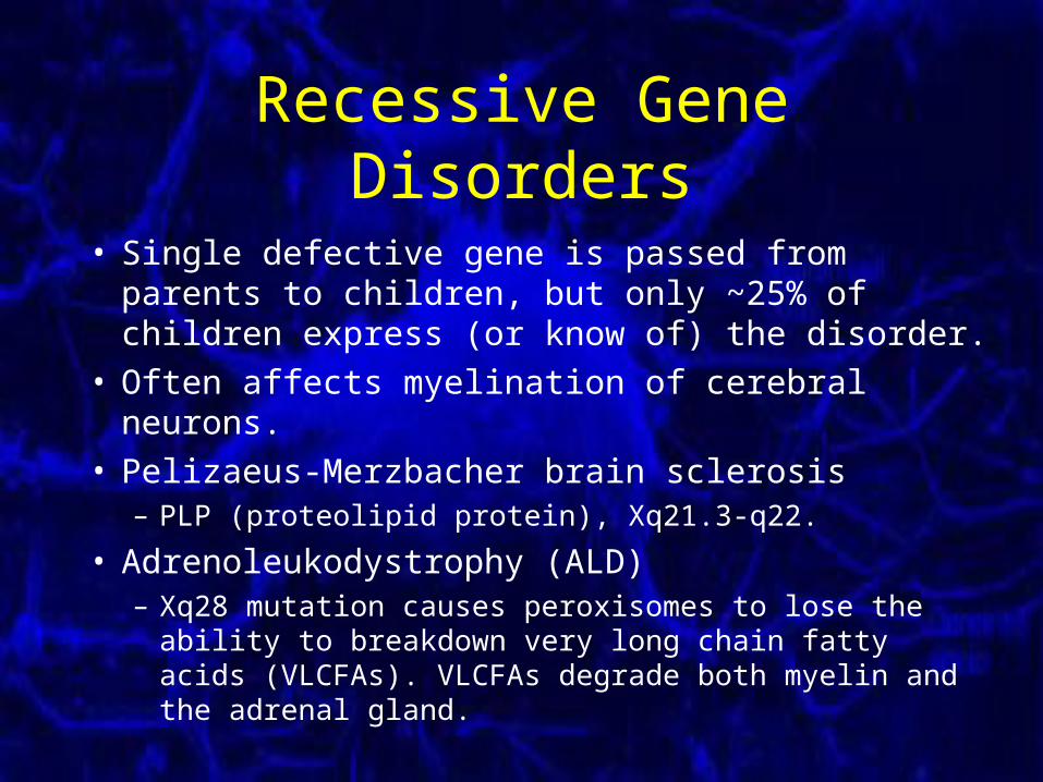
Recessive Gene Disorders
• Single defective gene is passed from parents to children, but only ~25% of children express (or know of) the disorder.
• Often affects myelination of cerebral neurons.• Pelizaeus-Merzbacher brain sclerosis
– PLP (proteolipid protein), Xq21.3-q22.
• Adrenoleukodystrophy (ALD)– Xq28 mutation causes peroxisomes to lose the ability to
breakdown very long chain fatty acids (VLCFAs). VLCFAs degrade both myelin and the adrenal gland.

Polygenetic Disorders
• Requires the presence of multiple defective genes, along with environmental influences.
• Most psychological disorders & personality traits.
• Genetics can also protect against disorders.
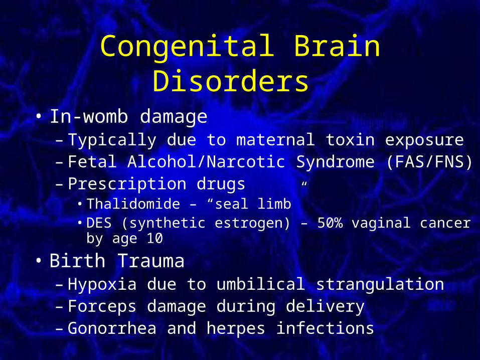
Congenital Brain Disorders
• In-womb damage – Typically due to maternal toxin exposure– Fetal Alcohol/Narcotic Syndrome (FAS/FNS)– Prescription drugs
• Thalidomide – “seal limb”• DES (synthetic estrogen) – 50% vaginal cancer by age 10
• Birth Trauma– Hypoxia due to umbilical strangulation– Forceps damage during delivery– Gonorrhea and herpes infections
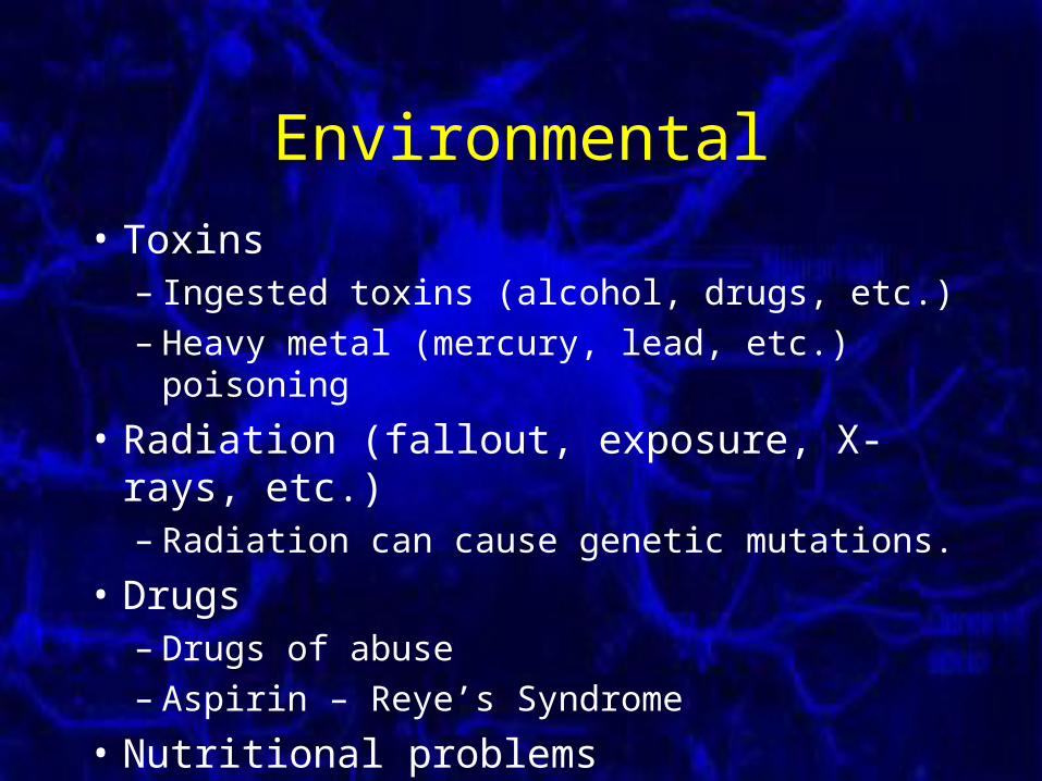
Environmental
• Toxins– Ingested toxins (alcohol, drugs, etc.)– Heavy metal (mercury, lead, etc.) poisoning
• Radiation (fallout, exposure, X-rays, etc.)– Radiation can cause genetic mutations.
• Drugs– Drugs of abuse– Aspirin – Reye’s Syndrome
• Nutritional problems
Neoplasms (Cancers)
• Mass of new tissue (= “new growth”).• Physiologically useless.• Growth is usually not controlled.• Neoplasms account for a relatively high
proportion of neurological disease.• Brain is 2nd only to uterus as tumor area.• Brain tumors typically not from nerves.
– Gliomas account for 45% of brain neoplasms
Neoplasms
• Two types of tumors– Benign (-omas)
• Regular, well-defined shape.• Does not intrude into surrounding tissue.• Effects only by pressure on the brain.
– Malignant (-carcinomas)• Irregularly shaped.• Intrudes (infiltrates) into surrounding tissue.• Tend to be recurrent.
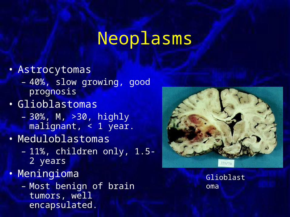
Neoplasms
• Astrocytomas– 40%, slow growing, good
prognosis
• Glioblastomas– 30%, M, >30, highly malignant,
< 1 year.
• Meduloblastomas– 11%, children only, 1.5-2 years
• Meningioma– Most benign of brain tumors,
well encapsulated.
Glioblastoma
Neoplasms
• Metastatic Tumors – Cancerous cells migrate from other parts of the
body, normally the lung or breast. – It is not uncommon for lung cancers to first be
noticed in the brain.– Normally multiple implant sites.– All malignant.– Very hard to treat.
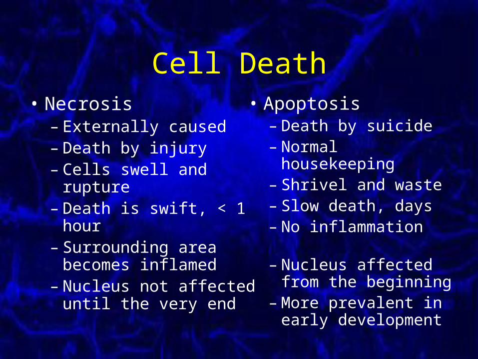
Cell Death• Necrosis
– Externally caused– Death by injury– Cells swell and rupture– Death is swift, < 1 hour– Surrounding area
becomes inflamed– Nucleus not affected
until the very end
• Apoptosis– Death by suicide– Normal housekeeping – Shrivel and waste– Slow death, days– No inflammation
– Nucleus affected from the beginning
– More prevalent in early development
Cerebrovascular Damage
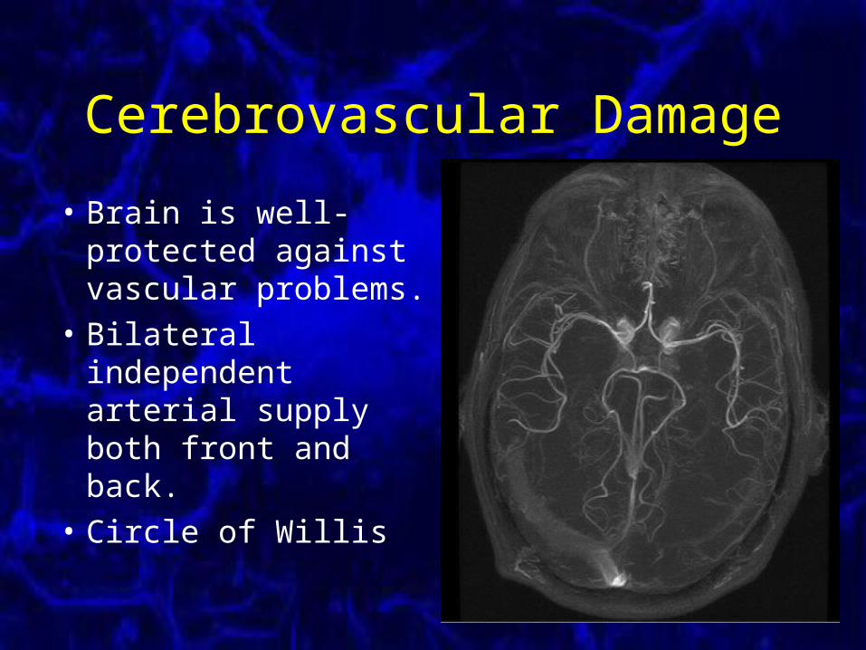
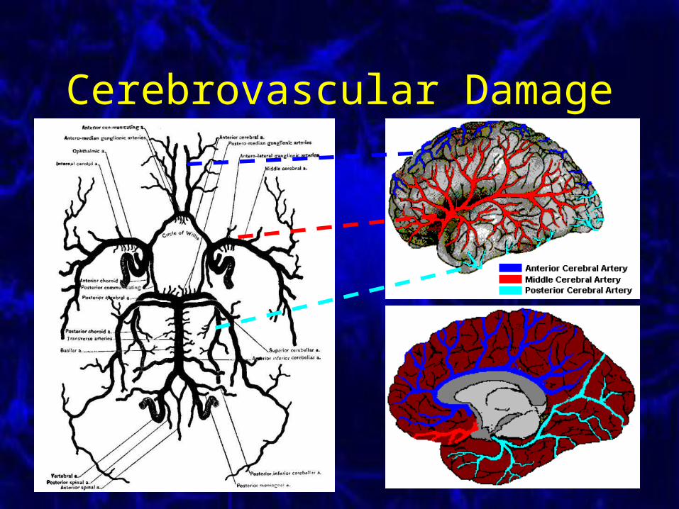
• Brain is well-protected against vascular problems.
• Bilateral independent arterial supply both front and back.
• Circle of Willis
Cerebrovascular Damage
Cerebrovascular Damage
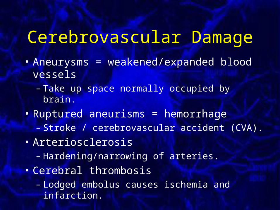
• Aneurysms = weakened/expanded blood vessels– Take up space normally occupied by brain.
• Ruptured aneurisms = hemorrhage– Stroke / cerebrovascular accident (CVA).
• Arteriosclerosis– Hardening/narrowing of arteries.
• Cerebral thrombosis– Lodged embolus causes ischemia and infarction.
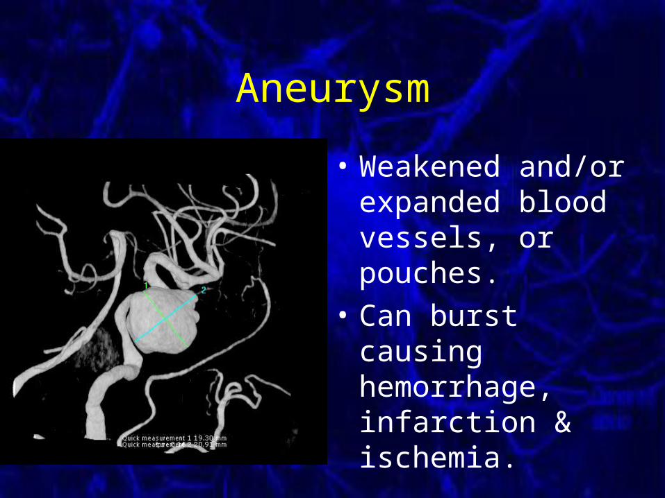
Aneurysm
• Weakened and/or expanded blood vessels, or pouches.
• Can burst causing hemorrhage, infarction & ischemia.
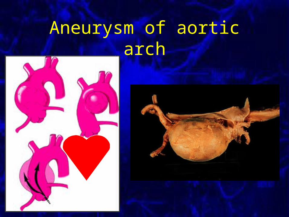
Aneurysm of aortic arch
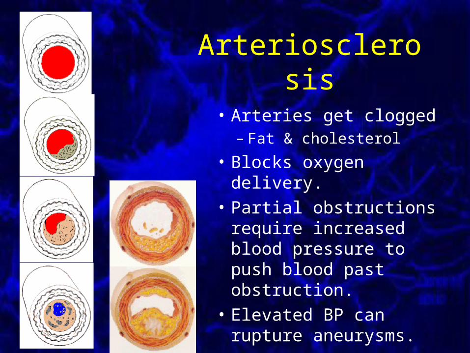
Arteriosclerosis
• Arteries get clogged – Fat & cholesterol
• Blocks oxygen delivery.
• Partial obstructions require increased blood pressure to push blood past obstruction.
• Elevated BP can rupture aneurysms.
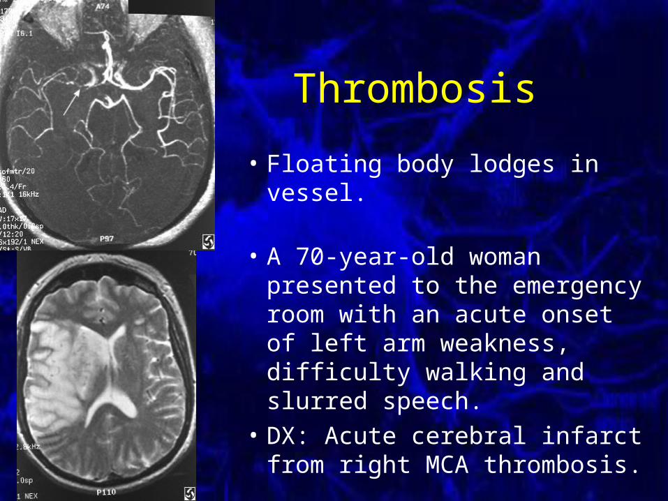
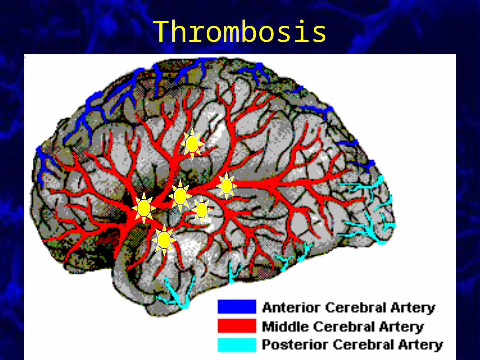
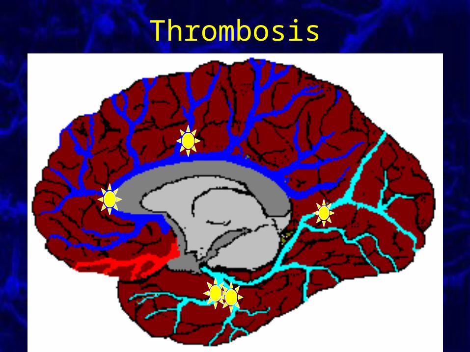
Thrombosis
• Floating body lodges in vessel.
• A 70-year-old woman presented to the emergency room with an acute onset of left arm weakness, difficulty walking and slurred speech.
• DX: Acute cerebral infarct from right MCA thrombosis.
Thrombosis
Thrombosis
Cerebrovascular Damage• Formerly assumed that most damage was
due to decreased oxygen and/or glucose.• Now appears that most cerebrovascular
damage is due to excess glutamate release by blood-deprived neurons.
• Excess Ca++ and Na+ influx, especially through NMDA receptors, kills cells.
• Hippocampus is particularly affected.• Ca++ channel & NMDA receptor blockers
helpful in preventing post-stroke damage.
Head Impact Injuries
• Car accidents• Household accidents
– Falling down stairs– Bike/scooter/Rollerblade accidents
• Shaken baby syndrome• Physical abuse
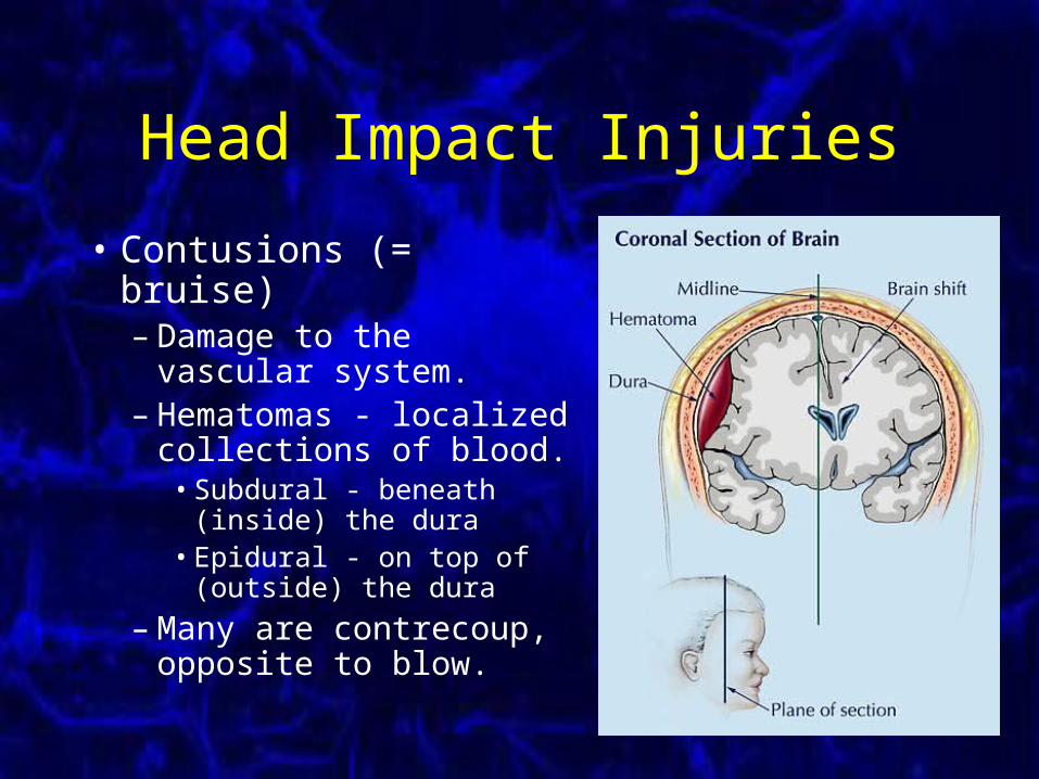
Head Impact Injuries
• Contusions (= bruise)– Damage to the vascular
system.– Hematomas - localized
collections of blood.• Subdural - beneath
(inside) the dura• Epidural - on top of
(outside) the dura
– Many are contrecoup, opposite to blow.
Head Impact Injuries
• Concussion– Disturbance of consciousness without evidence
of contusion or other structural damage.
• Common assumption is temporary disruption with no long-term damage.– Punch-drunk syndrome suggests otherwise.– Boxers show cerebral scarring with dementia.
Other CNS Injuries
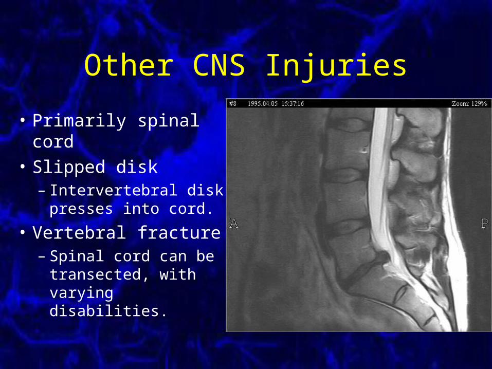
• Primarily spinal cord
• Slipped disk– Intervertebral disk
presses into cord.
• Vertebral fracture– Spinal cord can be
transected, with varying disabilities.
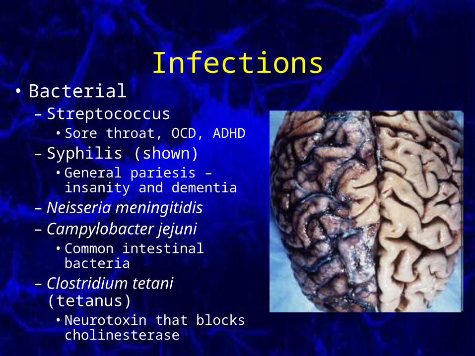
Infections• Bacterial
– Streptococcus• Sore throat, OCD, ADHD
– Syphilis (shown)• General pariesis –
insanity and dementia
– Neisseria meningitidis– Campylobacter jejuni
• Common intestinal bacteria
– Clostridium tetani (tetanus)• Neurotoxin that blocks
cholinesterase
Infections
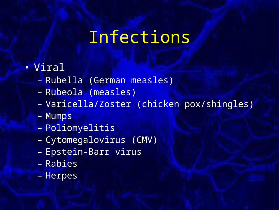
• Viral– Rubella (German measles)– Rubeola (measles)– Varicella/Zoster (chicken pox/shingles)– Mumps– Poliomyelitis– Cytomegalovirus (CMV)– Epstein-Barr virus– Rabies– Herpes
Infections
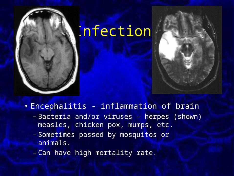
• Encephalitis - inflammation of brain– Bacteria and/or viruses – herpes (shown)
measles, chicken pox, mumps, etc.– Sometimes passed by mosquitos or animals.– Can have high mortality rate.
Infections
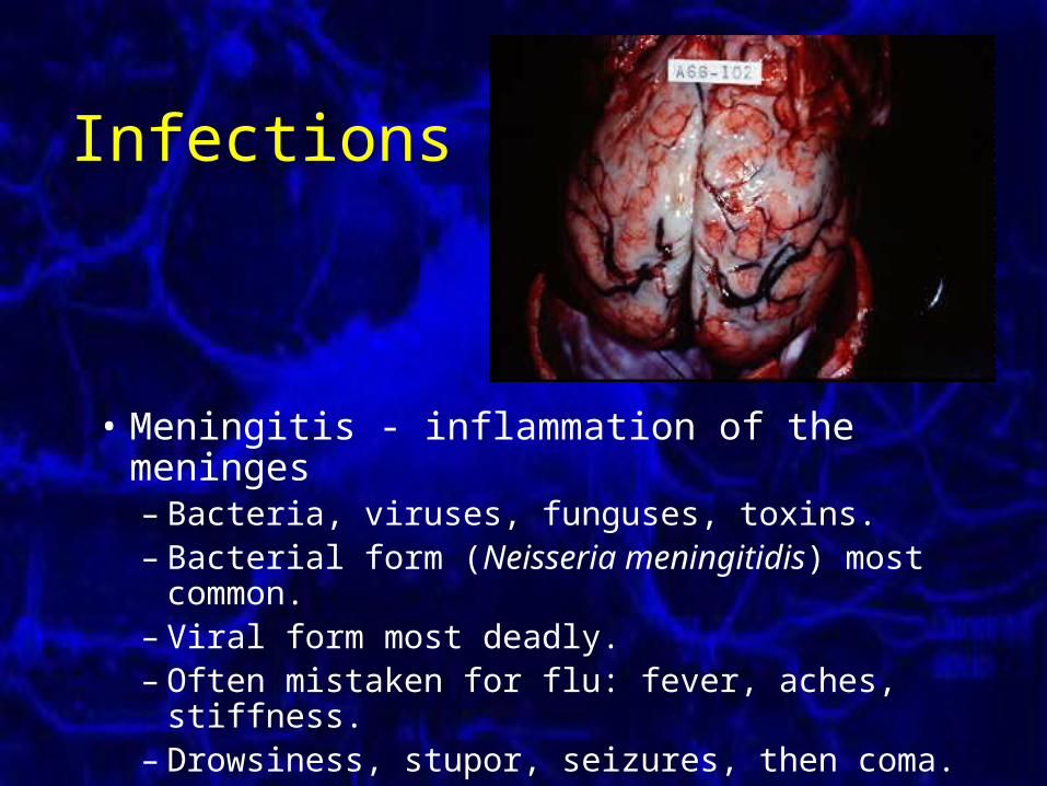
• Meningitis - inflammation of the meninges– Bacteria, viruses, funguses, toxins.– Bacterial form (Neisseria meningitidis) most
common.– Viral form most deadly.– Often mistaken for flu: fever, aches, stiffness.– Drowsiness, stupor, seizures, then coma.
Effects of Brain Damage
• Kill neurons– Damage to nerve itself– Damage to glial cells/altered environment– Damage to pre-/post-synaptic nerves
• Crowd out or put pressure on the brain
• Disable neurons– Often by interfering with myelin
Recovery of Function
Recovery of Function
• What happens after (or during) brain damage?– Degeneration– Regeneration– Reorganization
• Can anything be done to aid recovery?
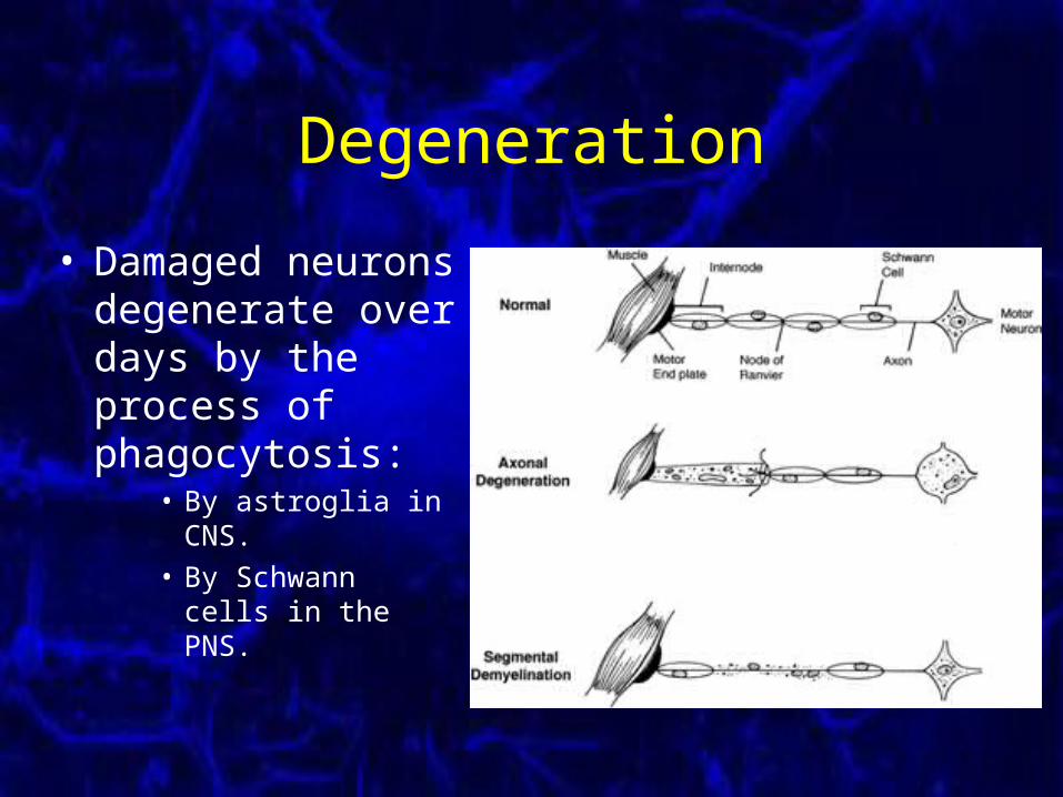
Degeneration
• Damaged neurons degenerate over days by the process of phagocytosis:
• By astroglia in CNS.
• By Schwann cells in the PNS.
Degeneration
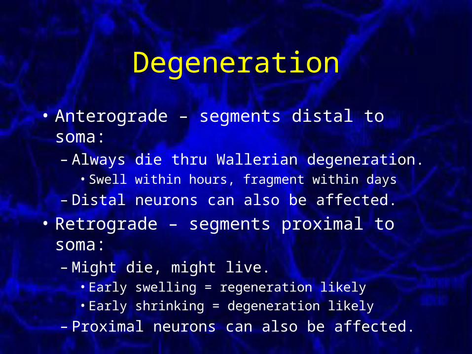
• Anterograde – segments distal to soma:– Always die thru Wallerian degeneration.
• Swell within hours, fragment within days
– Distal neurons can also be affected.
• Retrograde – segments proximal to soma:– Might die, might live.
• Early swelling = regeneration likely
• Early shrinking = degeneration likely
– Proximal neurons can also be affected.
Regeneration
• Damaged neurons may regrow.
• Regeneration is well developed in invertebrates.– Accurate in both the CNS and PNS.– Accurate even when not in a myelin channel.
• Mammals seem to lose their regeneration capabilities during maturation.– Almost non-existent in mammalian CNS.– Hit-or-miss in mammalian PNS.
Regeneration
• Regeneration in mammals depends on the balance of two competing factors:– Growth promoting factors (higher in PNS)
• Neural growth factor (NGF)
• Brain-derived neurotrophic factor (BDNF)
– Growth inhibiting factors (higher in CNS)• Nogo
• Myelin-associated glycoprotein (MAG)
• Chondroitin sulfate proteoglycan (CSPG)
• Oligodendrocyte myelin glycoprotein (OMG)
Regeneration in PNS
• If myelin sheaths remain intact, nerve can regrow at mm/day to original destination.
• If nerve sections are separated by more than a couple mm, they can grow back thru the wrong myelin tunnels.
• If nerves sections are widely separated, both sections will die, or regenerating axon tips will grow wildly into a spaghetti patch.
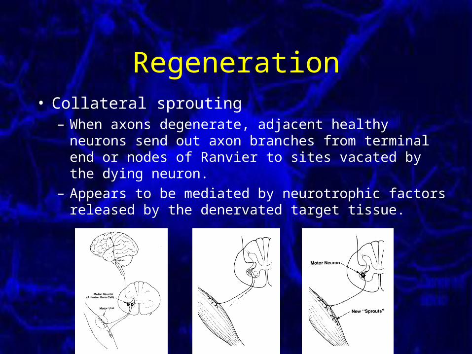
Regeneration• Collateral sprouting
– When axons degenerate, adjacent healthy neurons send out axon branches from terminal end or nodes of Ranvier to sites vacated by the dying neuron.
– Appears to be mediated by neurotrophic factors released by the denervated target tissue.
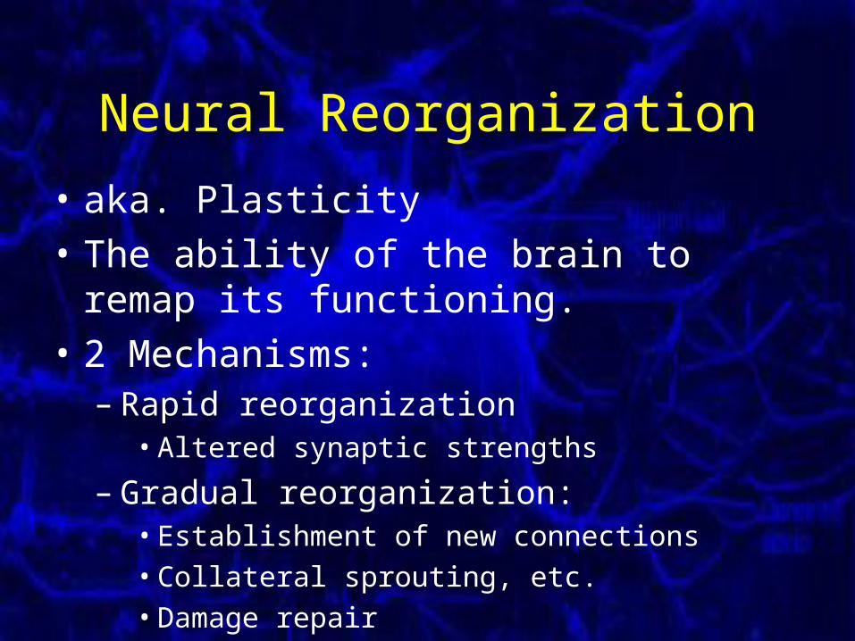
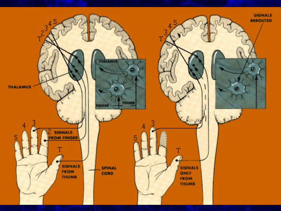
Neural Reorganization
• aka. Plasticity
• The ability of the brain to remap its functioning.
• 2 Mechanisms:– Rapid reorganization
• Altered synaptic strengths
– Gradual reorganization:• Establishment of new connections
• Collateral sprouting, etc.
• Damage repair
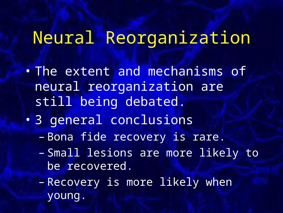
Neural Reorganization
• The extent and mechanisms of neural reorganization are still being debated.
• 3 general conclusions– Bona fide recovery is rare.– Small lesions are more likely to be recovered.– Recovery is more likely when young.
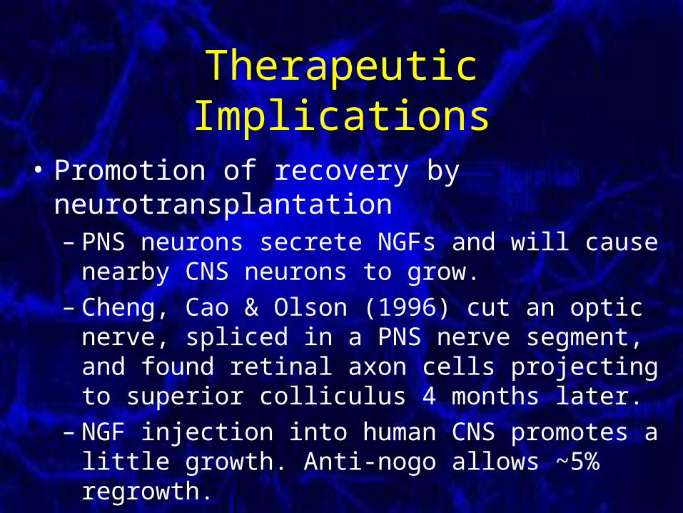
Therapeutic Implications
• Promotion of recovery by neurotransplantation– PNS neurons secrete NGFs and will cause nearby
CNS neurons to grow.– Cheng, Cao & Olson (1996) cut an optic nerve,
spliced in a PNS nerve segment, and found retinal axon cells projecting to superior colliculus 4 months later.
– NGF injection into human CNS promotes a little growth. Anti-nogo allows ~5% regrowth.
Therapeutic Implications
• Promotion of recovery by rehabilitative training.– At least in motor cortex, greater use = less loss
of function (or greater recovery).– Recovery of monkeys with hand motor cortex
lesions improved with hand motor tasks.– Humans with spinal cord injuries learn to walk
better with rehabilitative treatment than with just physiotherapy.
Therapeutic Implications
• Promotion of recovery by genetic engineering.– The promise of the future.– Since CNS nerves do not grow because of lack of
neural growth factor (NGF), the idea is to introduce some from stem cells or altered viruses.
– Nogo also prevents the growth of nerves. Maybe it can be artificially inhibited. Experimental nogo antagonists have been found.
Myelination Disorders
Myelination Disorders
• Dysmyelination – poor myelin formation– Early in life, mostly genetic.– Many disorders
• Many affect connection between myelin layers.
• Demyelination – myelin destroyed– Later in life, some genetic, mostly autoimmune.
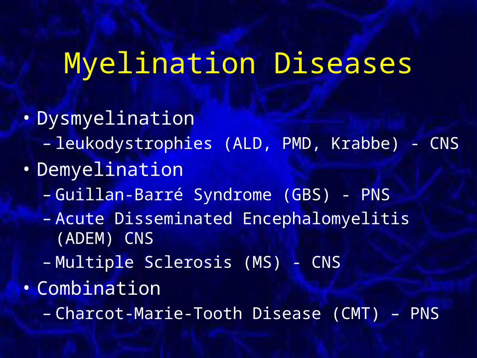
Myelination Diseases
• Dysmyelination– leukodystrophies (ALD, PMD, Krabbe) - CNS
• Demyelination– Guillan-Barré Syndrome (GBS) - PNS– Acute Disseminated Encephalomyelitis (ADEM) CNS– Multiple Sclerosis (MS) - CNS
• Combination– Charcot-Marie-Tooth Disease (CMT) – PNS
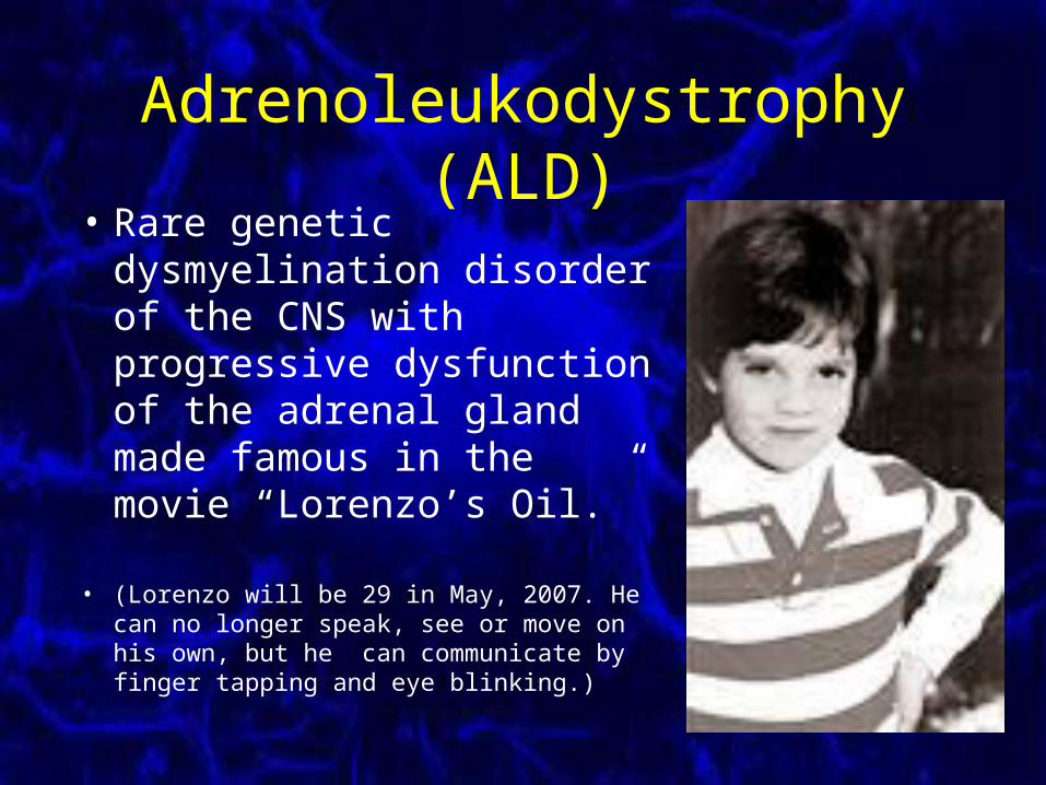
Adrenoleukodystrophy (ALD)• Rare genetic
dysmyelination disorder of the CNS with progressive dysfunction of the adrenal gland made famous in the movie “Lorenzo’s Oil.”
• (Lorenzo will be 29 in May, 2007. He can no longer speak, see or move on his own, but he can communicate by finger tapping and eye blinking.)

Adrenoleukodystrophy (ALD)• Etiology
– A genetic defect on Xq28 causes peroxisomes to lose the ability to breakdown very long chain fatty acids (VLCFAs). The accumulating VLCFAs degrade both myelin and the adrenal gland.
• Course– Poor prognosis, typical 1-10 years till death.
– Progresses to blind, mute and tetraplegic.
• “Treatment”– Lorenzo’s Oil, lovastatin, bone marrow transplant
Acute Disseminated Encephalomyelitis (ADEM)
• CNS demyelination, especially in corpus callosum and white matter in the brain around veins.– analogous to Guillan-Barré Syndrome in PNS.
• Probably autoimmune, often appears 1-3 weeks after an antecedent infection or vaccination.
• measles, mumps, rubella, varicella, smallpox, herpes, etc.
• Monophasic, 30% mortality or 2 week recovery.• A significant number will later develop MS.• SX: fever, visual neuritis, ataxia, stupor, seizures
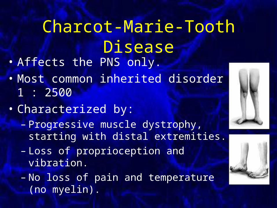
Charcot-Marie-Tooth Disease
• Affects the PNS only.
• Most common inherited disorder 1 : 2500
• Characterized by:– Progressive muscle dystrophy, starting with
distal extremities.– Loss of proprioception and vibration.– No loss of pain and temperature (no myelin).
Charcot-Marie-Tooth Disease• Hereditary
– Variety of genes, some dominant, some recessive.
• 2 major types:– Demyelinating or hypertrophic type
• The myelin dies off or doesn’t form properly.
• Includes CMT 1, 3, 4 and most X forms.
– Axonal type• The axon dies off, leaving the myelin intact.
• Includes CMT 2, 5, 6 and some X forms.
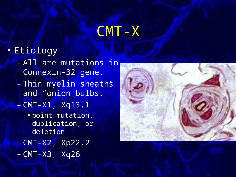
CMT-X• Etiology
– All are mutations in Connexin-32 gene.
– Thin myelin sheaths and “onion bulbs.”
– CMT-X1, Xq13.1• point mutation,
duplication, or deletion
– CMT-X2, Xp22.2– CMT-X3, Xq26
Multiple Sclerosis• Autoimmune disease of the CNS causes:
– Multiple white matter lesions (plaques). – Multiple episodes of neurological deficits.
• 1:1000, F = 2xM, MZ = 25%, onset 20-50
• Most common neurological disorder in young Canadians.
• Mild neuritis and paresthesias progress to ataxia and paraplegia, with cognitive problems.
Multiple Sclerosis
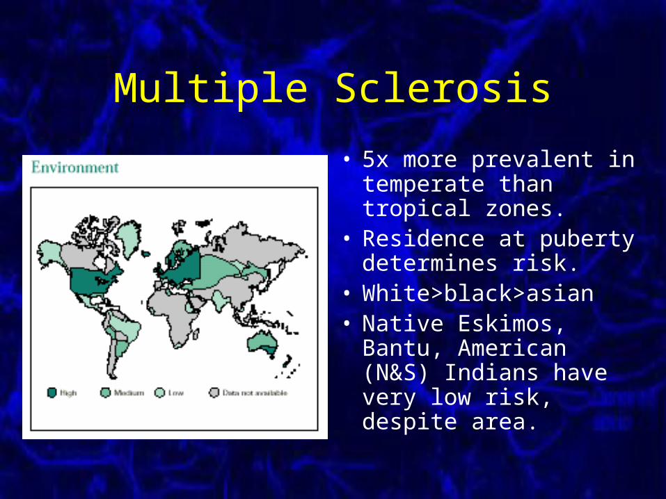
• 5x more prevalent in temperate than tropical zones.
• Residence at puberty determines risk.
• White>black>asian• Native Eskimos, Bantu,
American (N&S) Indians have very low risk, despite area.
Multiple Sclerosis• Etiology
– Autoimmune attack assumed.– There is some genetic and environmental
predisposition.– The DNA of HHV-6 (childhood roseola) has
been found in plaques.– Proteins of the coronavirus have been found to
mimic the structure of MBP.– Antibodies to nerve cells are often found in MS
patients.
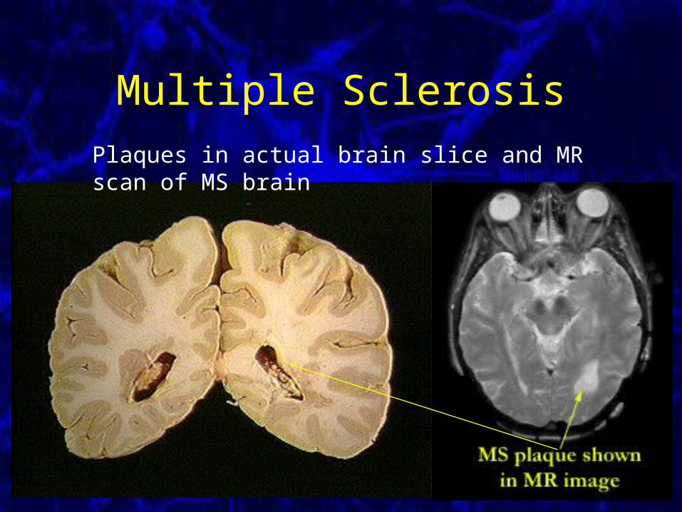
Multiple Sclerosis
Plaques in actual brain slice and MR scan of MS brain