brain inflammation is accompanied by peripheral
TRANSCRIPT
SHORT REPORT Open Access
Brain inflammation is accompanied byperipheral inflammation in Cstb−/− mice, amodel for progressive myoclonus epilepsyOlesya Okuneva1,2,3†, Zhilin Li3†, Inken Körber1,2,3*, Saara Tegelberg1,2,3, Tarja Joensuu1,2,3, Li Tian3,4
and Anna-Elina Lehesjoki1,2,3
Abstract
Progressive myoclonus epilepsy of Unverricht-Lundborg type (EPM1) is an autosomal recessively inherited childhood-onset neurodegenerative disorder, characterized by myoclonus, seizures, and ataxia. Mutations in the cystatin B gene(CSTB) underlie EPM1. The CSTB-deficient (Cstb−/−) mouse model recapitulates key features of EPM1, includingmyoclonic seizures. The mice show early microglial activation that precedes seizure onset and neuronal loss and leadsto neuroinflammation. We here characterized the inflammatory phenotype of Cstb−/− mice in more detail. We foundhigher concentrations of chemokines and pro-inflammatory cytokines in the serum of Cstb−/− mice and higher CXCL13expression in activated microglia in Cstb−/− compared to control mouse brains. The elevated chemokine levels werenot accompanied by blood-brain barrier disruption, despite increased brain vascularization. Macrophages in the spleenand brain of Cstb−/− mice were predominantly pro-inflammatory. Taken together, these data show that CXCL13expression is a hallmark of microglial activation in Cstb−/− mice and that the brain inflammation is linked to peripheralinflammatory changes, which might contribute to the disease pathology of EPM1.
Keywords: Cystatin B, Chemokine, CXCL13, Macrophage, M1/M2, Vascularization
IntroductionProgressive myoclonus epilepsy of Unverricht-Lundborgtype (EPM1, OMIM 254800) is an autosomal recessivelyinherited neurodegenerative disorder with onset from 6to 16 years of age and characterized by action-activatedand highly incapacitating myoclonus, tonic-clonic epilep-tic seizures, and ataxia [1]. EPM1 is caused by loss-of-function mutations in the cystatin B (CSTB) gene [2, 3],which encodes an inhibitor of lysosomal cysteine cathep-sins [4]. CSTB is highly expressed in immune cells, e.g., inblood leukocytes, hepatic lymphocytes, placental macro-phages, and microglia [5–9], and it is upregulated in vitroby pro-inflammatory stimulation [8, 10, 11]. In immunecells, the function of CSTB has been linked to chemotaxis[8], expression and secretion of cytokines, and release of
nitric oxide [10, 12, 13], implying a role in the immuneresponse. CSTB function has also been associated withdiverse cellular processes, such as regulation of apoptosis[14, 15], bone resorption [16, 17], protection of neuronsfrom oxidative stress [18], and cell cycle progression [19].A CSTB-deficient mouse model (Cstb−/−) mimics key
features of EPM1, including myoclonic seizures, ataxia[20], and progressive gray and white matter loss [21].The brain pathology of Cstb−/− mice is characterized bymicroglial activation in asymptomatic mice of 2 weeks ofage, followed by widespread activation of astrocytes aswell as progressive neuronal death and brain volume lossfrom 1 month of age onwards [22]. Moreover, activatedcultured Cstb−/− microglia secrete higher levels of che-mokines, such as chemokine (C-C motif ) ligand (CCL)2,CCL3, and chemokine (C-X-C motif ) ligand (CXCL)1,than control microglia [8]. Gene expression profiling ofcultured Cstb−/− microglia revealed impaired interferonsignaling and also showed altered chemokine expression[23]. Finally, a striking upregulation of Cxcl13 in gene
* Correspondence: [email protected]†Equal contributors1Folkhälsan Institute of Genetics, Haartmaninkatu 8, 00014 Helsinki, Finland2Research Program’s Unit, Molecular Neurology, University of Helsinki,Haartmaninkatu 8, 00014 Helsinki, FinlandFull list of author information is available at the end of the article
© The Author(s). 2016 Open Access This article is distributed under the terms of the Creative Commons Attribution 4.0International License (http://creativecommons.org/licenses/by/4.0/), which permits unrestricted use, distribution, andreproduction in any medium, provided you give appropriate credit to the original author(s) and the source, provide a link tothe Creative Commons license, and indicate if changes were made. The Creative Commons Public Domain Dedication waiver(http://creativecommons.org/publicdomain/zero/1.0/) applies to the data made available in this article, unless otherwise stated.
Okuneva et al. Journal of Neuroinflammation (2016) 13:298 DOI 10.1186/s12974-016-0764-7

expression profiling of postnatal day 30 (P30) Cstb−/−
mouse cerebellum was detected [24].We here confirm the increased CXCL13 expression
also on protein level and show that the inflammatoryprocesses in the Cstb−/− brain are linked to peripheralinflammation, which is characterized by increased levelsof chemokines and pro-inflammatory cytokines in theserum combined with relatively more pro-inflammatorymacrophages, and increased amounts of B lymphocytesin the spleen.
Materials and methodsMiceCSTB-deficient mice (Cstb−/−) were obtained from TheJackson Laboratory (129-Cstbtm1Rm/SvJ; stock no. #003486).Wild-type mice of the same age and background were usedas controls. The research protocols were approved by theAnimal Ethics Committee of the State Provincial Office ofSouthern Finland (decision no. ESAVI/7039/04.10.03/2012,ESAVI/5995/04.10.07/2013, and ESAVI/6288/04.10.07/2015).
Measurement of chemokines and cytokines in mouseserumBlood samples were obtained by intracardiac punctureof anesthetized P14 and P30 Cstb−/− and control mice.The blood was allowed to clot at room temperature (RT)for 15 min and centrifuged at 2000g for 13 min. Theserum was collected and kept at −80 °C until use. Thechemokine and cytokine concentrations were assessedusing a combination of mouse CXCL10, interleukin(IL)-1α, CXCL1, IL-6, IL-10, IL-18, IL-1β, IL-12, inter-feron (IFN)-γ, IFN-α, CCL2, CCL3, CCL4, tumor necrosisfactor α (TNFα), colony stimulating factor 2 (GM-CSF),and TGF-β1 FlowCytomix Simplex kits for flow cytometry(eBioscience). The CXCL13 concentration was determinedusing the Quantikine® mouse CXCL13/BLC/BCA-1 Im-munoassay ELISA kit (R&D Systems).
Tissue processing for histochemical analysisAnesthetized mice (150 mg/kg pentobarbital) were per-fused with phosphate-buffered saline (PBS) (pH 7.4) and4 % paraformaldehyde (PFA)/PBS for 10 min each. Thebrains were dissected, immersion fixed in 4 % PFA/PBSfor 48 h, and cryoprotected in 30 % sucrose/0.05 %NaN3/Tris-buffered saline (TBS) for 3 days. Coronal orsagittal 40-μm sections were cut using a cryostat LeicaCM3050 S (Leica Microsystems) and stored in 15 % su-crose/0.05 % NaN3/30 % ethylene glycol/TBS.
ImmunohistochemistryAdjacent 1-in-12 series of coronal free-floating sections(n = 5 per genotype and age) were incubated with 50 mMNH4Cl for 30 min to reduce non-specific background
staining and blocked with 15 % fetal calf serum (FCS)diluted in TBS/0.3 %Triton X-100 (TTX) for 1 h. Thesections were incubated with the primary antibodiesrabbit anti-ionized calcium-binding adaptor molecule 1(IBA1; Wako) combined with goat anti-CXCL13, goatanti-CXCL10 (both R&D Systems), or rabbit anti-CXCL1(Novus Biologicals) in 10 % FCS/TTX for 72 h at 4 °C.The secondary antibodies anti-rabbit Alexa Fluor 488 andanti-goat Alexa Fluor 594 (Invitrogen) were applied for2 h at RT, and mounted sections were examined using afluorescence microscope.
Evaluation of brain vascularityHistochemical detection of blood vessels was performedas described previously [25]. Adjacent 1-in-12 series ofsagittal free-floating sections of non-perfused Cstb−/−
and control brains (P14 and P30) were incubated in3,3′-diaminobenzidine (DAB) to detect endogenousperoxidase expression of erythrocytes. From each brain(n = 4 per genotype and age), eight sections were analyzed.Per each brain section, the vascularization was quantifiedfrom eight black and white bright-field images (×40, fivefrom cortex and three from cerebellum) as relative DAB-positive section area using ImageJ software.
Measurement of BBB permeabilityBlood-brain barrier (BBB) integrity was analyzed basedon its permeability for fluorescein [26, 27] and serum al-bumin [28]. To measure the fluorescein uptake into thebrain, Cstb−/− and control P30 mice were injected i.p.with 100 μl (5 ml per kg) of 100 mg/ml fluorescein so-dium salt (NaF, Sigma-Aldrich) in sterile PBS. After 1 h,the mice were perfused with PBS until the liquid leavingthe right atrium was colorless. The excised brains werefreed from the meninges and the fourth ventricular chor-oid plexus and weighed. After homogenization in 500 μlPBS and mixing with a vortex for 2 min, 500 μl of 60 %trichloroacetic acid (Sigma-Aldrich) was added to precipi-tate protein. Homogenized samples were kept at 4 °C for30 min and centrifuged at 18,000g at 4 °C for 10 min.Fluorescence intensity of the supernatants was measuredat excitation 440 nm and emission 525 nm using a micro-plate reader (WALLAC Victor 2). Fluorescein concen-trations were calculated based on a sodium fluoresceinstandard curve (10 to 200 ng/ml) and expressed asnanogram per milligram brain tissue [29]. For albuminstaining, adjacent 1-in-12 series of coronal free-floatingsections were incubated with 50 mM NH4Cl for30 min, blocked with 15 % FCS/TTX for 1 h, and incu-bated for 24 h at 4 °C protected from light with goatanti-mouse FITC-conjugated serum albumin IgG (AlphaDiagnostic International) diluted in 10 % FCS/TTX.Mounted sections were examined using a fluorescencemicroscope.
Okuneva et al. Journal of Neuroinflammation (2016) 13:298 Page 2 of 10

Isolation of brain mononuclear cells and nucleatedsplenocytesP14 and P30 mice were euthanized with CO2, perfusedwith ice-cold PBS, and the brain and spleen were dis-sected. Brain mononuclear cells were isolated as describedpreviously [8]. Splenocytes were collected from spleens bygently grinding through a 40-μm cell strainer, erythrolyzedusing VersaLyse lysing solution (Beckman Coulter), andwashed with ice-cold PBS.
Flow cytometryThe above isolated cells were blocked with 10 % normalrat serum/PBS on ice for 30 min. The brain mononuclearcells were stained with a combination of anti-mouseantibodies CD206-FITC +MHCII-PE + F4/80-PE/Cy7 +CD45-APC and the splenocytes with a combination ofCD11b-FITC + CD45-PE + F4/80-PE/Cy7 +Gr-1-APC orCD206-FITC +MHCII-PE + F4/80-PE/Cy7 + CD45-APC(all from BioLegend) on ice, protected from light, for30 min. Cells were washed and resuspended in 500 μlPBS/1 % FCS/0.02 % NaN3. The flow cytometric datawere acquired with a two-laser, six-color Gallios flowcytometer and analyzed by Kaluza analysis 1.3 software(Beckman Coulter). Brain mononuclear cells were definedas follows: microglia CD45+F4/80+, macrophages CD45hiF4/80+, M1-type macrophages CD45hiF4/80+MHCII+CD206−,and M2-type macrophages CD45hiF4/80+MHCII−/+CD206+.Splenocytes were defined as follows: granulocytesCD45+F4/80−/+Gr-1++, monocytes CD45+F4/80−Gr-1+,monocyte-derived macrophages CD45+CD11b+F4/80+Gr-1−, tissue-resident macrophages CD45+F4/80++Gr-1−/+,M1-type macrophages CD45+F4/80+MHCII+CD206−, andM2-type macrophages CD45+F4/80+MHCII−/+CD206+.Cell populations were calculated as percentages amongtotal leukocytes or macrophages.
Statistical analysesStatistical analyses were performed using unpaired, two-sided t test or two-way analysis of variance (ANOVA)test with Sidak’s multiple comparison test for comparisonbetween genotypes. All data are presented as mean ± SEMand a value of p < 0.05 is considered statisticallysignificant.Further methods are available in the Supporting Infor-
mation (Additional file 1).
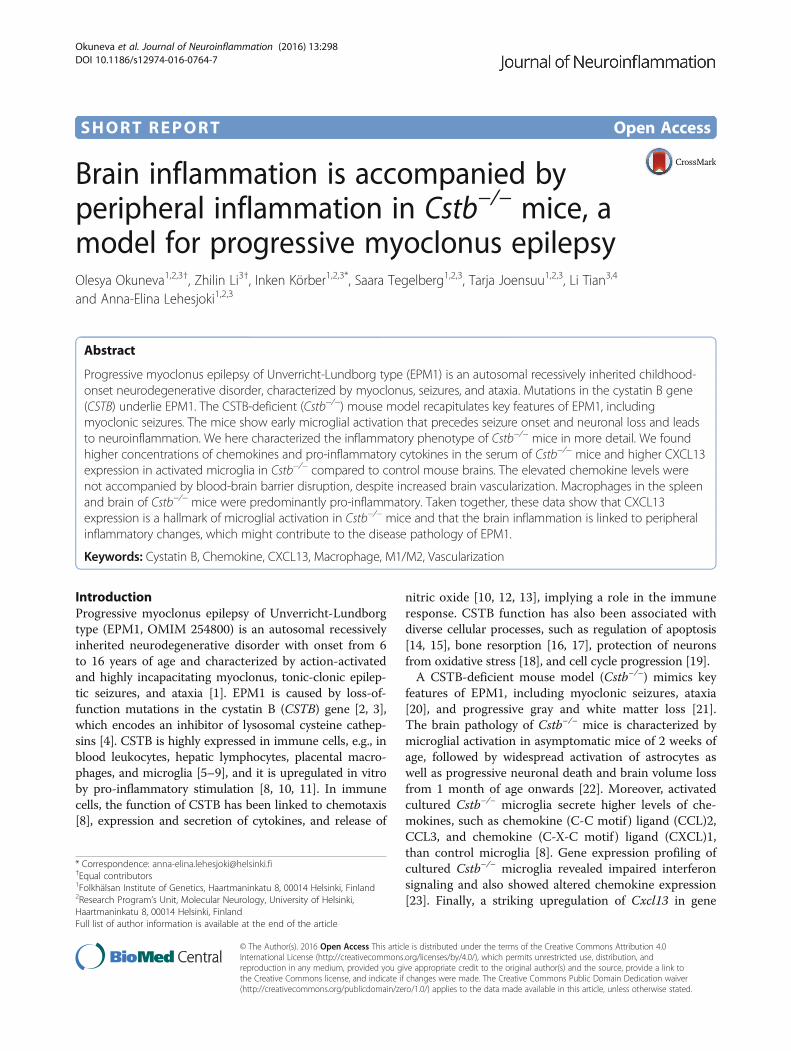
ResultsPro-inflammatory cytokine levels are high in the serum ofyoung Cstb−/− miceTo characterize peripheral inflammatory changes in pre-symptomatic and early symptomatic Cstb−/− mice, wedetermined the concentrations of 17 cytokines and che-mokines in the serum of Cstb−/− and control mice at P14and P30. At P14, the concentrations of pro-inflammatory
chemokines CXCL1 and CXCL10, as well as pro-inflammatory cytokines IL-1α and IL-18, were signifi-cantly higher in Cstb−/− than in control mice (Fig. 1a).In contrast, the concentration of anti-inflammatorycytokine TGF-β1 was reduced. The levels of CXCL1,CXCL10, and TNF-α were higher in the serum of P30Cstb−/− than in control mice, whereas the level of TGF-β1did not differ between genotypes (Fig. 1b). The level ofCXCL13 did not differ at P14, but was increased at P30.In conclusion, these data imply the presence of systemicinflammation, characterized by increased level of chemo-kines and pro-inflammatory cytokines already in pre-symptomatic Cstb−/− mice at P14.
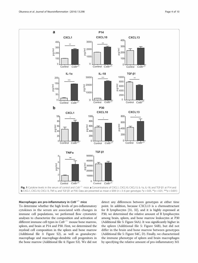
Expression of the pro-inflammatory chemokine CXCL13 ishighly increased in Cstb−/− microgliaAs the expression and secretion of chemokines have pre-viously been shown to be altered in cerebellar tissue andprimary microglia of Cstb−/− mice [8, 23, 24], we focusedour further analyses on brain expression of chemokinesCXCL1, CXCL10, and CXCL13, which were increased inthe sera of mice at P30. Using immunohistochemistry inCstb−/− and control mice, we did not detect expressionof CXCL1 and only low level of CXCL10 at P14 and P30(data not shown). Expression of CXCL13 was higher inCstb−/− than control brain at both time points (Figs. 2and 3). In P14 Cstb−/− brain tissue, CXCL13 immunopo-sitivity was restricted to the piriform cortex, the CA3area of the hippocampus, and the dorsal and ventral partof the anterior pretectal nucleus (Fig. 2), whereas theother cortical areas or the cerebellum did not expressCXCL13 (data not shown). At P30, CXCL13 was highlyexpressed also in other regions of the cortex and in thecerebellum (Fig. 3). CXCL13 immunopositivity co-localizedwith IBA1 immunopositivity, marking Cstb−/− microgliathat have an activated morphology.
Brain vascularization is enhanced and the BBB is intact inyoung Cstb−/− miceChemokines are involved in the regulation of angiogen-esis [30]. Therefore, we analyzed the vascularization inCstb−/− and control mice at P14 and P30 in non-perfusedbrains by determining the relative area positive for histo-chemical DAB staining, which detects endogenous erythro-cyte peroxidase (Fig. 4a). At P14, the extent of brainvascularization did not differ significantly between ge-notypes, but it was more intense in Cstb−/− than in controlmice at P30 (Fig. 4b). To determine whether this increasedvascularization is associated with higher BBB permeability,we measured the BBB integrity based on the presence ofperipherally injected sodium fluorescein or endogenousserum albumin in the brain tissue at P30. Neither methodrevealed differences in BBB permeability between Cstb−/−
and control mice (Additional file 2: Figure S1).
Okuneva et al. Journal of Neuroinflammation (2016) 13:298 Page 3 of 10
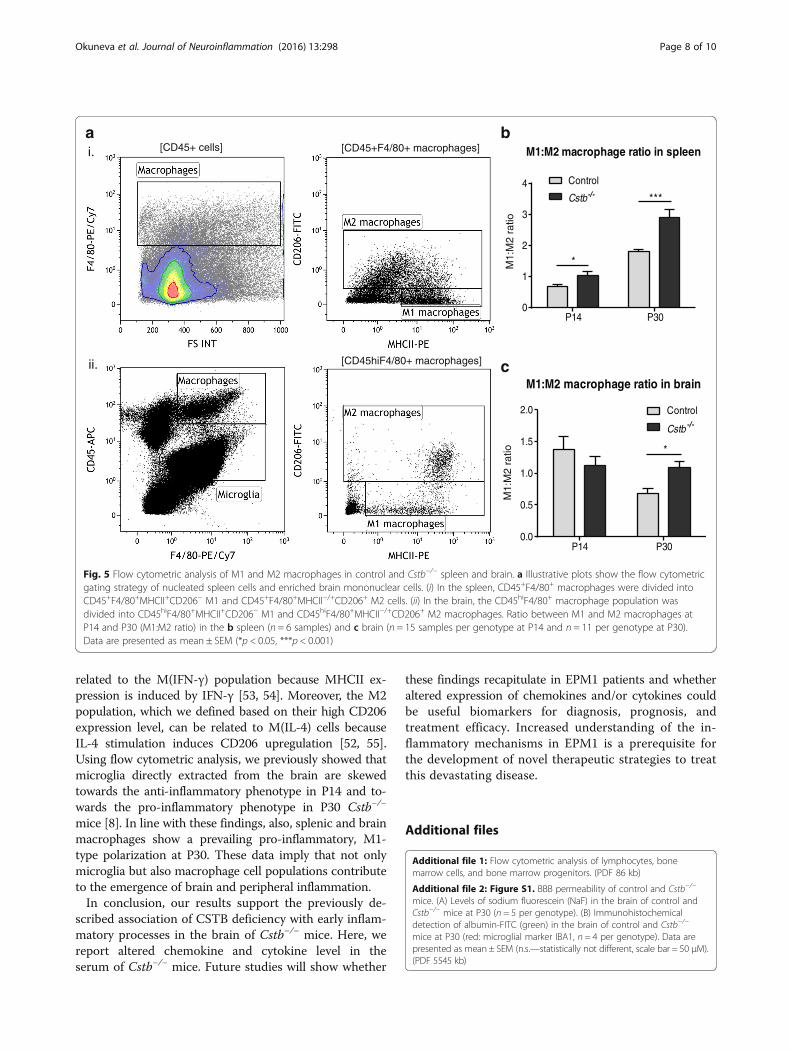
Macrophages are pro-inflammatory in Cstb−/− miceTo determine whether the high levels of pro-inflammatorycytokines in the serum are associated with changes inimmune cell populations, we performed flow cytometricanalyses to characterize the composition and activation ofdifferent immune cell types in Cstb−/− mouse bone marrow,spleen, and brain at P14 and P30. First, we determined themyeloid cell composition in the spleen and bone marrow(Additional file 3: Figure S2), as well as granulocyte-macrophage and macrophage-dendritic cell progenitors inthe bone marrow (Additional file 4: Figure S3). We did not
detect any differences between genotypes at either timepoint. In addition, because CXCL13 is a chemoattractantfor B lymphocytes [31, 32], and it is highly expressed atP30, we determined the relative amount of B lymphocytesamong brain, spleen, and bone marrow leukocytes at P30(Additional file 5: Figure S4A). It was significantly higher inthe spleen (Additional file 5: Figure S4B), but did notdiffer in the brain and bone marrow between genotypes(Additional file 5: Figure S4C, D). Finally, we characterizedthe immune phenotype of spleen and brain macrophagesby specifying the relative amount of pro-inflammatory M1
Fig. 1 Cytokine levels in the serum of control and Cstb−/− mice. a Concentrations of CXCL1, CXCL10, CXCL13, IL-1α, IL-18, and TGF-β1 at P14 andb CXCL1, CXCL10, CXCL13, TNF-α, and TGF-β1 at P30. Data are presented as mean ± SEM (n = 3–6 per genotype; *p < 0.05, **p < 0.01, ***p < 0.001)
Okuneva et al. Journal of Neuroinflammation (2016) 13:298 Page 4 of 10

and anti-inflammatory M2 macrophages from the totalamount of macrophages in each tissue (Fig. 5a andAdditional file 6: Figure S5) and determined the ratio(M1:M2) between both types. The ratio was higher inCstb−/− mice than in controls at P14 and P30 in thespleen (Fig. 5b). In the brain of P30 Cstb−/− mice, themacrophages were also more polarized towards thepro-inflammatory M1 type than control macrophages(Fig. 5d).
DiscussionIn this study, we show that altered levels of chemokinesin the serum and brain of young Cstb−/− mice, whichindicate systemic inflammation already in pre-symptomaticmice, is linked to increased brain vascularization in thepresence of a seemingly intact BBB. Moreover, we showthat high CXCL13 expression is a hallmark of activatedCstb−/− microglia and that macrophages in the Cstb−/−
spleen and brain are pro-inflammatory.
P14 Control piriform cortex
CXCL13
CXCL13
CXCL13
IBA1
IBA1
IBA1
P14 Cstb piriform cortex-/-
P14 Cstb CA3 hippocampus-/-
CXCL13 IBA1
P14 Control pretectum
CXCL13
CXCL13 IBA1
IBA1
P14 Cstb pretectum-/-
P14 Control CA3 hippocampus
CA3
Pir
APTD
APTV
CA3
Pir
APTD
APTV
CA3
Pir
APTD
APTV
i.
ii.
iii.
Fig. 2 Immunohistochemical detection of CXCL13 in control and Cstb−/− mouse brain at P14. CXCL13-positive microglia are shown by doubleimmunofluorescence staining of CXCL13 (red) with the microglial marker IBA1 (green) in the following brain areas: i piriform cortex, ii CA3 areaof the hippocampus, and iii pretectum of control and Cstb−/− mice. Representative CXCL13- and IBA1-double-positive cells in the merged imageare marked with arrows. Scale bar = 50 μM
Okuneva et al. Journal of Neuroinflammation (2016) 13:298 Page 5 of 10
Traumatic brain injury, epileptic seizures, ischemia,multiple sclerosis, and neurodegenerative diseases, whichare characterized by a higher prevalence or a reducedthreshold for seizures, are all associated with the expres-sion and secretion of cytokines [33, 34]. Cytokines andchemokines are released primarily by cells of the immunesystem and vascular endothelial cells, and they can activelycross the BBB or stimulate endothelial cells to express me-diators that activate brain cells [35, 36]. Previously, it hadbeen shown that the levels of pro-inflammatory cytokinesIL-18, IL-1β, and TNFα are increased in the serum ofadult Cstb−/− mice after peripheral LPS injection [12].Interestingly, we identified elevated levels of IL-18 andTNFα already in young Cstb−/− mice without activation ofinflammation with LPS, whereas no alterations in the levelof IL-1β were seen.
We also identified increased serum levels of chemo-kine CXCL1, CXCL10, and CXCL13 in Cstb−/− mice.Whether these chemokines are secreted from immunecells or endothelial cells requires further studies. Expres-sion of CXCL13, which binds CXCR4 receptor and regu-lates B cell migration [32], has been reported to beenhanced in inflammatory CNS diseases, such as multiplesclerosis and encephalitis [37–40]. Our previous gene ex-pression analysis of P30 Cstb−/− cerebellar tissue revealeda striking (29-fold) upregulation of Cxcl13 [24]. On thecontrary, in transcriptomics profiling of in vitro-culturedCstb−/− microglia, a slight downregulation of Cxcl13 wasobserved [23], suggesting that the CXCL13 upregulationin Cstb−/− microglia might be specific to the brain in vivo.In line with other studies, which have shown CXCL13expression in activated mouse microglia and in blood-
Fig. 3 Immunohistochemical detection of CXCL13 in control and Cstb−/− mouse brain at P30. CXCL13-positive microglia are shown by doubleimmunofluorescence staining of CXCL13 (red) with the microglial marker IBA1 (green) in the cortex and cerebellum of control and Cstb−/−
mice. Representative CXCL13- and IBA1-double-positive cells in the merged image are marked with arrows. The inserts show enlargements ofone double immuno-positive cell from both brain regions. Scale bar = 50 μM
Okuneva et al. Journal of Neuroinflammation (2016) 13:298 Page 6 of 10

derived human monocytes and macrophages [41–44],we detected increased expression of CXCL13 in IBA1-positive microglia. Therefore, CXCL13 serves as a markerfor activated microglia in Cstb−/− mice. Interestingly, theexpression of CXCL13 at P14 in Cstb−/− microglia was re-stricted to the piriform cortex, CA3 area of the hippocam-pus, and pretectum, but was more widespread at P30.Chemokines can regulate the integrity of the BBB
[45, 46]. In particular, they affect angiogenesis and BBBpermeability [30, 45, 46]. The chemokines CXCL10 andCXCL13 have been reported to be angiostatic, i.e., inhibit-ing the generation of vessels, whereas CXCL1 is angiogenicinducing vessel formation [30, 47]. Our results imply moreintense brain vascularization in P30 Cstb−/− mice, whichmight be mediated by the higher CXCL1 concentration inserum. In addition, CXCL10 and CXCL13 could be up-regulated in the serum and in the brain, respectively, tocounteract the angiogenic effect of CXCL1. Despite theelevated levels of cytokines in the serum of Cstb−/−
mice and the previously shown higher presence of mac-rophages, T cells, and granulocytes in the brain [8], wedid not detect a compromised BBB yet at P30. However,it is likely that the BBB integrity will be impaired inolder Cstb−/− mice as a consequence of a prolonged in-flammation in the brain.
Although CXCL13 has been reported to function as aB cell chemoattractant [31, 32], we did not detect agreater proportion of B cells in the Cstb−/− brain. In linewith our finding, the B cell infiltration after experimentalautoimmune encephalomyelitis has been shown to benormal in the brain of CXCL13-deficient mice [48]. Wedid find an increased B cell population in the spleen ofCstb−/− mice, but the mechanism and significance of thisfinding warrant further studies.In response to inflammatory stimuli or pathogens, micro-
glia and macrophages can be broadly classified intopro- (M1) or anti-inflammatory (M2) activated [49–51].Pro-inflammatory activation is linked to the release ofpro-inflammatory cytokines and mediators, whereas anti-inflammatory cells promote tissue repair and survival.However, microglia and macrophages adopt various inter-mediate phenotypes in vivo depending on the nature ofthe activating stimuli. Therefore, the M1-M2 classificationdoes not reflect the full spectrum of the intermediate andmutually non-exclusive “activation” states in vivo. A re-cent report by Murray et al. [52] revised the nomenclaturefor macrophages in vitro based on the activating stimuli.In relation to this framework, the M1 population in ourstudy, which we identified based on their low mannose re-ceptor (CD206) and high MHCII expression level, can be
Fig. 4 Brain vascularization of control and Cstb−/− mice. a Histochemical detection of brain vessels in the cortex of control and Cstb−/− mice atP14 and P30 was performed using DAB, which detects erythrocytes based on their endogenous peroxidase expression. b Vascularization is quantifiedat P14 and at P30 as relative DAB-positive area in 64 images from each of four control and four Cstb−/− brains. Data are presented as mean ±SEM (**p < 0.01, scale bar = 50 μM)
Okuneva et al. Journal of Neuroinflammation (2016) 13:298 Page 7 of 10
related to the M(IFN-γ) population because MHCII ex-pression is induced by IFN-γ [53, 54]. Moreover, the M2population, which we defined based on their high CD206expression level, can be related to M(IL-4) cells becauseIL-4 stimulation induces CD206 upregulation [52, 55].Using flow cytometric analysis, we previously showed thatmicroglia directly extracted from the brain are skewedtowards the anti-inflammatory phenotype in P14 and to-wards the pro-inflammatory phenotype in P30 Cstb−/−
mice [8]. In line with these findings, also, splenic and brainmacrophages show a prevailing pro-inflammatory, M1-type polarization at P30. These data imply that not onlymicroglia but also macrophage cell populations contributeto the emergence of brain and peripheral inflammation.In conclusion, our results support the previously de-
scribed association of CSTB deficiency with early inflam-matory processes in the brain of Cstb−/− mice. Here, wereport altered chemokine and cytokine level in theserum of Cstb−/− mice. Future studies will show whether
these findings recapitulate in EPM1 patients and whetheraltered expression of chemokines and/or cytokines couldbe useful biomarkers for diagnosis, prognosis, andtreatment efficacy. Increased understanding of the in-flammatory mechanisms in EPM1 is a prerequisite forthe development of novel therapeutic strategies to treatthis devastating disease.
Additional files
Additional file 1: Flow cytometric analysis of lymphocytes, bonemarrow cells, and bone marrow progenitors. (PDF 86 kb)
Additional file 2: Figure S1. BBB permeability of control and Cstb−/−
mice. (A) Levels of sodium fluorescein (NaF) in the brain of control andCstb−/− mice at P30 (n = 5 per genotype). (B) Immunohistochemicaldetection of albumin-FITC (green) in the brain of control and Cstb−/−
mice at P30 (red: microglial marker IBA1, n = 4 per genotype). Data arepresented as mean ± SEM (n.s.—statistically not different, scale bar = 50 μM).(PDF 5545 kb)
P14 P300
1
2
3
4
M1:M2 macrophage ratio in spleen
Control
Cstb-/- ***
*
P14 P300.0
0.5
1.0
1.5
2.0
M1:M2 macrophage ratio in brain
Control
Cstb -/-
*
c
b[CD45+F4/80+ macrophages]
[CD45hiF4/80+ macrophages]
M1:
M2
ratio
M1:
M2
ratio
a[CD45+ cells]i.
ii.
Fig. 5 Flow cytometric analysis of M1 and M2 macrophages in control and Cstb−/− spleen and brain. a Illustrative plots show the flow cytometricgating strategy of nucleated spleen cells and enriched brain mononuclear cells. (i) In the spleen, CD45+F4/80+ macrophages were divided intoCD45+F4/80+MHCII+CD206− M1 and CD45+F4/80+MHCII−/+CD206+ M2 cells. (ii) In the brain, the CD45hiF4/80+ macrophage population wasdivided into CD45hiF4/80+MHCII+CD206− M1 and CD45hiF4/80+MHCII−/+CD206+ M2 macrophages. Ratio between M1 and M2 macrophages atP14 and P30 (M1:M2 ratio) in the b spleen (n = 6 samples) and c brain (n = 15 samples per genotype at P14 and n = 11 per genotype at P30).Data are presented as mean ± SEM (*p < 0.05, ***p < 0.001)
Okuneva et al. Journal of Neuroinflammation (2016) 13:298 Page 8 of 10

Additional file 3: Figure S2. Flow cytometric analysis of myeloid cellsfrom control and Cstb−/− mouse spleen and bone marrow. (A) Illustrativeplots show the flow cytometric gating strategy of enriched nucleatedcells from spleen and bone marrow. In spleen, the CD45+ leukocyteswere divided into (i) CD45+F4/80−/+Gr-1++ granulocytes, CD45+F4/80−Gr-1+
monocytes, CD45+F4/80++Gr-1−/+ tissue-resident macrophages and (ii)CD45+CD11b+F4/80+Gr-1− monocyte-derived macrophages. In the bonemarrow, the CD45+ leukocytes were divided into (iii) CD45+F4/80−/+Gr-1++
granulocytes, CD45+F4/80−Gr-1+ monocytes, and CD45+F4/80+Gr-1−/+
macrophages. (B) Percentages of granulocytes, monocytes, and tissue-resident and monocyte-derived macrophages in the total CD45+
leukocyte population in spleen of control and Cstb−/− mice at P14 andP30. (C) Percentages of granulocytes, monocytes, and macrophages inthe total CD45+ leukocyte population in the bone marrow of control andCstb−/− mice at P14 and P30. Data are presented as mean ± SEM (n = 15samples per genotype at P14, and n = 11 samples per genotype at P30).(PDF 1769 kb)
Additional file 4: Figure S3. Analysis of granulocyte-macrophageprogenitors (GMP) and macrophage-dendritic cell progenitors (MDP)in control and Cstb−/− mice. (A) Illustrative plots show the flow cytometricgating strategy of progenitor cells from bone marrow. GMP cells arerepresented as Lin−c-Kit+Sca-1−CD16/32+CD115− and MDP cells asLin−c-Kit+Sca-1−CD16/32+CD115+. Percentages of (B) GMP and (C)MDP cells in the total bone marrow leucocytes of control and Cstb−/−
mice at P14 and P30. Data are presented as mean ± SEM (n = 6 samples pergenotype; each sample containing cells from one mouse). (PDF 1712 kb)
Additional file 5: Figure S4. Analysis of B lymphocytes from the spleen,brain, and bone marrow of control and Cstb−/− mice. (A) Illustrative plotsshow the flow cytometric gating strategy of lymphocytes from the (i) spleen,(ii) brain, and (iii) bone marrow. Percentage of B lymphocytes in the totalCD45+ cell population in the (B) spleen, (C) brain, and (D) bone marrow ofcontrol and Cstb−/− mice at P30. Data are presented as mean ± SEM (n = 5samples per genotype; each sample containing cells from one mouse;***p < 0.001). (PDF 3206 kb)
Additional file 6: Figure S5. Flow cytometric analysis of M1 and M2macrophages in control and Cstb−/− mouse spleen and brain. Percentagesof M1 and M2 macrophages in the total macrophage population in the(A and B) spleen and (C and D) brain of control and Cstb−/− mice at P14 andP30. (*p < 0.05, **p < 0.01, ***p < 0.001). (PDF 309 kb)
AbbreviationsBBB: Blood-brain barrier; CSTB: Cystatin B; EPM1: Progressive myoclonusepilepsy of Unverricht-Lundborg type
AcknowledgementsThe authors thank Ms. Paula Hakala for coordinating the mouse breeding.We acknowledge Dmitry Molotkov and the Biomedicum Imaging Unit stafffor microscopy service and assistance.
FundingThis work was supported by Folkhälsan Research Foundation, Academy ofFinland (project 1256107 and 1283085 (L.T)), Sigrid Jusélius Foundation,Medicinska Understödsföreningen Liv och Hälsa r.f (Life and Health MedicalFund), and the Doctoral Program in Biomedicine (I.K and Z.L).
Availability of data and materialsAll raw data in this manuscript are available on request.
Authors’ contributionsOO performed the serum cytokine and the vascularization assays, OO, IK, andST conducted the IHC and BBB assays, and ZL performed the flow cytometricexperiments. LT, TJ, and AEL were responsible for the study design andsupervision. OO, IK, and AEL wrote the manuscript. All authors critically revisedthe manuscript and approved the final version.
Competing interestsThe authors declare that they have no competing interests.
Consent for publicationNot applicable.
Ethics approvalAll research protocols were approved by the Animal Ethics Committee of theState Provincial Office of Southern Finland (decision no. ESAVI/7039/04.10.03/2012, ESAVI/5995/04.10.07/2013, and ESAVI/6288/04.10.07/2015).
Author details1Folkhälsan Institute of Genetics, Haartmaninkatu 8, 00014 Helsinki, Finland.2Research Program’s Unit, Molecular Neurology, University of Helsinki,Haartmaninkatu 8, 00014 Helsinki, Finland. 3Neuroscience Center, Universityof Helsinki, Viikinkaari 4, 00014 Helsinki, Finland. 4Beijing HuilongguanHospital, Peking University teaching hospital, Beijing, China.
Received: 27 September 2016 Accepted: 16 November 2016
References1. Kälviäinen R, Khyuppenen J, Koskenkorva P, Eriksson K, Vanninen R, Mervaala E.
Clinical picture of EPM1-Unverricht-Lundborg disease. Epilepsia. 2008;49:549–56.2. Lalioti MD, Scott HS, Antonarakis SE. What is expanded in progressive
myoclonus epilepsy? Nat Genet. 1997;17:17.3. Pennacchio LA, Myers RM. Isolation and characterization of the mouse
cystatin B gene. Genome Res. 1996;6:1103–9.4. Turk V, Bode W. The cystatins: protein inhibitors of cysteine proteinases.
FEBS Lett. 1991;285:213–9.5. Haves-Zburof D, Paperna T, Gour-Lavie A, Mandel I, Glass-Marmor L, Miller A.
Cathepsins and their endogenous inhibitors cystatins: expression andmodulation in multiple sclerosis. J Cell Mol Med. 2011;15:2421–9.
6. Lenarcic B, Krizaj I, Zunec P, Turk V. Differences in specificity for theinteractions of stefins A, B and D with cysteine proteinases. FEBS Lett.1996;395:113–8.
7. Luciano-Montalvo C, Ciborowski P, Duan F, Gendelman HE, Melendez LM.Proteomic analyses associate cystatin B with restricted HIV-1 replication inplacental macrophages. Placenta. 2008;29:1016–23.
8. Okuneva O, Körber I, Li Z, Tian L, Joensuu T, Kopra O, Lehesjoki AE.Abnormal microglial activation in the Cstb(−/−) mouse, a model forprogressive myoclonus epilepsy, EPM1. Glia. 2015;63:400–11.
9. Rinne A, Jarvinen M, Dorn A, Alavaikko M, Jokinen K, Hopsu-Havu VK.Low-molecular cysteine protease inhibitors in the human palatal tonsil.Anat Anz. 1986;161:215–30.
10. Maher K, Zavrsnik J, Jeric-Kokelj B, Vasiljeva O, Turk B, Kopitar-Jerala N.Decreased IL-10 expression in stefin B-deficient macrophages is regulatedby the MAP kinase and STAT-3 signaling pathways. FEBS Lett. 2014;588:720–6.
11. Suzuki T, Hashimoto S, Toyoda N, Nagai S, Yamazaki N, Dong HY, Sakai J,Yamashita T, Nukiwa T, Matsushima K. Comprehensive gene expressionprofile of LPS-stimulated human monocytes by SAGE. Blood. 2000;96:2584–91.
12. Maher K, Jeric Kokelj B, Butinar M, Mikhaylov G, Mancek-Keber M, Stoka V,Vasiljeva O, Turk B, Grigoryev SA, Kopitar-Jerala N. A role for stefin B(cystatin B) in inflammation and endotoxemia. J Biol Chem. 2014;289:31736–50.
13. Verdot L, Lalmanach G, Vercruysse V, Hartmann S, Lucius R, Hoebeke J,Gauthier F, Vray B. Cystatins up-regulate nitric oxide release frominterferon-gamma-activated mouse peritoneal macrophages. J Biol Chem.1996;271:28077–81.
14. Kopitar-Jerala N, Schweiger A, Myers RM, Turk V, Turk B. Sensitization ofstefin B-deficient thymocytes towards staurosporin-induced apoptosis isindependent of cysteine cathepsins. FEBS Lett. 2005;579:2149–55.
15. Sun L, Wu Z, Hayashi Y, Peters C, Tsuda M, Inoue K, Nakanishi H. Microglialcathepsin B contributes to the initiation of peripheral inflammation-inducedchronic pain. J Neurosci. 2012;32:11330–42.
16. Laitala-Leinonen T, Rinne R, Saukko P, Väänänen HK, Rinne A. Cystatin B asan intracellular modulator of bone resorption. Matrix Biol. 2006;25:149–57.
17. Manninen O, Puolakkainen T, Lehto J, Harittu E, Kallonen A, Peura M,Laitala-Leinonen T, Kopra O, Kiviranta R, Lehesjoki AE. Impaired osteoclasthomeostasis in the cystatin B-deficient mouse model of progressivemyoclonus epilepsy. Bone Reports. 2015;3:76–82.
18. Lehtinen MK, Tegelberg S, Schipper H, Su H, Zukor H, Manninen O, Kopra O,Joensuu T, Hakala P, Bonni A, Lehesjoki AE. Cystatin B deficiency sensitizesneurons to oxidative stress in progressive myoclonus epilepsy, EPM1. JNeurosci. 2009;29:5910–5.
Okuneva et al. Journal of Neuroinflammation (2016) 13:298 Page 9 of 10
19. Ceru S, Konjar S, Maher K, Repnik U, Krizaj I, Bencina M, Renko M, Nepveu A,Zerovnik E, Turk B, Kopitar-Jerala N. Stefin B interacts with histones andcathepsin L in the nucleus. J Biol Chem. 2010;285:10078–86.
20. Pennacchio LA, Bouley DM, Higgins KM, Scott MP, Noebels JL, Myers RM.Progressive ataxia, myoclonic epilepsy and cerebellar apoptosis in cystatinB-deficient mice. Nat Genet. 1998;20:251–8.
21. Manninen O, Koskenkorva P, Lehtimäki KK, Hyppönen J, Könönen M,Laitinen T, Kalimo H, Kopra O, Kälviäinen R, Gröhn O, et al. White matterdegeneration with Unverricht-Lundborg progressive myoclonus epilepsy:a translational diffusion-tensor imaging study in patients and cystatinB-deficient mice. Radiology. 2013;269:232–9.
22. Tegelberg S, Kopra O, Joensuu T, Cooper JD, Lehesjoki AE. Early microglialactivation precedes neuronal loss in the brain of the Cstb−/− mouse modelof progressive myoclonus epilepsy, EPM1. J Neuropathol Exp Neurol.2012;71:40–53.
23. Körber I, Katayama S, Einarsdottir E, Krjutškov K, Hakala P, Kere J, Lehesjoki AE,Joensuu T. Gene-expression profiling suggests impaired signaling via theinterferon pathway in Cstb−/− microglia. PLoS One. 2016;11:e0158195.
24. Joensuu T, Tegelberg S, Reinmaa E, Segerstråle M, Hakala P, Pehkonen H,Korpi ER, Tyynelä J, Taira T, Hovatta I, et al. Gene expression alterations inthe cerebellum and granule neurons of Cstb(−/−) mouse are associated withearly synaptic changes and inflammation. PLoS One. 2014;9:e89321.
25. Rigau V, Morin M, Rousset MC, de Bock F, Lebrun A, Coubes P, Picot MC,Baldy-Moulinier M, Bockaert J, Crespel A, Lerner-Natoli M. Angiogenesis isassociated with blood–brain barrier permeability in temporal lobe epilepsy.Brain. 2007;130:1942–56.
26. Kaya M, Ahishali B. Assessment of permeability in barrier type of endotheliumin brain using tracers: Evans blue, sodium fluorescein, and horseradishperoxidase. Methods Mol Biol. 2011;763:369–82.
27. Morrey JD, Olsen AL, Siddharthan V, Motter NE, Wang H, Taro BS, Chen D,Ruffner D, Hall JO. Increased blood-brain barrier permeability is not a primarydeterminant for lethality of West Nile virus infection in rodents. J Gen Virol.2008;89:467–73.
28. Garbuzova-Davis S, Louis MK, Haller EM, Derasari HM, Rawls AE, Sanberg PR.Blood-brain barrier impairment in an animal model of MPS III B. PLoS One.2011;6:e16601.
29. Aggarwal A, Khera A, Singh I, Sandhir R. S-nitrosoglutathione preventsblood-brain barrier disruption associated with increased matrixmetalloproteinase-9 activity in experimental diabetes. J Neurochem.2015;132:595–608.
30. Romagnani P, Lasagni L, Annunziato F, Serio M, Romagnani S. CXCchemokines: the regulatory link between inflammation and angiogenesis.Trends Immunol. 2004;25:201–9.
31. Gunn MD, Ngo VN, Ansel KM, Ekland EH, Cyster JG, Williams LT. A B-cell-homing chemokine made in lymphoid follicles activates Burkitt's lymphomareceptor-1. Nature. 1998;391:799–803.
32. Legler DF, Loetscher M, Roos RS, Clark-Lewis I, Baggiolini M, Moser B. Bcell-attracting chemokine 1, a human CXC chemokine expressed in lymphoidtissues, selectively attracts B lymphocytes via BLR1/CXCR5. J Exp Med.1998;187:655–60.
33. Galic MA, Riazi K, Pittman QJ. Cytokines and brain excitability. FrontNeuroendocrinol. 2012;33:116–25.
34. Murta V, Ferrari CC. Influence of peripheral inflammation on the progressionof multiple sclerosis: evidence from the clinic and experimental animalmodels. Mol Cell Neurosci. 2013;53:6–13.
35. Banks WA. The blood-brain barrier in neuroimmunology: tales of separationand assimilation. Brain Behav Immun. 2015;44:1–8.
36. Rochfort KD, Cummins PM. The blood-brain barrier endothelium: a targetfor pro-inflammatory cytokines. Biochem Soc Trans. 2015;43:702–6.
37. Festa ED, Hankiewicz K, Kim S, Skurnick J, Wolansky LJ, Cook SD, Cadavid D.Serum levels of CXCL13 are elevated in active multiple sclerosis. Mult Scler.2009;15:1271–9.
38. Kothur K, Wienholt L, Mohammad SS, Tantsis EM, Pillai S, Britton PN, Jones CA,Angiti RR, Barnes EH, Schlub T, et al. Utility of CSF cytokine/chemokines asmarkers of active intrathecal inflammation: comparison of demyelinating.Anti-NMDAR and Enteroviral Encephalitis. PLoS One. 2016;11:e0161656.
39. Kuenz B, Lutterotti A, Ehling R, Gneiss C, Haemmerle M, Rainer C,Deisenhammer F, Schocke M, Berger T, Reindl M. Cerebrospinal fluid B cellscorrelate with early brain inflammation in multiple sclerosis. PLoS One. 2008;3:e2559.
40. Liba Z, Kayserova J, Elisak M, Marusic P, Nohejlova H, Hanzalova J, Komarek V,Sediva A. Anti-N-methyl-D-aspartate receptor encephalitis: the clinical course inlight of the chemokine and cytokine levels in cerebrospinal fluid. JNeuroinflammation. 2016;13:55.
41. Chapman KZ, Ge R, Monni E, Tatarishvili J, Ahlenius H, Arvidsson A, Ekdahl CT,Lindvall O, Kokaia Z. Inflammation without neuronal death triggers striatalneurogenesis comparable to stroke. Neurobiol Dis. 2015;83:1–15.
42. Esen N, Rainey-Barger EK, Huber AK, Blakely PK, Irani DN. Type-I interferonssuppress microglial production of the lymphoid chemokine, CXCL13. Glia.2014;62:1452–62.
43. Huang C, Sakry D, Menzel L, Dangel L, Sebastiani A, Krämer T, Karram K,Engelhard K, Trotter J, Schäfer MK. Lack of NG2 exacerbates neurologicaloutcome and modulates glial responses after traumatic brain injury. Glia.2016;64:507–23.
44. Krumbholz M, Theil D, Cepok S, Hemmer B, Kivisäkk P, Ransohoff RM,Hofbauer M, Farina C, Derfuss T, Hartle C, et al. Chemokines in multiplesclerosis: CXCL12 and CXCL13 up-regulation is differentially linked to CNSimmune cell recruitment. Brain. 2006;129:200–11.
45. Chai Q, She R, Huang Y, Fu ZF. Expression of neuronal CXCL10 induced byrabies virus infection initiates infiltration of inflammatory cells, production ofchemokines and cytokines, and enhancement of blood–brain barrierpermeability. J Virol. 2015;89:870–6.
46. Roberts TK, Eugenin EA, Lopez L, Romero IA, Weksler BB, Couraud PO,Berman JW. CCL2 disrupts the adherens junction: implications forneuroinflammation. Lab Invest. 2012;92:1213–33.
47. Scapini P, Morini M, Tecchio C, Minghelli S, Di Carlo E, Tanghetti E, Albini A,Lowell C, Berton G, Noonan DM, Cassatella MA. CXCL1/macrophageinflammatory protein-2-induced angiogenesis in vivo is mediated byneutrophil-derived vascular endothelial growth factor-A. J Immunol.2004;172:5034–40.
48. Rainey-Barger EK, Rumble JM, Lalor SJ, Esen N, Segal BM, Irani DN. Thelymphoid chemokine, CXCL13, is dispensable for the initial recruitment of Bcells to the acutely inflamed central nervous system. Brain Behav Immun.2011;25:922–31.
49. Butovsky O, Talpalar AE, Ben-Yaakov K, Schwartz M. Activation of microgliaby aggregated beta-amyloid or lipopolysaccharide impairs MHC-II expressionand renders them cytotoxic whereas IFN-gamma and IL-4 render themprotective. Mol Cell Neurosci. 2005;29:381–93.
50. Mills CD, Kincaid K, Alt JM, Heilman MJ, Hill AM. M-1/M-2 macrophages andthe Th1/Th2 paradigm. J Immunol. 2000;164:6166–73.
51. Mulder R, Banete A, Basta S. Spleen-derived macrophages are readily polarizedinto classically activated (M1) or alternatively activated (M2) states.Immunobiology. 2014;219:737–45.
52. Murray PJ, Allen JE, Biswas SK, Fisher EA, Gilroy DW, Goerdt S, Gordon S,Hamilton JA, Ivashkiv LB, Lawrence T, et al. Macrophage activation andpolarization: nomenclature and experimental guidelines. Immunity.2014;41:14–20.
53. Goñalons E, Barrachina M, García-Sanz JA, Celada A. Translational control ofMHC class II I-A molecules by IFN-gamma. J Immunol. 1998;161:1837–43.
54. King DP, Jones PP. Induction of Ia and H-2 antigens on a macrophage cellline by immune interferon. J Immunol. 1983;131:315–8.
55. Stein M, Keshav S, Harris N, Gordon S. Interleukin 4 potently enhancesmurine macrophage mannose receptor activity: a marker of alternativeimmunologic macrophage activation. J Exp Med. 1992;176:287–92.
Okuneva et al. Journal of Neuroinflammation (2016) 13:298 Page 10 of 10