brain mr image specification using neural network
DESCRIPTION
IOSR Journal of Computer Engineering (IOSR-JCE) vol.17 issue.3 version.6TRANSCRIPT

7/18/2019 Brain MR Image Specification Using Neural Network
http://slidepdf.com/reader/full/brain-mr-image-specification-using-neural-network 1/6
IOSR Journal of Computer Engineering (IOSR-JCE)e-ISSN: 2278-0661,p-ISSN: 2278-8727, Volume 17, Issue 3, Ver. VI (May – Jun. 2015), PP 52-57www.iosrjournals.org
DOI: 10.9790/0661-17365257 www.iosrjournals.org 52 | Page
Brain MR Image Specification Using Neural Network
Sonali B. Gaikwad1
, Prof. Madhuri S. Joshi2
1,2(CSE department, JNEC/BAMU University, Aurangabad, Maharashtra, India)
Abstract: Abnormal growth of the cell in the brain is the brain tumor. Brain tumor is common and serious
disease. The proposed method for tumor classification in magnetic resonance brain image is the humaninspection. Magnetic Resonance Imaging (MRI) plays an intrinsic role in the brain tumor disease diagnostic
application. Various types of tumor that leads decision complicated. So that correct classification of brain
tumor is important to detect the types of tumor. In this paper, Probabilistic Neural network (PNN) is used for
brain tumor classification. Decision making was performed in two steps: 1) Feature extraction using Principal
Component Analysis (PCA). And 2) Classification is done by Probabilistic neural network (PNN). Brain tumor
is classified into three classes: Normal, Benign and Malignant. PNN is faster and provide good classification
accuracy.
Keywords: Brain Tumor Classification, Principle Component Analysis (PCA), Probabilistic Neural Network
(PNN), MRI .
I. IntroductionA brain tumor is an intracranial solid neoplasm or abnormal growth of cells within the brain or the
central spinal canal [4]. The main purpose of brain tumor classification is correctly classify the MR image inorder to detect which type of tumor that suffered by the patients. Due to tumor there is problem of blood
circulation in the brain. Therefore blood tubercles are forms. There are many kind of test are used to detect brain
tumor such as MRI, Computed Tomography (CT) scan, Biopsy and many more. Among the entire test MRI has
great potential for classification of tumor. MRI, in comparison with other diagnostic imaging modalities, such as
computerized tomography, provides superior contrast and resolution for different brain tissues [1]. Additionally,
MR images encapsulate valuable information regarding numerous tissue parameters (proton density, spin-lattice
(T1) and spin – spin (T2) relaxation times, flow velocity, and chemical shift), which lead to more accurate brain
tissue characterization [3].The correct classification approach leads to the right decision and provide respective treatment to the
patients. The key of brain tumor cure is to detect the tumor in its early stage. So that good classification is
required. There are various methods to detect brain tumor. In this paper, we studied Probabilistic Neural
Network (PNN) algorithm for classification of brain tumor.
In proposed method, the first stage is to extract features from brain MR images using PCA and then
train the PNN for classification. The principal component analysis (PCA) method transforms the existing
attributes into new ones considered to be crucial in classification [2]. Multiclass classification models for
classifying brain tumor are proposed by [1], [3], [7].
The paper is organized in several sections: In the first section, a brief review about brain tumor. In thesecond section, we discussed Principal Component Analysis for feature extraction. PNN are discussed and
implemented in the third section. Methodology of the proposed system is discussed in fourth section. Dataset are
discussed in fifth section. In the final section we discussed experimentation and results.
II. Principal Component Analysis
In this paper, features are extracted using PCA. The principal Component Analysis (PCA) is one of themost successful techniques that have been used in image recognition and compression [1]. The purpose of PCA
is to reduce the large dimensionality of the data [1].
Feature extractions are done for training as well as testing phase. The main purpose of MR image
recognition system is to identify maximum similarities between training MR images and test MR image.
In the training phase, feature vectors are extracted from each training MR images. Let Ω1 be a training image of
image 1 which has a pixel resolution of M x N ( M rows, N columns). In order to extract PCA features of Ω1, first
convert the image into a pixel vector Φ 1 by concatenating each of the M rows into a single vector. The length
(or , dimensionality) of the vector Φ1 will be M x N . In this paper, the PCA algorithm is used as a dimensionality
reduction technique which transforms the vector Φ1 to a vector ω1 which has a dimensionality d where d << M x
N . For each training image Ωi, these feature vectors ωi are calculated and stored.
7/18/2019 Brain MR Image Specification Using Neural Network
http://slidepdf.com/reader/full/brain-mr-image-specification-using-neural-network 2/6
Brain MR Image Specification Using Neural Network
DOI: 10.9790/0661-17365257 www.iosrjournals.org 53 | Page
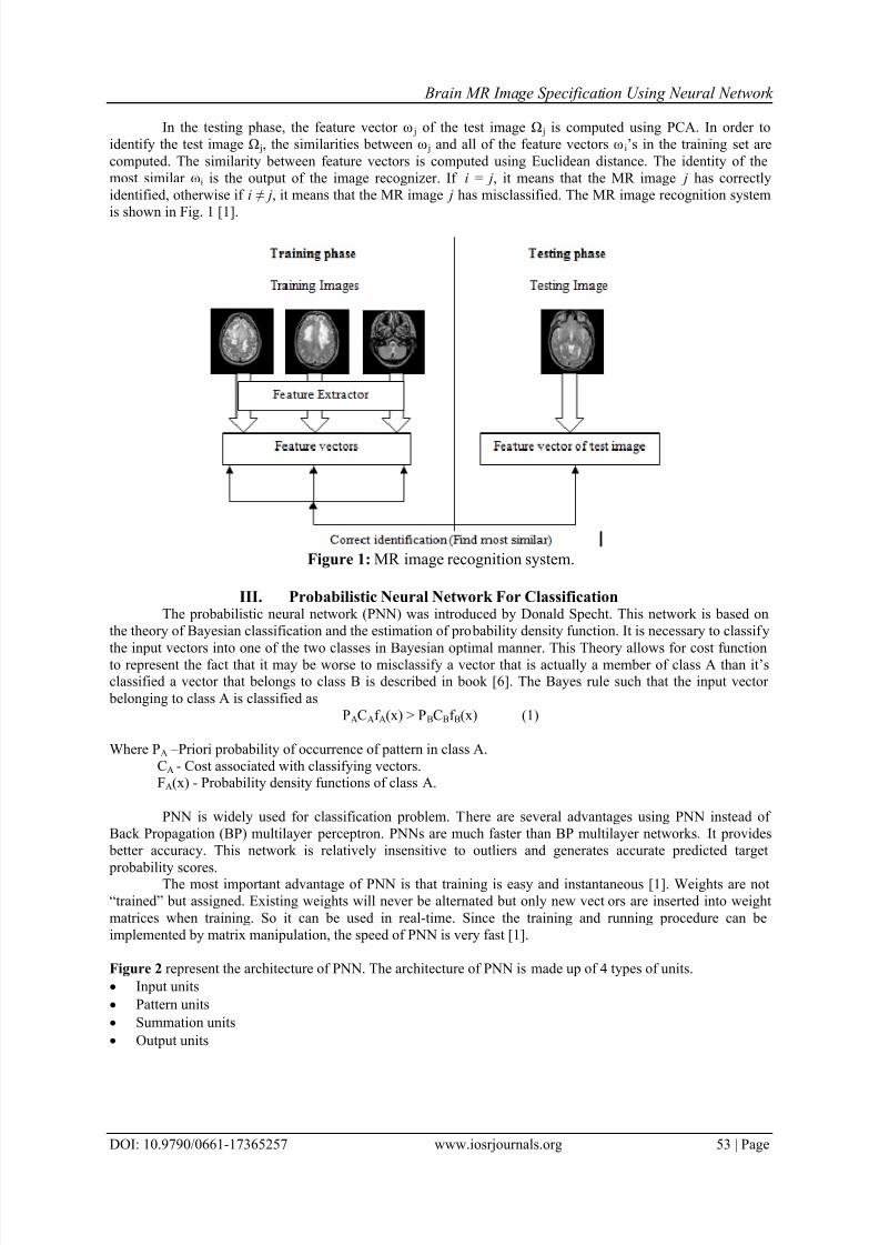
In the testing phase, the feature vector ω j of the test image Ω j is computed using PCA. In order to
identify the test image Ω j, the similarities between ω j and all of the feature vectors ωi’s in the training set are
computed. The similarity between feature vectors is computed using Euclidean distance. The identity of the
most similar ωi is the output of the image recognizer. If i = j, it means that the MR image j has correctly
identified, otherwise if i ≠ j, it means that the MR image j has misclassified. The MR image recognition system
is shown in Fig. 1 [1].
Figure 1: MR image recognition system.
III. Probabilistic Neural Network For ClassificationThe probabilistic neural network (PNN) was introduced by Donald Specht. This network is based on
the theory of Bayesian classification and the estimation of probability density function. It is necessary to classify
the input vectors into one of the two classes in Bayesian optimal manner. This Theory allows for cost functionto represent the fact that it may be worse to misclassify a vector that is actually a member of class A than it’sclassified a vector that belongs to class B is described in book [6]. The Bayes rule such that the input vector
belonging to class A is classified as
PACAf A(x) > PBCBf B(x) (1)
Where PA – Priori probability of occurrence of pattern in class A.
CA - Cost associated with classifying vectors.FA(x) - Probability density functions of class A.
PNN is widely used for classification problem. There are several advantages using PNN instead of
Back Propagation (BP) multilayer perceptron. PNNs are much faster than BP multilayer networks. It provides
better accuracy. This network is relatively insensitive to outliers and generates accurate predicted target
probability scores.The most important advantage of PNN is that training is easy and instantaneous [1]. Weights are not
“trained” but assigned. Existing weights will never be alternated but only new vectors are inserted into weight
matrices when training. So it can be used in real-time. Since the training and running procedure can be
implemented by matrix manipulation, the speed of PNN is very fast [1].
Figure 2 represent the architecture of PNN. The architecture of PNN is made up of 4 types of units.
Input units
Pattern units
Summation units
Output units

7/18/2019 Brain MR Image Specification Using Neural Network
http://slidepdf.com/reader/full/brain-mr-image-specification-using-neural-network 3/6
Brain MR Image Specification Using Neural Network
DOI: 10.9790/0661-17365257 www.iosrjournals.org 54 | Page
Figure 2: Architecture of PNN.
I. Input units: Hear input unit x(p), p=1,2,…..P are connected to all pattern units.
II. Pattern units: Create pattern unit ZP .Weight vector for unit ZP is computed as WP=x(p). Unit ZP is either
ZA or ZB unit. III. Summation units: Connect the pattern unit to summation unit. If x(p) belongs to class A, connect pattern
unit ZP to summation unit SA. Else connect pattern unit ZP to summation unit SB. The weight used by the
summation unit for class B is
VB= - PBCBmA (2)
PACAmB
IV. Output units: It sums the signals from f A and f B. Input vector is classified as class A if the total input to
decision unit is positive. y is the final output.
V.
IV. Methodology
The main purpose of proposed method to detect tumor automatically from the MR images.
The PNNclassifier presented good accuracy, very small training time, robustness to weight changes, and negligible
retraining time [1].The proposed method is made up of six steps which are starting from input data to output. In
the first step, use MR image as input image. Convert input image into graylevel and resize it into 256 x 256 size.
This process is performed in second step. In the third step, perform PCA for feature extraction. Features are
extracted from training as well as testing MR images. In the forth step, PNN is train for classification of MR
images. Finally performance based on the result will be analyzed at the end of the development phase. The
proposed brain tumor classification method is shown in Fig. 3. The final output of the proposed method is either
normal, benign or malignant.
Figure 3: The proposed method of PCA-PNN.

7/18/2019 Brain MR Image Specification Using Neural Network
http://slidepdf.com/reader/full/brain-mr-image-specification-using-neural-network 4/6
Brain MR Image Specification Using Neural Network
DOI: 10.9790/0661-17365257 www.iosrjournals.org 55 | Page
V. DatasetIn this study, it has obtained some brain MR images from Government hospital of Aurangabad and
most of the images from Sahyadri Hospital of Pune, Maharashtra, India. Dataset contains 70 training samples(Normal=25, Benign=25, and Malignant=20) and 35 testing samples for classify tumor as normal, benign, or
malignant.
VI. Results And DiscusionsIn this proposed method, various individualize experiments were performed and the sizes of the
training and testing sets were determined the classification accuracy.
The dataset was divided into two datasets- one is the training and another is testing dataset. The first
training dataset was used to train MR images for normal, benign and malignant. This dataset contain 90 MR
images. The testing dataset was used to verify the accuracy of trained PNN network for brain tumor
classification.
The MATLAB R2009a is used to implement PNN. In this proposed method smoothing factor was used to
examine the accuracy of the classifier. The spread value (SV) was used as smoothing factor and examined the
classifier accuracy when different values of spread value were used. If S.V. is near zero, the network will act as
a nearest neighbour classifier, and the network will take into account several nearby design vectors if its value
becomes larger [1].
In this study, PNN were examined using testing datasets. Clinical and performance measurements withrespect to normal, benign and malignant are shown in Table 1 and Table 2 respectively. In both the table the
accuracy of the classifier was examined for different values of the S.V.
True positives (TP) refer to the positive tuples that were correctly labeled by the classifier, while true negatives
(TN) are the negative tuples that were correctly labeled by the classifier. False positives (FP) are the negative
tuples that were incorrectly labelled. Similarly false negatives (FN) are the positive tuples that were incorrectlylabeled [5].
Table 1. Clinical measurements of PNN with respect to normal, benign and malignant.
The sensitivity and specificity measures can be used for correct classification. Sensitivity is also
referred to as the true positive (recognition) rate (that is, the proportion of positive tuples that are correctly
identified), while specificity is the true negative rate (that is, the proportion of negative tuples that are correctly
identified) [5]. These measures are defined as
Sensitivity = t_pos (3)
pos
Specificity = t_neg (4)
neg
The accuracy is a function of sensitivity and specificity:
Accuracy=Sensitivity pos + Specificity neg (5)
(pos+neg) (pos+neg)

7/18/2019 Brain MR Image Specification Using Neural Network
http://slidepdf.com/reader/full/brain-mr-image-specification-using-neural-network 5/6
Brain MR Image Specification Using Neural Network
DOI: 10.9790/0661-17365257 www.iosrjournals.org 56 | Page
Table 2. Performance measurements of PNN with respect to normal, benign and malignant.
In Table 2, 70 training samples and 35 testing samples have been used. The classifier accuracy varies
from 57% to 97.14%. With 90 training samples and 35 testing samples, the results remain unchanged.Therefore 70 training samples can suffice to get the acceptable accuracy and it also reduces the classifier
training time.
Where S.V. =107, accuracy is 97.14 %.
In this work, Table 1 and Table 2 shows clinical and performance measurements related to normal,
benign and malignant. PNN was developed to examine the accuracy with different spread value ranges from 102
to 107as shown in Table 2. Classifier accuracy reached to 97.14% when S. V. was 10
7.
Figure 4 show normal MR image which indicate MR brain image without tumor. Figure 5 and Figure 6
represent benign and malignant MR images.
Figure 7 shown the accuracy with respect to S.V for different classes of brain tumor.
Figure 4: Normal MR image. Figure 5: Benign MR image. Figure 6: Malignant MR image.
Figure 7: Accuracy of normal, benign and malignant.

7/18/2019 Brain MR Image Specification Using Neural Network
http://slidepdf.com/reader/full/brain-mr-image-specification-using-neural-network 6/6
Brain MR Image Specification Using Neural Network
DOI: 10.9790/0661-17365257 www.iosrjournals.org 57 | Page
VII. ConclusionIn this paper brain tumor MR image is automatically classified into normal, benign and malignant
category using PNN classifier. Maximum accuracy of 97.14% is obtained with spread value =107 as shown in
Table 2. This work will act as supportive tool for radiologists and will help doctor for fast diagnosis based on
which the treatment plan can be decided.
ReferencesJournal Papers:[1]. M.F. Othman and M. A. M. Basri, “Probabilistic Neural Network for brain tumor Classification”, IEEE Second International
Conference on Intelligent Systems, Modelling and Simulation, 136 – 138, 2011.
[2]. N. Kwak, and C. H. Choi, “Input Feature Selection for Classification Problems”, IEEE Transactions on Neural Networks,
13(1), 143 – 159, 2002.[3]. P. Georgiadis and G. Kagadis, “Non-linear Least Squares Features Transformation for Improving the Performance of Probabilistic
Neural Networks in Classifying Human Brain Tumors on MRI” ICCSA 2007, LNCS 4707, pp. 239 – 247, 2007, Springer-Verlag
Berlin Heidelberg 2007.[4]. D. SRIDHAR, “Brain Tumor Classification Using Discrete Cosine Transform and Probabilistic Neural Network”, 2013 IEEE
International Conference on Signal Processing, Image Processing and Pattern Recognition, 978-1-4673-4862-1/13, 2013 IEEE.
[5]. Kumar, J. Sachdeva, “Classification of brain tumors using PCA-ANN” 978-1-4673-0126-8/11 2011 IEEE.
Books:
[1].
J. Han and M. Kamber, “Data Mining: Concepts and Techniques”, 2006. [2]. S. N. Sivanandam, S. N Deepa, “Introduction to Neural Network using MATLAB 6.0”, 2006.