brain research center in the world - mcgill...
TRANSCRIPT

Brain research center Brain research center in the Worldin the World
Top 11Top 11Lingzhong.Fan

Laboratory of Neuro Imaging, UCLAMartinos Center for Biomedical Imaging, HarvardVan Essen Lab,Washington University in St. Louis The Section for Biomedical Image Analysis, PennResearch Imaging Institute, University of Texas McConnell Brain Imaging Center, McGill Wellcome Trust Centre for Neuroimaging, UCL FMRIB Center, OxfordMRC Cognition and Brain Sciences Unit, CambridgeMax Planck Institute for Human Cognitive and Brain Sciences Institute of Neuroscience and Medicine (INM), Julich
OutlineOutline

ICBMICBM
• International Consortium for Brain Mapping (ICBM)
– The International Consortium for Brain Mapping (ICBM) was formed in 1993 with a grant from the NIMH.
– This consortium is composed of four core research sites, UCLA, Montreal Neurologic Institute, University of Texas at San Antonio, and the Institute of Medicine, Juelich/Heinrich HeineUniversity - Germany.
http://www.loni.ucla.edu/ICBM/About/

ResearchResearch
• The primary goal of ICBM has been and remains, the development of a probabilistic reference system for the human brain as an important neuroinformatics tool for use by the neuroscience community.
• To this end we have been incrementing existing data sets, analysis software and data base capabilities, expanding the range of studies with the inclusion of additional in vivo and post mortem data sets, and integrating the existing structural, functional and structure-function atlases that we have produced.

Organization of Human Brain Mapping (OHBM)Organization of Human Brain Mapping (OHBM)
• The OHBM is the primary international organization dedicated to neuroimagingresearch.
• The organization was created in 1995 and has since evolved in response to the explosion in the field of human functional neuroimaging and its movement into the scientific mainstream.
http://www.humanbrainmapping.org

Future MeetingsFuture Meetings
2010 Barcelona, SpainJune 6 – June 10
2012 Beijing, ChinaJune 2012
2011 Quebec City Quebec City, CanadaJune 26 – June 30
2013 Seattle, WA – USAJune 16 – June 20

http://http://brainmapping.orgbrainmapping.org//
• Dedicated to the communication of news, science, and information of interest to the brain mapping community, and to sharing and promoting the science of brain mapping.
http://brainmapping.org/http://ccn.ucla.edu/wiki/index.php/Principles_of_Neuroimaging_A

Laboratory of Laboratory of NeuroNeuro Imaging (LONI)Imaging (LONI)
LONI seeks to improve understanding of the brain in health and disease. The laboratory is dedicated to the development of scientific approaches for the comprehensive mapping of the brain structure and function.
Laboratory of Laboratory of NeuroNeuro ImagingImagingDepartment of Neurology, UCLA School of MedicineDepartment of Neurology, UCLA School of Medicine635 Charles E. Young Drive South, Suite 225635 Charles E. Young Drive South, Suite 225
Los Angeles, CA 90095Los Angeles, CA 90095--73347334

Laboratory of Laboratory of NeuroNeuro Imaging (LONI)Imaging (LONI)
• LONI was originally established to study cerebral metabolism with the goal of understanding the relationship between brain structure and function using image data.
• Work progressed into three-dimensional reconstruction and visualization. This enabled the study of functional anatomy in the same geometric configuration as that found in the living animal.
• As these reconstructions became more sophisticated, their application to computational atlases became possible.
http://www.loni.ucla.edu/

PeoplePeople
Arthur Togahttp://www.loni.ucla.edu/About_Loni/people/indiv_detail.php?people_id=1
Paul [email protected]
http://www.loni.ucla.edu/~thompson/thompson.html
Elizabeth [email protected]
http://www.loni.ucla.edu/~esowell/edevel/
Katherine [email protected]
http://users.loni.ucla.edu/~narr/

SoftwareSoftware
• The LONI Pipeline is a free workflow application primarily aimed at NeuroimagingResearchers.
– The LONI Pipeline Processing Environment is a simple, efficient, and distributed computing solution to these problems enabling software inclusion from different laboratories in different environments.
– With the LONI Pipeline, users can create workflows that take advantage of all the greatest Neuroimagingtools available, quickly.

ResearchResearch

MartinosMartinos Center for Biomedical ImagingCenter for Biomedical Imaging
Creating and applying innovative imaging technologiestoward more comprehensive understanding and better care of the human mind and body.
Athinoula A. Martinos Center for Biomedical Imaging149 Thirteenth Street, Suite 2301Charlestown, Massachusetts 02129Fax: 617 726-7422

MartinosMartinos CenterCenter
• The Martinos Center's dual mission includes translational research and technology development
– A particular area of innovation at the Center is Multimodal Functional Neuroimaging which involves the integration of imaging technologies.
– We are also world leaders in the development of primate neuroimaging techniques.
http://www.nmr.mgh.harvard.edu/martinos/noFlashHome.php

PeoplePeople
Bruce R Rosen, MD, PhDProfessor in Radiology at Harvard Medical School
Director, Athinoula A. Martinos Center for Biomedical Imaging
Bruce Fischl, PhDDirector, Computational Core
Computational & Data Processing Resources

Neurorecovery Laboratory Center for Functional Neuroimaging Technologies
Neural Systems GroupCenter for the Development of a Virtual Tumor (CViT)
Molecular Imaging LaboratoryCenter for Biomarkers in Imaging (MGH/HST)
MEG Core LaboratoryCardiovascular MR Program
CRC Biomedical Imaging CoreCenter for Acupuncture Neuroimaging
Low-field Imaging LaboratoryBiomedical Informatics Research Network
Language and Reading Research Lab Biomaterials Laboratory
Laboratory of Aging and EmotionAnalog Brain Imaging Laboratory
PET-MAG-NET Network for Multimodal Imaging
Center for Neuroimaging of Aging & Degenerative Disease
Perceptual Learning and Sleep LaboratoryCenter for Morphometric Analysis
Research UnitResearch Unit

SoftwareSoftware
• Freesurfer & FS-Fast • The Freesurfer package is tools for segmentation, surface reconstruction and processing of surface models
of the human cerebral cortex. It includes FS-Fast fMRI data analysis tools.
• HomER• HomER (Hemodynamic Evoked Response) graphical interface for visualization and analysis of Near
Infra-Red Spectroscopy (NIRS) data
• MNE• Minimum Norm Estimates software for MEG source modeling.
• PMI Toolbox• Photon Migration Imaging (PMI) Toolbox for solving diffuse optical imaging (DOI) forward and inverse
problems
• WRST Analysis Toolbox• Wavelet Regularized Spatiotemporal Analysis Toolbox for single subject fMRI

Van Essen LabVan Essen LabDepartment of Anatomy and Neurobiology at Washington University Department of Anatomy and Neurobiology at Washington University
Medical SchoolMedical School
Our laboratory develops and uses computerized brain mapping techniques to study the structure, function, and development of cerebral cortexin humans and nonhuman primates.
Department of Anatomy and Neurobiology, Washington University School of Medicine,
660 S. Euclid Ave., Box 8108, St. Louis, MO 63110, USA.

PeoplePeople
http://thalamus.wustl.edu/index.php

PeoplePeople
Professor of Neurobiology and Department HeadDavid Van Essen
http://brainvis.wustl.edu/
John Harwell
Ping Gu

ResearchResearch
• Human Cortical Development In collaboration with Terrie Inder, Jeff Neil, Jason Hill, and others, we study human cortical development in premature as well as term-born infants.
– Our objectives are to better understand normal cortical maturation and to characterize cortical abnormalities that correlate with abnormal childhood development.
• Cortical Structure and Function in Disease. – We use surface-based approaches to characterize abnormalities in cortical
structure and function in a variety of disease conditions, including autism, schizophrenia, and Williams Syndrome.
• Interspecies Comparisons. – We use interspecies surface-based registration to compare cortical organization in
macaques, humans, and great apes (Orban et al., 2004, Van Essen, 2004, and Van Essen and Dierker, 2007).

PALS and Other AtlasesPALS and Other Atlases
• The Population-Average, Landmark- and Surface-based (PALS) atlas approach involves surface-based and volume-based representations of cortical shape, each available as population averages and as individual subject data.
Surface-based and volume-based sulcal identity maps for individuals and the population average.
Population-average representations of cortical shape.

SoftwareSoftware
• Caret is a free, open-source, software package for structural and functional analyses of the cerebral and cerebellar cortex.

The Section for Biomedical Image Analysis (SBIA)
3600 Market St.Suite 380Philadelphia, PA 19104
https://www.rad.upenn.edu/sbia/index.html

SBIASBIA
• The Section for Biomedical Image Analysis (SBIA) is devoted to the development of computer-based image analysis methods, and their application to a wide variety of clinical research studies.

PeoplePeople
Christos DavatzikosDirector Section of Biomedical Image Analysis
Professor, Department of Radiology

ResearchResearch
• Image analysis methodologies include image registration, segmentation, population-based statistical analysis, biophysical modeling of anatomical deformations, and high-dimensional pattern classification.
• Clinical research studies span a variety of clinical areas and organs, and are performed within a wide network of collaborations from within and outside Penn.
– They include brain diseases such as Alzheimer's and schizophrenia, evaluation of treatment effects in large clinical trials, diagnosis of cardiac diseases, and diagnosis prostate, breast and brain cancer.
• SBIA also performs small animal imaging research aiming to understand brain development in mouse models.

SoftwareSoftware
• HAMMER• TetSplit• SSD• Atrophy Simulation• Optimized Prostate Cancer Detection• Mouse Brain Maturation Atlas• DTI GUI• COMPARE• BRAID• White Matter Lesion Segmentation• CLASSIC

University of Texas Health Science University of Texas Health Science Center: Center:
Research Imaging InstituteResearch Imaging Institute
Research Imaging CenterUTHSCSA7703 Floyd Curl DriveSan Antonio, TX 78229USA
Talairach Label Data

PeoplePeople
Jack Lancaster, Ph.D.Professor, Radiology
[email protected] Fox, MD
Neuroimaging Core Directorhttp://ric.uthscsa.edu/lancasterj.php

Research Imaging InstituteResearch Imaging Institute
• The mission of the RII is to perform basic, clinical and translational research using noninvasive, biomedical imaging methods for measuring the structure and function of living organisms. Neuroscience research is given highest priority.
– Positron Emission Tomography (Paul Jerabek, Chief);– Magnetic Resonance Imaging (Timothy Duong, Chief);– Human Electrophysiology (Shalini Narayana, Chief);– Biomedical Image Analysis (Jack Lancaster, Chief);– Translational Imaging (M. Duff Davis, Chief);

TalairachTalairach SoftwareSoftware
• The Talairach software, generally known as the TalairachDaemon, was created and developed by Jack Lancaster and Peter Fox at the Research Imaging Center of the University of Texas Health Science Center San Antonio (UTHSCSA).
– Talairach Client: a Java application for finding individual and batch labels as well as command-line tools for accessing the daemon.
– Talairach Applet: a web application for the daemon which includes graphical overlays and nearest gray matter searches.
– Talairach Daemon: a high-speed database server for querying and retrieving data about human brain structure over the internet.
http://www.talairach.org/

Montreal Neurological Institute (MNI)McConnell brain mapping centerMcConnell brain mapping center
The Problem of Neurology is to Understand Man Himself !McConnell Brain Imaging Center Montreal Neurological Institute, room: WB-325 3801 University St Montreal (QC), H3A 2B4 CANADA

Research UnitResearch Unit
• The Montreal Neurological Institute and Hospital is a unique academic medical centre dedicated to neuroscience.
• Here multidisciplinary teams of basic and clinical scientists generate fundamental information about the nervous system and apply that knowledge to understanding and treating neurological diseases.
http://www.mni.mcgill.ca/

McConnell brain mapping center (BIC)McConnell brain mapping center (BIC)
• The BIC is a multidisciplinary research centre dedicated to advancing our understanding and treatment of neurological diseases by creating and using imaging methods to study the human nervous system.
• Research interests include MR and PET imaging, image post processing, MR spectroscopy, small animal imaging, and imaging of epilepsy, human dopamine, and multiple sclerosis.
http://noodles.bic.mni.mcgill.ca/Main/HomePage
http://wiki.bic.mni.mcgill.ca/

People in BICPeople in BIC
http://www.mni.mcgill.ca/neuro_team/mbic/andrea_bernasconi/
Andrea Bernasconi, PhD
Louis Collins, PhD
http://web.me.com/bruce.pike/Bruce_Pike_-_MNI/Welcome.html
http://noodles.bic.mni.mcgill.ca/PersonalCollinsdlouis/HomePage
http://www.bic.mni.mcgill.ca/~alan/
Alan C. Evans, PhD
Bruce Pike, PhD Alain Dagher, PhD

ResearchResearch
• The CIVET project was initiated in order to create an environment that allows for easy use of all the important software tools available at the BIC by researchers that are not inclined to delve into the coding and developing of code, as well as offer a flexible platform fordevelopers.
• The objective is to make it possible for someone with little or no programming background to make full use of the available software for automated structural (anatomical) research, while simultaneously allowing developers to have maximal capacity to customize, add or improve various functions to the platform.

SoftwareSoftware
• Applications– BrainView - all things related to the
pretty little brain spinning application– BrainRender - tutorial on how to
volume render MINC files – Register– DisplayManual– WindowsBicSoftware
• MINC programs– conglomerate– minctotag– mni_autoreg– nlfit_smr– postf– volume_io– nu_correct (see also
WikiNuCorrectFaq) – CLASP

Brain TemplateBrain Template

CIVETCIVET

Cortical ThicknessCortical Thickness

NIHPDNIHPD
1. Images of T1W, T2W, DTI Fiber Orientations, Fractional Anisotropy at various stages of development. 2. Image Animation of a T1W image from 3 months to 11 months.
3. Cortical thickness output
The overarching goal of the Pediatric MRI Study is to foster a bThe overarching goal of the Pediatric MRI Study is to foster a better understandingetter understandingof normal brain maturation as a basis for understanding a typicaof normal brain maturation as a basis for understanding a typical brain development l brain development associated with a variety of disorders and diseases.associated with a variety of disorders and diseases.
http://www.bic.mni.mcgill.ca/nihpd/info/index.html

WellcomeWellcome Trust Centre for Trust Centre for NeuroimagingNeuroimaging at UCLat UCL
http://en.wikibooks.org/wiki/SPM
Functional Imaging Laboratory,12 Queen Square, London, WC1N 3BG, UK.tel:+44 (0)20 78337491 or +44 (0)20 78373611 x4381fax:+44 (0)20 78131420email:john @ fil.ion.ucl.ac.uk

Functional Imaging Laboratory (FIL) Functional Imaging Laboratory (FIL)
• The Wellcome Trust Centre for Neuroimaging at UCL bring together clinicians and scientists who study higher cognitive function using neuroimagingtechniques.
• Our goal is to understand how thought and perception arise from brain activity, and how such processes break down in neurological and psychiatric disease.
• Our research groups study all aspects of higher cognitive function including vision, memory, language and reasoning, emotion, decision making and motor control.
http://www.fil.ion.ucl.ac.uk

PeoplePeople
Karl Friston PhD Ray Dolan PhD
John Ashburner PhDJon Driver PhD
http://www.fil.ion.ucl.ac.uk/

Principal areas of investigationPrincipal areas of investigation
• Cognition & emotion (Professor Ray Dolan)
Attention (Professor Jon Driver)
Computational Neuroscience (Dr John Ashburner)
Imaging neuroscience & theoretical neurobiology(Professor Karl Friston FRS)
Consciousness & higher brain function(Emeritus Professor Chris FrithFRS)
• Memory & space (Professor Eleanor Maguire)
Language (Professor Cathy Price)
Visual awareness (Professor Geraint Rees)
MRI Physics (Dr NikolausWeiskopf)
Methods (Dr Will Penny)
MEG (Dr Gareth Barnes)

ResearchResearch
• Statistical Parametric Mapping refers to the construction and assessment of spatially extended statistical processes used to test hypotheses about functional imaging data. These ideas have been instantiated in software that is called SPM.
• The SPM software package has been designed for the analysis of brain imaging data sequences. The sequences can be a series of images from different cohorts, or time-series from the same subject. The current release is designed for the analysis of fMRI, PET, SPECT, EEG and MEG.

Oxford Centre for Functional MRIOxford Centre for Functional MRIof the Brainof the Brain
http://www.fmrib.ox.ac.uk/
FMRIB Centre, John Radcliffe Hospital, Oxford OX3 9DU, UK

FMRIB CentreFMRIB Centre
• The FMRIB Centre is a multi-disciplinary neuroimagingresearch facility, which focuses on the use of Magnetic Resonance Imaging (MRI) and related technologies.
• The centre is composed of research groups in all aspects of brain imaging research, including physics, analysis, basic science and clinical neuroscience.

ResearchResearch
• Research groups– FMRIB is a recognised world-class MR imaging laboratory that
integrates research into key neurological and neuroscientific problems with cutting-edge developments in MR physics and data analysis.
– Our core research strengths include the following areas of translational neuroscience: Pain, Plasticity in Disease, Cognition, in vivo Neuroanatomy, MR Physics, and Image Analysis.
AnalysisPhysicsPainConnectivityPlasticity in Disease
Language and DevelopmentVisionNeurodegenerationCognitionPsychiatry

PeoplePeople
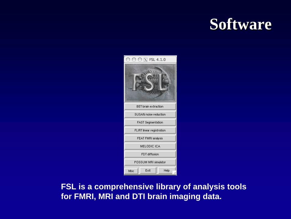
FSL is a comprehensive library of analysis tools for FMRI, MRI and DTI brain imaging data.
SoftwareSoftware

MRC Cognition and Brain Sciences UnitMRC Cognition and Brain Sciences Unit
MRC Cognition and Brain Sciences Unit,15 Chaucer Road,Cambridge,CB2 7EF http://imaging.mrc-cbu.cam.ac.uk/imaging/CbuImaging

CBUCBU
• The CBU now constitutes one of the largest concentrations of cognitive scientists and neuroscientists on a single site anywhere in the world, with nearly 100 active scientists, students and research staff.
• A priority in the CBU research strategy over the last 5 years has been to develop a strong research programmein neuroimaging, working closely with the Wolfson Brain Imaging Centre, and more than half of the scientific staff and students are actively involved in neuroimaging projects.
http://www.mrc-cbu.cam.ac.uk

PeoplePeople
Michael [email protected]
William Marslen-Wilson
Unit Director, Speech and Language Group

Principal areas of investigationPrincipal areas of investigation
• Attention • Emotion • Speech and language • Memory and perception• Methods research and infrastructure
http://www.fmrib.ox.ac.uk/fsl/

Max Planck Institute for Human Cognitive and Brain SciencesStephanstraße 1A, 04103 Leipzig, Germany
Max Planck Institutefor Human Cognitive and Brain Sciences
Max Planck Institute
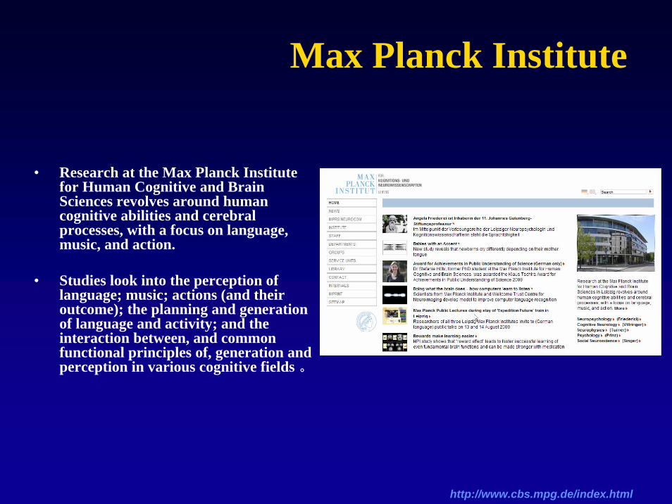
• Research at the Max Planck Institute for Human Cognitive and Brain Sciences revolves around human cognitive abilities and cerebral processes, with a focus on language, music, and action.
• Studies look into the perception of language; music; actions (and their outcome); the planning and generation of language and activity; and the interaction between, and common functional principles of, generation and perception in various cognitive fields 。
http://www.cbs.mpg.de/index.html

PeoplePeople
Prof. Dr Angela D. FriedericiNeuropsychology
Prof. Dr Wolfgang PrinzPsychology
Prof. Dr Robert TurnerBrain Mapping, both Functional and Anatomical
Prof. Dr Arno VillringerCognitive Neurology

ResearchResearch
• Research at the Max Planck Institute for Human Cognitive and Brain Sciences revolves around human cognitive abilities and cerebral processes, with a focus on language, music, and action.
– Neuropsychology ( Professor Angela D. Friederici) – Cognitive Neurology ( Professor Arno Villringer) – Neurophysics ( Professor Robert Turner) – Psychology ( Professor Wolfgang Prinz) – Social Neuroscience ( Prof. Dr. Tania Singer)

SoftwareSoftware
• Lipsia– Leipzig Image Processing and Statistical Inference Algorithms – a tool
for fMRI data analysis • registration and normalization • preprocessing • statistical evaluation • region of interest analysis • timecourse analysis • visualization, rendering • converters to various data formats
http://neuro.debian.net/pkgs/lipsia.html

Institute of Neuroscience and Institute of Neuroscience and Medicine (INM)Medicine (INM)
The INM is devoted to brain research.
http://www.fz-juelich.de/inm/index.php?index=3
Institute for Neuroscience and Medicine (INM-1)Research Center Juelich52425 Jülich

INMINM
• The Institute of Neuroscience and Medicine, INM-1, is devoted to experimental studies about multimodal mapping of the human brain.
• Our aim is the development of a new, three-dimensional realistic brain model on the basis of cytoarchitectonic, molecular and functional data as well as connectivity.
PeoplePeople
•
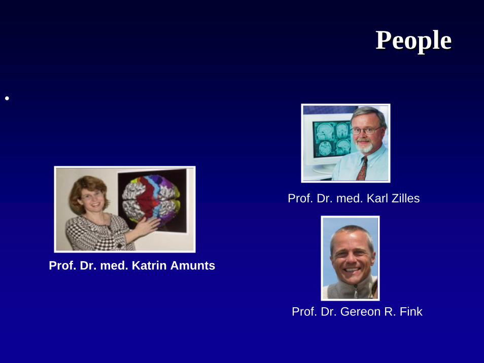
Prof. Dr. med. Katrin Amunts
Prof. Dr. med. Karl Zilles
Prof. Dr. Gereon R. Fink

ResearchResearch
• Important Research Fields:
Brain Imaging PhysicsCognitive NeurologyComputational and Systems NeuroscienceHuman Brain MappingMolecular Organisation of the Human CortexNeuromodulation

ResearchResearch
• Mapping of the human cerebral cortex based on the analysis of its neurochemical(receptorarchitecture) and histological (cyto- and myeloarchitecture) structure
• Mapping the cerebral cortex of non-human primates based on the analysis of their neurochemical (receptorarchitecture) and histological (cyto- and myeloarchitecturestructure
• Analysis of the disease-related changes in the densities of neurotransmitter receptors in brain tissue from patients with focal temporal lobe epilepsy or hepatic encephalopathy
• Combination of (receptor-) architectural data with results obtained from functional imaging studies ( SPM Anatomy Toolbox)
• Methodical developments towards the multimodal integration of structural and functional information concering the human brain in order to enable analysis of the principals of cortical (hierarchical) organisation

ResearchResearch
Molecular Molecular OrganisationOrganisation of the Human Cortexof the Human Cortex

国内国内
http://psychbrain.bnu.edu.cn/Org.htmhttp://restfmri.net/forum/index.php相关科研人员:臧玉峰,贺永,等
http://www.rccm.org.cn/http://www.nlpr.ia.ac.cn/jiangtz相关科研人员:蒋田仔,等
http://www.hmrrc.org.cn/相关科研人员:龚启勇,等

ThanksThanks
Research Center for Sectional and Imaging Anatomy