brain spots on imaging tests - mud group - brain spots on imagng tests mud...brain spots on imaging...
TRANSCRIPT
Brain Spots on Imaging Tests To Be or Not to Be Concerned
Metropolitan Underwriting Charles Levy, MD Discussion Group Aviva USA 1/29/13
CT and MRI
2 most common forms of brain imaging today
• As with any diagnostic test
– Risks of imaging have to be weighed against benefits of identifying treatable disease
• With acceptable sensitivity and specificity
2
CT
• Used to be modality of choice for non-invasive assessment of the brain – Now study of choice mainly in cases
• Trauma • Acute neurologic emergencies
– Acute Stroke – Intracranial hemorrhage – Intracranial or spinal trauma – Detection of fine bone detail and skull fractures
3
CT (2)
• Advantage
– Faster than MRI • Study of choice in acute situations
• Disadvantages
– 15X more radiation than a chest x-ray – Less contrast resolution for soft tissue
abnormalities or normal structures than MRI – Requires contrast injection for angiography
4

MRI
• Advantages – No radiation exposure – Does not require contrast injection for
angiography – More sensitive for detection of
• Normal brain anatomic structures • CNS and intracranial lesions • Cranial nerves • Posterior fossa structures • Pituitary • Acute and chronic bleeding in the brain
5
MRI (2)
• More sensitive for detection of (cont.) • All soft tissue abnormalities
– FLAIR images allow better discrimination between solid tissue and edema
– DWI images improve » detection of acute ischemic stroke » differentiating acute stroke from other
processes that cause acute neurologic events
• Also excels at detecting brain lesions in asymptomatic individuals
6
MRI (3)
• Has replaced CT in brain imaging – Except in patients with contraindications to MRI
7
MRI (4)
• Contraindications to MRI: – Metallic fragments, clips or devices in the brain,
eye, spinal cord • Includes intracranial clips
– Magnetically activated implanted devices: • Cardiac pacemakers, insulin pumps, neuro-
stimulators, cochlear implants • Metal outside the brain and eye is NOT a
contraindication: – Cardiac valves, inferior vena cava filters, biliary and
vascular stents, IUD's, metallic prostheses
8
MRI (5)
• Disadvantages due to its increased use and
superior tissue definition/resolution – Studies in sick and healthy persons both show
MRI to be oversensitive • Displays even incidental white matter foci without
underlying pathology • Other incidental findings
9
MRI (7)
Definition: Incidental findings on brain imaging are
defined as previously undetected abnormalities of potential relevance
• Unexpectedly discovered • Unrelated to the purpose of the imaging
10

Case #1
40 yom $750,000 Term • on NBExam noted dizziness for the past few years for
which an MRI was done as part of the w/u • 1/11 MRI: Impression: Solitary indeterminate right
frontal white matter abnormal foci. This is nonspecific but could represent focal gliosis from prior inflammation or possibly vasculitis, the sequelae of migraine headaches and less likely demyelination or atypical infection. O/w normal MRI.
• episodic confusion, problems focusing and confused, panic sensation, may pass out. Sx remain intermittent and similar thru end of APS 1/12.
• EEG, MRA, w/u for inflammatory dis all neg, no history of seizures
11

Case 2
38 yof, $500,000 Term • 1/10 c/o headaches, paresthesias left side of face, vision
blurred with focus on small objects • APS notes thru 6/11 with no recurrence of symptoms • also hx of obesity, impaired glucose tolerance,
hypertension poorly controlled in ’10 but better since. • 1/21/10 MRI: 3 foci of hyperintense T2 signal in the
white matter of the left frontal lobe measuring 3 mm in size. Nonspecific in etiology of the type often seen in neurologically asymptomatic patients, demyelinating disease, small vessel vascular change, migraine headaches.
12
Case #3
50 yom $20 million UL • 9/11 MVA, concussion, dizziness, hard to read, vertigo
– Bilateral high frequency hearing loss, ENG abnormal, less reactive left labyrinth
– EBCT score +25 • 10/11 MRI: a few punctate scattered non-
specific supratentorial white matter abnormalities
13
Incidental MRI Findings (4) Study 1 11/07
• 2000 participants
– Ages 45-96 (mean 63.3) – 54.7% female
• Ages 45-59: 94.6% were found to have asymptomatic “white matter changes”
• >age 75: increased to 98% – 272 (13%) were found to have incidental
findings other than “white matter changes”
14
Incidental MRI Findings (5) Study 1 11/07 p.2
– Other than “white matter changes include:
• Asymptomatic stroke – Lacunar infarct > cortical infarct
• Aneurysms • Benign tumors (esp. meningiomas) • Arachnoid cysts • Cavernous hemangiomas • 1 urgent finding
– possible low grade glioma
15
Incidental Findings Brain MRI Study 2 8/09
• 15, 559 participants • Incidental finding in 1 out of every 37
asymptomatic people scanned (2.7%) – 2.0% non-neoplastic – 0.7% incidental neoplastic findings
• These 420 incidental findings are in addition to already excluded – White matter hyperintensities – Microbleeds – Silent infarcts
16

Trends in Both Studies
• Increasing prevalence of all neoplastic incidental brain findings with age – Probably related to meningiomas
• Increasing prevalence of “white matter changes”
with age
• Extremely small number of incidental findings that required specific treatment – MRI has been available for 20-30 yrs
• Long term prognosis of some incidental findings not fully known yet
17
White Matter Hyperintensities
• Most frequent incidental findings in brain MRIs
• Common finding on neuro-imaging associated with – Aging – Medical illness – Some invasive medical procedures – Hypertension – Migraine headaches – Multiple sclerosis
18
White Matter Hyperintensities (2)
• Most important to consider in context and with
the significant differentiating elements – Number – Size – Location – Presence or absence of edema – Reaction to contrast medium
• Including Gadolinium – Evolution in time
19
White Matter Hyperintensities (3)
• Have been concluded to be associated with increased risk of – Overall increased risk of cerebrovascular events – Dementia and faster decline in cognitive function
• However, they can not be considered in isolation
from clinical data and other diagnostic test results – Think of them more as a predictive test like an
EBCT or carotid IMT (Intima Media Thickness) • Than as an indicator of a specific problem to be
treated now
20
Conclusions
• Circle back to the case studies in light of the information about – Frequency of incidental findings of white matter
hyperintensities without relationship to specific disease or need for specific treatment
– Possibility that these incidental white matter hyperintensities constitute a risk factor rather than a marker of a specific disease entity
21
CVA
• Advantages MRI over CT – Better defines intracerebral hemorrhages
• old and new.
– more sensitive than CT for the early diagnosis of brain infarction.
– better determines the precise location and size of the infarction and
– better follows the lesion over time.
22
CVA (2)
• Advantages MRI over CT (p.2) – Lacunar infarcts and small cortical strokes are seen
with higher sensitivity. – FLAIR images show infarcts earlier after onset of
symptoms.
– DWI images are useful in distinguishing acute from chronic ischemic changes
23
CVA (3)
• Does a normal CT or MRI rule out stroke? – No.
• It is important to remember that in patients with ischemia who do not yet have infarction, both CT and MRI may be normal.
• Repeating the scan in 48 hours will most likely demonstrate the stroke lesion.
• Sub-acute infarct (1 to 8 weeks): – Contrast enhancement slowly decreases in time but
can persist for 8 weeks, with decreasing mass effect and abnormal signal intensity.
24
TIA vs CVA
• Transient Ischemic Attack – Acute episode of temporary neurologic
dysfunction • Resulting from focal cerebral, spinal or retinal
ischemia • Not associated with permanent cerebral infarction
– Sudden onset – Duration < 24 hours
• Clinical symptoms typically last < 1 hour • CVA and not TIA if:
– Clinical signs or symptoms last >24 hours – Evidence of infarction on imaging
25

Cerebrovascular Lesions
• Subclinical vascular pathologic changes – Silent cortical infarcts – Lacunar infarcts – White Matter Hyperintensities
• Linked to – Increased risk of stroke – Cognitive decline
• Acute ischemic stroke patients – 11-29% found to have unrelated additional
infarcts 26
Cerebrovascular Lesions (2)
• Silent infarct: – Incidentally found lesions with appearance
typical of infarction – Without clinical history compatible with clinical
stroke – Strong relationship with age and other stroke
risk factors • Suggests they may themselves be risk factors for
significant cerebrovascular disease – Important risk factor for
• Further stroke • Dementia
27
Cerebrovascular Lesions (3)
• Global cognitive function significantly worse – With silent brain infarcts on baseline MRI than
without
28
Lacunar stroke
• Small deep infarcts that result from occlusion of a penetrating artery
• Account for about a quarter of all ischemic strokes • Commonly had been regarded as benign vascular
lesions with a favorable long-term prognosis • Recent studies have shown that is only the case early
in the disease course but in the years after the infarct – increased risk of death, mainly from cardiovascular
causes. – risk of recurrent stroke after lacunar infarct is similar
to that for most other types of stroke – increased risk of developing cognitive decline and
dementia.
Conclusions
• MRI more sensitive than CT for detection of all cerebrovascular ischemic abnormalities – Symptomatic CVA – Silent CVA – Lacunar infarct
• MRI detects acute infarct earlier – If not seen on initial MRI
• Repeat may be warranted in 48 hours if neurologic deficit persists without adequate alternative explanation
30
Conclusions (2)
• Infarct/CVA not necessarily ruled out (or TIA
ruled in) with negative (even negative repeat) imaging if symptoms last >24 hours
• Silent infarcts, lacunar infarcts and white matter hyperintensities increase the risk for additional cerebrovascular insults/cognitive decline
31
Aneurysms
American Heart Assn. guidelines for
unruptured cerebral aneurysms • Taking into consideration
– Age – Past history of cerebral aneurysm – Family history – Genetic/familial conditions
32
Aneurysms (2)
– All symptomatic aneurysms should be treated – Incidental aneurysms
• <10 mm without history of SAH should be treated conservatively
• Anterior circulation aneurysms less likely to rupture • small aneurysms approaching the 10-mm diameter
size should be considered for treatment if: – unique features – Family history of aneurysm or SAH – Past history of aneurysm or SAH
33
Aneurysms (3)
• Factors that favor surgery include – A young patient – Long life expectancy – Previously ruptured aneurysms – Family history of aneurysm – Large aneurysms – Symptomatic – Observed aneurysm growth
34
Aneurysms (4)
• Factors that favor conservative management
include – Older patient age – Decreased life expectancy – asymptomatic small aneurysms
• Especially anterior circulation
35
Subarachnoid Hemorrhage (SAH) with Negative Angiography • Ruptured cerebral aneurysm is the most
common cause of spontaneous SAH • The aneurysm may not be visualized in up to
17.5% of cases where there is one due to – Small aneurysm size – Vascular spasm of the parent artery – Thrombosis of the aneurysm
• A second angiogram should be performed a week later – If negative
• Risk of non-visualized aneurysm is low • Prognosis is good
36
Conclusions
• Increased sensitivities of MRIs will find more asymptomatic aneurysms – Not all unoperated aneurysms have the same
mortality risk profile • Size • Location • Age • Family history • Past history
• One negative imaging study after SAH may not be adequate to rule out an underlying aneurysm
37
Venous angioma
• Currently called Developmental Venous Anomalies ( DVA) in Medical Literature
• Still called Venous Angioma in UW Guides
Will be referred to as DVA in this presentation and in the Medical Records you obtain
38
DVA/Venous Angioma (2)
• Congenital anomalies of the intracranial venous
drainage. • Highest prevalence rate of all intracranial
vascular malformations – Commonest intracranial vascular malformation seen
at autopsy – Previously considered to be rare until the advent of
CT and MRI scanning – Now considered to be NORMAL VARIANT of cerebral
venous system
39
DVA/Venous Angioma (3)
• Presentation (reason the scan was done)
variable and non-specific – Most are incidental findings on MRI – Headache most common symptom prompting the
MRI – Most of the symptoms prompting the scan not related
to the lesion
40
DVA/Venous Angioma (4)
• Isolated DVA represents no additional mortality risk
– Because these anomalies provide a useful and important blood draining function, in no case should they be excised or radiated – Do not require routine removal
• UW Manuals suggest a different risk profile between supratentorial vs infratentorial lesion – Due to different risk profile from bleeding in the
brainstem (infratentorial) region – May not be consistent with latest medical literature
findings
41
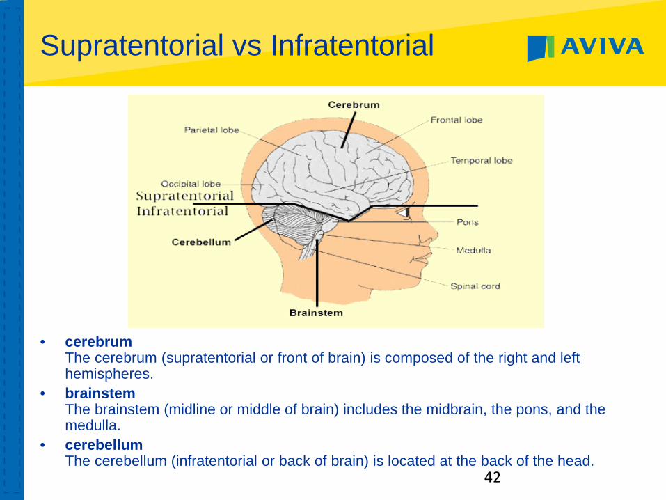
Supratentorial vs Infratentorial
• cerebrum The cerebrum (supratentorial or front of brain) is composed of the right and left hemispheres.
• brainstem The brainstem (midline or middle of brain) includes the midbrain, the pons, and the medulla.
• cerebellum The cerebellum (infratentorial or back of brain) is located at the back of the head.
42
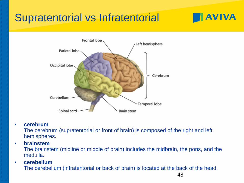
Supratentorial vs Infratentorial
• cerebrum The cerebrum (supratentorial or front of brain) is composed of the right and left hemispheres.
• brainstem The brainstem (midline or middle of brain) includes the midbrain, the pons, and the medulla.
• cerebellum The cerebellum (infratentorial or back of brain) is located at the back of the head.
43

DVA/Venous Angioma (5)
• Often associated with Cavernous Angiomas – (also known as Cavernous Hemangiomas)
• Cavernous Angiomas should be treated
– If >10 mm in size – Multiple lesions – If any evidence of prior bleed
• Including hemosiderin deposition • Infratentorial location
– If possible leaving associated DVA intact
44
Multiple Sclerosis on MRI
• Classic MS brain lesion – T2 hyperintense lesion
• Called plaques • Can be new, old or reactivated lesions
– If Gadolinium contrast used
• Differentiates new or reactivated lesions – By contrast “enhancement”
» Enhancement lasts 4-6 weeks • Also picks up very new lesions not yet
hyperintense 45
Multiple Sclerosis on MRI (2)
• Diagnosis of MS – Neurological disturbance of kind seen in MS
• Minimum duration 24 hours – 2 or more MRI lesions
Or
– Asymptomatic – 1 Gadolinium enhancing lesion or 9 hyperintense
MRI lesions
46
Meningioma
While repeated scanning has become routine for asymptomatic (and incidental) meningioma
• 94% remain asymptomatic • 63% do not grow Factors to consider • Location • Stability • Age of applicant • Size
47
Meningioma (2)
History of resected meningioma MRI f/u: Pre and postcontrast brain MRI • Findings:
– Again identified are post surgical changes. Right frontal craniectomy/cranioplasty, right frontal lobe surgical cavity with residual T2/FLAIR signal abnormality, stable residual right frontal dural enhancement
• Stable brain MRI findings since prior MRI. No recurrent tumor
48

Conclusions:
• Additional MRI incidental findings: – Isolated DVA produces no additional mortality
risk – Findings suggestive of MS need to be taken in
context as described • And diagnostic criteria followed before diagnosis or
risk profile for MS attached – Post-op changes on the MRI f/u of a resected
meningioma should not be confused with recurrent tumor
49