branches on the tree of life: protists · branches on the tree of life: protists written, produced...
TRANSCRIPT

.
.
A New DVD for Biology Teaching from BioMEDIA ASSOCIATES ©2004
Branches on the Tree of Life:PROTISTS
Written, produced and photographed by Bruce RussellAll photos in this guide are from the DVD
Cosmerium, a green alga Arcella
The goal of this program is to show a representative sample of the great diversity ofprotists, and to show why they need a new classification reflecting our growingunderstanding of their long evolutionary history. The protists shown can be found inhabitats such as: roadside puddles, park duck ponds, aquariums, birdbaths and inthe gut of termites. We hope that these observations will encourage students tocollect pond water samples and see for themselves this amazing hidden world.The live photography was accomplished using a variety microscope techniques,including dark field (good for showing the natural color of subjects) and the mostadvanced forms of differential interference contrast (DIC gives contrast totransparent structures that would be invisible in normal bright field microscopy).While these forms of imaging living protists are particularly revealing for someaspects of micro-organism biology, all of the organisms seen here can be studied
BRANCHES ON THE TREE OF LIFE: PROTISTS
Micrasterias
successfully with student microscopes.
IMPORTANT NOTE: This program packs a lot ofinformation and a wealth of observational data intonine different modules. We recommend that thematerial be viewed module by module, stopping fordiscussion and replay as needed. “Discussion starters”for the various modules are provided in this guide.
Chapters on this DVD:
What is a Protist?The EuglenidsDiatoms and Their Unlikely RelativesAmoebas and HeliozoansGreen ProtistsColonial ProtistsInsidersCiliated ProtistsParamecium(Additional Opservations):Flagellated ProtistsFlagellate “Unknowns” to identifyAmoeboid ProtistsAmoeba “Unknowns” to identifyCiliated ProtistsCiliate “Unknowns” to identify
CHAPTER CONTENTS
Campanella, a colonial ciliate
What is a Protist.... Cells with a nucleus, are known as “eukaryotic cells”.Single cell eukaryotes are called Protists. One of theproblems in biology is how to classify protists. Analysis oftheir DNA and RNA shows that some lines of protistsseparated very far back in time, diverging from the earliesteukaryotes. Also, some lines of single cell life (green algae)are more closely related to multicellular organisms (plants)than to other lines of protists..... The lesson here is that single celled eukaryotes showsuch extreme evolutionary divergence, and that the old ideaof lumping them into a single kingdom needs correcting.Biologists are now attempting to create a protist classificationthat looks more closely at their genetic material. Theultimate goal is to understand how the various lines ofprotists diverged from each other, and how theseindependently living cells and multicellular organisms arerelated.
Questions for Discussion:Should biological classification reflect genetic relationships?Why did biologists lump all single cell eukaryotes (and their near multicellular relatives) into asingle kingdom Protista?What should be the criteria for grouping organisms into a kingdom?
BRANCHES ON THE TREE OF LIFE: PROTISTS

..Euglena rubra
The Euglenids.... In green euglenids, the red eyespot and corresponding photosensitive region near the base of the flagellumcreate a guidance system that allows Euglena to home in on a light source and swim in that direction until itreaches a light level suitable for photosynthesis. The euglenid shown is E. rubra, which can live at the surface infull sunlight, a niche unavailable to other euglenids due to the high levels of ultra violet radiation. E. rubrasurvives the radiation using a shield of red pigment. The pigment can be withdrawn when the sun isnÕt shining,giving the chloroplasts more light for photosynthesis. A swimming E. rubra shows the red pigment as aconcentrated core in the middle of the cell. Several other species are shown hinting at the diversity of greeneuglenids..... Non-green euglenids include Astasia, which evolved from a green euglenid that lost its chloroplasts. Withoutphotosynthesis, Astasia obtains its nutrition by absorbing nutrient molecules directly from its surroundings. Inanother relative, Peranema, the exceptionally robust flagellum works like a propeller, pulling the cell through thewater. Peranema is a predator, capturing and engulfing smaller euglenids. Two rods, located in the mouth area,are used to hold prey during engulfment..... In the euglenid line, as in most other flagellated protists, individuals divide longitudinally. Division begins withduplication of the basal body at the base of the flagellum creating a cell with two flagella that then splits rightdown the middle. Today’s euglenids are modern representative of an ancient line of life, so different from otherprotists that some biologists have suggested placing them in a kingdom of their own.
Questions for Discussion:How might a euglenid loose its chloroplasts?How could such a cell, deprived of photosynthesis, obtain nutrition?What are the dangers to a single cell exposed to full sunlight?How many kinds of euglenids can be found in accessible habitats near your school?
Diatoms and Their Unlikely Relatives.... Diatoms are important producers in aquatic food chains. Indiatoms, the photosynthetic pigment is yellow, not green.Diatoms convert the products of photosynthesis into oildroplets--an energy reserve the cell can draw on when the sunisn’t shining. But the unique thing about a diatom is its house.These cells remove dissolved silica from the water and use it toconstruct finely sculptured glass cases..... Diatoms are not alone on their evolutionary branch. Thelesson here is that even though organisms my look very different(diatoms and water molds), their genetic material may indicatethat they occupy the same branch on the tree of life. Regroupingorganisms on this branch is such a new game that biologists areyet to agree upon a name. One proposal for a branch name is“Chromista,” considered by some to be a phylum and by others, akingdom.
Questions for Discussion:How do diatoms move?What advantage might oil droplets be to a planktonic diatom?In the intricately patterned glass houses of diatoms what function might the sculpturing serve?Should evolutionary relationships be used as a basis for classification, even though the relativeslook nothing alike?
..
BRANCHES ON THE TREE OF LIFE: PROTISTS

Amoeba, extending pseudopod Actinosphaerium, a heliozoan
Amoebas and Heliozoans.... This observational module shows amoeboid protists moving and feeding by the extension of pseudopodia. Allof the amoeba observations are in real time. Shell-building amoebas such as Difflugia are common in pond watersamples..... Heliozoans have long been classified with the amoebas, but genetic analysis places them on their own limb ofthe eukaryote tree. Their spines, made of microtubules, are often broken by a thrashing prey, but are quicklyrebuilt.
Question for Discussion:Does it appear that the diatoms ingested by the amoeba have undergone digestion, or do theirglass cases protect them from digestive enzymes.How do amoebas move?Is there more than a single method of amoeba movement?How does a heliozoan’s spines actually trap prey?
Spirogyra zygotes
Green Protists.... In unpolluted habitats it is common to find adiversity of unicellular green algae includingcrescent-shaped Closterium, a brilliant green cell withdancing crystals of gypsum in vacuoles at its tips..... Spirogyra is a filamentous alga composed ofbarrel-shaped cells with a band shaped chloroplast thatspirals around the cell. Conjugation in Spirogyra usuallyoccurs before seasonal disruption of the habitat.Conjugation results in zygotes that will assure a newpopulation of Spirogyra when conditions improve.
Questions for Discussion:What is causing the gypsum crystals todance?Are there male and female strands ofSpirogyra?What environmental factors mighttrigger conjugation in Spirogyra?
BRANCHES ON THE TREE OF LIFE: PROTISTS

Volvox eggs Volvox, daughter colony development
Colonial Protists.... Colonial green protists let us imagine how simple plants evolved from single-celled green protists. In thesame drop of pond water you can find: small colonies such as Gonium--usually with 16 cells; Eudorina--a 32 cellscolony; and the queen of colonial protists--Volvox, with thousands of flagellated cells lining the sphere..... Volvox shows the beginnings of cellular differentiation, a key event in embryology of multicellular organisms.Within the Volvox colony germinal cells divide, producing daughter colonies that break out in an asexual cyclethat repeats generation after generation. As winter comes on some daughters will produce packets of sperm,others eggs. The result of fertilization is a thick walled zygote that will carry Volvox through freezing and drying.
Questions for Discussion:How would you explain the appearance of Volvox in temporary rain pools (vernal pools)?Aside from difference in size and number of cells how does Volvox differ from Gonium?What survival advantage might have driven the evolution of colonies from single cells?
Plasmodium, in field of red blood cells Trychonympha, from a termite gut
Insiders.... There are probably at least as many kinds if protists living inside animals as there are living in ponds,puddles, and oceans..... Plasmodium (three are seen in a stained blood smear) is a parasite of the red blood cells multiplying andbreaking out in a daily cycle that produces the alternating chills and fever of malaria..... Another blood parasite, Trypanosoma (shown live in a drop of blood) produces an often fatal disease--sleepingsickness. Certain amoeboid protists colonize mammalian intestines, causing tissue damage and diarrhea. But notall insiders are harmful..... A termite’s gut is packed solid with flagellated protists (Trichonympha) that aid in breaking down wood.Without its helpers, a termite would continue to eat wood, but die of starvation. A variety of protists live in thetermite gut, but not all of them digest wood. The relationships among the various species are still being workedout.
Questions for Discussion:How might blood parasites, such as Plasmodium and Trypanosoma evolve from free living protistancestors?At some point were their ancestors parasites of insects?How do newly hatched termites become “infected” with their wood digesting symbionts?
BRANCHES ON THE TREE OF LIFE: PROTISTS

Ciliated Protists.... In our view, ciliates are the dinosaurs of micro-space. Colpidium is representative of a large line of ciliatedprotists in which the cilia are uniformly distributed in rows. Lying just below the cilia rows are the mitochondriathat supply the ATP that fuels the ciliary beat.
.... Animation shows cilia structure and explains how the power stroke is produced. The next section shows eightorganisms (common in pond water samples) that demonstrate the diversity of ciliates.
LacrymariaLacrymaria hides in the bushes, extending its neck to feed. This remarkable behavior is madepossible by myonemes--muscle-like fibers that spiral around the cell. (Q. - In what ways mightthe highly extendible neck be advantageous for Lacrymaria?)
EuplotesEuplotes, has tendril-like locomotor structures called “cirri.” The cirri can be used to walk oversurfaces. (Q. - How might “walking” benefit a protist?)
Euplotes taking a stroll
VorticellaIn Vorticella, a band of cilia-stiffened membrane is used to generate feeding currents. Vorticella isa bacteria feeder, bringing in food from a distance. The bacteria selected are engulfed in a foodvacuole that pinches off and circulates through the cell while the bacteria digest. If disturbed,Vorticella can snap in--the advantages of living on a contractile stalk. When it's time to divide,one daughter retains the stalk while the other develops a band of swimming cilia and breaks free.The swimmer is attracted to the stalks of other vorticellids where it will settle down, grow a newstalk, and join the group. (Q. - What advantages might Vorticella enjoy by living in groups?)
Vorticella, contractile stalk Vorticella group on duckweed rootlet
UrocentrumUrocentrum turbo, has a unique method for remaining in choice feeding areas. Urocentrum’s trickis an invisible tether line that it attaches to some nearby object.(Q. - How is Urocentrum able to compete with other bacteria feeders such as Vorticella andParamecium?)
BRANCHES ON THE TREE OF LIFE: PROTISTS

\Urocentrum, linked by tethers Stentor
StentorThere are many Stentor species (several are shown). Like other ciliates, stentors join at theirmouths and exchange micronuclei in a form of sexual behavior known as conjugation.Q. - In large Stentor species the macronucleus appears as a string of beads. Can thisconfiguration be functional?
TrichodinaTrichodina lives as a commensal on Hydra, feeding on the bacteria that collect around hydra'sregurgitated meals. Trichodina hangs on by means of an amazing adaptation--a multi-toothedbasal disc.Q. - What advantages might Trichodina derive by living on Hydra? Could Hydra derive anybenefits from its guests? How might the protist be harmful to its host?
Trichodina attachment disk Coleps feeding on Paramecium carcass
ColepsNo community would exist for long without scavengers, and in the microworld Coleps is theJackal. These are feasting on a paramecium carcass.Q. - How does Coleps attach in order to ingest food?
SuctoriansLooking in pond water you may find a ciliate that has no ciliaÑa suctorian. Upon contact, thesuctorian’s tentacle tips fuses with the prey’s membrane and with some pressure adjustments,the suctorian sucks in its meal. Suctorians exhibit the very unprotist-like behavior of giving birth.As the ciliated larva emerges from the birth pore, it turns on the power--a response that helps itavoid touching its mother's tentacles.Q. - Why do biologist place suctorians (organisms without cilia) on the ciliate line of evolution?
DidiniumDidinium has an appetite for just one type of prey--Paramecium. It takes Didinium approximately40 seconds to swallow a Paramecium. In a laboratory dish, the feeding frenzy will continue untilnearly all the paramecia have been eaten. With no more food, the didinia form cysts, lyingdormant while they wait for a new population of paramecia to build up.Q. - In a pond, how can Paramecium, which normally does not encyst, avoid being wiped out by aDidinium attack? What might signal an encysted Didinium that it is time to excyst and go hunting?
BRANCHES ON THE TREE OF LIFE: PROTISTS

Paramecium.... Paramecium can be cultured by adding some boiled hay (or dry grass clippings) to pond, or aquarium water.
Paramecium, excretory crystals Didinium attacks a Paramecium
.... With plenty of organisms to examine one can observe:
• How Paramecium feeds on bacteria by phagocytosis and digests them in food vacuoles.• How the cell gets rid of water entering by osmosis by means of its contractile vacuoles.• How it eliminates undigested material through its anal pore.• How it deals with metabolic wastes by crystalizing them.• How it avoids getting trapped in microscopic mazes.• How it reproduces asexually, with division of its macronucleus and the duplication of its variousorganelles. Periodically, Paramecium enters into a sexual process, whereby individuals fusetogether at their oral grooves and exchange micronuclei. This mixing of genes revitalizes theParamecium population which can then continue on reproducing asexually for hundred ofgenerations.
Paramecium dividing by fission
BRANCHES ON THE TREE OF LIFE: PROTISTS

Additional ObservationsThe remaining sections on this DVD show a variety of protists with background music but with no narration.This exciting state-of-the-art microscope footage is provided for teachers who would like to involve studentsin observing living protists, an activity that leads to discussion and research. These can be used as anintroduction to the study of protists (and the narrated portion of this DVD), or played as “ambiance” (toinspire student interest and questioning) during laboratory work with protists. One thing everyone agreesupon is that observing this remarkable footage of living microorganisms has tremendous learning benefits.
Flagellated Protists
Opalina Opalina showing many nucleii
Opalina: 100-200µ Although Opalina appears to be a ciliate, it is more closely related to certainflagellated organisms. Opalinids are intestinal parasite of amphibians. The species shown was taken from atree frog tadpole. Note that Opalina is a multinucleated cell with hundreds of nuclei, a rare phenomenon inprotists. Because most of the opalinid population occurs in the hostÕs rectum, Opalina is considered to bemore commensal than a parasite.
To obtain opalinids for microscope study, without killing their host, use a glass eyedropper filled with 0.7%saline solution. The fire-polished end of the dropper can be gently inserted in the host’s cloaca. The idea is toprovide a saline enema while sucking back a sample of the rectal content including any commensals it maycontain. For a frog, the procedure may be a little unsettling, but certainly better than a postmortemextraction.
Questions for Discussion: How might a multinucleated cell evolve? What kinds of nutrientsare available to Opalina? How do tadpoles become infected?
Hexamita: around 20µ Hexamita is a diplomonad, related to Giardia, the notorious intestinal parasiteknown to travelers who indulge in untreated drinking water and spend the remainder of their trip in hospitalbathrooms. The dark-field shot shows the typical rapid dancing behavior of unrestrained Hexamita. Thesecame from a jar of putrid pond water in which a large freshwater bryozoan colony was decomposing (note theabundance of large spiral bacteria). Hexamita species are also common in the large intestine of frogs andsalmonid fish.
Questions for Discussion: How might the transition from a free-living Hexamita to aparasite be accomplished? Might free living Hexamita have evolved the other wayaround--from parasite to free-living?
BRANCHES ON THE TREE OF LIFE: PROTISTS

Dinobryon
Dinobryon: cells around 40µ Dinobryon colonies are found in the plankton of lakes. Colonial life mayprovide several advantages over living as a single isolated cell. Larger size may help prevent predation.Branching may help orient and stabilize the colony in the open water. Each new individual, resulting fromdivision, builds on the rim of their sister and so derives all the benefits of colonial life.
Questions for Discussion: Based on the video observation, in what group of protists would
you place Dinobryon?
Codosiga on duckweed rootlet Codosiga, collar and flagellum
Codosiga: cells around 20µ Choanoflagellates such as Codosiga live attached to other objects. The firstscene shows a colony of Codosiga attached to a duckweed rootlet. These cells have transparent collars (seenin optical section). A long flagellum emerges from the collar where it creates food-trapping currents.Choanoflagellates are virtually identical to the collar cells of sponges, suggesting a close relationship. In fact,these little cells may be our closest living protistan relative.
Questions for Discussion: What steps might a colonial choanoflagellate have undergone in
becoming a sponge-like organism?
BRANCHES ON THE TREE OF LIFE: PROTISTS

Gymnodinium, a dinoflagellate
Gymnodinium: around 40µ Gymnodinium is a common dinoflagellate found in lakes and ponds. Thecells have a thin cellulose shell and two flagella. One flagellum is contained in a groove that circles the celland produces rotation. The other flagellum projects posteriorly and provides forward locomotion. A surprisingnew finding, based on genetic analysis, is that dinoflagellates are more closely related to ciliates than to otherflagellated protists.
Four “Unknowns” to identify:
#1 Ceratium is a dinoflagellate common in the plankton of lake, ponds and oceans. Howmight CeratiumÕs structure be adaptive for planktonic life?#2 Actinocystis is a colonial flagellate that produces a branching extension tube. Thecolonies break off and sheds individual cells which may start up new colonies. Whatadvantages might gained by producing an extension?#3 Phacus is a green flagellate with many species, some twisted, some flat. To what groupof flagellates might Phacus be related?#4 Synura is a colonial flagellate. Spherical colonies of flagellated cells are common in boththe green and yellow-brown lines of life--and there are a few clear ones as well such asActinocystis, #2 above.
Amoeboid Protists
Pelomyxa: 100-600µ These giant amoebas seldom put out pseudopods, creeping along by means ofcytoplasmic waves. The one shown does indeed extend a broad pseudopod, but this may be in response tothe coverglass pressing down on the organism. Pelomyxa is one of the few eukaryotic organisms lackingmitochondria, the ATP producing organelles that supply energy for cell use. Pelomyxa’s energy needs are metby symbiotic bacteria living in its cytoplasm. Some of these aerobic symbionts can be seen in the clear zoneat Pelomyxa’s advancing surface.
Question for Discussion: In what ways might Pelomyxa be a model for the evolution ofmitochondria?
BRANCHES ON THE TREE OF LIFE: PROTISTS

Hydramoeba
Hydramoeba: around 150µ We had been filming Hydra’s feeding and reproductive behavior. Oursubjects, which numbered in the hundreds, suddenly went into an obvious decline. They stopped eating thecopepods we sent their way and their tentacles withered to nubs. The day after we first noticed the decline allbut a few had simply vanished. Examining the remaining sickly Hydras showed them to be stuffed withparasitic amoebas which oozed out when the Hydra was pressed by the coverglass. Other amoebas clung tothe sickly hydraÕs tentacles and body wall. Higher magnification showed that in addition to feeding onendothelial cells, the amoebas were ingesting the hydraÕs nematocystsÑthe stinging cells used to captureprey.
Questions for Discussion: At what stage in the evolution of multicellular organisms did they
become hosts for unicellular parasites? How might such relationships begin? How can aparasite benefit by killing its host?
Vampyrella
Vampyrella: Named for its ability to enter and absorb the cytoplasm of algae cells, Vampyrella is anamazing find. Entering and emptying one cell in an algal filament, Vampyrella then moves to next and ondown the line.
Questions for Discussion: Many filamentous algas (Spirogyra, Zygonema) produce a
jelly-like coating. What evolutionary pressures might account for these jelly sheaths?
Raphidocystis: This small heliozoan is easily recognized by its two types of axiopodia, longaxiopods with beads that move up and down the axiopod, and shorter processes with wineglass tips.
Choanocystis: This delicate heliozoan demonstrates the amazing ability of axiopods to repairthemselves. The real-time observation shows how the microtubules that make up the axiopod quicklyreassemble and repair the broken structure.
BRANCHES ON THE TREE OF LIFE: PROTISTS

Two "unknowns" to identify.
#5 Acanthocystis, a heliozoan filled with symbiotic algae (Chlorella). The fork-tippedprocesses are distinctive for this genus.#6 Thecamoeba, an amoeba with a thick pellicle that forms folds. These slow movingamoebas ingest filaments of cyanobacteria.
Ciliated ProtistsNassula: 200µ Jellybeanshaped Nassula is from anancient line of ciliates, possiblygoing back to a time whencyanobacteria were a major foodsource for early eukaryotic cells.Nassula continues this ancientdiet today by engulfing strandsof Oscillatoria. Its food habitsmake it one of the most colorful
ciliates, due to Oscillatoria fragments in various stages ofdigestion. When food runs low, or when the pond dries up,Nassula encysts, awaiting better times.
Questions for Discussion:Does it appearthat Nassula is restricted in its diet? Mostciliates engulf their food by phagocytosis, aprocess of surrounding the food withplasma membrane as it is taken into thecell. How then does Nassula, which engulfsits meal en toto break up the cyanobacteriastrands and surround them withmembrane?
Homalozoon: 400µ These large ciliates have a “nose” filled with toxicysts used to stun and kill smallciliate prey. In the dark-field observation, the “nose” touches a small ciliate which immediately disintegrates.Homalozoon then expands its anterior end and engulfs the prey. Homalozoon is adapted for living on, andmoving slowly over, surfaces. In a petri dish it is often difficult to dislodge the cell, which adheres to the glasssurface.
Questions for Discussion: How would you describe HomalozoonÕs ecological niche? How
does it “stick” to surfaces?
BRANCHES ON THE TREE OF LIFE: PROTISTS
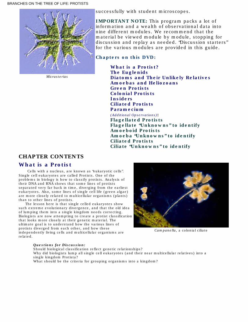
Loxodes: two species are shown,Loxodes magnus (500-700µ) and Loxodesvorax (150µ). After several hours ofobservation, we have yet to see Loxodesactually engulf food. The cytostome islocated in the in-curved region and appearsto have an extension that may “unzip” toengulf large items. Loxodes has an unusualfeatureÑa row of pinkish bodies known asMullerÕs vesicles. The pink bodies arebarium salt crystals.
Questions for Discussion: What functionmight the Muller’s vesicles serve?
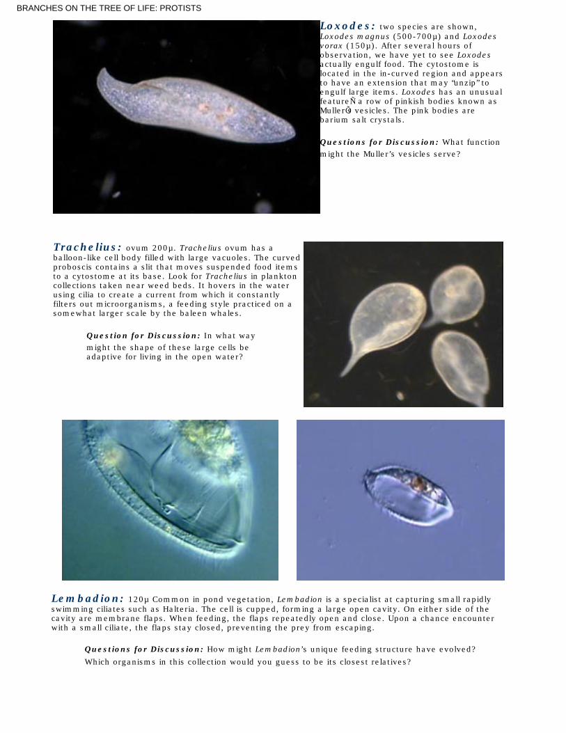
Trachelius: ovum 200µ. Trachelius ovum has aballoon-like cell body filled with large vacuoles. The curvedproboscis contains a slit that moves suspended food itemsto a cytostome at its base. Look for Trachelius in planktoncollections taken near weed beds. It hovers in the waterusing cilia to create a current from which it constantlyfilters out microorganisms, a feeding style practiced on asomewhat larger scale by the baleen whales.
Question for Discussion: In what waymight the shape of these large cells beadaptive for living in the open water?
......
Lembadion: 120µ Common in pond vegetation, Lembadion is a specialist at capturing small rapidlyswimming ciliates such as Halteria. The cell is cupped, forming a large open cavity. On either side of thecavity are membrane flaps. When feeding, the flaps repeatedly open and close. Upon a chance encounterwith a small ciliate, the flaps stay closed, preventing the prey from escaping.
Questions for Discussion: How might Lembadion’s unique feeding structure have evolved?
Which organisms in this collection would you guess to be its closest relatives?
BRANCHES ON THE TREE OF LIFE: PROTISTS

Bursaria trunkatella 500-1000µ, Bursaria swimsthrough the water with its cavernous food trap sweeping upprotists in its path. The actual cell mouth is at the rear of thecavity. Rows of cilia lining the narrowing food channelprevent prey from escaping. The macronucleus is long,curving around the cell interior. Contractile vacuoles aresmall and numerous lying near the surface. Bursaria is oftenfound after seasonal rains fill temporary pools or catchbasins, where it survives drying and freezing within theprotection of thick walled cysts.
Spirostomum: The species shownhere is a small one, extending to around300µ. Some species of Spiostomum reach alength of 4mm. The species shown has asingle macronucleus, but its longer relativeshave multiple macronuclei strung togetherlike a string of beads. A feeding groove,with associated cilia, extends part way downthe elongated cell with a cytostome at itsend. A collecting tube runs the length of thecell emptying into a contractile vacuole thatfills the posterior of the cell. Myonemes(muscle-like contractile fibers visible in theobservation) run the length of these longcells. Spirostomum’s worm-like shapeseems well suited for living in the tangle ofbottom debris where bacteria and smallprotists are usually plentiful. A thickpopulation of Spirostomum resembles aplate of animated spaghetti. When disturbedSpirostomum suddenly contracts to about aquarter of its extended length. Find theplace in the observation where the cells contract.
Questions for Discussion: How might the ability to contract benefit these extremely longcells?
Blepharisma: 130-200µ common in pond water. Two species are shown. Most ciliates are colorless,except for the colored food items they have ingested. Blepharisma is the rare exception. Cultured in brightsunlight it may appear colorless, but most individuals found in the shady aquatic jungles of ponds are pink.Blepharisma has a feeding channel, lined on one side by a membranous veil that traps small organisms anddirect them to its mouth. On the other side of the channel, tufts of long cilia, beating in waves, help steersmall food organisms into the cytostome. The second species shown in the observation cultures algae in itscytoplasm, a kind of symbiosis practice by a number of other large ciliates.
Strobilidium gyrans: 40µ Think of it as an escaped airplane motor running at full throttle. A ring ofextremely powerful fused cilia create a feeding current that brings bacteria and small algae cells toStrobilidium's centrally located cytostome. Strobilidium secretes a tether, allowing it to remain in choicefeeding zones. The tether is produced from a “tail” projection covered with “teeth” that appear to be used tosnip the tether, allowing the cell go shooting away should danger threaten or local conditions deteriorate. Interms of body length traveled over time Strobilidium may well be world’s fastest protist. Under full power itcan exceed 150 body lengths per second, not bad for a cell 40 micrometers in length moving through aviscous medium. Strobilidium is commonly found in aquatic plant infusions and seems more at home in welloxygenated water than in putrid cultures. This may be due to the high oxygen demands imposed by itspowerful ciliature--the propellers that create its long-reaching feeding currents, and its remarkable speed.
Questions for Discussion: What is the advantage of living on a tether?
BRANCHES ON THE TREE OF LIFE: PROTISTS

Halteria: 25-40µ, Common in stagnant pond watercultures. Halteria is shaped like a fat flower vase. Theanterior is surrounded by cirri that produce a smooth glidingmotion. Around the mid-section are groups of long stiff cirrithat produce an entirely different form of locomotion--thesudden jumps for which this ciliate is noted. Bacteria andsmall algae cells are taken in through a curved slit borderedby a row of cirri that sweep in the food. These entertaininglittle cells go bouncing through their environment--but towhat purpose? Filming several caught up in Stentor’sfeeding currents we discovered once again how structure,behavior, and function are inevitably related.
Campanella: cells 200µ, colonies up to 4 cm,Campanella colonies are by far the largest we have seenin nature. In our lab’s outdoor fish pond, they often formblobs several centimeters in diameter. The cells areextremely robust, with rings of cilia-stiffened membranethat create a lot of local turbulence. The branching stalks(which began from a single individual) contain nomyonemes and so can not contract. The individual cells,however, contain contractile proteins and jerk into tightballs if disturbed.
Epistylis: cells 100µ, Members of this genus are oftenfound living on other organisms ranging from turtles tocopepods. Like Campanella, the stalk does not contract andfunctions as a pedestal that holds the feeding individualsabove their attachment site.
Six "unknowns" to identify.
Lionotus 100-200µ. Lionotus can be distinguished from other elongated ciliates byhaving cilia restricted to the outer curve of its “neck”. The inside curve lacks cilia but isequipped with trichites distributed along the length of the cytostome--a long slitrunning along the slender neck. If you find a population of Lionotus, try to determinehow it feeds and the kinds of food it goes after.
BRANCHES ON THE TREE OF LIFE: PROTISTS

Dileptus 250-300µ One of the stealthy predators of the microworld, Dileptushides in the bushes, extending its neck to snare passing rotifers and large protists.Once brought to the cytostome located at the base of the neck, the prey is engulfedwith the aid of trichites.
Glacoma 50µ A number of small ciliates have cytostomes containing flappingmembranes. See Colpidium in the narrated section of this program, and for anextreme case, Lembadion seen in the additional ciliates section
Cyclidium 25-30µ These tiny ciliates, almost always seen in masses, remind one ofa cage of mice, all scampering about in total confusion. The illusion is enhanced by aparticularly long cilium at the posterior end of the cell. Stop on clear frames to seeCyclidium’s structures, particularly the undulating membrane that channels food to itsmouth and the long stiff cilia that produce the jumping locomotion characteristic ofthese small cells.
Metopus Two species are shown. Sizes vary from 100µ to 200µ . The anterior ofMetopus is folded over, causing these cells to spiral when free swimming. The twistcreates a trap for food organisms that are then transported to the mouth. Metopusthrives in low oxygen environments.
Carchesium cells 120µ, colonies up to 6mm Carchesium forms a branchingcolony in which the various branches contract independently. The observation shows anewly formed colony with just a few individuals. Over the next month, the colony grewto approximately a centimeter in diameter with thousands of individuals distributed onfive main branches. Touching a few individuals on one branch caused a chain reactionwith all members of that branch contracting causing the sub-branch to snap back intoa ball. Occasionally the response would spread to the other branches and the grapesized colony would snap down to pea size before returning to feeding.
. To learn more about these fascinating organisms, collect water from local sources. Jars of pond water thatcontain some decaying vegetation and kept at room temperature for several days will often teem withpopulations of protists..... To identify organisms found in natural collections we recommend the classic How to Know the Protozoaby T. L. Jahn, and Guide to Microlife by Rainis and Russell..... To further explore the lives of these fascinating organisms see The MicroNaturalists Notebook, a monthlyfeature on our web site eBioMEDIA.com..... A full listing of the Biology of--- series video and DVD programs (18 topics, from Viruses to Chordates)can be found at eBioMEDIA.com.
All text and images ©2004, BioMEDIA ASSOCIATES.Limited Educational use may be allowed. No other use of this material is allowed without written
permission.See our PERMISSION page for details.
BRANCHES ON THE TREE OF LIFE: PROTISTS