brasilicardin a. a novel tricyclic metabolite with potent immunosuppressive activity from...
TRANSCRIPT
Brasilicardin A. A Novel Tricyclic Metabolite with PotentImmunosuppressive Activity from Actinomycete Nocardia
brasiliensis
Hideyuki Shigemori,1a Hisayuki Komaki,1b Katsukiyo Yazawa,1b Yuzuru Mikami,1b
Akira Nemoto,1c Yasushi Tanaka,1c Takuma Sasaki,1d Yasuko In,1e Toshimasa Ishida,1e andJun′ichi Kobayashi*,1a
Graduate School of Pharmaceutical Sciences, Hokkaido University, Sapporo 060-0812, Japan,Research Center for Pathogenic Fungi and Microbial Toxicoses, Chiba University,
Chiba 260-8673, Japan, Research Laboratory, Higeta Shoyu Co., Ltd., Chiba 288-8680, Japan,Cancer Research Institute, Kanazawa University, Kanazawa 920-0934, Japan, and
Osaka University of Pharmaceutical Sciences, Takatsuki 569-1094, Japan
Received April 16, 1998
A novel tricyclic metabolite, brasilicardin A (1), with potent immunosuppressive activity has beenisolated from the cultured broth of the actinomycete Nocardia brasiliensis IFM 0406, and thestructure including absolute stereochemistry was established on the basis of spectroscopic data,chemical means, and X-ray analysis. Brasilicardin A (1) is the first tricyclic compound consistingof an anti/syn/anti-perhydrophenanthrene skeleton with a rhamnose, an N-acetylglucosamine, andan amino acid moiety.
In our investigations on new bioactive metabolites frompathogenic actinomycete Nocardia strains, we havereported an anthracycline,2 benz[a]anthraquinones,3 andan indole alkaloid4 with cytotoxic and antimicrobialactivities isolated from N. brasiliensis IFM 0075 and IFM0089. We recently isolated a new 32-membered mac-rolide, brasilinolide A, possessing immunosuppressiveactivity from N. brasiliensis IFM 0406.5 Further searchfor unique bioactive metabolites from the same strainresulted in the isolation of brasilicardin A (1), a noveltricyclic metabolite with potent immunosuppressive ac-tivity, consisting of an anti/syn/anti-perhydrophenan-threne skeleton with a rhamnose, an N-acetylgluco-samine, and an amino acid moiety. The structurecontaining the relative stereochemistry of 1 was eluci-dated on the basis of 2D NMR data and chemical means.The presence of L-rhamnose and D-glucosamine wasidentified by GC and amino acid analyses and opticalrotations of hydrolysis products of 1. The absolutestereochemistry of 1 was established by modified Mosh-er’s methods and CD exciton chirality applied for deriva-tives of the hydrolysis product (2) of 1, as well as X-rayanalysis for the 4-bromobenzoyl derivative of 2. In thispaper we describe the isolation, structure elucidation, andbiological activities of 1.
Results and Discussion
The actinomycete N. brasiliensis IFM 0406 was grownin the broth [glycerol (2.0%), polypeptone (1.0%), andmeat extract (0.5%) in H2O, pH 7.0]. Cultures were
incubated at 32 °C for 4 days with stirring at 250 rpm.The supernatant of the fermentation broth was passedthrough a Diaion HP-20 column, which was subsequentlyeluted with MeOH. The MeOH-soluble portion wassuspended in H2O and then partitioned with CHCl3. Theaqueous layer was chromatographed on a DEAE-Toyo-pearl 650M column and then subjected to a CM Toyopearl650M column. The active fraction was further purifiedby reversed-phase HPLC to give brasilicardin A (1, 3.6mg).
HRFABMS analysis of brasilicardin A (1), a colorlessamorphous solid ([R]30
D +15.0°), revealed the molecularformula to be C45H68N2O16 [m/z 893.4646 (M + H)+, ∆-0.1 mmu]. Interpretation of 1H and 13C NMR data(Table 1) of 1 in CD3OD indicated the presence of onecarboxyl, one ester, and one amide carbonyl, one trisub-stituted olefin, two hemiacetal carbons, 10 oxymethines,three sp3 quaternary carbons, two aromatic quaternarycarbons, five sp3 methines, four sp2 methines, one oxy-methylene, five sp3 methylenes, and one methoxy, oneacetyl, and six methyls. Two anomeric protons (δH 5.06and 4.58) and carbons (δC 103.62 and 104.54) indicatedthat 1 possessed two sugar moieties. The presence of3-hydroxybenzoate was revealed by the proton signalsat δH 7.51 (t, J ) 1.4 Hz, H-9′), 7.10 (dd, J ) 7.6 and 1.4Hz, H-11′), 7.38 (t, J ) 7.6 Hz, H-12′), and 7.59 (dd, J )7.6 and 1.4 Hz, H-13′), and a fragment ion peak at m/z121 [(HO)PhCO] in the FABMS spectrum. UV absorp-tions at 212 (ε 15000) and 239 (5200) nm also supportedthe presence of the benzoate group. Detailed analysisof the 1H-1H COSY spectrum of 1 implied connectivitiesof C-1-C-3, C-5-C-7, C-9-C-12, C-14-C-17, C-1′-C-6′,and C-1′′-C-6′′. HMBC correlations of H3-19 and H3-20to C-3, C-4, and C-5 revealed that both methyl groups(Me-19 and Me-20) were attached to C-4. The presenceof Me-21 at C-10 and Me-22 at C-8 was indicated byHMBC correlations of H3-21 to C-1, C-5, C-9, and C-10and H3-22 to C-7, C-8, C-9, and C-14. The 1H-13C long-range correlations of H3-23 to C-12, C-13, and C-14
(1) (a) Hokkaido University. (b) Chiba University. (c) Higeta ShoyuCo., Ltd. (d) Kanazawa University. (e) Osaka University of Pharma-ceutical Sciences.
(2) Mikami, Y.; Yazawa, K.; Ohashi, S.; Maeda, A.; Akao, M.;Ishibashi, M.; Kobayashi, J. J. Antibiot. 1992, 45, 995-997.
(3) Tsuda M.; Sato, H.; Tanaka, Y.; Yazawa, K.; Mikami, Y.Kobayashi, J. J. Chem. Soc., Perkin Trans. 1 1996, 1773-1775.
(4) Kobayashi, J.; Tsuda, M.; Nemoto, A.; Tanaka, Y.; Yazawa, K.;Mikami, Y. J. Nat. Prod. 1997, 60, 719-720.
(5) Shigemori, H.; Tanaka, Y.; Yazawa, K.; Mikami, Y.; Kobayashi,J. Tetrahedron 1996, 52, 9031-9034.
6900 J. Org. Chem. 1998, 63, 6900-6904
S0022-3263(98)00711-7 CCC: $15.00 © 1998 American Chemical SocietyPublished on Web 09/15/1998
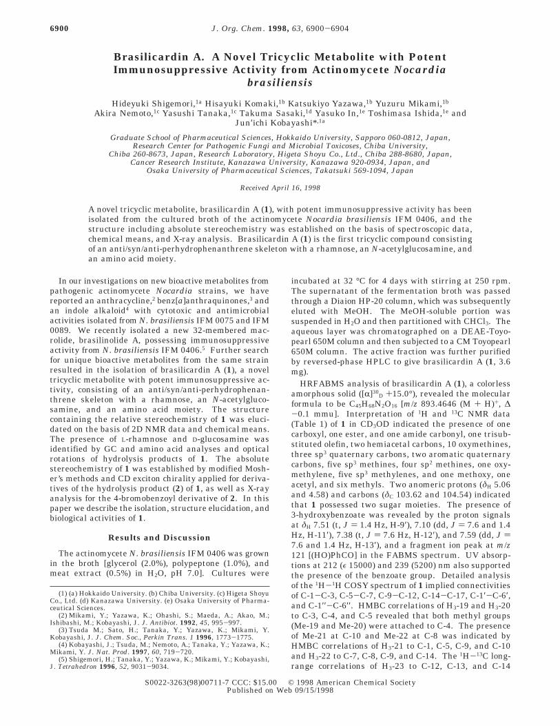
revealed that a vinyl methy group (Me-23) was attachedto C-13. These spectral data suggested that 1 possesseda perhydrophenanthrene skeleton (Chart 1).6 A methoxygroup (δH 3.53) was attached at C-16 judging from anHMBC correlation between H-16 (δH 3.81) and themethoxy carbon (δC 58.90). The presence of a carboxylgroup at C-17 was indicated by an HMBC correlation of
H-17 to C-18, while an amino group was also attachedat C-17 (δC 55.36) from comparison with R-carbon chemi-cal shifts (δC 50-60) of amino acids. 1H-1H couplingconstants (J1′,2′ ) 1.1 Hz, J2′,3′ ) 3.1 Hz, J3′,4′ ) 9.8 Hz,and J4′,5′ ) 9.8 Hz) indicated that a sugar moiety (C-1′-C-6′) was rhamnose, while another sugar moiety wasassigned as glucosamine on the basis of 1H-1H couplingconstants (J1′′,2′′ ) 8.5 Hz, J2′′,3′′ ) 10.0 Hz, and J3′′,4′′ )8.1 Hz) and ROESY correlations of 1′′/3′′, 1′′/5′′, 2′′/4′′,4′′/6′′a, and 4′′/6′′b (Figure 1). The presence of therhamnose was firmly identified on the basis of GCanalysis of the trimethylsilyl derivatives of the metha-nolysis product of 1, while the presence of a glucosaminewas identified by amino acid analysis of the acid hydroly-sate of 1. An HMBC correlation of H-4′ to C-7′ revealedthat 3-hydroxybenzoate was attached at C-4′, while anacetyl group (δH 1.54) was attached at the amino groupof glucosamine by an HMBC correlation of H-2′′ (δH 3.58)to the acetyl carbonyl (δC 174.55). The N-acetylglu-cosamine was connected at C-3′ of the rhamnose from1H-13C long-range correlation between H-3′ and C-1′′. AnHMBC correlation between H-2 and C-1′ indicated thatthe rhamnose was attached at C-2. Stereochemistry ateach anomeric position of the sugar moieties was as-signed as R for rhamnose and â for glucosamine, respec-tively, on the basis of the one-bond 1H-13C couplingconstants7 obtained by an INEPT experiment8 of 1 inCD3OD (C-1′, 1JC,H ) 171.0 Hz; C-1′′, 1JC,H ) 165.4 Hz)9
and 1H-1H coupling constants (J1′,2′ ) 1.1 Hz; J1′′,2′′ )8.5 Hz). The existence of the â-glycoside bond at C-1′′was consistent with ROESY correlation observed betweenH-1′′ and H-5′′. Relative stereochemistry of the anti/syn/anti-perhydrophenanthrene skeleton (Figure 1) was elu-cidated by ROESY data. ROESY correlations of H-2/H3-19, H-3/H-5, H-3/H3-20, and H-1b/H3-21 indicated thatring A had a chair conformation, while a boat conforma-tion of ring B was assigned from ROESY correlation ofH-5/H3-22. Relative stereochemistry of the amino acidmoiety (C-16-C-18) was elucidated to be erythro betweenC-16 and C-17 by comparison of 1H-1H coupling constant(J16,17 ) 2.6 Hz) with those of O-methylthreonine (JR,â
<1 Hz) and allo-O-methythreonine (JR,â ) 3.6 Hz). Thusthe relative stereostructure of brasilicardin A was as-signed to be 1.
Methanolysis of brasilicardin A (1) yielded the aglycone2, methyl R-glucosamine (3), methyl R-rhamnopyranoside(4), and methyl 3-hydroxybenzoate (5) (Scheme 1). The
(6) Nishizawa, M.; Takenaka, H.; Hayashi, Y. J. Org. Chem. 1986,51, 806-813.
(7) Bock, K.; Pedersen, C. J. Chem. Soc., Perkin Trans. 2 1974, 293-297.
(8) Morris, G. A.; Freeman, R. J. Am. Chem. Soc. 1979, 101, 760-762.
(9) In hexapyranoses 1J (C, equatrial H) is usually around 170 Hz,while 1J (C, axial H) is around 160 Hz. The former corresponded to anR-anomer and the latter to a â-anomer: Hansen, P. E. Prog. NMRSpectrosc. 1981, 14, 175-296.
Table 1. 1H and 13C NMR Data of Brasilicardin A (1) inCD3OD
1Ha 13Caposition J (Hz) HMBCb (1H)
1(a) 1.49 m 44.53 t 211(b) 1.82 m2 3.73 t 9.0 80.31 d 33 3.06 d 9.7 83.83 d 2, 19, 204 41.70 s 3, 19, 205 1.69 m 19.22 d 6b, 9, 19, 20, 216(a) 1.69 m 46.75 t6(b) 1.78 m7 1.41 m 31.78 t 6b, 228 39.05 s 9, 14, 15, 229 1.32 m 47.87 d 11b, 12, 14, 21, 2210 38.09 s 1a, 9, 2111 1.93 brs 27.57 t 912 5.39 brs 124.08 d 11b, 14, 2313 139.04 s 11b, 14, 2314 1.61 m 52.81 d 12, 22, 2315(a) 1.41 m 32.46 t 14, 1615(b) 1.49 m16 3.81 dd 11.4, 3.3 81.07 d 14, 17
OMe 3.53 s 58.90 q 1617 4.46 d 3.5 55.36 d18 170.62 s 1719 0.97 s 17.82 q 1820 1.04 s 29.74 q 1821 1.14 s 29.42 q 922 1.09 s 23.06 q 1423 1.69 s 23.13 q 121′ 5.06 d 1.1 103.62 d 22′ 4.46 dd 3.1, 1.1 72.60 d 1′3′ 4.12 dd 9.8, 3.1 80.35 d 1′, 2′4′ 5.31 t 9.8 74.64 d 2′, 3′, 6′5′ 4.05 dq 9.8, 6.2 68.58 d 1′, 4′, 6′6′ 1.16 d 6.2 18.30 q 4′7′ 167.50 s 4′8′ 132.96 s 9′, 12′9′ 7.51 t 1.4 117.94 d 10′, 13′10′ 159.50 s 9′, 12′11′ 7.10 dd 7.6, 1.4 122.04 d 13′12′ 7.38 t 7.6 131.38 d13′ 7.59 dd 7.6, 1.4 122.50 d 9′, 11′1′′ 4.58 d 8.5 104.54 d 3′2′′ 3.58 dd 10.0, 8.5 58.02 d
NHAc 1.54 s 23.42 q174.55 s 2′′
3′′ 3.41 dd 10.0, 8.1 75.80 d 2′′4′′ 3.35 m 72.33 d 3′′, 6′′b5′′ 3.33 m 78.30 d 6′′a6′′(a) 3.73 d 11.6 63.07 t6′′(b) 3.93 dd 11.6, 1.6
a δ in ppm. b Delay time (∆) for C-H long-range coupling wasset to 50 ms.
Chart 1
Figure 1. Relative stereochemistry of brasilicardin A (1).
Brasilicardin A J. Org. Chem., Vol. 63, No. 20, 1998 6901
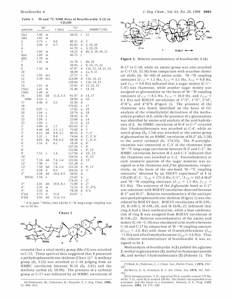
absolute configurations of compounds 3 and 4 wereassigned as D and L, respectively, by comparison of opticalrotations for 3 ([R]D +49°) and for 4 ([R]D -39°) with thoseof synthetic 3 ([R]D +49°) and 4 ([R]D -46°). Sincebrasilicardin A (1) was elucidated to possess a 1,2-diolfunctionality at C-2 and C-3, the CD exciton chiralitymethod10 was applied to determine absolute stereochem-istry of the perhydrophenanthrene skeleton. The 1H-1H coupling constant (J2,3 ) 10.3 Hz) between H-2 (δH
5.38) and H-3 (δH 5.10) in compound 6, which was de-rived from compound 2 by 4-dimethylaminobenzoylation(Scheme 2, A), indicated that the two 4-dimethylami-nobenzoate groups at C-2 and C-3 were trans diequatri-ally disposed. The CD spectrum of 6 showed well-splitintense Cotton effects, ∆ε +43.8 at 322 nm and ∆ε -24.6at 297 nm, indicating that the projection of the two4-dimethylaminobenzoate groups at C-2 and C-3 shouldbe clockwise. These results allowed assignment of theS configurations for C-2 and C-3. To determine theabsolute configuration at C-17 of brasilicardin A (1), com-pound 2 was converted into the (S)- and (R)-2-meth-oxy-2-trifluoromethylphenylacetamides (MTPA amides)(7 and 8, respectively) (Scheme 2, B).11 The values of∆δ [δ(S-MTPA amide) - δ(R-MTPA amide)] (Figure 2)indicated that the absolute configuration at C-17 of 2 wasS. Furthermore, compound 2 was converted into thep-bromobenzamide (9) (Scheme 2, C), which was crystal-lized from EtOH to give prisms of space group P212121.The crystal structure was solved by the direct methodand refined by a full-matrix least-squares method to R) 0.053 and Rw ) 0.156 using 1845 (I > 2σ (I)) observedreflections. The absolute stereostructure of 9 was estab-lished by the X-ray analysis as shown in Figure 3, whichcorresponded to that of 1 elucidated by NMR and CDdata. Thus, the absolute stereochemistry of 1 wasdetermined as shown in Chart 1.
Brasilicardin A (1) is a novel tricyclic metabolite con-taining a rhamnose, an N-acetylglucosamine, and an
amino acid moiety from the broth of N. brasiliensis IFM0406. This is the first isolation of perhydrophenanthrenederivative with two sugar units from natural sources. Theanti/syn/anti-perhydrophenanthrene skeleton such as 1is very rare, although only one example, isoaplysin-20from a sea hare,12,13 has been reported. On the otherhand, tricyclic diterpenoids and sesterterpenoids pos-sessing an anti/anti/anti-perhydrophenanthrene skeletonhave been isolated from a plant14 and a marine nudi-branch.15 Biosynthetically the anti/syn/anti-perhydro-phenanthrene skeleton with a boat form for ring B maybe derived from cyclization of (E,E,E)-geranylgeranylacetate.6 Brasilicardin A (1) exhibited a potent immu-nosuppressive activity in mouse mixed lymphocyte assay(IC50, 0.07 µg/mL), while the IC50 values of cyclosporin Aand ascomycin, known immunosuppresive agents, in theassay were 0.016 and 0.04 µg/mL, respectively. Prelimi-nary study suggests that the mode of action of 1 mightbe different from those of cyclosporin A or FK506.Compound 1 showed cytotoxicity against murine leuke-mia L1210 cells (IC50, 1.2 µg/mL), human epidermoidcarcinoma KB cells (IC50, 1.3 µg/mL), and adriamycin-resistant murine leukemia P388/ADM cells (IC50, 0.22 µg/mL), while 1 exhibited antifungal activity against Paecil-omyces variotti (MIC, 25 µg/mL). Testing of 1 in thehuman tumor screen using 38 types of cell lines16,17 gavea striking pattern of differential cytotoxicity. COMPARE
(10) Harada, N.; Nakanishi, K. In Circular Dichroic Spectroscopy-Exciton Coupling in Organic Stereochemistry; University ScienceBooks: Mill Valley, CA, 1983.
(11) Kusumi, T.; Fukushima, T.; Ohatani, I.; Kakisawa, H. Tetra-hedron Lett. 1991, 32, 2939-2942.
(12) Yamamura, S.; Terada, Y. Tetrahedron Lett. 1977, 2171-2172.(13) Nishizawa, M.; Takenaka, H.; Hirotsu, K.; Higuchi, T.; Hayashi,
Y. J. Am. Chem. Soc. 1984, 106, 4290-4291.(14) Kamaya, R.; Masuda, K.; Suzuki, K.; Ageta, H.; Hsu, H.-Y.
Chem. Pharm. Bull. 1996, 44, 690-694.(15) Kubanek, J.; Graziani, E. I.; Andersen, R. J. J. Org. Chem. 1997,
62, 7239-7246.(16) Yamori, T. Jpn. J. Cancer Chemother. 1997, 24, 129-135.(17) Boyd, M. R. Prin. Pract. Oncol. 1989, 3, 1-12.
Scheme 1
Scheme 2
Figure 2. 1H NMR chemical shift differences (∆δ) for MTPAamides of compound 2. ∆δ (ppm) ) δ[(S)-MTPA amide (7) -(R)-MTPA amide (8)].
Figure 3. Perspective ORTEP drawing of the X-ray structureof 9.
6902 J. Org. Chem., Vol. 63, No. 20, 1998 Shigemori et al.

pattern-recognition analyses of the mean-graph profilesof 1 did not reveal any significant correlations to theprofiles of known antitumor compounds contained in thestandard agent database.16,17 The mean panel GI50
concentration of 1 was approximately 0.43 µM. Detailedbioactivities of 1 will be reported elsewhere.
Experimental Section
Cultivation. The voucher specimen (Nocardia brasiliensisIFM 0406, deposit No. FERM BP-5498)18 was deposited at theCenter for Pathogenic Fungi and Microbial Toxicoses, ChibaUniversity. The actinomycete N. brasiliensis IFM 0406 wasgrown in the broth [glycerol (2.0%), polypeptone (1.0%), andmeat extract (0.5%) in H2O, pH 7.0]. Cultures were incubatedin a 150 L jar fermentor at 32 °C for 4 days with stirring at250 rpm and 150 L/min aeration rate and were centrifuged.
Extraction and Separation. The supernatant of thefermentation broth (15 L) was passed through a Diaion HP-20 column (5 × 30 cm) and washed with 2 M NaCl(aq) (4 L)and H2O (4 L) and then eluted batchwise with MeOH/H2O (1:1, 4 L) and MeOH (2 L). The eluant with MeOH wassuspended in H2O and partitioned with CHCl3. The aqueouslayer was chromatographed on a DEAE-Toyopearl 650Mcolumn (2.5 × 10 cm) eluted with 20 mM Tris-HCl (pH 8). Theactive fraction (32 mg) was subjected to a CM Toyopearl 650Mcolumn (2.5 × 10 cm) eluted with 20 mM NaOAc-HCl (pH 4).The fraction (17 mg) was further purified by reversed-phaseHPLC (Capcell pak C18 SG120, Shiseido Co. Ltd., 3 × 25 cm,flow rate 3.0 mL/min, 18%f42% MeCN/H2O containing 0.15%TFA) to give brasilicardin A (1, 3.6 mg).
Brasilicardin A (1): A colorless amorphous solid: mp 270-273 °C; [R]30
D +15.0° (c 0.50, MeOH); IR (KBr) νmax 3432, 2934,1676, 1454, 1378, 1291, 1203, 1075, 893, 839, 801, 755, 722,and 570 cm-1; UV (MeOH) λmax 212 (ε 15000), 239 (5200), and300 (1900) nm; 1H and 13C NMR (see Table 1); FABMS(positive, glycerol matrix) m/z 893 (M + H)+ and 121; HR-FABMS m/z 893.4646 (M + H)+, calcd for C45H69N2O16,893.4647; HMBC correlations (see Table 1); ROESY correla-tions (CD3OD, H/H) 1b/21, 2/19, 2/21, 2/1′, 3/5, 3/20, 5/22, 6b/20, 11/22, 12/23, 14/17, 14/22, 16/16-OMe, 17/16-OMe, 4′/6′,1′′/3′′, 1′′/5′′, 2′′/4′′, 4′′/6′′a, and 4′′/6′′b.
Sugar Analysis of 1 by GC. Compound 1 (0.5 mg) wasdissolved in 5% HCl/MeOH (0.5 mL) and heated at 65 °C for15 h in a sealed tube. After evaporation of the solvent by astream of nitrogen, the residue was dissolved in pyridine (50µL) and treated with hexamethyldisilazane (10 µL) andtrimethylsilyl chloride (5 µL) at room temperature for 30 min.Solvent was removed by a nitrogen stream, and the residuedissolved in hexane was used for GC analysis [1.5% OV-17glass column (3 mm × 2 m); N2 as a carrier gas; the programrate 120-200 °C at 0.5 °C/min], showing a peak at tR 4.4 min,which corresponded to that of rhamnose (4.4 min) but notfucose (5.3 min). The TMS/Me derivatives of 6-deoxyhexoses,rhamnose and fucose, were prepared as authentic specimens.
Methanolysis of 1. Compound 1 (2.0 mg) was dissolvedin 5% HCl/MeOH (1.0 mL) and heated at 65 °C for 10 h in asealed tube. After evaporation of the solvent by a stream ofnitrogen, the residue was separated by a silica gel column(CHCl3/MeOH, 14:1 f BuOH/AcOH/H2O, 8:1:1) to affordcompounds 2 (0.6 mg, yield 61%), 3 (0.3 mg), 4 (0.2 mg), and5 (0.2 mg).
Compound 2: A colorless amorphous solid; [R]26D +58° (c
0.1 MeOH); IR (film) νmax 3369, 2929, 1735, 1560, 1438, 1377,1261, 1101, 802, and 755 cm-1; 1H NMR (CDCl3) δ 5.30 (1H,s, H-12), 3.89 (1H, d, J ) 2.6 Hz, H-17), 3.75 (3H, s, MeO),3.72 (1H, td, J ) 9.5 and 5.2 Hz, H-2), 3.42 (1H, m, H-16),3.41 (3H, s, MeO), 2.95 (1H, d, J ) 9.5 Hz, H-3), 1.89 (1H, m,H-11a), 1.86 (3H, s, H-23), 1.80 (1H, m, H-11b), 1.72 (1H, m,H-6a), 1.71 (1H, m, H-14), 1.70 (1H, m, H-1a), 1.60 (1H, m,
H-6b), 1.55 (1H, m, H-7a), 1.54 (1H, m, H-5), 1.52 (1H, m,H-14), 1.42 (1H, m, H-15a), 1.35 (1H, m, H-15b), 1.34 (1H, m,H-1b), 1.28 (1H, m, H-7b), 1.24 (1H, m, H-9), 1.05 (3H, s, H-21),0.99 (6H, s, H-20 and H-22), and 0.92 (3H, s, H-19); 13C NMR(CDCl3) δ 173.91 (s, C-18), 137.79 (s, C-13), 121.97 (d, C-12),83.88 (d, C-3), 82.97 (d, C-16), 69.47 (d, C-2), 57.88 (q, MeO),55.35 (d, C-17), 51.98 (q, MeO), 50.98 (d, C-14), 45.95 (d, C-9),43.23 (t, C-1), 43.08 (t, C-6), 39.56 (s, C-4), 37.26 (s, C-8), 36.54(s, C-10), 30.79 (t, C-15), 29.93 (t, C-7), 28.61 (q, C-20), 28.26(q, C-21), 26.02 (t, C-11), 22.59 (q, C-22), 22.26 (q, C-23), 17.65(d, C-5), and 16.64 (q, C-19); EIMS m/z 437 (M+), 378, 349,331, 317, 299, 281, 207, and 89; HREIMS m/z 378.2972 (M -CO2Me)+, calcd for C23H40NO3, 378.3008.
Tris-4-dimethylaminobenzoyl Derivative (6) from 2.The mixture of compound 2 (0.2 mg) and excess 4-dimeth-ylaminobenzoyl chloride in dry pyridine (0.2 mL) was heatedat 80 °C for 2 h. After complete removal of pyridine in vacuo,the residue was purified by a silica gel column (hexane/acetone,5:2) to give compound 6 (0.2 mg, yield 50%), a colorlessamorphous solid: [R]27
D +94.0° (c 0.085, MeOH); IR (KBr) νmax
1739, 1702, 1608, 1530, and 1447 cm-1; UV (hexane) λmax 310(ε 39400) and 224 (14100) nm; CD (hexane) 322 (∆ε +43.8)309 (0), and 297 (-24.6) nm; 1H NMR (CDCl3) δ 7.85 (2H, d,J ) 10.0 Hz, 4-(Me2N)Bz), 7.78 (2H, d, J ) 8.9 Hz, 4-(Me2N)-Bz), 7.70 (2H, d, J ) 9.0 Hz, 4-(Me2N)Bz), 6.65 (2H, d, J ) 9.0Hz, 4-(Me2N)Bz), 6.57 (2H, d, J ) 8.9 Hz, 4-(Me2N)Bz), 6.55(2H, d, J ) 10.0 Hz, 4-(Me2N)Bz), 5.38 (1H, td, J ) 10.3 and5.3 Hz, H-2), 5.35 (1H, m, H-17), 5.30 (1H, brs, H-12), 5.15(1H, m, H-16), 5.10 (1H, d, J ) 10.3 Hz, H-3), 3.79 (3H, s,MeO), 3.50 (3H, s, MeO), 3.09 (3H, s, NMe2), 3.08 (3H, s, MeN),3.07 (3H, s, MeN), 3.06 (3H, s, MeN), 3.05 (3H, s, MeN), 3.04(3H, s, MeN), 2.00 (1H, m, H-11a), 1.85 (1H, m, H-11b), 1.82(1H, m, H-1a), 1.81 (1H, m, H-14), 1.75 (1H, m, H-6a), 1.63(3H, s, H-23), 1.58 (1H, m, H-5), 1.52 (1H, m, H-7a), 1.42 (1H,m, H-15a), 1.35 (1H, m, H-15b), 1.34 (1H, m, H-1b), 1.32 (1H,m, H-6b), 1.28 (1H, m, H-7b), 1.24 (1H, m, H-9), 1.20 (3H, s,H-21), 1.15 (3H, s, H-19), 1.05 (3H, s, H-22), and 0.95 (3H, s,H-20); FDMS m/z 878 (M+); HRFDMS m/z 878.5161 (M+), calcdfor C52H70N4O8, 878.5194.
Preparation of (S)- or (R)-MTPA Amide (7 or 8) ofCompound 2. A solution of compound 2 (0.16 mg) in CH2Cl2
(30 µL) was treated with DCC (0.15 mg) and (S)-MTPA acid(0.17 mg), and then the mixture was stirred at room temper-ature for 1 h. The reaction mixture containing the precipitatesof urea was concentrated, and the residue was separated bypreparative SiO2 TLC (hexane/EtOAc, 1:1) to afford the (S)-MTPA amide (7, 0.16 mg) of 2. The (R)-MTPA amide (8) of 2was also prepared from 2 and (R)-MTPA acid by the sameprocedure as 7.
Compound 7: A colorless oil; [R]27D +104° (c 0.12, MeOH);
IR (KBr) νmax 3432, 1742, 1707, 1631, 1561, 1516, and 1450cm-1; UV (MeOH) λmax 207 (ε 7500) nm; 1H NMR (CDCl3) δ7.592 (2H, m, Ph), 7.363 (3H, m, Ph), 7.205 (1H, brs, NH-17),5.265 (1H, s, H-12), 5.207 (1H, d, J ) 2.6 Hz, H-17), 3.779(3H, s, CO2Me-18), 3.697 (1H, td, J ) 9.5 and 5.2 Hz, H-2),3.544 (1H, m, H-16), 3.494 (3H, s, MeO-MTPA), 3.394 (3H, s,MeO-16), 2.957 (1H, d, J ) 9.5 Hz, H-3), 1.89 (1H, m, H-11a),1.80 (1H, m, H-11b), 1.72 (1H, m, H-6a), 1.71 (1H, m, H-14),1.70 (1H, m, H-1a), 1.60 (1H, m, H-6b), 1.55 (1H, m, H-7a),1.54 (1H, m, H-5), 1.479 (3H, s, H-23), 1.42 (1H, m, H-15a),1.35 (1H, m, H-15b), 1.34 (1H, m, H-1b), 1.28 (1H, m, H-7b),1.24 (1H, m, H-9), 1.05 (3H, s, H-21), 0.99 (3H, s, H-20), 0.974(3H, s, H-22), and 0.92 (3H, s, H-19); EIMS m/z 653 (M+) and621 (M - MeOH)+; HREIMS m/z 653.3544 (M+), calcd forC35H50NO7F3, 653.3539.
Compound 8: A colorless oil; [R]30D +76° (c 0.16, CHCl3);
IR (KBr) νmax 3431, 1742, 1705, 1627, 1577, 1516, and 1450cm-1; UV (MeOH) λmax 207 (ε 6400) nm; 1H NMR (CDCl3) δ7.549 (2H, m, Ph), 7.405 (3H, m, Ph), 7.295 (1H, brs, NH-17),5.322 (1H, s, H-12), 5.065 (1H, d, J ) 2.6 Hz, H-17), 3.739(3H, s, CO2Me-18), 3.699 (1H, td, J ) 9.5 and 5.2 Hz, H-2),3.593 (1H, m, H-16), 3.433 (3H, s, MeO-MTPA), 3.443 (3H, s,MeO-16), 2.961 (1H, d, J ) 9.5 Hz, H-3), 1.89 (1H, m, H-11a),1.80 (1H, m, H-11b), 1.72 (1H, m, H-6a), 1.71 (1H, m, H-14),1.70 (1H, m, H-1a), 1.624 (3H, s, H-23), 1.60 (1H, m, H-6b),
(18) Tanaka, Y.; Komaki, H.; Yazawa, K.; Mikami, Y.; Nemoto, A.;Tojyo, T.; Kadowaki, K.; Shigemori, H.; Kobayashi, J. J. Antibiot. 1997,50, 1036-1041.
Brasilicardin A J. Org. Chem., Vol. 63, No. 20, 1998 6903
1.55 (1H, m, H-7a), 1.54 (1H, m, H-5), 1.42 (1H, m, H-15a),1.35 (1H, m, H-15b), 1.34 (1H, m, H-1b), 1.28 (1H, m, H-7b),1.24 (1H, m, H-9), 1.05 (3H, s, H-21), 0.99 (3H, s, H-20), 0.987(3H, s, H-22), and 0.92 (3H, s, H-19); EIMS m/z 653 (M+) and621 (M -MeOH)+; HREIMS m/z 621.3248 (M-MeOH)+, calcdfor C34H46NO7F3, 621.3277.
4-Bromobenzoyl Derivative (9) from 2. A solution ofcompound 2 (1.6 mg) in CH2Cl2 (100 µL) was treated with DCC(1.9 mg) and 4-bromobenzoic acid (1.9 mg), and the mixturewas stirred at room temperature for 8 h. The reaction mixturecontaining the precipitates of urea was concentrated, and theresidue was separated by preparative SiO2 TLC (CHCl3/MeOH,12:1) to afford compound 9 (1.6 mg, yield 70%), a colorless plate(from EtOH): mp 286-287 °C; [R]27
D +118° (c 0.32, CHCl3);IR (KBr) νmax 3435, 1741, 1648, 1592, 1540, 1482, and 1439cm-1; UV (MeOH) λmax 241 (ε 16900) and 210 (15500) nm; 1HNMR (CDCl3) δ 7.68 (2H, d, J ) 8.5 Hz, 4-BrBz), 7.59 (2H, d,J ) 8.5 Hz, 4-BrBz), 6.63 (1H, d, J ) 8.0 Hz, NH-17), 5.33(1H, brs, H-12), 5.17 (1H, dd, J ) 8.1 and 3.5 Hz, H-16), 3.80(3H, s, CO2Me-18), 3.71 (1H, td, J ) 9.5 and 4.3 Hz, H-2), 3.62(1H, dd, J ) 8.0 and 3.5 Hz, H-17), 3.48 (3H, s, MeO-16), 2.97(1H, d, J ) 9.5 Hz, H-3), 1.91 (1H, brd, J ) 17.0 Hz, H-11a),1.83 (1H, brdd, J ) 17.0 and 13.5 Hz, H-11b), 1.74 (1H, m,H-6a), 1.72 (1H, m, H-14), 1.71 (1H, m, H-1a), 1.60 (1H, m,H-6b), 1.55 (3H, s, H-23), 1.55 (1H, m, H-7a), 1.54 (1H, m, H-5),1.42 (1H, m, H-15a), 1.35 (1H, m, H-15b), 1.34 (1H, m, H-1b),1.28 (1H, m, H-7b), 1.25 (1H, m, H-9), 1.05 (3H, s, H-21), 1.00(3H, s, H-20), 0.99 (3H, s, H-22), and 0.90 (3H, s, H-19); EIMSm/z 621 and 619 (M+, 1:1); HREIMS m/z 619.2512 (M+), calcdfor C32H46NO6
79Br, 619.2509.X-ray Crystallography of 9. Compound 9 was obtained
as a colorless prism from EtOH. Compound 9 was crystallizedas an orthorhombic system, space group P212121 with onemolecule per asymmetric unit. Cell constants were a ) 12.431-(1) Å, b ) 21.490(3) Å, c ) 12.094(3) Å, â ) 90.00°, and V )3230.9(7) Å3. All unique reflections with 3° < 2θ < 126° were
collected on a Rigaku AFC-5 diffractometer using graphite-monochromated Cu KR radiation and employing ω-2θ scanmode. In 2941 collected reflections 1845 reflections (I > 2σ-(I)) were judged as observed and used for the structuredetermination and refinement. The structure was solved bydirect methods and refined by a full-matrix least-squaresmethod with anisotropic thermal parameters [SHELXL93program].19 The positions of H atoms were obtained from adifference Fourier map and were included in the final refine-ment. The residual factors were R ) 0.053 and Rw ) 0.156for 1845 observed reflections.20
Acknowledgment. The authors thank the Screen-ing Committee of New Anticancer Agents supported bya Grant-in-Aid for Scientific Research on Priority Area“Cancer” from the Ministry of Education, Science,Sports, and Culture of Japan for testing in the humantumor screen. This work was partly supported by aGrant-in-Aid for Scientific Research from the Ministryof Education, Science, Sports, and Culture of Japan.
Supporting Information Available: All spectra of 1,NMR spectra of compounds 2, 6, 7, 8, and 9, and X-raycrystallographic data of 9 (18 pages). This material is con-tained in libraries on microfiche, immediately follows thisarticle in the microfilm version of the journal, and can beordered from the ACS; see any current masthead page forordering information.
JO9807114
(19) Sheldrick, G. M. SHELXL93, Program for the Refinement ofCrystal Structure; University of Gottingen: Gottingen, 1993.
(20) Lists of structure factors, anisotropic distancement parameters,H atom coordinates, and complete geometry have been deposited withthe IUCr (Reference: AS1088).
6904 J. Org. Chem., Vol. 63, No. 20, 1998 Shigemori et al.