breast disorders2 8-11
TRANSCRIPT

Disorders of the BreastUNC School of Medicine
Obstetrics and Gynecology ClerkshipCase Based Seminar Series
Objectives for Disorders of the Breast
Describe the symptoms and physical examination findings of benign or malignant conditions of the breast
Demonstrate the performance of a clinical breast examination
Discuss the steps in evaluation of common breast complaints: mastalgia, mass, nipple discharge
Discuss the initial management options for benign and malignant conditions of the breast
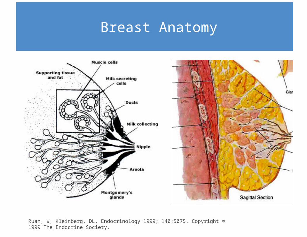
Primarily adipose tissue, glandular tissue, and suspensory ligaments
Composed of 15-25 radially arranged lobes of parenchyma, each associated with a major lactiferous duct
Each major duct extends from the nipple to terminate in a “terminal duct-lobular unit” via branching ducts of diminishing caliber
Breast Anatomy
Breast Anatomy
Ruan, W, Kleinberg, DL. Endocrinology 1999; 140:5075. Copyright © 1999 The Endocrine Society.

History: Change in general appearance of breast (size, symmetry) New or persistent skin changes New nipple inversion Breast pain (cyclic vs. noncyclic, duration, location in breast) Breast mass (how it was discovered, duration, change in size, location) Relationship of mass to menstrual cycles Nipple discharge (unilateral vs. bilateral, color) Medications (e.g. hormones) Risk factors for breast cancer
Evaluation: History
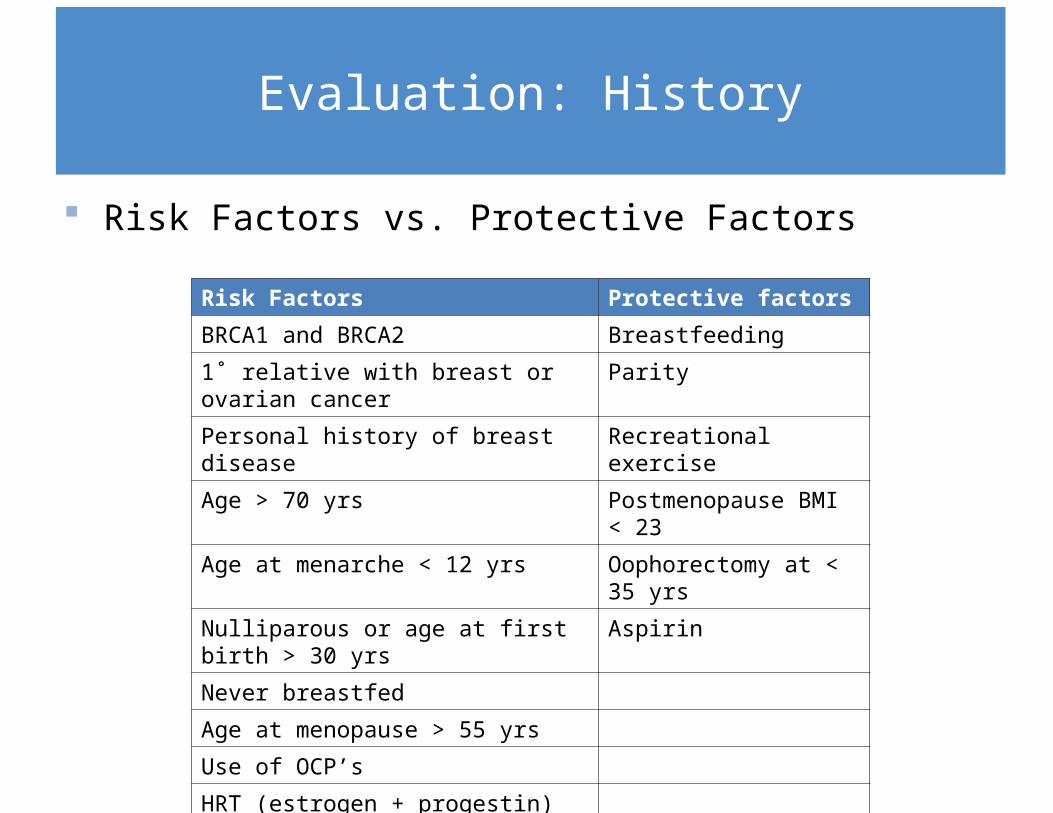
Risk Factors vs. Protective Factors
Evaluation: History
Risk Factors Protective factors
BRCA1 and BRCA2 Breastfeeding
1˚ relative with breast or ovarian cancer Parity
Personal history of breast disease Recreational exercise
Age > 70 yrs Postmenopause BMI < 23
Age at menarche < 12 yrs Oophorectomy at < 35 yrs
Nulliparous or age at first birth > 30 yrs Aspirin
Never breastfed
Age at menopause > 55 yrs
Use of OCP’s
HRT (estrogen + progestin)
Radiation exposure to chest
EtOH
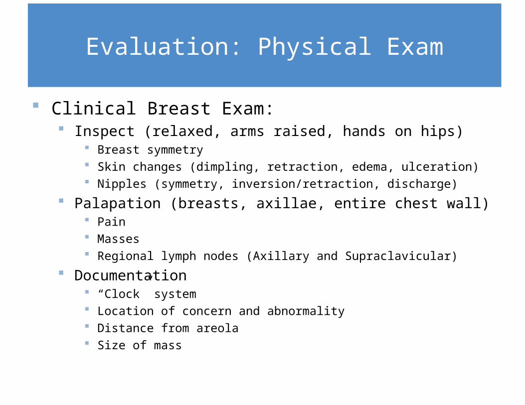
Clinical Breast Exam: Inspect (relaxed, arms raised, hands on hips)
Breast symmetry Skin changes (dimpling, retraction, edema, ulceration) Nipples (symmetry, inversion/retraction, discharge)
Palapation (breasts, axillae, entire chest wall) Pain Masses Regional lymph nodes (Axillary and Supraclavicular)
Documentation “Clock” system Location of concern and abnormality Distance from areola Size of mass
Evaluation: Physical Exam
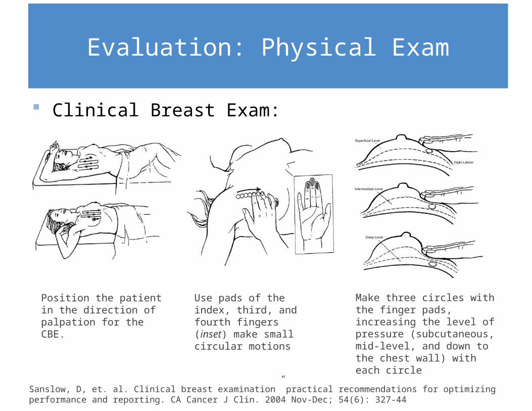
Clinical Breast Exam:
Evaluation: Physical Exam
Use pads of the index, third, and fourth fingers (inset) make small circular motions
Make three circles with the finger pads, increasing the level of pressure (subcutaneous, mid-level, and down to the chest wall) with each circle
Position the patient in the direction of palpation for the CBE.
Sanslow, D, et. al. Clinical breast examination” practical recommendations for optimizing performance and reporting. CA Cancer J Clin. 2004 Nov-Dec; 54(6): 327-44

Benign vs. Malignant
Chief Complaint Benign Characteristics Malignant Characteristics
Breast mass Multiple lesions Single lesion
“Rubbery” Hard
Mobile Immovable
Well circumscribed border Irregular borders
Nipple discharge Bilateral Unilateral
Multiductal Uniductal
Milky Bloody, Clear, or Colored
Spontaneous
Persistent
Skin changes Retraction
Dimpling
Thickening
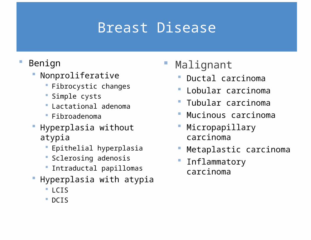
Benign Nonproliferative
Fibrocystic changes Simple cysts Lactational adenoma Fibroadenoma
Hyperplasia without atypia Epithelial hyperplasia Sclerosing adenosis Intraductal papillomas
Hyperplasia with atypia LCIS DCIS
Breast Disease
Malignant Ductal carcinoma Lobular carcinoma Tubular carcinoma Mucinous carcinoma Micropapillary carcinoma Metaplastic carcinoma Inflammatory carcinoma
Approximately 45% of women have mild breast pain, and 21% have severe breast pain in their lifetime
Breast cancer is found in 1.2 – 6.7% of women presenting with breast pain
Mastalgia: Incidence
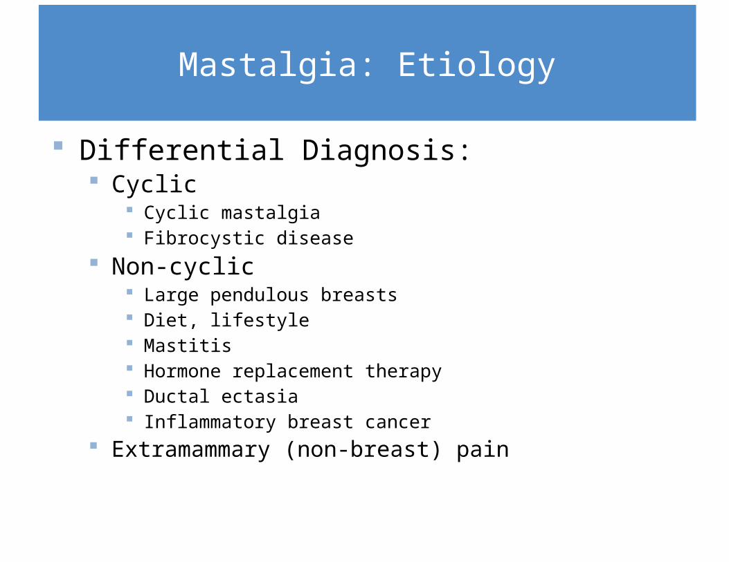
Differential Diagnosis: Cyclic
Cyclic mastalgia Fibrocystic disease
Non-cyclic Large pendulous breasts Diet, lifestyle Mastitis Hormone replacement therapy Ductal ectasia Inflammatory breast cancer
Extramammary (non-breast) pain
Mastalgia: Etiology
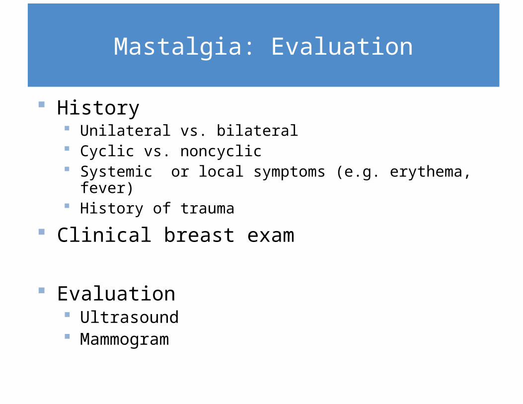
History Unilateral vs. bilateral Cyclic vs. noncyclic Systemic or local symptoms (e.g. erythema, fever) History of trauma
Clinical breast exam
Evaluation Ultrasound Mammogram
Mastalgia: Evaluation
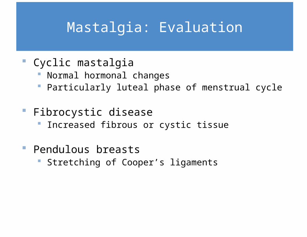
Cyclic mastalgia Normal hormonal changes Particularly luteal phase of menstrual cycle
Fibrocystic disease Increased fibrous or cystic tissue
Pendulous breasts Stretching of Cooper’s ligaments
Mastalgia: Evaluation
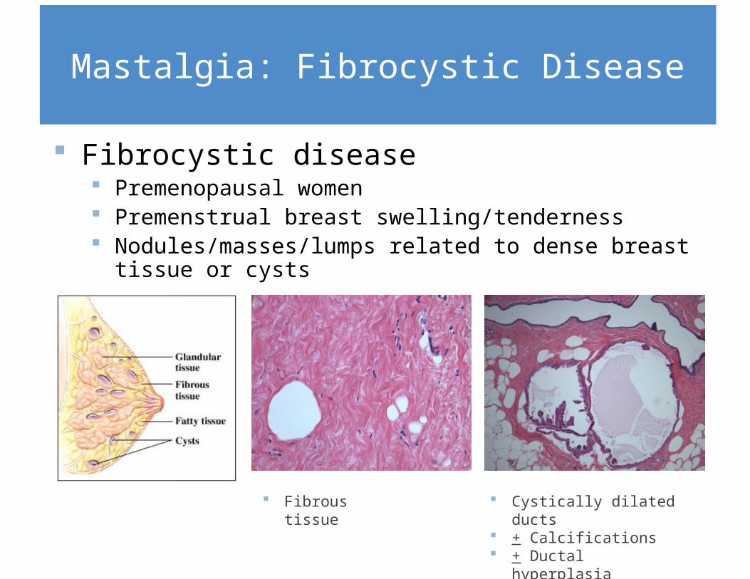
Fibrocystic disease Premenopausal women Premenstrual breast swelling/tenderness Nodules/masses/lumps related to dense breast tissue or cysts
Mastalgia: Fibrocystic Disease
Fibrous tissue Cystically dilated ducts + Calcifications + Ductal hyperplasia
Treatment: Lifestyle
Eliminate caffeine Low fat diet
Symptomatic Support garments (well-fitting, supportive bra, sports bra) Compresses
Medication NSAID’s OCP’s, Progestogens Danazol Bromocriptine GnRH agonists Tamoxifen - IF severe mastalgia
Mastalgia: Management
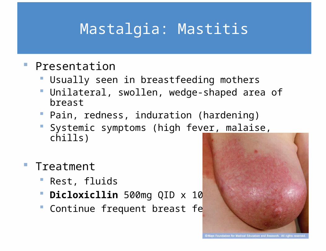
Presentation Usually seen in breastfeeding mothers Unilateral, swollen, wedge-shaped area of breast Pain, redness, induration (hardening) Systemic symptoms (high fever, malaise, chills)
Treatment Rest, fluids Dicloxicllin 500mg QID x 10-14d Continue frequent breast feeding
Mastalgia: Mastitis
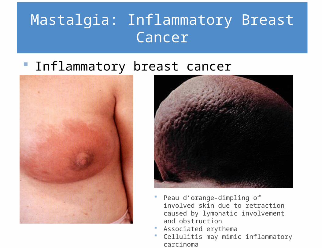
Inflammatory breast cancer
Mastalgia: Inflammatory Breast Cancer
Peau d’orange-dimpling of involved skin due to retraction caused by lymphatic involvement and obstruction
Associated erythema Cellulitis may mimic inflammatory carcinoma
More than 90% of palpable breast masses in women in their 20’s to early 50’s are benign
Differential Diagnosis: Fibrocystic changes Fibroadenoma Fat necrosis Phyllodes tumor Intraductal papilloma Breast cancer
Breast Mass: Etiology

History How it was discovered Duration Change in size Location Relationship of mass to menstrual cycles
Clinical breast exam
Breast Mass: Evaluation

Fibroadenoma Solitary, firm, rubbery, mobile mass Women < 30 yrs Slow growing (? hormonally mediated)
Breast Mass: Fibroadenoma
Fibroadenoma gross specimen Firm, tan, lobulated Well circumscribed mass Variable size
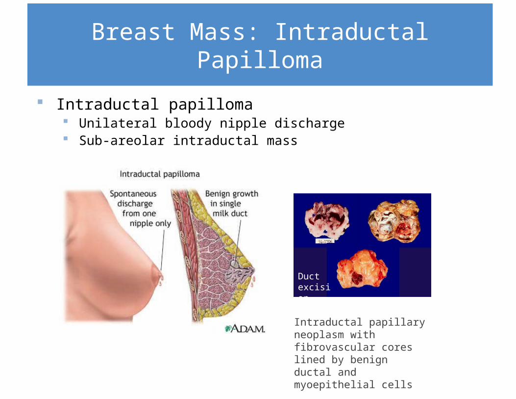
Intraductal papilloma Unilateral bloody nipple discharge Sub-areolar intraductal mass
Breast Mass: Intraductal Papilloma
Intraductal papillary neoplasm with fibrovascular cores lined by benign ductal and myoepithelial cells
Duct excision
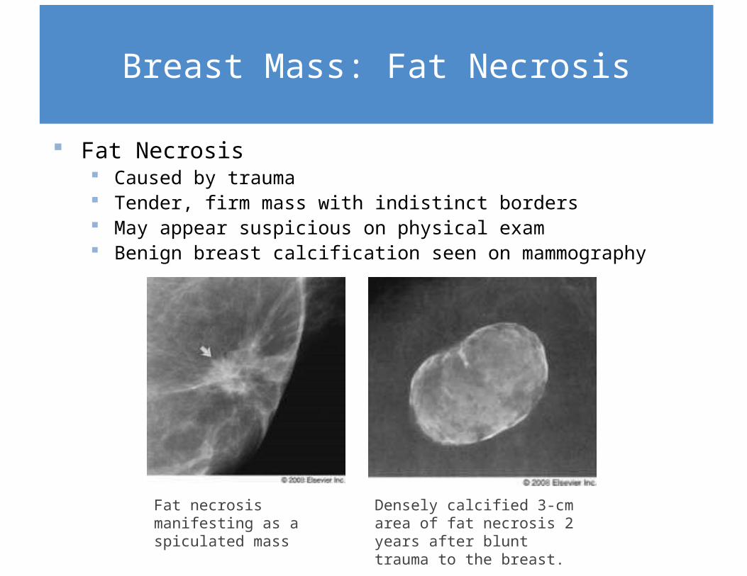
Fat Necrosis Caused by trauma Tender, firm mass with indistinct borders May appear suspicious on physical exam Benign breast calcification seen on mammography
Breast Mass: Fat Necrosis
Fat necrosis manifesting as a spiculated mass
Densely calcified 3-cm area of fat necrosis 2 years after blunt trauma to the breast.

Initial evaluation < 30 yr – Diagnostic ultrasound + Diagnostic mammogram > 30 yr – Diagnostic mammogram
Further evaluation Simple cyst
Symptomatic – Aspirate Asymptomatic – Observe for 2-4 months
Complicated cyst – Ultrasound-guided aspiration Solid mass – Core needle biopsy (CNB) or Excision No specific findings – Re-examine after two cycles
Breast Mass: Evaluation

Breast Ultrasound
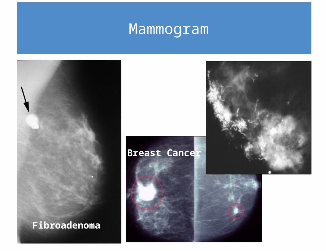
Fibroadenoma
Breast Cancer
Mammogram
Etiology Lactation Physiologic nipple discharge
Hyperprolactinemia Hypothyroidism Medication related Neurogenic stimulation
Pathologic Intraductal papilloma Ductal ectasia DCIS
Nipple Discharge: Etiology
History Unilateral vs. bilateral Spontaneous vs. provoked discharge Appearance of discharge Medications (e.g. antipsychotics, antidepressants) History of trauma History of amenorrhea History of hypogonadism (e.g. hot flashes, vaginal dryness)
Clinical breast exam Attempt to elicit discharge, identify involved duct(s) Evaluate discharge for gross blood or guaiac positivity
Nipple Discharge: Evaluation
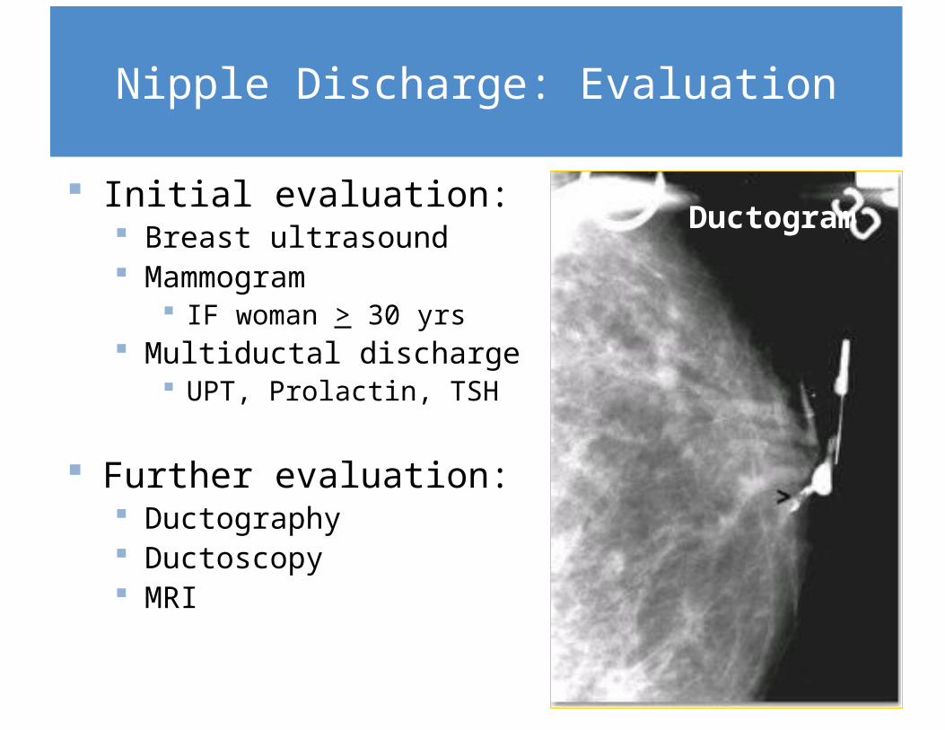
Initial evaluation: Breast ultrasound Mammogram
IF woman > 30 yrs Multiductal discharge
UPT, Prolactin, TSH
Further evaluation: Ductography Ductoscopy MRI
Nipple Discharge: Evaluation
Ductogram
Management Physiologic nipple discharge
Directed at underlying cause Pathologic nipple discharge
Refer to surgeon Terminal duct excision Central (total) terminal duct excision Resection of intraductal papilloma
Nipple Discharge: Management
Pathologic finding on CNB or excision biopsy DCIS/LCIS Invasive carcinoma
Refer to surgical oncologist
Treatment modalities: Radiation Chemotherapy Lumpectomy Mastectomy Hormonal therapy
Malignant Breast Disease
Bottom Line Concepts It is important to evaluate breast complaints thoroughly to ensure that breast
cancers, as well as benign breast lesions, are diagnosed and treated promptly. Evaluation of a woman presenting with a breast complaints requires careful
assessment of symptoms and risk factors for developing breast cancer. The clinical breast exam include inspection and palpation of the breast tissue,
chest wall, and regional lymph nodes. Documentation should included both positive and negative findings.
Women with breast problems can present with any combination of symptoms including breast mass or thickening, breast pain, nipple discharge, or skin changes.
Typically, women presenting with a suspicious breast mass who are > 30 yrs should receive a diagnostic mammogram, whereas women younger than 30 should receive a diagnostic ultrasound.
Negative imaging should not stop further investigation is a suspicious lump is felt on clinical exam.
Masses that are solid on ultrasound imaging require biopsy to exclude cancer and provide a histological diagnosis.

References and Resources
APGO Medical Student Educational Objectives, 9th edition, (2009), Educational Topic 40 (p84-85).
Beckman & Ling: Obstetrics and Gynecology, 6th edition, (2010), Charles RB Beckmann, Frank W Ling, Barabara M Barzansky, William NP Herbert, Douglas W Laube, Roger P Smith. Chapter 31 (p283-294).
Hacker & Moore: Hacker and Moore's Essentials of Obstetrics and Gynecology, 5th edition (2009), Neville F Hacker, Joseph C Gambone, Calvin J Hobel. Chapter 29 (p326-331).