breast imaging “the good, the bad & the ugly” & how to
TRANSCRIPT

BREAST IMAGING “THE GOOD, THE BAD & THE UGLY”
& HOW TO NAVIGATE THE SYSTEM IN THE
CHANGING FACE OF MEDICINE 2015
PHILIP GETSON, D.O. BOARD CERTIFIED THERMOLOGIST
FEBRUARY 25, 2015 T.D.I./ H.T.A. WEBINAR

THE GOOD
THERMOGRAPHY

THE BAD
MAMMOGRAPHY

…AND THE UGLY…

1928
The year in which Dr. D.T. Quigley wrote an article in Radiology warning physicians to handle “cancerous breasts with care- for fear of accidentally disseminating cells” and spreading cancer-40 years before the first mammogram!

RADIATION
In 2005 The National Toxicology Program classified X-radiation and gamma radiation as known human carcinogens. Sources of X-radiation include X-rays, CT scans, fluoroscopy, mammography and other medical radiological procedures. These forms of radiation can break the chemical bonds in molecules including DNA molecules, thereby disturbing their normal functioning

NEW YORK TIMES FRONT PAGE NEWS…
On November 16, 2009 the U.S. Preventative Task force Breast Screening Recommendations for the General Public was released, changing the long standing position on mammography.
It stated that : “Rather than benefiting from screening, women with cancer may incur harm when undergoing mammography, additional imaging and biopsies”

A study in the British Medical Journal in December 2011 confirmed that …
“breast cancer screening (mammograms)
may cause women harm especially during the early years after they start screening. This harm is largely due to surgeries, such as lumpectomies and mastectomies and other (often unnecessary) interventions”

NORDIC COCHRANE CENTRE REPORT
“If 2000 women are screened regularly for 10 years, ONE will benefit from the screening as she will avoid dying from breast cancer
At the same time, 10 healthy women will, as a consequence, become cancer patients and will be treated unnecessarily. These women will have either a part of their breast or the whole breast removed, and they will often receive radiotherapy, and sometimes chemotherapy”

NORDIC COCHRANE CENTRE REPORT
Furthermore, about 200 healthy women will experience false alarm. The psychological strain until one knows whether or not it was cancer, and even afterwards, can be severe.

NORDIC COCHRANE CENTRE REPORT
According to the Nordic Cochrane Center: "It therefore no longer seems reasonable to
attend for breast cancer screening (mammography)”
In fact, by avoiding going to screening (mammography), a woman will lower her risk of getting a breast cancer diagnosis."

A Surgeon’s Opinion
In a Newsweek article dated December 12, 2011,
Dr Susan Love a breast surgeon at UCLA Medical Center stated that at least 30 percent of tumors found on mammograms would go
away if you did absolutely nothing!

In the New England Journal of Medicine on November 22, 2013…
Archie Bleyer, M.D. and Gilbert Welsh, M.D. two gynecologists, concluded that from 1976 -2008 screening mammography accounted for an increase in breast cancer diagnosis of 122 cases per 100,000 women only 8 of which would have progressed to late stage breast cancer. Further they estimated breast cancer was over-diagnosed (tumors that were detected on screening that would never have led to clinical symptoms) in 1.3 million U.S. women in the last 30 years. In 2008 they estimated that more than 70,000 women were over-diagnosed; this accounted for 31% of all diagnosed breast cancers
Additionally Dr Welsh concluded that : “…few breast cancers are
destined to kill no matter what we do!”

In the British Medical Journal of February 2014, an article titled:
“Twenty five year follow-up for breast
cancer incidence and mortality of the Canadian National Breast Screening Study: randomized screening trial”
concluded:

“Annual mammography in women aged 40-59 does not reduce mortality from breast cancer beyond that of physical examination or usual care when adjuvant therapy for breast cancer is freely available.
Overall 22% (160/484) of screen detected invasive breast cancers were over-diagnosed, representing one over-diagnosed breast cancer for every 424 women who received mammography screening in the trial”

B.M.J. Article concluded…
“Assuming that nearly all over-diagnosed cancers in the Canadian National Breast
Screening Study were non-palpable, 50% (106/212) of mammogram detected,
non-palpable cancers were over-diagnosed.”

A FEW WORDS ON DENSE BREASTS
Extensive density can hide tumors, referred to as masking, Bonnie Rush, president, Breast Imaging Specialists, presented at AHRA 2014. - likened it to:
“Trying to find a snowball in a snowstorm.”
The estimated sensitivity of mammography is 80-85% in women who do NOT have dense
breasts and 62-68% in women who do (40-50% of all women)
An American College of Radiology Imaging Network/National Cancer Institute found that in women with greater than 50 percent breast density, the ability of the radiologist to see an abnormality that may be cancer is directly affected, offering only 60 percent sensitivity with high breast density.
The ACR took notice of the severity of the issue and, in 1992, recommended that the
mammography report begin with a description of breast composition, and when the breast tissue is greater than 50 percent dense, a disclaimer should be included regarding the decreased sensitivity of the study, Rush explained.

Breast density considerations July 17, 2014: A bill was introduced into congress setting a Federally mandated
minimum standard for notifying women if they have dense breast tissue The Breast Density and Mammography reporting Act would require that
mammogram reports include whether a woman has dense breast tissue. This is state law in 21 states (with 10 others pending) and in New Jersey since May
1, 2014 where reports read: “Your mammogram may show that you have dense breast tissue as determined by
the Breast Imaging and Reporting and Data System established by the American Academy of Radiology. Dense breast tissue is very common and is not abnormal. However, in some cases, dense breast tissue can make it harder to find a cancer on a mammogram and may also be associated with a risk factor for breast cancer.”

Dr. Charles B. Simone a former clinical assistant professor of immunology and pharmacology at the National Cancer
Institute stated…
“Mammograms increase the risk for developing breast cancer and raise the risk of spreading or metastasizing an existing growth”.

DR SIMONE ADDED…
Since mammographic screening was introduced in 1983, the incidence of DCIS (ductal carcinoma in situ) which represents 12% of all breast cancers has increased by 328% and 200% of this increase is due to the use of mammography. The increase for women screened mammographically under the age of 40 has gone up 3000% !

D.C.I.S. In July, 2013 a working group from the National Cancer Institute published an
opinion in JAMA that stated that DCIS (DUCTAL CARCINOMA IN SITU) should be re-named to exclude the word carcinoma thereby causing less fear in patients. They recommended calling it IDLE (indolent lesions of epithelial origin)
“We need a 21st century definition of cancer instead of a 19th century definition
of cancer which is what we’ve been using” said Dr Otis W. Brawley, the Chief Medical Officer for the American Cancer Society
“Ductal Carcinoma in situ is not cancer so why are we calling it cancer” said
Dr Laura J Esserman director of the Carol Franc Buck Breast Care Center at U.C.S.F. who is a professor of surgery and radiology at that institution.

Early Detection ?
By the time a tumor has grown to sufficient size to be detected by a mammogram, it has been growing for seven to ten years and has achieved more than 40 doublings of the malignant cell colony!

Growth of Cancer Cells 90 days 2 cells 1 year 16 cells 2 years 256 cells 3 years 896 cells 4 years 65,536 cells 5 years 1,048,576 cells 6 years 16,777,216 cells 7 years 268,435,456 cells 8 years 4,294,967,296 cells (32X)

What About the New 3-D Mammograms? (Tomosynthesis)
In a study published in Radiology Today in 2011 the following information was imparted:
“Because the digital breast tomosynthesis exam requires at least two additional exposures over a standard mammogram the total radiation dose from the combined 2D and tomosynthesis examination is three times that of a standard mammogram”

FINALLY…
In April of 2014 the Medical board of
Switzerland recommended to the government that no more mammography centers be allowed to open in the country. By year’s end, they further recommended that ALL mammography centers in the country be closed and that no further mammograms be done

It is an indisputable fact that a MAN invented mammographic equipment.
How do we know? Because if a woman had invented the apparatus
she would also have created….


THERMOGRAPHY
A NON-INVASIVE EARLY DETECTION BREAST HEALTH RISK ASSESSMENT
TOOL

THERMOGRAM
“Heat Picture”


WHAT IS THERMOGRAPHY?
Thermography uses state of the art, FDA approved infrared technology to provide an image of the body’s physiologic responses. This is a totally safe, non-radiologic, non-invasive painless test with absolutely NO known adverse effects and NO contraindications. It can be used at any age and provides a screening tool far superior to others in the early stages of some diseases.

BREAST HEALTH ASSESSMENT
Thermography detects the physiologic changes in the breast tissue that have been shown to correlate with cancerous or pre-cancerous states. Since thermal imaging detects changes at the cellular level, studies suggest that this test can detect activity 8-10 years before any other test. Thermography has been determine to have a sensitivity of 90% and when used as a part of a comprehensive multifaceted approach (clinical examinations, thermography and anatomic testing) will lead to early detection of 95% of early stage cancers. This increases the long term survival rate by as much as 60%.

The First Recorded Use Of Thermobiological Diagnostics
The Department of Health Education And Welfare – 1972
Released a position paper in 1972 in which the director, Thomas Tiernery, wrote, “The medical consultants indicate that thermography, in its
present state of development, is beyond the experimental state as a diagnostic procedure in the following areas:
Archer, F., Gros, C.: Classification Thermogaphique des Cancers Mammaries. Bull Cancer 58:351-362, 1971
1. Pathology of the Female Breast 2. Extra-Cranial Vessel Disease 3. Peripheral Vascular Disease 4. Musculoskeletal Injury

1982 Thermography Established
The Bureau of Medical Devices, Food And Drug Administration, Health And Human Services On Thermography……
Acting Bureau Director “thermography can be a useful adjunct for diagnosis in
many areas, including musculoskeletal injuries.”
1982 Federal Drug Administration proposed classifications for thermographic systems.
Published classifications in Federal Register, Vol. 147, No. 20 - p. 4419

Proper Protocol For Imaging According to the Guidelines of the
American Academy of Thermology
Stable ambient room temperature 68-71 degrees F Patient equilibration to the room temperature – 10 minutes Windows in exam room to be covered – Avoid Direct Sunlight Air vents redirected to prevent cold air blowing on patient Cold stress patient to capture sympathetic reaction – and compare pre and post cold stressed images

Patient Profile for Thermograms Patients at high risk by family history Patients with breast implants Patients with dense breasts Patients with surgical scars Patients with inconclusive anatomic studies Patients with conditions making breast compression
impossible Patients wishing to avoid radiation/compression

A study published in the American Journal of Radiology in January 2003 concluded that
… (thermography) could help prevent most unnecessary breast biopsies.
“Infrared imaging offers a safe noninvasive procedure that would be valuable as an adjunct to mammography in determining whether a lesion is benign or malignant”

“The warning patterns seen by thermography have been found to resolve and return to normal after only a few short months of healthy diet and lifestyle changes…”
“Thus, thermography is the first tool we have that shows promise at being able to pick up breast cancers so early-at a stage that involves only precancerous physiologic changes-that women can reverse these changes and avoid getting breast cancer by making a few simple diet and lifestyle modifications”

TESTIMONIALS
“Thermography is a breast health screening modality that actually helps prevent breast cancer, not just diagnose it early. I highly recommend this approach.” Christianne Northrup M.D.


TESTIMONIALS
Thermographic breast screening is brilliantly simple.” Joseph Mercola, M.D.


Patient Presented with palpable mass- after the thermogram suggested cancer, ultrasound and
mammogram were normal –biopsy showed cancer
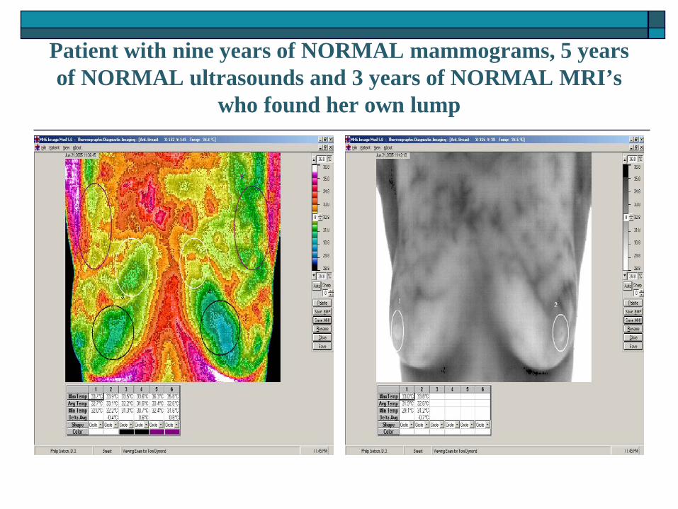
Patient with nine years of NORMAL mammograms, 5 years of NORMAL ultrasounds and 3 years of NORMAL MRI’s
who found her own lump
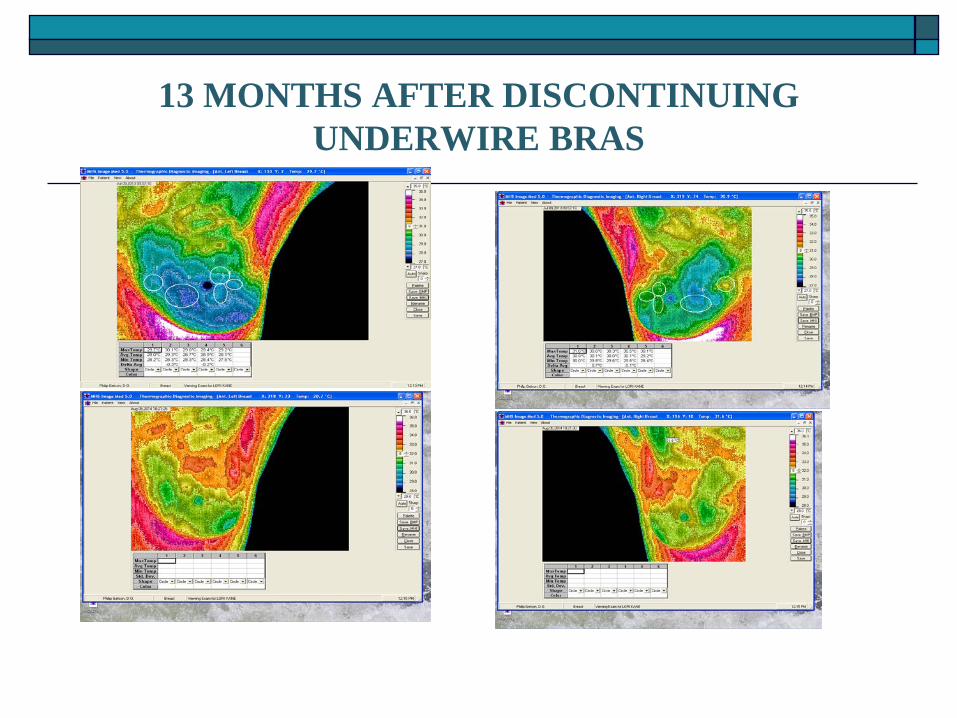
13 MONTHS AFTER DISCONTINUING UNDERWIRE BRAS

FOUR MONTHS OF GLUTEN FREE EATING

Effect of Lifestyle Change on Breast Health- 3 months after eliminating caffeine, sugar and
red meat

BEFORE AND AFTER IMAGES OF A PATIENT WHO LOST 50 POUNDS, CHANGED HER DIET AND BEGAN
A DAILY EXERCISE REGIMEN

OTHER USES FOR THERMOGRAPHY
NEUROMUSCULAR
THYROID – FACIAL- DENTAL

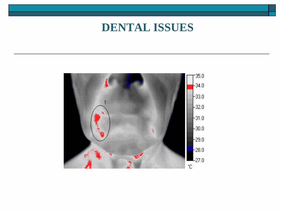
DENTAL ISSUES

Patient with long standing hypothyroidism

OTHER BREAST SCREENING STUDIES
Ultrasound- Uses sound waves which “bounce off” the
breast to create a black and white image. Good for differentiating solid from cystic masses
Magnetic Resonance Imaging (MRI) – Uses magnets to “pull apart” the body’s water molecules to create and image. No radiation. In breast studies this is done following an intravenous injection of gadolinium, a water soluble contrast dye
P.E.T. SCANS A PET scan uses radiation, or nuclear medicine
imaging, to produce 3-dimensional, color images of the functional processes within the human body. PET stands for positron emission tomography. The machine detects pairs of gamma rays that are emitted indirectly by a tracer (positron-emitting radionuclide), which is placed in the body on a biologically active molecule. The images are reconstructed by computer analysis. Modern machines often use a CT X-ray scan which is performed on a patient at the same time in the same machine.

Insurance Issue #1 The radiology community has established a “rule” that
states that “ you cannot have an ultrasound without first having a mammogram” This is NONSENSE. There is no such rule or law. It is an attempt to force women into getting mammograms. Slowly but surely there are more centers who are acceding to the wishes of their female patients and doing the ultrasound without the mammogram.

Insurance Issue #2 MRI’s are costly. A breast study can run
anywhere from $800-$2000. Interestingly, the centers will offer huge discounts for cash patients.
Virtually all insurers require prior authorization for MRI’s
However, Horizon BC/BS will NOT authorize an MRI UNLESS an individual has already had a biopsy that showed cancer

Insurance Issue #3 Medicare will retrospectively pay for MRI’s with
just cause. These causes include, but are not limited to prior MRI, prior diagnosis of breast cancer, inconclusive but suspicious other studies, Birad gene positivity and in some cases, the inability to undergo mammography due to concurrent medical conditions

Diet & Lifestyle modification
EAT ORGANIC FOODS- CONSIDER GLUTEN FREE ELIMINATE PROCESSED FOOD, WHITE SUGAR, WHITE FLOUR, WHITE
SALT ELIMINATE COMMERCIAL HOUSEHOLD CLEANING PRODUCTS AND
TOXIC GARDEN PESTICIDES DRINK PURE FILTERED WATER REFUSE SYNTHETIC HORMONE TREATMENTS SEEK NATURAL APPROACHES TO HEALTHCARE DETOXIFY THE BODY EMPOWER YOURSELF WITH A POSITIVE OUTLOOK - ADOPT AN
ATTITUDE OF GRATITUDE SUPPLEMENT YOUR DIET WITH APPROPRIATE VITAMINS AND
NUTRITIONAL SUPPORT EXERCISE FIND A HEALTHY AVENUE FOR STRESS RELEASE SUCH AS
MEDITATION, YOGA, GARDENING READING ETC. MAINTAIN HEALTHY RELATIONSHIPS EXPLORE YOUR SPIRITUALITY

TDINJ.COM & HEALTH THROUGH AWARENESS.COM
The websites that will provide information regarding thermography as well as helpful diet and lifestyle tips for better breast health
100 Brick Rd Suite 206
Marlton, NJ 08053 856-596-5834