breast pathology post-neoadjuvant chemotherapy · breast pathology post-neoadjuvant chemotherapy...
TRANSCRIPT

Breast Pathology Post-Neoadjuvant Chemotherapy
Megan Troxell, MD/PhD
Stanford Pathology

2 mm

Objectives
• Develop a framework for gross analysis of post-chemotherapy breast specimens.
• Recognize histologic features of tumor bed and post-chemo carcinoma, including pitfalls
• Understand reporting schemes, utility and clinical impact of residual tumor burden

Neoadjuvant chemotherapy
Kummel. BJS 2014; 101: 912–24
• Equivalent long term outcome whether chemotherapy before or after surgery
• Neoadjuvant: – Can assess response/non-response
• Degree of response: prognostic for survival
– Response is short term endpoint for clinical trials • Tissue collection for research before/during/after
– Downsize tumor for breast conserving surgery
• Chemo most efficacious in Her2+ or triple negative tumors, (vs ER+)
• Now standard of care for locally advanced breast cancer

Mod Pathol 2015; 28:1185–1201
More extensive diagrams

• Prognostic information from degree of pathologic response?
• Propose RCB as continuous variable: – primary tumor dimensions,
– residual cellularity of tumor bed
– axillary nodal burden
RCB

Initial diagnostic pathology Breast • Adequate core biopsy essential
– Caution if limited tumor or extensive DCIS
• ER, PR, Her2 & other markers – % tumor cellularity for some
studies
• Clip placement essential!
Hint: core is often ~6 months ago if neoadjuvant Rx
Axilla • Status impacts local/systemic
therapy • Routine axillary ultrasound • FNA or core bx for clinical,
radiologic abnormal nodes – Clip!?? Recommended by NCCN’15
• Up-front surgical SLN bx not recommended – Invalidates ypN & RCB – Precludes assessment of nodal
response

Steps to evaluate post-chemo breast
1. Recognize its post-chemo
2. Identify gross tumor/tumor bed & document size
3. Judiciously sample (map)
4. Assess residual size (largest contiguous and span), cellularity & standard parameters
5. Evaluate lymph nodes
6. Report as per local custom/mandates

Essential clinical info with specimen • “At an absolute minimum, the specimen must be
clearly marked as post-NAST; pre-NAST location and size of the tumor must be indicated.” (Bossuyt)
& five other elements
Was the axillary node clipped??

Provenzano Research samples only if grossly obvious residual invasive cancer. (Bossuyt)
Patterns of residual CA

Specimen sampling • CAP: “Special attention to finding and evaluating the tumor
bed is necessary for these specimens.” • Provenzano:“It is strongly recommended that an image of
the sliced specimen be recorded (radiograph, photograph, photocopy, or drawing) and then used as a map for the sections taken, so that the histopathologic findings of any residual disease in the breast can be more easily understood.”
• Bossuyt: “Overly exhaustive sampling and histologic evaluation of the entire tumor bed are not required and not as efficient or informative as informed mapping of the specimen.”

Bossuyt et al. Ann Oncol. 2015;26:1280-91

Specimen sampling • Small specimens: submit entirely, mapped
– Document if tumor bed at specimen edge (esp if residual scattered ca)
• Large specimens: guided by pretreatment size & location, mapped – Provenzano: full face of pretreatment area every 1 cm
• If very large, 5 blocks per every 1-2 cm, up to 25 blocks • Good clinical judgment on a case-by-case basis
– RCB: Submit the largest cross-sectional area for histology – FDA: at least one block per cm of pre-treatment tumor size, or at least
10 blocks in total (greater of) – Sahoo ‘09: ~1 section/cm of original tumor size
• Multiple pretreatment lesions: as above & sample between lesions – Intervening invasive CA or DCIS? – Largest is used for RCB and ypT stage

Localizing post-chemo Once you have the slides, document at least one of: 1) Tumor/lesion compatible with findings on prior
biopsy -- If abundant tumor, I don’t mandate section of biopsy site -- Caution: second ‘occult tumor’
2) Tumor bed 3) Biopsy site or clip -- Rarely, minimal histologic evidence -- But clips can migrate, or displace during sectioning….

Tumor bed
Grossly: fibrous rubbery area; here with residual tumor Sahoo & Lester. Surgical Pathology Clinics 2012:5;749–74

Mastectomy: clips at 12:00 (or nipple bed) Stitch axillary tail

Mastectomy: clips at 12:00 (or nipple bed) Stitch axillary tail Need to know location, size of pre-chemo cancer(s) Any additional lesions on imaging
Section nipple, ink, slice, photo or Xray in order, then map sections taken

Pre-chemo cores heterogeneity, focal geographic necrosis (not shown)
Post-chemo Poor response

Continued
Medial
Pre-chemo core
Post-chemo tumor bed: DCIS (below)


Continued Safety pins mark lesions ID’ed fresh
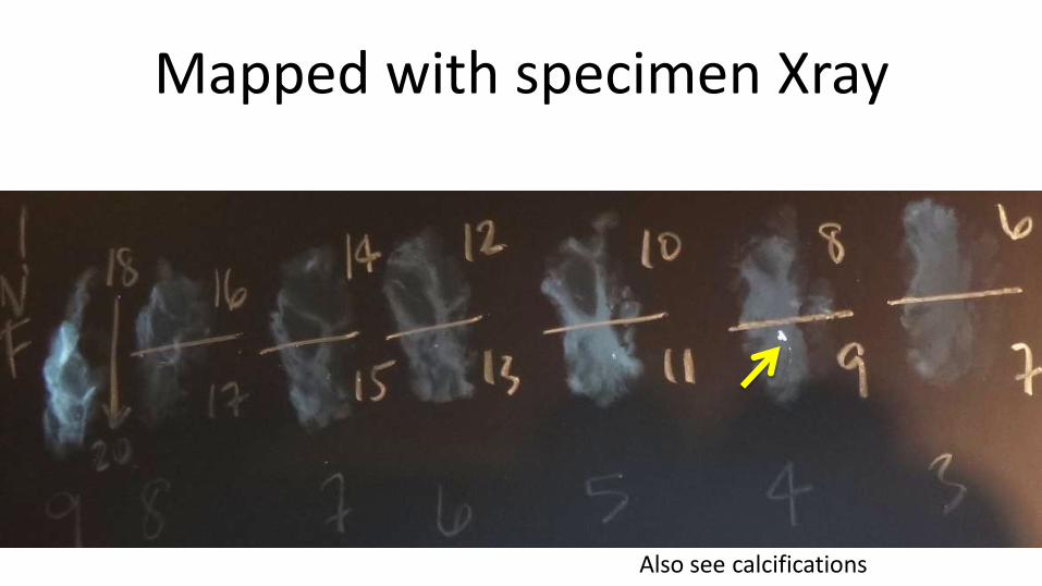
Mapped with specimen Xray
Also see calcifications


Lumpectomy with 3 clips!

What is this in the specimen?
Cox. Ann Surg Oncol 2016;23:1824–1830 Gilcrease. AJSP 2016; 40:1375–1379

What is this in the specimen?
Cox. Ann Surg Oncol 2016;23:1824–1830 Gilcrease. AJSP 2016; 40:1375–1379
Savi SCOUT
Mango. AJR 2016; 207:W1–W4
Titanium Encapsulated Radioactive Seed

New wire-loc alternatives
• Savi SCOUT
• Radioactive seeds
• Intraoperative US
• Magseed
Cox. Ann Surg Oncol 2016;23:1824–1830

• I125 localized w/ gamma probe • Mayo: 0/2000 • MD Anderson: 2/1400 • Case 1
– Specimen radiograph shows parts of Ti cap in 2 slices!!
• Case 2: large ossified mass – Seed cut with bone saw – Titanium cap not damaged – Why the &^%$##!! was
localization used?
• If immobilized with forceps, seeds can severed with scalpel blade
• “Use forceps with finesse;” no scissors
• Added recommendations: – Know where seed is; use same
gamma probe as is used in OR – “as blade approaches the location of
the seed, slow and careful slicing of the tissue”
– Dedicated grossing bench; do not discard anything until seed retrieved
Gilcrease. AJSP 2016; 40:1375–1379

Histologic tumor bed • Fibrosis +/- elastosis • Prominent vessels • Fewer to absent normal epithelial structures • Inflammation
– histiocytes, lymphs, giant cells
• Hemosiderin • Calcification
Find: 1) Tumor 2) Tumor bed 3) Bx site or clip

Tumor bed:
vessels, fibrosis,
paucity of normal


Histiocyte rich tumor
bed

Normal breast
Edge of tumor bed; same specimen
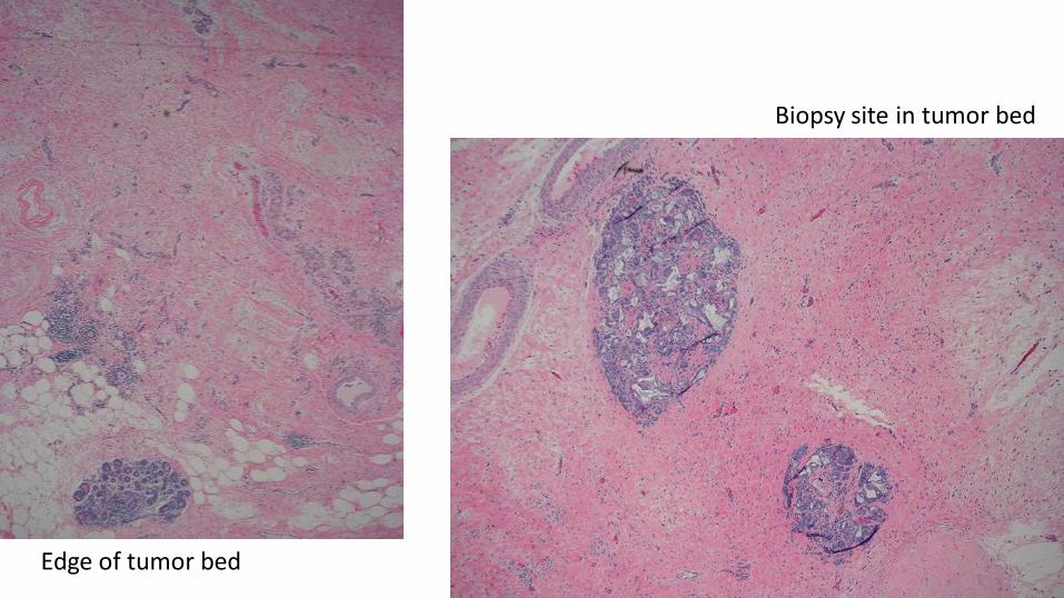
Edge of tumor bed
Biopsy site in tumor bed


Left: parenchymal atrophy post-chemo Below: LCIS unperturbed by chemo

Bowtie clip M clip


Lack of ‘tumor bed’ stromal change

Tumor Bed …...


History of G3 IDC, triple
negative
Find: 1) Tumor 2) Tumor bed 3) Bx site or clip

History of G3 IDC, triple
negative, prior
Several foci like this post-chemo
Find: 1) Tumor 2) Tumor bed 3) Bx site or clip

Prior axillary LN core biopsy

Biopsy site
Find: 1) Tumor 2) Tumor bed 3) Bx site or clip

Changes in carcinoma histology • Histiocytoid appearance • Cytoplasmic vacuolization, eosinophilia • Nuclear hyperchromasia, pleomorphism
– Multinucleation
• Decreased mitotic activity • Lobular-like growth pattern • Retraction artifact • Heterogeneity (selection by chemo?)
– Expect cellularity to vary across tumor Sahoo, Provenzano
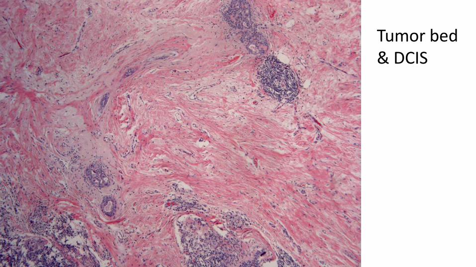
Tumor bed & DCIS

Tumor bed & DCIS

Tumor bed & DCIS


Atypia vs. DCIS?

Grading post chemo
• Often increased nuclear pleomorphism
• Often decreased mitotic rate
• Should still be graded
– Nottingham/mSBR/Elston-Ellis
– Tubules, nuclei, mitosis

Treated carcinoma
• Metaplasia due to chemotherapy?
• Squamous elements relatively resistant to chemotherapy?

Keratin
ER

Different morphologies across tumor

Heterogeneity post-chemo
Pre-chemo core

Residual histiocytoid IDC & DCIS

Dermal tumor
Heterogeneous tumor vs. histiocytes?

Residual invasive carcinoma vs histiocytes?

Residual invasive carcinoma vs histiocytes?
Keratin

Left: Core bx IDC3, met to LN ER+, PR- Her2+ Right: Post-chemo tumor bed with no residual IDC
Multiple tumors: different chemo-sensitivity

Left: Core bx ILC, E-cad negative Right: Post-chemo residual ILC
Same patient

Same patient
Chemo response: Her2+ > Triple Neg >> ER+Her2-

Post-chemo reporting: CAP (AJCC R) In the Breast • No known presurgical therapy • No definite response to presurgical
therapy in the invasive carcinoma • Probable or definite response to
presurgical therapy in the invasive ca • No residual invasive carcinoma is
present in the breast after presurgical therapy
In the Lymph Nodes • No known presurgical therapy • No lymph nodes removed • No definite response to presurgical
therapy in metastatic carcinoma • Probable or definite response to
presurgical therapy in metastatic ca • No lymph node metastases. Fibrous
scarring, possibly related to prior lymph node metastases with pathologic complete response
• No lymph node metastases and no prominent fibrous scarring in the nodes

System Score in breast Correlate w/ core?
LN included? # categories partial
B-18 Any treatment effect on invasive No Yes, size met 1
Chevallier Presence of invasive with sclerosis, fibrosis
No Yes 1
Sataloff Presence of invasive Presence of treatment effect
No Yes +/- TE 2
UICC Product of 2 dimensions No No 3
Miller-Payne Presence of invasive,cellularity Yes No 3
AJCC (y) Size of invasive No Yes, # Up to 4
MNPI Size of invasive, grade No Yes, # 3
Pinder % tumor remaining in breast Yes Yes, evidence of response
3
RCB size in 2-dimensions, cellularity No Yes, # & size 4

• Prognostic information from degree of pathologic response?
• Propose RCB as continuous variable: primary tumor dimensions, residual cellularity of tumor bed, and axillary nodal burden – Cohort of 382 patients; 2 different neoadjuvant chemo regimens
– Validation cohort of 141
RCB

Residual Cancer Burden (RCB)
• Tumor size, 2 dimensions
• % residual invasive cellularity
• Number of residual involved lymph node
• Size of largest metastasis

©2007 by American Society of Clinical Oncology
Symmans W F et al. JCO 2007;25:4414-4422
Illustrative examples of how residual tumor bed would be defined.

©2007 by American Society of Clinical Oncology
Symmans W F et al. JCO 2007;25:4414-4422
Illustrative examples of how residual tumor bed would be defined.

Size reporting: difference
RCB
• Largest area of residual invasive cancer (A, span)
• Does not need to be contiguous
• Two dimensions
• Favored by Provenzano
Provenzano
AJCC 7th (ypT)
• Largest contiguous focus of invasive cancer (B)
– Use ‘m’ for scattered foci
• One dimension

Residual tumor %
cellularity
©2007 by American Society of Clinical Oncology
Symmans W F et al. JCO 2007;25:4414-4422

Residual Cancer Burden (RCB)
Symmans. http://www3.mdanderson.org/app/medcalc/index.cfm?pagename=jsconvert3 See website for instructions, helpful diagrams
Purists include all cancer cellularity, subtract DCIS (I just score invasive)

Post-chemo synopsis: example Focality of residual invasion: Present as scattered single cells/small clusters.
Present as multiple residual foci/masses. Present as single contiguous residual mass/focus.
Span of residual invasion: ______ cm x ______ cm
Size of largest contiguous focus: ______ cm
Average cellularity of invasive carcinoma in tumor bed:
______ %
Changes consistent with treatment effect:
Present Indeterminate No definite

http://www3.mdanderson.org/app/medcalc/index.cfm?pagename=jsconvert3
Instructions and several helpful downloads (diagrams)
RCB

Residual LVI • Should not be considered pCR • Ensure ID & adequate sampling of
tumor bed – Most patients have other residual
disease (LN)
• Confirm LVI, not DCIS, not retraction artifact (IHC)
• Don’t include in residual invasive cancer size
• LVI at margin: separately comment • Lack of data for current reporting
recommendations
• LVI per Rosen/CAP: – Outside the border of the invasive
carcinoma – Tumor emboli do not conform to the
contours of space; invasive with retraction has exactly the same shape.
– Endothelial cell nuclei should be seen lining the space
– Lymphatics often adjacent to blood vessels
• ‘Extensive LVI’ – Provenzano: one or more foci in more
than one block – Stanford: 3 foci – NCCN: not defined – Colleoni:
• focal: one focus of in one tumor block only • Moderate: more than one focus in one
block • Extensive: one or more foci in more than
one tumor block

LVI
LVI with ‘satellite’

LVI D2-40
Her2

Endothelial markers CD31, CD34, ERG Lymphatic markers LYVE-1, D2-40 (podoplanin)
Human Pathology. 2008;39:175-83


Tumor in lymphatics relatively
resistant to chemotherapy
(as is DCIS)

• Retrospective study – Predominanty AC-4 cycles
– 14% no residual cancer cells (pCR)
– 10% DCIS only (pCR)
– 4% pure IL tumor
– 3.4% predominantly IL
– 69% residual IDC
• IL 3-fold higher risk of death
AJSP. 2009;33:256-63

• LVI was associated with poorer PFS and OS
– independent of post-surgical stage/nodal status
– No pathology review!
Breast Cancer Res Treat 2016; 157:555–64
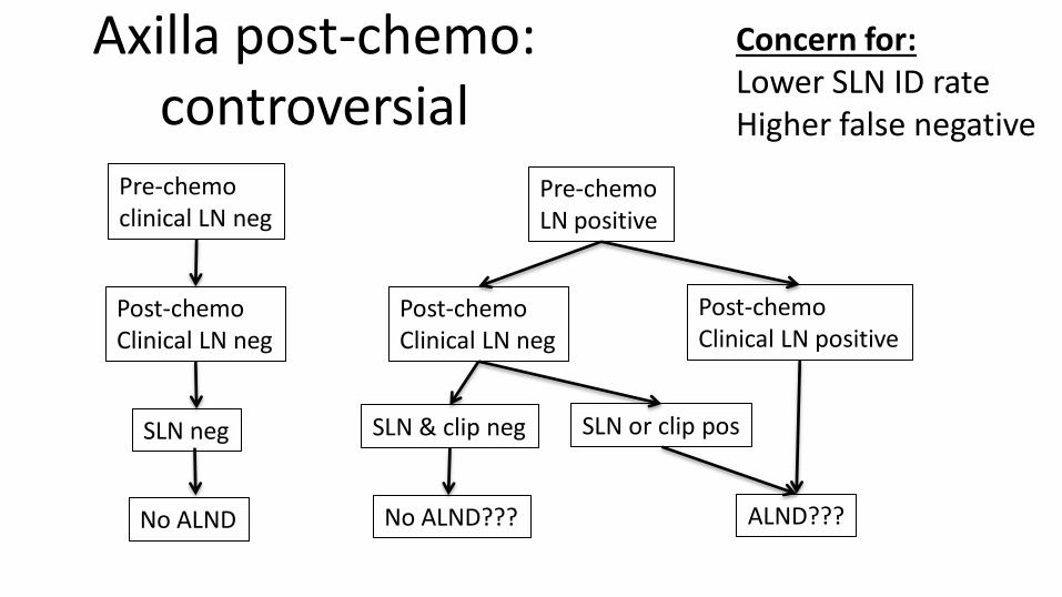
Axilla post-chemo: controversial
Pre-chemo clinical LN neg
Post-chemo Clinical LN neg
SLN neg
No ALND
Pre-chemo LN positive
Post-chemo Clinical LN neg
SLN & clip neg
No ALND???
SLN or clip pos
ALND???
Post-chemo Clinical LN positive
Concern for: Lower SLN ID rate Higher false negative

Axillary Lymph Nodes • Up to 40% of patients convert to node-negative • May be more difficult to ID post chemo
– If so, submit fibrotic & perivascular areas in axilla
• Handle nodes with standard protocols – 2mm gross sections
• Report with standard protocols – #, sizes, ECE – AND treatment effect/fibrosis

SLN: fibrosis, no residual CA

SLN: fibrosis, Exclude residual tumor cells

Post-chemo SLN: Granulomas?


Post-chemo SLN Germinal center?

Post-chemo SLN Germinal center?

SLN FS Both foci cancer?

SLN FS How about now?

SLN permanents Right: CA Left: clip

SLN frozen section

SLN Permanents Top: tumor & fibrosis Bottom: clip

Another type of clip in SLN
“In our opinion, it is best to exercise caution….and maintain a low threshold for deferral of the final diagnosis to permanent sections in order to avoid false-positive & unecessary ALND.” Brogi. Histopath. 2015;68:152-67.

LN reporting post chemo AJCC (ypN)
• Isolated tumor cells reported as node negative (ypN0itc)
• But not regarded as pCR
WHO
• Isolated tumor cells node positive
Provezano (opinion)
• Any residual disease in LN (mi, itc) should NOT be classified as pCR
• If no associated fibrosis, report as in adjuvant setting (ypN0itc)
• If fibrosis, likely macro- or micromet with response – Describe in comment
– Measure entire area, including tumor cells & intervening stroma
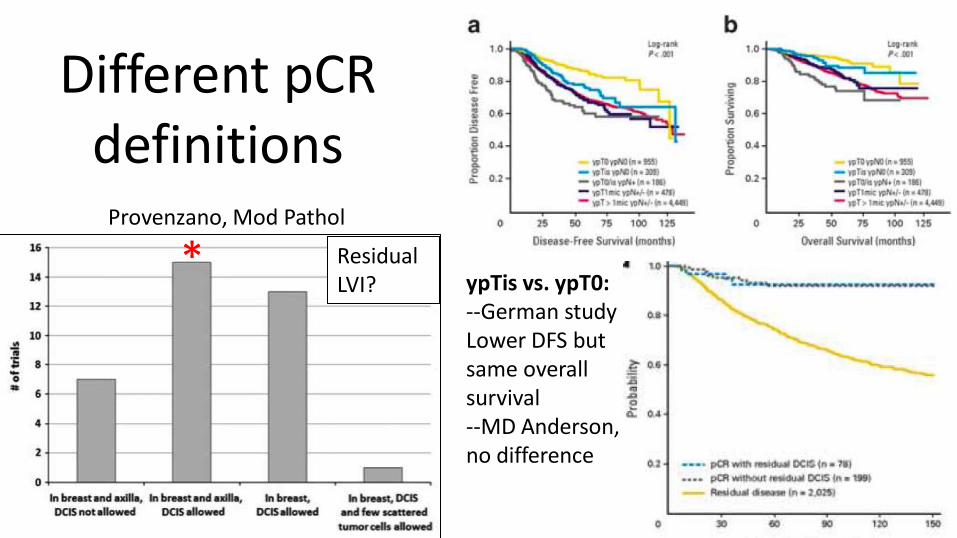
Different pCR definitions
* ypTis vs. ypT0: --German study Lower DFS but same overall survival --MD Anderson, no difference
Provenzano, Mod Pathol
Residual LVI?

Complete Pathologic Response (pCR)
pCR NOT pCR
Insufficient evidence
Comment
DCIS X x Definitions vary for DCIS
LCIS X
LVI X X LVI w/o LN disease very rare
LN: macro- & micro- met
X Residual CA in LN worse prognosis irrespective of breast
LN: isolated tumor cells
X Mi & itc difference significance than adjuvant setting
From Bossuyt

Retesting of biomarkers Provenzano • Routine retest not
recommended – Positive ER/PR/Her2 core
• Consider retest – Negative or equivocal
result on core – Outside biopsy/markers – Heterogeneous or multiple
tumors – No response to therapy
• Discordance post-chemo – ER: ~15% – PR: ~30% – Her2: 6-9%
• Due to: – Technical failures – Intratumoral heterogeneity – Changes due to therapy
(?selection)
• Working group: 6/20 retest routinely

Left: Core biopsy Her2+
Right: Tumor bed with fibrosis, lymphs, histiocytes, no tumor

Steps to evaluate post-chemo breast
1. Recognize its post-chemo
2. Identify gross tumor/tumor bed & document size
3. Judiciously sample (map)
4. Assess residual size (largest contiguous and span), cellularity & standard parameters
5. Evaluate lymph nodes
6. Report as per local custom/mandates

Selected references • Provenzano et al. Standardization of pathologic evaluation and reporting
of postneoadjuvant specimens in clinical trials of breast cancer: recommendations from an international working group. Modern Pathology (2015) 28, 1185–1201
• Bossuyt et al. Recommendations for standardized pathological characterization of residual disease for neoadjuvant clinical trials of breast cancer by the BIG-NABCG collaboration. Annals of Oncology 26: 1280–1291, 2015
• Lakhani et al eds. WHO Classification of Tumours of the Breast. 2012 • Symmans WF et al. Measurement of residual breast cancer burden to
predict survival after neoadjuvant chemotherapy.JCO 2007;25:4414-4422 • See also on-slide references

End