breast reconstruction following mastectomy or lumpectomy · breast reconstruction following...
TRANSCRIPT

Page 1 of 37 Medical Coverage Policy: 0178
Medical Coverage Policy
Effective Date ............................................. 1/15/2018 Next Review Date ....................................... 1/15/2019 Coverage Policy Number .................................. 0178
Breast Reconstruction Following Mastectomy or Lumpectomy Table of Contents Coverage Policy .................................................. 1 Overview.............................................................. 4 General Background ........................................... 4 Coding/Billing Information ................................. 21 References ........................................................ 25
Related Coverage Resources Botulinum Therapy Breast Implant Removal Complex Lymphedema Therapy (Complete
Decongestive Therapy) Injectable Fillers Panniculectomy and Abdominoplasty Pneumatic Compression Devices and Compression
Garments Prophylactic Mastectomy Reduction Mammoplasty Redundant Skin Surgery Scar Revision Surgical Treatment of Chest Wall Deformities Tissue-Engineered Skin Substitutes
INSTRUCTIONS FOR USE The following Coverage Policy applies to health benefit plans administered by Cigna Companies. Certain Cigna Companies and/or lines of business only provide utilization review services to clients and do not make coverage determinations. References to standard benefit plan language and coverage determinations do not apply to those clients. Coverage Policies are intended to provide guidance in interpreting certain standard benefit plans administered by Cigna Companies. Please note, the terms of a customer’s particular benefit plan document [Group Service Agreement, Evidence of Coverage, Certificate of Coverage, Summary Plan Description (SPD) or similar plan document] may differ significantly from the standard benefit plans upon which these Coverage Policies are based. For example, a customer’s benefit plan document may contain a specific exclusion related to a topic addressed in a Coverage Policy. In the event of a conflict, a customer’s benefit plan document always supersedes the information in the Coverage Policies. In the absence of a controlling federal or state coverage mandate, benefits are ultimately determined by the terms of the applicable benefit plan document. Coverage determinations in each specific instance require consideration of 1) the terms of the applicable benefit plan document in effect on the date of service; 2) any applicable laws/regulations; 3) any relevant collateral source materials including Coverage Policies and; 4) the specific facts of the particular situation. Coverage Policies relate exclusively to the administration of health benefit plans. Coverage Policies are not recommendations for treatment and should never be used as treatment guidelines. In certain markets, delegated vendor guidelines may be used to support medical necessity and other coverage determinations.
Coverage Policy Coverage for breast reconstruction* and breast prostheses following mastectomy or lumpectomy is governed by federal and/or state mandates. Breast Reconstruction *Please note: Coverage for breast reconstruction services following mastectomy and lumpectomy is available to both females and males. In addition, a diagnosis of breast cancer is not required for breast reconstruction services to be covered, and the timing of reconstructive services is not a factor in coverage.

Page 2 of 37 Medical Coverage Policy: 0178
Breast reconstruction following mastectomy or lumpectomy is considered medically necessary for EITHER of the following:
• breast reconstruction procedures performed on the diseased/affected breast (i.e., breast on which the mastectomy/lumpectomy was performed), including:
areolar and nipple reconstruction areolar and nipple tattooing autologous fat transplant (i.e., liposuction, lipoinjection, lipofilling, lipomodeling) breast implant removal and subsequent reimplantation capsulectomy capsulotomy implantation of tissue expander implantation of U.S. Food and Drug Administration (FDA)-approved internal breast prosthesis oncoplastic reconstruction reconstructive surgical revisions tissue/muscle reconstruction procedures (e.g., flaps), including, but not limited to, the following:
o deep inferior epigastric perforator (DIEP) flap o latissimus dorsi (LD) myocutaneous flap o Ruben’s flap o superficial inferior epigastric perforator/artery (SIEP/SIEA) flap o superior or inferior gluteal free flap o thoracodorsal artery perforator (TDAP) flap o transverse rectus abdominus myocutaneous (TRAM) flap o transverse upper gracilis (TUG) flap
• breast reconstruction procedures performed on the nondiseased/unaffected/contralateral breast,
in order to produce a symmetrical appearance, including:
areolar and nipple reconstruction areolar and nipple tattooing augmentation mammoplasty augmentation with implantation of FDA-approved internal breast prosthesis when the unaffected
breast is smaller than the smallest available internal prosthesis autologous fat transplant (i.e., liposuction, lipoinjection, lipofilling, lipomodeling) breast implant removal and subsequent reimplantation when performed to produce a symmetrical
appearance breast reduction by mammoplasty or mastopexy capsulectomy capsulotomy reconstructive surgery revisions to produce a symmetrical appearance
Intraoperative assessment of tissue perfusion is considered an integral part of the breast reconstruction procedure and not separately reimbursable. The following products* are considered medically necessary when used in association with a medically necessary breast reconstruction procedure:
• AlloDerm™ • AlloMax™ • DermACELL™ • FlexHD® Acellular Hydrated Dermis • NeoForm™ Dermis

Page 3 of 37 Medical Coverage Policy: 0178
The following products* when used in association with a breast reconstruction procedure are considered experimental, investigational or unproven (this list may not be all-inclusive):
• Biodesign® Nipple Reconstruction Cylinder • Cortiva™ • DermaMatrix Acellular Dermis • hMatrix® • Juvederm® • Permacol® • Phasix™ Mesh • Radiesse® • Repriza® • SERI™ Surgical Scaffold • Strattice™
Reconstructive Tissue Matrix • SurgiMend® • Veritas Collagen Matrix
*Note: Refer to the table in Appendix A for a list of products and the associated CPT and HCPCS codes. The following breast reconstruction procedures are considered experimental, investigational or unproven for this indication:
• autologous fat transplant with the use of adipose-derived stem cells • vascularized lymph node transfer (VLNTx) • xenograft cartilage grafting
Suction lipectomy or ultrasonically-assisted suction lipectomy (liposuction) for correction of surgically-induced donor site asymmetry (e.g., trunk or extremity) that results from one or more flap breast reconstruction procedures is considered cosmetic in nature and not medically necessary. Removal of either a saline-filled OR silicone gel-filled breast implant when associated with breast reconstruction following mastectomy or lumpectomy for ANY indication, including for the purpose of producing a symmetrical appearance of the nondiseased breast is considered medically necessary. Refer to the Breast Implant Removal Medical Coverage Policy for additional information on breast implant removal. Following removal of a breast implant, the subsequent surgical implantation of a new U.S. Food and Drug Administration (FDA)-approved breast implant is considered medically necessary for EITHER of the following:
• breast reconstruction of a diseased or affected breast following mastectomy or lumpectomy • creation of a symmetrical appearance in the contralateral/nondiseased breast following mastectomy or
lumpectomy in the opposite breast External Breast Prostheses and Mastectomy Bras External breast prostheses and mastectomy bras following mastectomy or lumpectomy are covered under the core medical benefits of the plan. An external breast prosthesis or mastectomy bra for any other indication is considered not medically necessary. This Medical Coverage Policy does not address treatments for lymphedema. For information on these treatments, refer to the separate Cigna Medical Coverage Policies Pneumatic Compression Devices and Compression Garments, and Complex Lymphedema Therapy (Complete Decongestive Therapy).

Page 4 of 37 Medical Coverage Policy: 0178
Overview This Coverage Policy addresses reconstructive breast surgery and external breast prostheses and mastectomy bras following mastectomy or lumpectomy. General Background Breast reconstruction was originally designed to reduce post-mastectomy complications and to establish symmetry between the surgical breast and the contralateral breast. Surgical procedures that are performed to establish symmetry can include: breast reduction; breast augmentation with an FDA-approved breast implant; and/or areola-with-nipple reconstruction and nipple-area tattooing. Breast reconstruction after mastectomy has evolved over the last century to become an integral component of therapy for patients with breast cancer. Reconstruction can occur immediately after a mastectomy, or be delayed for weeks or years until a patient undergoes radiation, chemotherapy, or determines whether they want breast reconstruction. Prosthetic Reconstruction Breast Implants: Breast implants can be inserted at the same time as the mastectomy (e.g., direct-to-implant breast reconstruction or one-stage immediate breast reconstruction) or in two stages, using an implanted tissue expander in the first stage followed by removal of the expander and insertion of a permanent breast implant (e.g., two-stage reconstruction or two-stage delayed reconstruction). The FDA-approved implant is placed either deep in the breast on the pectoral fascia (submammary) or beneath the pectoralis major. The advantages of tissue expander implant reconstruction are the reliability, simplicity, and avoidance of donor-site morbidity. Complications associated with the use of breast implants can occur in the immediate perioperative period or years later. Such complications include exposure, extrusion, or infection of the implants. Longer term problems also include asymmetry, capsular contracture, malposition of the implant, rupture, and pain. These conditions, when they become clinically significant, may require removal of the implant (Roehl, et al., 2012; American Cancer Society [ACS], 2017; Roostaeian, et al., 2012). Indications for implant reconstruction include: bilateral reconstruction; individuals requiring augmentation in addition to reconstruction; individuals not suited for long surgery; a lack of abdominal tissue; individual unwilling to have additional scars on either their back or abdomen; and a small breast mound with minimal ptosis. Relative contraindications to implant reconstruction include: young age (i.e., may need implant replaced multiple times); individual unwilling to follow up; very large or ptotic breast. The contraindications to implant reconstruction include: silicone allergy; fear of implants; previously failed implants; or need for adjuvant radiation therapy (Roehl, et al., 2012). Surgical complications associated with breast implantation are similar to those encountered with other breast surgeries: infection, bleeding, change in nipple sensation (e.g., hypersensitivity or hyposensitivity), malposition, delayed healing, and anesthetic accidents. Although implantable breast prostheses may be inserted for either reconstructive or cosmetic reasons, clinically significant post-implant complications may occur, necessitating removal of the implants. Local complications associated with implanted breast prostheses include: capsular contracture, persistent infection, silicone implant extrusion, tissue necrosis and silicone implant rupture. These conditions, when they become clinically significant, may require removal of the implant. Additionally, the presence of an implant may interfere with the diagnosis or treatment of breast cancer. Infections that may occur in or around an implant include wound infections, as well as infections within a capsular contracture or as a result of a ruptured implant. Removal of the implant may be necessary when the infection does not respond to antibiotics. Unstable or weakened tissue and/or interruption in wound healing may result in the implant breaking through the skin or extrusion. Necrotic tissue may form around the implant, requiring implant removal. Silicone gel-filled implant rupture may cause the contents to leak into the surrounding tissues.

Page 5 of 37 Medical Coverage Policy: 0178
U.S. Food and Drug Administration (FDA): In the FDA labeling for approved breast implants Mentor Corp., Santa Barbara, CA; Ideal Implant ®, Inc., Dallas, TX; Allergan Corp. (formerly Inamed), Irvine, CA and Sientra, Inc., Santa Barbara, CA are listed as manufacturers of silicone and saline breast implants. FDA-approved saline-filled implants:
• Allergan Medical RTV Saline-Filled Breast Implant • Ideal Implant Saline-Filled Breast Implant • Mentor Saline-Filled and Spectrum™ Breast Implants
The FDA approved saline-filled breast implants for breast augmentation in women age 18 or older and for breast reconstruction in women of any age. They are also used in revision surgeries, which correct or improve the result of an original surgery. FDA-approved silicone gel-filled breast implants:
• Allergan Natrelle® • Allergan Natrelle® 410 Highly Cohesive Anatomically Shaped Silicone-Filled Breast Implant • Mentor MemoryGel® • Mentor MemoryShape™ Silicone Gel-Filled Breast Implant • Sientra® Silicone Gel Breast Implant
The FDA labeling for silicone and saline breast implantation states breast implant surgery should not be performed in women with: an active infection, existing cancer or precancer of a breast that has not been adequately treated, or who are pregnant or nursing (FDA, 2012). In June 2011 the FDA released a report updating the clinical and scientific information for silicone gel-filled breast implants, including preliminary safety data from studies conducted by the manufacturers as a condition of their November 2006 approval. The conclusion in the report states that, “Based on the totality of the evidence, the FDA believes that silicone gel-filled breast implants have a reasonable assurance of safety and effectiveness when used as labeled. Despite frequent local complications and adverse outcomes, the benefits and risks of breast implants are sufficiently well understood for women to make informed decisions about their use. Manufacturers and physicians should continue to provide balanced and up-to-date information to women considering breast implants to help inform their decisions” (FDA, 2011). Tissue Expanders Following mastectomy, some individuals have inadequate elasticity in the remaining tissue to accommodate and support a breast implant. For these individuals, tissue expanders can be inserted under the chest muscle or skin. The expander is an empty balloon-like container that, over time, is injected with saline. This inflation causes the tissue to expand. The tissue expander is surgically removed once an adequate pocket has been established, and the permanent implant is then inserted. The most appropriate patients for this type of reconstruction are individuals who do not qualify for autogenous reconstruction, individuals who do not want additional scars from other donor sites, individuals who prefer a typically quicker postoperative recovery period, and individuals who have relatively small breasts. Contraindication for this type of reconstruction are mastectomy flaps that are too thin for adequate implant coverage and the completed or planned use of adjuvant radiation therapy because of higher implant complication rates (ACS, 2017; Hu, et al., 2007). Tissue Flap Procedures Autologous breast reconstruction procedures are safe and effective and are a well-established standard of care include tissue/muscle reconstruction procedures (e.g., flaps). Methods of autologous tissue breast reconstruction include local flaps and distant flaps. Local flaps rely on transposition of muscle, subcutaneous tissue, and skin into the mastectomy defect based on the attached native blood supply of the muscle (e.g., latissimus dorsi myocutaneous (LD) flap and the pedicled transverse rectus abdominus myocutaneous (TRAM) flap). Distant flap breast reconstruction requires the use of microvascular free-tissue transfer (e.g., free TRAM flap, deep inferior epigastric perforator [DIEP] flap, superficial inferior epigastric artery perforator [SIEP] flap, inferior or superior gluteal flap, superior gluteal artery perforator flap, Reubens flap, and the transverse upper gracilis (TUG) flap). Breast reconstruction using these donor sites relies on harvesting the flap with its vascular pedicle, which is

Page 6 of 37 Medical Coverage Policy: 0178
anastomosed using microsurgical technique to appropriate recipient vessels in the mastectomy site. The two most common types of tissue flap procedures are the TRAM flap and the LD flap. Other tissue flap surgeries are more specialized, and may not be available everywhere. The choice of procedure for a given individual is affected by her age, her health, her contralateral breast size and shape, her personal preference, and the expertise of the reconstructive surgeon (ACS, 2017, Roehl, et al., 2012; Spear et al., 2007; Mehrara et al., 2006; Alderman et al., 2006; Garvey et al., 2006; Bajaj et al., 2006; Wechselberger, et al., 2004; Behnam et al., 2003). Deep Inferior Epigastric Perforator (DIEP) Flap: A modification of the free TRAM flap is the deep inferior epigastric perforator (DIEP) flap. This flap does not harvest any muscle or fascia from the abdomen, and reportedly has significantly less donor-site morbidity than the usual TRAM flap. Patients are thought to have reduced postoperative pain, a lower risk of abdominal bulge or hernia, and less postoperative abdominal donor-site weakness. In reducing the amount of disturbance to the abdominal wall donor site, however, use of the DIEP flap unavoidably reduces the number of perforators supplying blood to the flap. This could potentially lead to a reduced supply of blood to the flap, thereby causing an increase in partial flap loss and fat necrosis (Kroll, 2000). Latissimus Dorsi Myocutaneous (LD) Flap: The LD flap moves muscle and skin from the back to reconstruct the breast. The LD flap is ideally suited for single-stage reconstruction for individuals with small breasts and a moderate degree of ptosis and for patients with no available abdominal donor site due to scars or lack of tissue. The LD flap can be used to correct lumpectomy defects which require a smaller implant or no implant. Some individuals may have weakness in their back, shoulder, or arm after this surgery. Relative contraindications to the LD flap include: planned postoperative radiation therapy, bilateral reconstruction, and significant breast ptosis. Contraindications to the LD flap include: previous lateral thoracotomy and individuals with large breast volume who do not desire reduction (Roehl, et al., 2012). Rubens Flap: The Rubens flap is based on the circumflex iliac vessels and is an option for individuals who have an excess of soft tissue over the hips. Because this reconstructive procedure is limited in bulk and skin envelope, and often requires a balancing procedure on the contralateral hip, it is not usually considered as a first option for breast reconstruction (Roehl, et al., 2012). Superficial Inferior Epigastric Perforator/Artery (SIEP/SIEA) Flap: The skin and fat of the lower abdomen are supplied by perforators (vessels that perforate the rectus abdominis muscle), including the superficial inferior epigastric artery (SIEA). For this type of reconstruction, an elliptical flap of tissue is transferred from the lower abdomen to the chest while still allowing a tension-free closure of the donor site in the abdomen. The apex of the triangular flap becomes the tail of the reconstructed breast. The internal mammary artery perforators or thoracodorsal vessels are often a good size match for the SIEA; these are anastomosed to the perforators of the graft using microsurgical technique. Construction of an SIEA flap presents several technical challenges and cannot be used in all cases (Hayes, 2015). Superior or Inferior Gluteal Free Flap: The superior or inferior gluteal free flap requires skin, fat, blood vessels, and muscle is removed from the gluteus maximus to reconstruct the breast. This technique is an option for when the abdomen is no longer an alternative for flap transfer. This flap is technically complex and has complications including: seroma, sciatica, unfavorable scar location, and asymmetrical buttock contour (Roehl, et al., 2012). Thoracodorsal Artery Perforator (TDAP) Flap: The TDAP flap is a rarely chosen source for autogenous tissue breast reconstruction. The TDAP flap is an evolution of the LD flap. The TDAP flap allows for collection of skin and soft tissue from the upper back without sacrifice of muscle tissue. The flap is based on proximal perforating vessels that originate from the thoracodorsal artery and vein. These vessels pass through the latissimus dorsi muscle and into the overlying skin and fat (DellaCroce, 2015). Transverse Rectus Abdominus Myocutaneous (TRAM) Flap: The TRAM flap is the most commonly performed autologous reconstructive procedure and is considered the gold standard in breast reconstruction because of the lower abdominal tissue’s similarities in consistency with breast tissue There are three types of TRAM flaps: unipedicle, bipedicle, or free. Pedicle flaps involve leaving the flap attached to its original blood supply and tunneling it under the skin to the breast area. Free flap involves cutting the flap free of skin, fat, blood vessels, and muscle from its original location and attaching the flap to blood vessels in the chest area. The

Page 7 of 37 Medical Coverage Policy: 0178
advantage of these procedures lies in the consistency of the reconstructed breast and its aesthetic appearance. These procedures are indicated for individuals with (Zenn, 2014, Roehl, et al., 2012):
• large tissue requirement after a radical mastectomy • history of radiation to the chest wall • small or large opposite breast that is difficult to match with an implant • previous failure of implant reconstruction • excess lower abdominal tissue
Abdominal complications resulting from this surgery include loss of abdominal strength, abdominal bulge and hernia formations. It is recommended that reconstruction be delayed when adjuvant chemotherapy is planned, as complications of the reconstruction can be detrimental in beginning the individual’s therapy. Numerous factors place an individual at higher risk for complications and are therefore considered relative contraindications to TRAM flap surgery (e.g., cardiac and/or pulmonary disease, diabetes, history of pulmonary embolus or deep venous thrombosis) (Zenn, 2014, Roehl, et al., 2012): Transverse Upper Gracilis (TUG) Flap: The TUG flap is taken from the upper inner thigh area. Part or all of the gracilis muscle is included with the flap to ensure the most reliable blood supply. This is a breast reconstructive method for those individuals who have limited flap donor sites. Candidates for TUG flap breast reconstruction include individuals desiring autogenous breast reconstruction with sufficient upper inner thigh tissue but who have had a previous abdominoplasty or a flap previously taken from their abdomen. Very thin or athletic individuals who may have insufficient abdominal donor tissue may be candidates for the TUG flap. This flap may be referred to as the TUG Perforator Flap which, as a perforator flap, it is a flap made of skin and fat only (no muscle). The TUG Myocutaneous Flap includes skin, fat, a portion of the gracillis muscle and the blood vessels associated with it to keep it alive. It is not usually considered as a first option for breast reconstruction. Intraoperative Assessment of Tissue Perfusion One of the reported causes of early complications following breast reconstructive procedures is considered to be inadequate tissue perfusion. Accurate and reliable intraoperative evaluation of tissue perfusion is needed to reduce complications and improve clinical outcomes. Besides clinical judgement, several technologies to assess tissue vascularity have been evaluated in studies and are used clinically (e.g., intraoperative laser angiography using indocyanine green (ICG); fluorescein, doppler) (Gurtner, et al., 2013). One device that is used is the SPY® Fluorescent Imaging System (Novadaq Technologies Inc.; Mississauga, Ontario) (FDA, 2008). Intra-operative assessment of tissue perfusion is considered an integral part of a breast reconstruction procedure. Reconstruction of the Nipple-Areolar Complex This portion of the breast reconstruction is usually performed as a second or third stage after the breast mound has been constructed. The recreation of the nipple-areolar complex involves various proposed techniques such as skin grafts, autologous and xenograft cartilage grafts, local tissue flaps, tissue-engineered structures, and tattooing and/or transplantation of nipple-areolar tissue from the opposite breast. It has been reported that within 12 months, most reconstructed nipples undergo a 50% reduction in projection. Therefore, the nipple should be made larger than desired during the initial surgery. The rebuilding of the nipple-areolar area is conducted first, and the tattooing procedure is done when swelling has subsided, usually 3–6 weeks after nipple creation. Successful nipple-areola reconstruction is expected to maintain nipple projection and areola size; however, longevity of this reconstruction is highly variable and is influenced by factors such as tissue thickness, scar contracture, trauma and radiation. Tattooing is commonly repeated (Chun, 2017; Beckenstein, 2014; Roehl, et al., 2012; Heitland, et al., 2006; Guerra, et al., 2003). Local tissue flaps are the most frequently performed methods of reconstruction. A common donor site is the medial thigh skin. Nipple reconstruction with local flaps is achieved with various techniques, each with its own proponents and benefits. These include the skate flap, bell flap, double opposing tab flap, star flap, top-hat flap, twin flap, propeller flap, S flap, rolled dermal-fat flap, and autologous cartilage. Acellular dermal matrices used alone or in conjunction with local flaps are being proposed as well as injectable materials for nipple reconstruction. Some have also advocated creating a more stable de-epithelialized skin base for the reconstructed nipple to minimize loss of projection (Chun, 2017; Beckenstein, 2014).

Page 8 of 37 Medical Coverage Policy: 0178
Loss of nipple projection commonly occurs a few years after reconstruction. This problem may be reduced with the use of bell and double opposing tab flaps. Secondary nipple reconstruction has been proposed by various procedures such as re-elevating the flap; inserting autologous dermal tissue, autologous or banked cartilage; and using filler injection or AlloDerm. Discoloration and uneven pigment distribution may occur over time and can usually be corrected with tattooing (Chun, 2017). In a systematic review, Winocour et al. (2016) reported the efficacy, projection, and complication rates of different materials used in nipple reconstruction. A total of 31 retrospective and prospective studies with controlled and uncontrolled conditions reporting on outcomes of autologous, allogeneic, and synthetic grafts in nipple reconstruction were included. The authors reported heterogeneity in the type of material used within each category and inconsistent methodology used in outcomes assessment in nipple reconstruction. Overall, the quality of evidence is low. Synthetic materials have higher complication rates and allogeneic grafts have nipple projection comparable to that of autologous grafts. The authors reported that further investigation with high-level evidence is necessary to determine the optimal material for nipple reconstruction. Xenograft Cartilage Grafting: The use of cartilage is another method of nipple reconstruction, particularly in prosthetic reconstruction where there might be a soft-tissue deficiency. The procedure is applicable to both unilateral and bilateral nipple reconstruction, is reported to be an easy procedure to perform, does not involve a donor site, and maintains long-lasting projection. A reported disadvantage of donated cartilage is that the resulting nipple is firm and unnatural in feel. If the grafts are placed too superficially and do not have a smooth contour, they can extrude through the skin, warranting revision and/or removal. Caution is recommended with thin skin flaps or irradiated tissue which can also make extrusion more likely. The use of simple nipple–areola tattooing is recommended for these patients. During the informed consent, it is recommended that the patient be warned that the cartilage is from an organ donor and there is a minor risk of infectious diseases (Beckenstein, 2014). Autologous cartilage grafting in breast reconstruction procedures is the standard of care. There is a lack of lack of evidence in the peer reviewed published literature on the long-term outcomes, safety and efficacy of Xenograft cartilage use in breast reconstructive procedures. Juvederm®: Juvederm (Allergan, Irvin, TX) Voluma® XC hyaluronic acid filler has been proposed to reshape nipples after reconstruction of the breast following mastectomy. On October 22, 2013, Juvederm Voluma XC received FDA premarket approval (PMA). The FDA indications for use state that the device is indicated for deep (subcutaneous and/or supraperiosteal) injection for cheek augmentation to correct age-related volume deficit in the midface in adults over the age of 21. Breast reconstruction is not specifically mentioned as an approved FDA indication in the FDA PMA approval order or in the PMA supplements (FDA, 2013). Evidence in the published, peer-reviewed scientific literature supporting the use of this product in breast reconstructive procedures is lacking and its role is unclear. Radiesse®: Radiesse (BioForm Medical, Inc., San Mateo, CA) has been proposed to reshape nipples after reconstruction of the breast following mastectomy. Radiesse injections consist of very small, smooth calcium hydroxylapatite (CaHA) microspheres that are suspended in a water-based gel carrier. Radiesse has received PMA approval by the FDA as a medical device for subdermal implantation for two indications: correction of moderate to severe facial wrinkles and folds such as nasolabial folds and the correction of facial fat loss in people with human immunodeficiency virus (FDA, 2006c). There remains a lack of evidence in the peer reviewed published literature on the long-term outcomes, safety and efficacy of Radiesse in breast reconstructive procedures. Cook Biodesign® Nipple Reconstruction Cylinder: The Cook Biodesign Nipple Reconstruction Cylinder (Cook Biotech Incorporated, West Lafayette, IN) is a porcine non-cross-linked, non-dermis-based biologic graft material that is marketed for breast procedures including breast reconstruction, breast revision and mastopexy. It may be used in combination with Biodesign® Tissue Generation Matrix. On June 20, 2011, the Cook Biodesign Nipple Reconstruction Cylinder received FDA 510(k) approval. The FDA indications for use for the Biodesign Nipple Reconstruction Cylinder states it is intended for implantation to reinforce soft tissue where weakness exists, in plastic and reconstructive surgery of the nipple. The cylinder is supplied sterile and is intended for one-time use. The Biodesign Nipple Reconstruction Cylinder is a rolled Small Intestinal Submucosa (SIS) mesh and available in sizes from 0.7 cm to 1.0 cm in diameter and 1.0 cm to 2.5 cm in length. The cylinder is a scaffold which

Page 9 of 37 Medical Coverage Policy: 0178
becomes infiltrated by the host cells during the body’s natural repair process. The device is implanted using a skin flap procedure that prevents migration of the device. The clinical performance of the Biodesign Nipple Reconstruction Cylinder was assessed in two case studies and anecdotal evidence of 186 device implants. Of the 188 implants, complications included device extrusion (number of extrusions not given). Follow-up periods ranged from 2 to 12 months. The clinical studies showed the Biodesign Nipple Reconstruction Cylinder as substantially equivalent to its predicates in its application (FDA, 2011). In the first multi-center prospective study, Collins et al. (2016) reported on the use of the Biodesign Nipple Reconstruction Cylinder (NRC) during reconstruction of the nipple after mastectomy in patients with a history of breast cancer and mastectomy. Unilateral or bilateral nipple reconstruction was performed. Skin flaps were raised, the NRC was placed beneath the flaps as a stent, and the site was protected for up to four weeks with a nipple shield. Nipple projection was measured for 12 months after surgery. Patient satisfaction was measured and adverse events were recorded. Follow-up examinations were performed at one week, and then at one, three, six, and 12 months after surgery. A total of 82 nipple reconstructions were performed in 50 patients. Related postoperative adverse events were minor, but reported in eight reconstructions (9.8%) representing seven patients (14.0%). Average projection at six and 12 months was 4.1 ± 1.6 mm and 3.8 ± 1.5 mm, respectively, compared with 10.5 ± 2.2 mm one week after surgery. Of patients completing the satisfaction questionnaire at 12 months, 70/75 (93.3%) of reconstructions were rated "pleased" or "very pleased" with the overall outcome. Overall, 45/46 (97.8%) patients would recommend nipple reconstruction to other women. This study is limited by the small homogenous sample size, lack of a control group and short term follow-up. There is a lack of evidence in the peer reviewed published literature regarding the long-term outcomes and efficacy of the Cook Biodesign Nipple Reconstruction Cylinder for use in breast reconstruction or for any other indication. Contralateral Breast Although the goal of breast reconstruction is to restore symmetry, the process may leave the opposite or contralateral breast larger or smaller than the surgical breast. To correct this asymmetry, a mastopexy or reduction mammoplasty may be performed on the contralateral breast. If the reconstructed breast is larger, then an augmentation mammoplasty with implant may be performed on the nondiseased breast (Roehl, et al., 2012). Oncoplastic Reconstruction Oncoplastic procedures are performed immediately or one to two weeks after lumpectomy, once final pathology is available. They include rearrangement of the remaining breast tissue through a variety of techniques, often adhering to breast reduction principles. In addition, more tissue can be brought into the breast to correct the volume deficit, often in the form of a latissimus dorsi flap. Indications for these procedures depend on the patient’s preoperative breast size, available remaining breast tissue, and overall goals for ultimate breast size and shape. All these procedures are done prior to radiation to prevent the contracture of the lumpectomy defect and distortion of the nipple-areolar complex (Roehl, et al., 2012). Radiation Tattoo Markers: Ink markers are tattooed as landmarks before radiotherapy of breast cancer with the purpose of obtaining a precise radiation field. These tattoos are permanent, but are the size of a freckle. Individuals may have these tattoo markers removed via laser or punch biopsy excision as a part of the overall breast reconstruction procedure (Bregnhoj, et al., 2010). Nonsurgical Options Some women may choose not to have breast reconstruction or are poor candidates for reconstruction. For these women, an external breast prosthesis and mastectomy bras are additional options (Hu, et al., 2007). Skin Substitutes During breast reconstruction, acellular dermal skin substitutes (i.e., AlloDerm®, AlloMax™, DermACELL® and FlexHD®) are primarily used in the setting of tissue expander and breast implant reconstruction. Patients should be in overall good health and have no underlying condition that would restrict blood flow or interfere with the normal healing process (e.g., uncontrolled diabetes, hypertension, previous surgery). These matrixes may be indicated when there is insufficient tissue expander or implant coverage by the pectoralis major muscle and additional coverage is required, as may be the case in a very thin patient; if there is viable but compromised or

Page 10 of 37 Medical Coverage Policy: 0178
thin post-mastectomy skin flaps that are at risk of dehiscence or necrosis; or if there is a need to re-establish the inframammary fold and lateral mammary fold landmarks. When used in appropriate candidates, these skin substitutes are proposed to improve control over placement of the inframammary fold and final breast contour, enhance use of available mastectomy skin, reduce the number of expander fills necessary, reduce time to complete expansion and eventual implant exchange, potential improved management of a threatened implant, reduce the need for explantation and the potential for reduction in the incidence of capsular contracture. However, there are ongoing concerns regarding the increased risk of seroma and infection, a higher risk of an implant having to be removed, and tissue flap death (ACS, 2017; Nguyen, et al., 2011; Sbitany and Serletti, 2011). There is a paucity of data comparing the skin substitute products directly. The products vary in many aspects, including the source of tissue, processing, storage, surgical preparation and available sizes. The most familiar product to most plastic surgeons, AlloDerm, was the first human dermis product available in 1994 (Cheng, et al., 2012). U.S. Food and Drug Administration (FDA) Depending on the purpose of the product and how it functions, skin substitutes are regulated by the FDA premarket approval (PMA) process, 510(k) premarket notification process, or the FDA regulations for banked human tissue. Products that are classified by the FDA under the PMA process as a class III, high-risk device require clinical data to support their claims for use. These devices may be used as a long-term skin substitute or a temporary synthetic skin substitute. They actively promote healing by interacting directly or indirectly with the body tissues. Other wound care devices are approved by the 510(k) process, and their primary purpose is to protect the wound and provide a scaffold for healing. They may or may not be integrated into the body tissue. Some devices are rejected by the body after approximately ten days to several weeks and removed prior to definitive wound therapy or skin grafting. The following is a list of skin substitutes, not derived from human tissue, that are FDA approved (this list may not be all-inclusive):
• Permacol • Strattice • SurgiMend
Donated skin that requires minimal processing and is not significantly changed in structure from its natural form is classified by the FDA as banked human tissue, is not considered a medical device, and does not require PMA or 510(k) approval. Donated skin is regulated by the American Association of Tissue Banks (AATB) and the FDA guidelines for banked human tissue. AATB oversees a voluntary accreditation program and the FDA focuses on preventing the transmission of communicable diseases by requiring donor screening and testing. Tissue establishments must register with the FDA and list each cell or tissue produced. An example of a banked human tissue product is AlloDerm, an acellular dermal matrix (FDA, 2004; Department of Health and Human Services, 2001). The following skin subsitutes are derived from human tissue and therefore subject to the rules and regulations for banked human tissue developed by the American Association of Tissue Banks (AATB) (this list may not be all-inclusive):
• Alloderm • Allomax • DermACell • DermaMatrix • FlexHD • hMatrix

Page 11 of 37 Medical Coverage Policy: 0178
• Repriza The safety and efficacy of the skin substitutes listed below are supported by the evidence in the published peer-reviewed scientific literature and/or are established treatment options for post-mastectomy breast reconstruction. AlloDerm™: AlloDerm (Allergan™, Parsippany, NJ [formerly LifeCell™ Corporation, Branchburg, NJ]) is an acellular dermal matrix allograft classified as banked human tissue by the FDA because it is minimally processed and not significantly changed in structure from the natural material. AlloDerm is an established treatment option and is supported by the evidence in the published peer-reviewed scientific literature for tissue repair during postmastectomy breast reconstruction (McCarthy, et al., 2012; Cheng, et al., 2012; Vardanian, et al., 2012; Jansen and Macadam, 2011; Salzberg, et al., 2011; Joanna, et al., 2011; Antony, et al., 2010; Haddock, et al., 2010; Spear, et al., 2008; Bindingnavele, et al., 2007; Breuing and Colwell, 2007; Zienowicz, et al., 2007; Gamboa-Bobadilla, 2006; Glasberg, et al., 2006; Salzberg, 2006; Breuing, et al., 2005; Nahabedian, 2005). Various forms of AlloDerm are available including AlloDerm™ Regenerative Tissue Matrix, AlloDerm Select™ Tissue Matrix and AlloDerm Select Duo™ Tissue Matrix Bilateral Pair (Allergan, 2017; Hayes, 2017). AlloMax™: AlloMax Surgical Graft (Bard Davol, Inc. Warwick, RI) is an acellular non-cross-linked human dermis allograft. Because AlloMax is a natural human product it is classified as banked human tissue and does not require FDA approval. It is regulated by the American Association of Tissue Banks and the FDA guidelines for banked human tissue. AlloMax Surgical Graft is available in multiple sizes. The AlloMax Surgical Graft for Breast Reconstruction (previously marketed as NeoForm™) is proposed for post-mastectomy breast reconstruction and is an established skin substitute for this indication (Venturi, et al., 2013; Bard, 2017). DermACELL™: DermACELL (LifeNet Health®, Virginia Beach, VA) is an acellular human dermis allograft collagen scaffold proposed for the treatment of breast reconstruction. LifeNet Health is registered with the FDA as an establishment producing tissue- and cellular-based products. MatrACELL® is a patented process that removes > 97% of donor DNA that renders Demacell acellular. Terminal sterilization is performed by low dose gamma irradiation. In December 2014, Novadaq Technologies was appointed the exclusive worldwide distributor of DermACELL (NOVADAQ, 2014, LifeNet Health, 2012). The use of DermACELL for breast reconstruction has evolved into an accepted standard of practice. Although the evidence supporting DermACELL for breast reconstruction is primarily in the form of case series and retrospective reviews, outcomes reported a significant improvement in time to drainage removal and fewer “red breast episodes” compared to AlloDerm (Pittman, et al., 2016). Zenn et al. (2016) reported that DermACELL was as good as Alloderm RTU in the occurrence of postoperative infection, implant loss, seroma and hematoma. Other studies have also reported favorable outcomes with DermACELL (Bullocks, et al., 2014; Vashi, 2014). Therefore, DermACELL has evolved into an accepted skin substitute for breast reconstruction (Chang and Liu, 2017; Pittman, et al., 2016; Zenn, et al., 2016; Bullocks, et al., 2014; Vashi, 2014). FlexHD® Acellular Hydrated Dermis: FlexHD Acellular Hydrated Dermis (Musculoskeletal Transplant Foundation, Edison, NJ) is a matrix derived from donated human allograft skin. The product is regulated by the American Association of Tissue Banks and the FDA guidelines for banked human tissue. FlexHD is indicated for the replacement of damaged or inadequate integumental tissue or for the repair, reinforcement or supplemental support of soft tissue defects. Flex HD is available in multiple sizes. Results of case series and retrospective reviews in the peer-reviewed literature support the safety and efficacy of FlexHD for use during postmastectomy breast reconstruction. FlexHD is an established skin substitute for this indication (Liu, et al., 2014; Seth, et al., 2013; Seth, et al., 2012; Brooke, et al., 2012; Rawlani, et al., 2011; Cahan, et al., 2011; Topol, et al., 2008). NeoForm™ Dermis: Neoform Dermis (Mentor Corp., Santa Barbara, CA) is a solvent-dehydrated, gamma-irradiated preserved human allograft dermis indicated for use as a soft tissue graft for horizontal and vertical soft tissue augmentation of thickness and length, such as breast reconstruction. NeoForm is classified as banked human tissue by the FDA. Although evidence in the published, peer-reviewed scientific literature supporting the use of this product in breast reconstruction is limited, Neoform Dermis is an established skin substitute used for tissue expansion in breast reconstruction following a mastectomy. Neoform is no longer available for distribution. Other Skin Substitutes

Page 12 of 37 Medical Coverage Policy: 0178
Additional skin substitutes have been proposed as treatment options in breast reconstruction as discussed below, but the evidence in the published peer-reviewed scientific literature does not support the safety and efficacy of the use of these substitutes. The number of available studies is limited and involves small, heterogeneous patient populations, short-term follow-ups, minimal comparisons to the established treatment method for the condition, and/or lack of a control group. In some cases, reported outcomes are inconsistent, and a consensus on patient selection criteria and the appropriate surgical approach and techniques that should be used have not been established. Cortiva™: Cortiva allograft dermis (RTI Surgical®, Inc., Alachua, FL) is a human non-crosslinked acellular dermal matrix (ADM). The manufacturer website states there is Cortiva and Cortiva 1mm allograft dermis. In a retrospective study, Keifer et al. (2016) evaluated prosthetic-based breast reconstruction in individuals using ADM types (AlloDerm [n=174] or Cortiva [n=124]) and use of a tissue expander or implant. Statistical analysis compared group demographics, risk factors, and early complications. This was the first study to evaluate the complications associated with Cortiva. Follow-up was limited to the first 60 days. The authors stated that although there is no significant difference in the overall complication rates, additional long-term studies are needed to evaluate complications such as capsular contractures. A clinical trial on the use of Alloderm versus Cortiva for post-mastectomy breast reconstruction is scheduled to begin in February 2017 with an estimated study completion date of April 2020 (Clinicaltrials.gov number NCT02891759). There is insufficient evidence to support the safety and efficacy of Cortiva as a skin substitute for breast reconstructive surgery. DermaMatrix Acellular Dermis: DermaMatrix (formerly manufactured by Synthes Inc., West Chester, PA) is an allograft derived from human skin and is classified by the FDA as banked human tissue. DermaMatrix is proposed for use for breast reconstruction postmastectomy. Per the manufacturer, as of June 2014, DermaMatrix is no longer available for distribution. hMatrix®: hMatrix Acellular Dermis (Bacterin International Holdings Inc., Belgrade, MT) is an acellular dermal scaffold processed from donated human skin. The dermis is processed using a proprietary method and the matrix is packaged and sterilized using low-dose gamma irradiation. hMatrix is regulated by the American Association of Tissue Banks and the FDA guidelines for banked human tissue. The product is stored and supplied frozen. Bacterin hMatrix PR for breast augmentation (Bacterin, 2013). hMatrix is available in four sizes. There is insufficient evidence to support the safety and efficacy of hMatrix as a skin substitute for breast reconstruction. Permacol®: The Permacol Crosslinked Porcine Dermal Collagen Surgical Mesh (Tissue Sciences Laboratories PLC, Hants, United Kingdom), a xenograft, is a fibrous flat sheet comprised of acellular porcine dermal collagen and elastin. According to the U.S. Food and Drug Administration (FDA) 510(k) approval, Permacol® is intended for use as a soft tissue patch to reinforce soft tissue where weakness exists and for the surgical repair of damaged or ruptured soft tissue membranes (FDA, 2000). Breast reconstruction is not specifically mentioned as an approved FDA indication. However, muscle flap reinforcement is an FDA-approved indication for use. Permacol is available in multiple sizes. Evidence in the published, peer-reviewed scientific literature supporting the use of this product in breast reconstruction is lacking and its role is unclear (Knabben, et al., 2017; Ramsden, et al., 2009). Phasix™ Mesh: Phasix Mesh (Bard Davol, Inc. Warwick, RI), is a fully resorbable poly-4-hydroxybutyrate (P4HB) polymer material proposed for use in breast reconstructive procedures. The P4HB is produced from a naturally occurring monomer and is processed into monofilament fiber then knitted into a surgical mesh. Phasix Mesh received FDA 510k approval on March 31, 2015 to reinforce soft tissue where weakness exists in patients undergoing plastic and reconstructive surgery, or for use in procedures involving soft tissue repair, such as the repair of hernia or other fascial defects that require the addition of a reinforcing or bridging material to obtain the desired surgical result (FDA, 2015). The FDA approved indication for Phasix has no specific language related to breast reconstruction. Phasix is available in multiple sizes as round, rectangular, and square implants (Hayes, 2017). Evidence in the published, peer-reviewed scientific literature supporting the use of this product in breast reconstruction is lacking and its role is unclear. Repriza® Acellular Dermal Matrix: Repriza (Promethean Lifesciences Inc., Pittsburg, PA) is a human skin, acellular dermal matrix. The product is regulated by the American Association of Tissue Banks and the FDA

Page 13 of 37 Medical Coverage Policy: 0178
guidelines for banked human tissue. Repriza is membrane free and proposed for implantation for reconstructive surgery including breast reconstruction, abdominal wall reconstruct and augmentation of soft tissue irregularities. The matrix is provided in 4X12 cm and 6X16 cm sheets. It can also be custom made. There is insufficient evidence to support the safety and efficacy of Repriza for breast reconstructive surgery. SERI™ Surgical Scaffold: SERI Surgical Scaffold (Sofregen Medical Inc., Cambridge, MA formerly Allergan, Medford, MA) has been proposed as a skin substitute for breast reconstruction (Jewell, et al., 2015). Breast reconstruction is not specifically mentioned as an approved FDA indication. The FDA 510(k) summary states that SERI is a “knitted, multi-filament, bioengineered, long-term bioresorbable scaffold. It is derived from silk that has been BIOSILK™ purified to yield ultra pure fibroin. The device is a mechanically strong and biocompatible bioprotein” (FDA, 2013). The FDA indications for use state, “SERI Surgical Scaffold is indicated for use as a transitory scaffold for soft tissue support and repair to reinforce deficiencies where weakness or voids exist that require the addition of material to obtain the desired surgical outcome. This includes reinforcement of soft tissue in plastic and reconstructive surgery, and general soft tissue reconstruction” (FDA, 2013). There is insufficient evidence in the published peer-reviewed scientific literature supporting the efficacy of SERI Surgical Scaffold for breast reconstruction. In May 2015 the FDA issued a warning letter to Allergan stating that the FDA approval of SERI Surgical Scaffold did not include the use of SERI Surgical Scaffold for breast reconstruction. Per the FDA, this indication falls outside of the intended use “because surgical mesh has not been cleared or approved for use in breast reconstruction using a tissue expander or implant”. The FDA requested Allergan “immediately cease activities that result in the misbranding or adulteration of the SERI Surgical Scaffold” for breast reconstruction” (FDA, 2015). Strattice™
Reconstructive Tissue Matrix or LTM Surgical Mesh: Strattice Reconstructive Tissue Matrix or LTM Surgical Mesh (Allergan™, Parsippany, NJ [formerly LifeCell™ Corporation, Branchburg, NJ]) is an acellular, xenographic tissue matrix derived from porcine dermis (FDA, 2007). It is FDA 510(k) approved as LTM-RC surgical mesh “for use as a soft tissue patch to reinforce soft tissue where weakness exists and for the surgical repair of damaged or ruptured soft tissue membranes. The implant is intended for the reinforcement of soft tissues repaired by sutures or suture anchors, during rotator cuff surgery. Indications for use also include the repair of hernias and/or body wall defects which require the use of reinforcing or bridging material to obtain the desired surgical outcome” (FDA, 2007). Strattice is proposed for use during postmastectomy breast reconstruction to support medial repair for breast pocket size and position. Strattice is available in seven sizes with various shapes. There is insufficient evidence in the published peer-reviewed scientific literature supporting the efficacy of Strattice. Studies are primarily in the form of retrospective reviews (Dikmans, et al., 2016; Barber, et al., 2015; Lardi, et al., 2014; Kilchenmann, et al., 2014; Maxwell, et al., 2014; Glasberg and Light, 2012). In June 2015 the FDA issued a warning letter to LifeCell Corporation stating that the FDA approval for Strattice did not include the use of Strattice for breast reconstruction. Per the FDA, this indication falls outside of the intended use “because surgical mesh has not been cleared or approved for use in breast reconstructive surgery applications”. The FDA requested that LifeCell “immediately cease activities that result in the misbranding or adulteration of the Strattice Reconstructive Tissue Matrix” for breast reconstruction. SurgiMend® Collagen Matrix: SurgiMend or SurgiMend Collagen Matrix (Integra® LifeSciences Corp., Plainsboro, NJ formerly TEI Biosciences Inc., Boston, MA) is an acellular dermal tissue matrix derived from fetal or neonatal bovine dermis. The matrix acts as a scaffold that is progressively integrated, remodeled, and replaced by the functional host tissue. Approved as a Class II, FDA 510(k) device, SurgiMend is “intended for implantation to reinforce soft tissue where weakness exists and for the surgical repair of damage or ruptured soft tissue membranes” specifically for plastic and reconstructive surgery, muscle flap reinforcement, and hernia repair (e.g., abdominal, inguinal, femoral, diaphragmatic, scrotal, umbilical, incisional) (FDA, 2009). SurgiMend is available in multiple sizes and thicknesses. SurgiMend PRS, a pure collagen product, is designed for plastic and reconstructive surgery and is available in multiple shapes, sizes and thicknesses (Integra LifeSciences Corp, 2017; Butterfield, et al., 2013, Gaster, et al., 2013, Ohkuma, et al., 2013; Endress, et al., 2012; Craft, et al., 2011; Cromwell, et al., 2009).

Page 14 of 37 Medical Coverage Policy: 0178
TEI has historically marketed SurgiMend for breast reconstruction. In May 2015, the FDA issued TEI a warning letter stating that TEI did not have FDA clearance or approval to market SurgiMend for breast reconstruction. Per the FDA, this indication falls outside of the intended use “because surgical mesh has not been cleared or approved for use in breast reconstructive surgery applications”. The FDA requested that TEI “immediately cease activities that result in the misbranding or adulteration of SurgiMend” for breast reconstruction (FDA, 2015). Veritas Collagen Matrix: Veritas Collagen Matrix (Synovis® Surgical Innovations, St. Paul, MN) is an implantable noncrosslinked biologic mesh made from bovine pericardium. Veritas is FDA approved as a surgical mesh under the 510(k) process for use as an implant for surgical repair of soft tissue deficiencies including: muscle flap reinforcement. There is also a Veritas Collagen Matrix Dry product that is FDA approved as a predicate device for the conventional Collagen Matrix (FDA, 2008; FDA, 2006). There is insufficient evidence to support the use of Veritas Collagen Matrix. The limited number of published studies investigating is primarily in the form of retrospective reviews. Systematic Review and Meta-Analysis: Ho et al. (2012) conducted a systematic review and meta-analysis to determine an aggregate estimate of risks associated with acellular dermal matrix (ADM)-assisted breast reconstruction. AlloDerm was used in the majority of studies. ADMs other than AlloDerm were used in one study (i.e., FlexHD, Strattice). Seven complications were studied including seroma, cellulitis, infection, hematoma, skin flap necrosis, capsular contracture, and reconstructive failure. Sixteen studies met the inclusion criteria. The pooled complication rates were seroma 6.9%, cellulitis 2.0%, infection 5.7%, skin flap necrosis 10.9%, hematoma 1.3%, capsular contracture 0.6% and reconstructive failure 5.1%. Five studies reported findings for both the ADM and non-ADM patients and were used in the meta-analysis to calculate pooled OR. ADM-assisted breast reconstructions had a higher likelihood of seroma, infection, and reconstructive failure than breast reconstructions without the use of ADM. The relation of ADM use to hematoma, cellulitis, was inconclusive. In the studies evaluated, ADM-assisted breast reconstructions exhibited a higher likelihood of seroma, infection, and reconstructive failure than prosthetic-based breast reconstructions using traditional musculofascial flaps. ADM is associated with a lower rate of capsular contracture. The authors reported that given the relatively low quality evidence that currently exists in the literature, additional randomized-controlled studies are needed to further evaluate the safety and efficacy of ADM in implant-based breast reconstruction. Kim et al. (2012) conducted a systematic review and meta-analysis to evaluate complication outcomes from human acellular dermal matrix (ADM) used as an adjunct to traditional submuscular tissue expander/implant breast reconstruction. Forty eight uncontrolled cohort studies met inclusion criteria. Thirteen studies had information only on human ADM matrix-based reconstruction, 29 had information only on submuscular-based reconstructions, and six reported complications on human ADM and submuscular techniques. A total of 2037 human ADM reconstructions and 12,847 submuscular reconstructions were included in the meta-analysis. A total of 877 human ADM and 3464 muscular reconstructions from six studies were used to calculate relative risks. Average follow-up time was 13.8 months in the human ADM group and 28.3 months in the submuscular cohort There was an increased rate of total complications, 15.4% vs. 14.0%; seroma, 4.8% vs. 3.5%; infection, 5.3% vs. 4.7%; and flap necrosis, 6.9% vs. 4.9% in the human ADM cohort compared to the submuscular reconstruction cohort. The rate of hematomas was greater in the submuscular cohort 1.5% vs. 1% in the human ADM. Meta-analysis revealed an increase in the risk of total complications (relative risk, 2.05; 95 percent confidence interval, 1.55 to 2.70), seroma (relative risk, 2.73; 95 percent confidence interval, 1.67 to 4.46), infection (relative risk, 2.47; 95 percent confidence interval, 1.71 to 3.57), and reconstructive failure (relative risk, 2.80; 95 percent confidence interval, 1.76 to 4.45) in the human ADM cohort. There was a trend toward increased risk of hematoma (relative risk, 2.06; 95 percent confidence interval, 0.86 to 4.95) and flap necrosis (relative risk, 1.56; 95 percent confidence interval, 0.85 to 2.85) in the human acellular dermal matrix cohort, but the results were not statistically significant. The majority of pooled complication analyses showed significant heterogeneity. The meta-analysis suggested that the use of human ADM increases complication rates compared to submuscular approach. Nguyen et al (2011) conducted a systematic review of the literature to assess the quality and quantity of evidence regarding the use of acellular dermal matrices (ADM) (e.g., AlloDerm) in prosthetic breast reconstruction. Eighteen articles in the form of case reports, prospective cohort studies and retrospective reviews met inclusion criteria. The authors analyzed the evidence for the following proposed advantages of ADM: decrease or eliminate the need for expanders to create a tissue pocket for an implant; reduction in postoperative

Page 15 of 37 Medical Coverage Policy: 0178
pain; decrease in operative time; increased initial fill volumes; fewer expansions; precise control of over the lateral and inframammary fold; ability to use more of the mastectomy skin flaps; faster time to completion of reconstruction; improved lower pole expansion; decreased incidence of capsular contractures; decreased rate of revisions; improved aesthetic outcome of the breast. The authors concluded that there was insufficient data to support any of the above proposed benefits of ADM for breast reconstruction due to the paucity of actual data and conflicting data. Some studies did not formally quantify or report applicable data; evidence was inconsistent due to the variations in size of the matrix, type of mastectomy, size and viability of the mastectomy flaps and surgeon judgment; and data was conflicting due to variations in surgical technique, patients’ physical characteristics, and number of expansions used. Professional Societies/Organizations: The 2013 American Society of Plastic Surgeons (ASPS) Evidence-Based Clinical Practice Guideline: Breast Reconstruction with Expanders and Implants discusses recommendations for acellur dermal matrix (ADM) stating that “evidence on acellular dermal matrix (ADM) in post-mastectomy expander/implant breast reconstruction is varied and conflicting. Surgeons should evaluate each clinical case individually and objectively determine the use of ADM” Level III Evidence Recommendation Grade: C. Grading C is described as an option with qualifying evidence Levels II, III, or IV evidence, but findings inconsistent. Implications for practice: Clinicians should be flexible in their decision-making regarding appropriate practice, although they may set bounds on alternatives; patient preference should be have a substantial influencing role. Use Outside of the US Skin substitutes are offered outside the US by several companies. Several products have received CE Mark approval (e.g., Integra® SurgiMend PRS Meshed, Strattice, Veritas Collagen Matrix). Products approved in the US may not be approved for use outside of the US and products approved outside the US may not be approved for use in the US. Also, the approved indications for the products may not be the same within and outside of the US. Integra LifeSciences has CE Mark approval for Surgimend PRS Mesh for pre- and sub-pectoral breast reconstruction in Europe. Strattice has CE Mark approval by the Netherlands-based notified body, KEMA, for its Strattice® Reconstructive Tissue Matrix. The CE Mark allows Strattice to be marketed in 27 Europena Union member states. Strattice is proposed for use in breast reconstruction. Native® (mbp, Germany) porcine acellular dermal matrix is proposed for use in Europe for breast reconstruction. Autologous Fat Transplant (Lipoinjection, Lipofilling, Lipomodeling) Despite various techniques of reconstruction with autologous tissues or prostheses, autologous fat transplant (i.e., lipoinjection, lipofilling, lipomodeling) has been proposed to replace volume after breast reconstruction or to fill defects in the breast following breast conservation surgery. It has been proposed as a stand alone procedure or as an adjunct to other breast reconstruction techniques. The use of autologous fat transplant for primary breast augmentation is controversial due to a lack of clarity regarding its safety and efficacy. It has been proposed that injection of fat into a previous tumor site may create a new environment for cancer and adjacent cells. Additionally, fat transplant to the breast has been discouraged since it has been reported that calcifications secondary to fat necrosis may mimic breast cancer or that radiological changes post fat grafting would obscure and delay the diagnosis of subsequent breast cancer. In breast reconstruction, unlike elsewhere in the body, fat is implanted in a poorly vascularized and loose space which may attribute to the poor results. Autologous fat transplant generally involves the transfer of fat from the abdomen or thighs into the breast under local anesthesia. The harvested fat is injected into the breast, usually in small parcels and thin strips, at different levels in the subcutis. It has been reported that a certain amount of fat resorption is expected in all cases of fat grafting. Clinically, volume loss has been reported between 40%-60% and usually within the first 4-6 months. Patients usually have 2-4 sessions of lipomodelling depending on their condition. The proposed benefit of the procedure is that it can restore volume to the breast without the morbidity associated with other reconstruction techniques. Although refinement in technique has aided reproducibility of favorable results, a standardized method of fat harvest, preparation, and injection is needed. Results are dependent on technique and surgeon expertise. It is recommended that breast reconstruction using autologous fat transfer be carried out by surgeons with specialist expertise and training in the procedure. Literature Review: The available literature consists mostly of case reports, case series and expert opinion and describes autologous fat transplant for various breast indications, both cosmetic and reconstructive. Although the

Page 16 of 37 Medical Coverage Policy: 0178
published evidence supporting the role of autologous fat transplant as a breast reconstruction procedure is not robust, limited data from several small studies indicate that autologous fat transplant raises no major safety concerns and may improve outcomes in a carefully selected subset of patients. Additionally, autologous fat transplant is widely used and accepted in clinical practice as a breast reconstruction procedure (De Decker, et al., 2016; Claro, et al., 2012; Parikh, et al., 2012; Saint-Cyr, et al., 2012; Rosing, et al., 2011; de Blacam, et al., 2011; Losken, et al., 2011; Petit, et al., 2011a; Petit, et al., 2011b; Illouz, et al., 2009; Hyakusoku, et al., 2009; Kanchwala, et al., 2009; American Society of Plastic Surgeons (Chan, et al., 2008; American Society for Dermatologic Surgery (ASDS); 2008; Coleman, et al., 2007; Spear, et al., 2005). Research is ongoing to distinguish benign from malignant lesions after fat grafting (Parikh, et al, 2012). Professional Societies/Organizations: The 2008 American Society for Dermatologic Surgery (ASDS) guidelines of care on injectable fillers states that, “One significant concern is the safety of fat transfer into the female breast. Calcifications and nodularity may develop and require the patient to undergo numerous tests and repeated evaluations to rule out an underlying malignancy” (Alam, et al., 2008). Use Outside the US: In 2012 The National Institute of Excellence (NICE) (United Kingdom).published an interventional procedure guidance and overview of breast reconstruction using lipomodelling after breast cancer treatment. The guidance states that the “current evidence on the efficacy of breast reconstruction using lipomodelling after breast cancer treatment is adequate and the evidence raises no major safety concerns. Therefore this procedure may be used provided that normal arrangements are in place for clinical governance, consent and audit. There is a theoretical concern about any possible influence of the procedure on recurrence of breast cancer in the long-term, although there is no evidence of this in published reports. NICE therefore encourages long-term data collection on this procedure. Patient selection should be carried out by a breast cancer multidisciplinary team. Breast reconstruction using lipomodelling after breast cancer treatment should only be carried out by surgeons with specialist expertise and training in the procedure” (NICE, 2012). The Australian Safety and Efficacy Register of New Interventional Procedures –Surgical (ASERNIP-S) published a systematic review on autologous fat transfer for cosmetic and reconstructive breast augmentation. The authors concluded that “the evidence base in this review is rated as poor, limited by the quality of the available evidence. Specific limitations of the evidence include absence of studies comparing autologous fat transfer to the nominated comparator procedures, as well as a lack of standardized reporting of outcomes. Autologous fat transfer for cosmetic and reconstructive breast augmentation is considered to be at least as safe as the nominated comparator procedures. It is important to note that this rating is based on indirect comparisons that have been made using overall complication rates. Important safety data examining the effect of microcalcifications following autologous fat transfer on subsequent breast cancer detection were not reported in the studies included in this review; therefore, safety in regards to this outcome cannot be determined. The efficacy of autologous fat transfer cannot be determined from the literature included in this review. Efficacy outcomes reported in the included autologous fat transfer studies varied from those reported for the nominated comparator procedures; therefore, it was not possible to compare efficacy. However, the inability of autologous fat transfer to achieve a volume increase comparable to that of prostheses or autologous tissue augmentation suggests that it is less efficacious than these comparator procedures. There is a need for controlled trials (ideally randomized), assessing the effects of microcalcifications following autologous fat transfer on immediate and long-term breast cancer detection, to be conducted. Studies to determine the maximal breast volume increase reliably achieved by autologous fat transfer would also be useful in order to define the patient population who would benefit most from the procedure, as well as which breast indications should be treated using autologous fat transfer” (Leopardi, et al., 2010). Autologous Fat Transplant with the use of Adipose-Derived Stem Cells Research has indicated that subcutaneous fat contains many stem and regenerative cells including cells important in tissue survival and vascularization. It is proposed that autologous adipose-derived regenerative cells (ADRCs) may increase graft survival. With the growing use of autologous fat grafting, published preclinical and clinical data describing cell enriched adipose tissue transplants in breast defect repairs and breast augmentation are increasing. After adipose harvesting using syringe liposuction, the tissue is processed with a system such as the Celution 800 System®, (Cytori Therapeutics, Inc., San Diego, CA) which washes the graft and isolates ADRCs (Kamakura, et al., 2011).

Page 17 of 37 Medical Coverage Policy: 0178
Literature Review: The available literature is limited and consists mostly of case reports and case series, both cosmetic and reconstructive. Optimal patient selection criteria have not been established through well-designed comparative clinical trials with long-term outcomes data (Kamakura, et al., 2011; Yoshimura, et al., 2008). Pérez-Cano et al. (2012) conducted a single-arm, prospective, multicenter clinical trial of 71 women who underwent breast conserving surgery for breast cancer and autologous adipose-derived regenerative cell (ADRC)-enriched fat grafting for reconstruction of defects =150 mL (the RESTORE-2 trial). Endpoints included patient and investigator satisfaction with functional and cosmetic results and improvement in overall breast deformity at 12 months post-procedure. Females presenting with partial mastectomy defects and without breast prosthesis were eligible. The RESTORE-2 protocol allowed for up to two treatment sessions and 24 patients elected to undergo a second procedure following the six-month follow-up visit. Of the 67 patients treated, 50 reported satisfaction with treatment results through 12 months. Sixty-one patients underwent radiation therapy as part of their treatment; two patients did not receive radiation and the status of radiation treatment was not known for the other four patients. Using the same metric, investigators reported satisfaction with 57 out of 67 patients. There were no serious adverse events associated with the ADRC-enriched fat graft injection procedure. There were no reported local cancer recurrences. The investigators reported improvement from baseline through 12 months in the degree of retraction or atrophy in 29 out of 67 patients, while 34 patients had no change and four patients reported worse symptoms. Post-radiation fibrosis at 12 months was reported as improved in 29 patients, while 35 patients had no change and three patients had worse symptoms. Management of atrophy was reported as improved in 17 patients, with 48 patients having no change and two patients reporting worse symptoms. The authors reported that future comparative studies are needed to determine the benefit of ADRC-enriched fat grafting as compared to traditional fat grafting in various clinical circumstances. In a case series study, Kamakura et al. (2011), reported on the use of autologous adipose-derived stem cell (ADSC) enriched fat grafting for breast augmentation (n=20) After the adipose tissue was harvested by liposuction, it was processed in the Celution 800 System® to wash and isolate the adipose-derived regenerative cells and produce a fat graft enriched with the regenerative cells. The average number of cells per gram of harvested adipose tissue was 3.4 x 10⁵, and the mean cell viability as measured with an automated cell counting system before graft delivery was 85%. Clinical outcomes measured included improvement in circumferential breast measurement from baseline state. There was improvement in circumferential breast measurement in all patients, and breast measurements were stable by three months after grafting. At nine months, the mean breast measurement had increased 3.3cm from preoperative measurements. Through nine months, overall patient satisfaction was 75%, and physician satisfaction 69%. The procedure was well-tolerated without any serious adverse events. Postoperative cyst formation was seen in two patients. This study was limited by small sample size, no control group and lack of long-term outcomes. The authors reported that to date a, a randomized, controlled study has not been performed to compare the outcomes of cell-enriched fat transfer with those of traditional fat transfer. Suction-Assisted Lipectomy Suction-assisted lipectomy of the trunk or extremity area is a procedure in which excess fat deposits are removed using a liposuction cannula with the goal of recontouring the body, thereby improving appearance. This procedure may be performed alone or as one component of the flap breast reconstruction procedure. Suction-assisted lipectomy, when performed alone and not as part of a medically necessary flap breast reconstruction procedure is considered cosmetic in nature. When the procedure is performed as part of a medically necessary flap breast reconstruction procedure, suction-assisted lipectomy of the trunk or extremity area is considered incidental to the primary procedure. Vascularized Lymph Node Transfers (VLNTx) for Lymphedema Lymphedema is the accumulation of protein-rich interstitial fluid within the skin and subcutaneous tissue Lymphedema is most commonly seen in breast cancer patients following axillary dissection (including sentinel lymph node dissection), radiation, or tumor recurrence. There is no cure for lymphedema. Manual therapies are the standard of care for lymphedema. Operative intervention has traditionally been recommended if medical therapy is ineffective in controlling lymphedema or preventing complications. The primary surgical method for treating lymphedema consists of removing the subcutaneous fat and fibrous tissue with or without creation of a dermal flap within the muscle to encourage superficial-to-deep lymphatic anastomoses. Surgery is proposed for impaired extremity function secondary to gross extremity size and weight, severe skin changes, and recurrent

Page 18 of 37 Medical Coverage Policy: 0178
lymphangitis (more than three episodes per year). Physiologic procedures attempt to re-establish lymphatic drainage, whereas excisional procedures debulk the limb, removing both fibrosclerotic and normal, lymph-producing tissue and fat. Physiologic procedures include lymphangioplasty, pedicle flap procedures (omental transposition, enteromesenteric bridge), and microsurgical anastomosis (lymph nodal-venous, lymphaticovenous (Charles procedure), buried dermal flap (Thompson procedure), staged subcutaneous excision beneath flaps (modified Homans’ procedure) and suction-assisted lipectomy. Surgeries are not commonly performed because of limited success rates and/or high rates of recurrence or complications (National Cancer Institute [NCI], 2015; Saaristo, et al., 2012; Stubblefield, 2011). An emerging treatment that has been proposed for the treatment of symptomatic lymphedema in postmastectomy patients is vascularized lymph node transfer (VLNTx). This is a microsurgical procedure in which normal lymph nodes and their associated adipose tissues are transferred to a region of the body with symptomatic lymphedema. The goal is to decrease limb size, alleviate subjective symptoms and potentially reduce daily compression requirements and slow progression of the disease. Raju et al. (2014) completed a review of the literature for VLNTx with updates and comparisons on current application, techniques, results, studies and possible future implications. The authors concluded that “Although the results with the use of VLNT for treatment of lymphedema have been largely positive, further exploration into standardized protocols for diagnosis, treatment optimization, and patient outcomes assessment is needed”. Literature Review: The available literature is comprised of a small randomized prospective control study, prospective and retrospective case series studies. Optimal patient selection criteria have not been established through well-designed comparative clinical trials with long-term outcomes data. In a prospective study, Gratzon et al. (2017) evaluated the clinical, psychosocial, and functional outcomes of patients who underwent VLNT to the axilla for the treatment of upper extremity lymphedema after breast cancer therapy (n=50). Patients were evaluated preoperatively and postoperatively at 1-, 3-, 6-, 9-, and 12-month intervals by circumferential measurements, pain/heaviness scales, and the LYMQOL questionnaire. Preliminary results showed a decrease in arm volumes by 34.57 % at 1 month, 52.03 % at 3 months, 42.34 % at 6 months, 65.23 % at 9 months, and 58.68 % at 12 months. Pain and heaviness consistently decreased over time at 12 months. Overall quality of life scores steadily improved at 12 months. There was a significant decrease in the number of infections of the affected arm postoperatively and a decreased need for physiotherapy. Complications occurred in 17 patients and consisted mainly of minor wound complications. The authors reported that a consensus of surgical and postoperative protocols for VLNT is needed among studies to assess adequately its utility in the treatment of lymphedema. Although preliminary results are promising, larger studies with longer follow-up are needed to evaluate the efficacy and safety of this procedure. In a randomized prospective control study, Dionyssiou et al. (2016) evaluated the effectiveness of free vascularized lymph node transfer (LNT) in stage II breast cancer-related lymphedema patients in comparison with non-surgical management. A total of 36 cases were included in this study and randomly divided in two groups: group A patients (n=18) underwent microsurgical LNT; followed by six months of physiotherapy and compression, while group B patients (n=18) were managed by physiotherapy and compression alone for six months. Patients of both groups removed their elastic garments after six months and were re-examined one year later. Limb volume reduction was observed in both groups; mean reduction was greater in group A (57%) than in group B (18%). Infection episodes in group A were significantly reduced compared to those in group B patients. All group A patients reported painless and feeling of heaviness-free extremities with overall functional improvement, while the corresponding changes in group B patients were no more than marginal. This study is limited by small sample size and short-term follow-up. In a case series study, Saaristo et al. (2012) describe a modified breast reconstruction flap containing lymph nodes from the groin area to reconstruct both the missing breast and the lymphatic network anatomy in the operated axilla. Breast reconstruction was completed in 87 patients. For all patients with lymphedema symptoms (n=9), a modified lower abdominal reconstruction flap containing lymph nodes and lymphatic vessels surrounding the superficial circumflex vessel pedicle was performed. Operation time, donor site morbidity, and postoperative recovery between the two groups (lymphedema breast reconstruction and breast reconstruction) were compared. The effect on the postoperative lymphatic vessel function was examined. The average operation time

Page 19 of 37 Medical Coverage Policy: 0178
was 426 minutes in the lymphedema breast reconstruction group and 391 minutes in the breast reconstruction group. The postoperative abdominal seroma formation was increased in patients with lymphedema. Postoperative lymphoscintigraphy demonstrated at least some improvement in lymphatic vessel function in five of six patients with lymphedema. The upper limb perimeter decreased in seven of nine patients. Physiotherapy and compression was no longer needed in three of nine patients. No edema problems were detected in the lymph node donor area. None of the operated patients with lymphedema reported pain, hernias, or edema symptoms in the donor area (low abdominal wall or lower limb). A total of three of nine patients with lymphedema have discontinued the use of compression and physiotherapy eight months to two years after the breast reconstruction and lymph node transfer. The authors reported that the lymph node transfer is still considered an experimental surgery and this study is the third report on the efficacy of the lymph node transfer in the treatment of lymphedema. In a case series study, Gharb et al. (2011) reported the outcome of vascularized lymph node transfer with hilar perforators compared with the conventional technique. A total of 21 patients affected by early stage II upper limb lymphedema were included in the study. A total of 11 patients received a free groin flap containing lymph nodes, and 10 patients received vascularized inguinal lymph nodes with hilar perforators. Mean follow-up was 46 and 40 months, respectively. Complications, secondary procedures, circumference of the limb, and subjective symptomatology were registered. There was no statistical difference in the limb circumference measurements between the two groups preoperatively. Differences between preoperative and postoperative measurements were statistically significant only in the perforator-based group at the levels below elbow, wrist, and midpalm (p=0.004, 0.002, 0.007, respectively). All the other differences were not statistically significant. The number of secondary procedures was significantly higher in the standard group (p=0.03). There were two cases of partial flap loss and donor site lymphorrhea in the standard group. In both the groups, visual analog scale scores improved after the operation. In a case series study, Lin et al. (2009) evaluated the outcome of vascularized groin lymph node transfer using the wrist as a recipient site in patients with post-mastectomy upper extremity lymphedema. A total of 13 consecutive patients underwent vascularized groin lymph node transfer for post-mastectomy upper extremity lymphedema. A vascularized groin lymph node nourished by the superficial circumflex iliac vessels was harvested and transferred to the dorsal wrist of the lymphedematous limb. The superficial radial artery and the cephalic vein were used as the recipient vessels. Outcome was assessed by upper limb girth, incidence of cellulitis, and lymphoscintigraphy. All flaps survived, and one flap required re-exploration, with successful salvage. No donor-site morbidity was encountered. At a mean follow-up of 56.31 ± 27.12 months, the mean reduction rate (50.55±19.26%) of the lymphedematous limb was statistically significant between the preoperative and postoperative groups (p<0.01). The incidence of cellulitis was decreased in 11 patients. Postoperative lymphoscintigraphy indicated improved lymph drainage of the affected arm, revealing decreased lymph stasis and rapid lymphatic clearance. In an initial report of this surgery which was performed in France, Becker et al. (2006) reported on retrospective data collected on 24 patients treated with inguinal lymph node transfers to the axillary region. Patients with lymphedema for more than five years underwent lymph node transplantation. In this case series, upper limb perimeter returned to normal in 10 cases, decreased in 12 cases, and remained unchanged in two cases. The 10 cases in which upper limb perimeter returned to normal were described as being “cured.” The authors reported that “no current gold standard for evaluation of lymphedema exists; hence, evaluating results of treatments remains difficult and appears controversial”. Long-term results were evaluated according to skin elasticity and existence of infectious disease, decrease or disappearance of the lymphedema assessed by measurements, effects observed on isotopic lymphangiography, and ability to stop or to discontinue physiotherapy after six months. Long-term results were also evaluated according to the duration of the lymphedema before surgery and occurrence of downstaging after surgery. Physiotherapy was discontinued after six months in 14 patients and after 12 months in one patient. In the nine other patients, physiotherapy remained necessary and was performed once weekly in seven patients. Physiotherapy was thus discontinued in 15 patients (62.5%). No results were reported after 12 months. In a Hayes Medical Technology Directory Report on Surgical Treatment of Lymphedema the authors summarized the evidence for vascularized lymph node transfer (VLNT) stating that “Some uncertainties about the safety and efficacy of VLNT remain. Outcomes did not consistently indicate that VLNT was successful for the

Page 20 of 37 Medical Coverage Policy: 0178
treatment of lymphedema; however, some evidence suggests that many patients were satisfied with their surgery. Benefits over conservative care are suggested by the results of a single randomized controlled trial (RCT) of patients who had been treated for breast cancer. More complications were reported for VLNT than for other surgeries. The overall quality of the evidence was rated as low” (Hayes, 2017). The American Board of Internal Medicine’s (ABIM) Foundation Choosing Wisely® Initiative: No relevant recommendations found. External Breast Prostheses and Mastectomy Bras Following Mastectomy or Lumpectomy Breast reconstruction has become an integral component of the treatment of patients with breast cancer who have undergone a mastectomy or lumpectomy. External breast prostheses are available for women who have uneven- or unequal-sized breasts and who decide not to, or are waiting to, undergo surgical breast reconstruction. They may choose to wear a breast prosthesis and mastectomy bra, or elect to wear a mastectomy garment that has the prosthesis already inserted in it. Prostheses can attach to the skin with a fabric backing and adhesive or may be worn unattached with a mastectomy bra. Prefabricated prostheses come in various shapes, sizes and skin tones. Custom-fabricated prostheses are custom-designed and special ordered for the individual. These are usually patterned after a mold that is taken of the breast and chest wall prior to surgery. In general, prefabricated prostheses can adequately meet the external prosthetic needs of most individuals. Therefore, custom-fabricated prosthetic garments would not generally be considered medically necessary. Reusable external nipple prostheses are available to cover flat or missing nipples. U.S. Food and Drug Administration (FDA) The FDA classifies external aesthetic restoration prostheses as Class I devices that are exempt from premarket notification (FDA, 2013). Federal Mandate The Women’s Health and Cancer Rights Act of 1998 (WHCRA) was enacted as a federal mandate in October 1998. The federal mandate defines coverage for breast reconstruction following mastectomy as:
• reconstruction of the breast on which the mastectomy was performed • surgery and reconstruction on the other breast to produce symmetrical appearance • prostheses and treatment of physical complications in all stages of mastectomy, including lymphedemas
Under this mandate, benefits for breast reconstruction services following mastectomy or lumpectomy must be provided to both men and women; a diagnosis of breast cancer cannot be required; and timing of breast reconstruction services is not a factor in coverage. In addition, the mandate prohibits any limitations to the number of prostheses or the length of time from the date of the mastectomy.
Appendix A Product/CPT/HCPCS Code Crosswalk
Product CPT® Code HCPCS Code
Considered Medically Necessary AlloDerm® 15777 Q4116 AlloMax™ 15777 C1781, Q4100 Dermacell™ 15777 Q4122 FlexHD® 15777 Q4128 NeoForm™ Dermis 15777 C1781, Q4100 Experimental, Investigational or Unproven and Not Medically Necessary Biodesign® Nipple Reconstruction Cylinder 19350 C1763 Cortiva™ 15777 Q4100, C9399 DermaMatrix Acellular Dermis 15777 C1781, Q4100 hMatrix 15777 C1781, Q4100
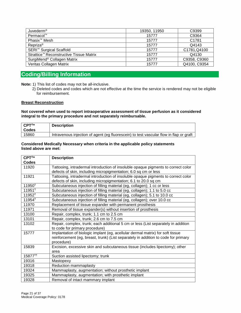
Page 21 of 37 Medical Coverage Policy: 0178
Juvederm® 19350, 11950 C9399 Permacol™ 15777 C9364 Phasix™ Mesh 15777 C1781 Repriza® 15777 Q4143 SERI™ Surgical Scaffold 15777 C1781,Q4100 Strattice™ Reconstructive Tissue Matrix 15777 Q4130 SurgiMend® Collagen Matrix 15777 C9358, C9360 Veritas Collagen Matrix 15777 Q4100, C9354
Coding/Billing Information Note: 1) This list of codes may not be all-inclusive. 2) Deleted codes and codes which are not effective at the time the service is rendered may not be eligible for reimbursement. Breast Reconstruction Not covered when used to report intraoperative assessment of tissue perfusion as it considered integral to the primary procedure and not separately reimbursable. CPT®* Codes
Description
15860 Intravenous injection of agent (eg fluorescein) to test vascular flow in flap or graft Considered Medically Necessary when criteria in the applicable policy statements listed above are met: CPT®* Codes
Description
11920 Tattooing, intradermal introduction of insoluble opaque pigments to correct color defects of skin, including micropigmentation; 6.0 sq cm or less
11921 Tattooing, intradermal introduction of insoluble opaque pigments to correct color defects of skin, including micropigmentation; 6.1 to 20.0 sq cm
11950† Subcutaneous injection of filling material (eg, collagen); 1 cc or less 11951† Subcutaneous injection of filling material (eg, collagen); 1.1 to 5.0 cc 11952† Subcutaneous injection of filling material (eg, collagen); 5.1 to 10.0 cc 11954† Subcutaneous injection of filling material (eg, collagen); over 10.0 cc 11970 Replacement of tissue expander with permanent prosthesis 11971 Removal of tissue expander(s) without insertion of prosthesis 13100 Repair, complex, trunk; 1.1 cm to 2.5 cm 13101 Repair, complex, trunk; 2.6 cm to 7.5 cm 13102 Repair, complex, trunk; each additional 5 cm or less (List separately in addition
to code for primary procedure) 15777 Implantation of biologic implant (eg, acellular dermal matrix) for soft tissue
reinforcement (eg, breast, trunk) (List separately in addition to code for primary procedure)
15839 Excision, excessive skin and subcutaneous tissue (includes lipectomy); other area
15877†† Suction assisted lipectomy; trunk 19316 Mastopexy 19318 Reduction mammaplasty 19324 Mammaplasty, augmentation; without prosthetic implant 19325 Mammaplasty, augmentation; with prosthetic implant 19328 Removal of intact mammary implant

Page 22 of 37 Medical Coverage Policy: 0178
19330 Removal of mammary implant material 19340 Immediate insertion of breast prosthesis following mastopexy, mastectomy or in
reconstruction 19342 Delayed insertion of breast prosthesis following mastopexy, mastectomy or in
reconstruction 19350††† Nipple/areola reconstruction 19357 Breast reconstruction, immediate or delayed, with tissue expander, including
subsequent expansion 19361 Breast reconstruction with latissimus dorsi flap, without prosthetic implant 19364 Breast reconstruction with free flap 19367 Breast reconstruction with transverse rectus abdominis myocutaneous flap
(TRAM), single pedicle, including closure of donor site 19368 Breast reconstruction with transverse rectus abdominis myocutaneous flap
(TRAM), single pedicle, including closure of donor site; with microvascular anastomosis (supercharging)
19369 Breast reconstruction with transverse rectus abdominis myocutaneous flap (TRAM), double pedicle, including closure of donor site
19370 Open periprosthetic capsulotomy, breast 19371 Periprosthetic capsulectomy, breast 19380 Revision of reconstructed breast 19499†††† Unlisted procedure, breast
†Note: Considered medically necessary when used to report autologous fat transplant without the use of adipose-derived stem cells or xenograft cartilage grafting. ††Note: Considered incidental to the primary procedure when used to report suction-assisted lipectomy of the trunk as part of a medically necessary flap breast reconstruction procedure. †††Note: Considered not medically necessary when used to report Cook Biodesign® Nipple Reconstruction Cylinder or Juvederm®. ††††Note: Considered medically necessary when used to report thoracodorsal artery perforator (TDAP) flap with a breast reconstruction procedure performed on the diseased/affected breast (i.e., breast on which the mastectomy/lumpectomy was performed). HCPCS Codes
Description
C1789 Prosthesis, breast (implantable) L8600 Implantable breast prosthesis, silicone or equal Q4116 Alloderm, per square centimeter Q4122 Dermacell, per square centimeter Q4128 Flex hd or allopatch hd, or matrix hd per square centimeter S2066 Breast reconstruction with gluteal artery perforator (GAP) flap, including
harvesting of the flap, microvascular transfer, closure of donor site and shaping the flap into a breast, unilateral
S2067 Breast reconstruction of a single breast with "stacked" deep inferior epigastric perforator (DIEP) flap(s) and/or gluteal artery perforator (GAP) flap(s), including harvesting of the flap(s), microvascular transfer, closure of donor site(s) and shaping the flap into a breast, unilateral
S2068 Breast reconstruction with deep inferior epigastric perforator (DIEP) flap or superficial inferior epigastric artery (SIEA) flap, including harvesting of the flap, microvascular transfer, closure of donor site and shaping the flap into a breast, unilateral
Considered medically necessary when used to report AlloMax™ or NeoForm™ Dermis used

Page 23 of 37 Medical Coverage Policy: 0178
in association with a medically necessary breast reconstruction procedure: CPT®* Codes
Description
C1781 Mesh (implantable) Q4100 Skin substitute, not otherwise specified
Considered medically necessary when used to report autologous fat transplant (i.e., liposuction, lipoinjection, lipofilling, lipomodeling) without the use of adipose-derived stem cells or xenograft cartilage grafting: CPT®* Codes
Description
19366 Breast reconstruction with other technique 19499 Unlisted procedure, breast
Experimental/Investigational/Unproven when used to report a product as not covered as listed in the policy statement above: CPT®* Codes
Description
11950††††† Subcutaneous injection of filling material (eg, collagen); 1 cc or less 11951††††† Subcutaneous injection of filling material (eg, collagen); 1.1 to 5.0 cc 11952††††† Subcutaneous injection of filling material (eg, collagen); 5.1 to 10.0 cc 11954††††† Subcutaneous injection of filling material (eg, collagen); over 10.0 cc
†††††Note: Experimental/Investigational/Unproven when used to report autologous fat transplant with the use of adipose-derived stem cells or xenograft cartilage grafting. HCPCS Codes
Description
C9358 Dermal substitute, native, non-denatured collagen, fetal bovine origin, (Surgimend collagen matrix), per 0.5 square centimeters
C9360 Dermal substitute, native, non-denatured collagen, neonatal bovine origin (Surgimend collagen matrix), per 0.5 square centimeters
C9364 Porcine implant Permacol, per square cm Q2026 Injection, Radiessee, 0.1 ml Q4134 Hmatrix, per square centimeter Q4143 Repriza, per square centimeter
Experimental/Investigational/Unproven when used to report Biodesign® Nipple Reconstruction Cylinder: HCPCS Codes
Description
C1763 Connective tissue, non-human (includes synthetic) Experimental/Investigational/Unproven when used to report Cortiva, DermaMatrix Aceullular Dermis, Juvederm®, Phasix™ Mesh, or SERI™ Surgical Scaffold: HCPCS Codes
Description
C1781 Mesh (implantable) C9399 Unclassified drugs or biologicals Q4100 Skin substitute, not otherwise specified

Page 24 of 37 Medical Coverage Policy: 0178
Experimental/Investigational/Unproven when used to report Strattice™ : HCPCS Codes
Description
Q4130 Strattice tm, per square centimeter Experimental/Investigational/Unproven when used to report Veritas Collagen Matrix: HCPCS Codes
Description
C9354 Acellular pericardial tissue matrix of non-human origin (Veritas), per square centimeter
Q4100 Skin substitute, not otherwise specified Experimental/Investigational/Unproven when used to report autologous fat transplant with the use of adipose-derived stem cells or xenograft cartilage grafting: CPT®* Codes
Description
19366 Breast reconstruction with other technique 19499 Unlisted procedure, breast
Experimental/Investigational/Unproven when used to report vascularized lymph node transfer (VLNTx): CPT®* Codes
Description
38999 Unlisted procedure, hemic or lymphatic system Cosmetic in nature/Not Medically Necessary when used to report correction of surgically-induced donor site asymmetry that results from flap breast reconstruction procedures: CPT®* Codes
Description
15877 Suction assisted lipectomy; trunk 15879 Suction assisted lipectomy; lower extremity
External Breast Prostheses and Mastectomy Bras Following Mastectomy or Lumpectomy Considered Medically Necessary when criteria in the applicable policy statements listed above are met: HCPCS Codes
Description
L8000 Breast prosthesis; mastectomy bra, without integrated breast prosthesis form, any size, any type
L8001 Breast prosthesis, mastectomy bra, with integrated breast prosthesis form, unilateral, any size, any type
L8002 Breast prosthesis, mastectomy bra, with integrated breast prosthesis form, bilateral, any size, any type
L8015 External breast prosthesis garment, with mastectomy form, post mastectomy L8020 Breast prosthesis, mastectomy form L8030 Breast prosthesis, silicone or equal, without integral adhesive L8031 Breast prosthesis, silicone or equal, with integral adhesive

Page 25 of 37 Medical Coverage Policy: 0178
L8032 Nipple prosthesis, reusable, any type, each L8035 Custom breast prosthesis, post mastectomy, molded to patient model L8039 Breast prosthesis, not otherwise specified
*Current Procedural Terminology (CPT®) ©2017 American Medical Association: Chicago, IL. References
1. Alam M, Gladstone H, Kramer EM, Murphy JP Jr, Nouri K, Neuhaus IM, et al; American Society for Dermatologic Surgery. ASDS guidelines of care: injectable fillers. Dermatol Surg. 2008 Jun;34 Suppl 1:S115-48.
2. Alderman AK, Kuzon WM Jr, Wilkins EG. A two-year prospective analysis of trunk function in TRAM
breast reconstructions. Plast Reconstr Surg. 2006 Jun;117(7):2131–8.
3. Allergan. SERI™ Surgical Scaffold. USA Product Labeling and Safety Information. 2017. Accessed Nov 9, 2017. Available at URL address: http://www.allergan.com/products/key-products/product-prescribing/labeling/seri_usa
4. Allergan. Juvederm®. Accessed Nov 9, 2017. Available at URL address: https://www.juvederm.com/
5. American Cancer Society (ACS): Breast reconstruction surgery. Last revised Sept 12, 2017. Accessed Nov 9, 2017. Available at URL address: http://www.cancer.org
6. American Society of Plastic Surgeons (ASPS). Evidence-Based Clinical Practice Guideline: Breast
Reconstruction with Expanders and Implants. March 2013. Accessed Nov 9, 2017. Available at URL address: http://www.plasticsurgery.org/for-medical-professionals/legislation-and-advocacy/health-policy-resources/evidence-based-guidelinespractice-parameters.html
7. Antony AK, McCarthy CM, Cordeiro PG, Mehrara BJ, Pusic AL, Teo EH, et al. Acellular human dermis
implantation in 153 immediate two-stage tissue expander breast reconstructions: determining the incidence and significant predictors of complications. Plast Reconstr Surg. 2010 Jun;125(6):1606-14.
8. Bacterin International Holdings Inc., Belgrade, MT. hMatrix®. Accessed Nov 9, 2017. Available at URL
address: http://bacterin.com/products/
9. Bajaj AK, Chevray PM, Chang DW. Comparison of donor-site complications and functional outcomes in free muscle-sparing TRAM flap and free DIEP flap breast reconstruction. Plast Reconstr Surg. 2006 Mar;117(3):737–46; discussion 747–50.
10. Barber MD, Williams L, Anderson ED, Neades GT, Raine C, Young O, et al. Outcome of the use of
acellular-dermal matrix to assist implant-based breast reconstruction in a single centre. Eur J Surg Oncol. 2015 Jan;41(1):100-5.
11. Bard Davol, Inc. AlloMax™ surgical graft for breast reconstruction. Accessed Nov 9, 2017. Available at
URL address: http://www.davol.com/products
12. Bard Davol, Inc. Phasix™ Mesh. Accessed Nov 9, 2017. Available at URL address: https://www.crbard.com/Davol/products/All-Products/Hernia-Repair-Fixation/
13. Baxter RA. Intracapsular allogenic dermal grafts for breast implant-related problems. Plast Reconstr Surg. 2003 Nov;112(6):1692-6; discussion 1697-8.
14. Beckenstein MS. Nipple Reconstruction. In: Grabb and Smith Plastic Surgery. 7th ed. Lippincott, Williams
and Wilkins. Philadelphia, PA; 2014. Ch 63.
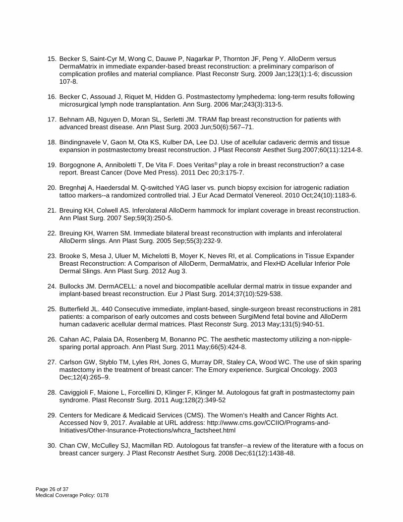
Page 26 of 37 Medical Coverage Policy: 0178
15. Becker S, Saint-Cyr M, Wong C, Dauwe P, Nagarkar P, Thornton JF, Peng Y. AlloDerm versus
DermaMatrix in immediate expander-based breast reconstruction: a preliminary comparison of complication profiles and material compliance. Plast Reconstr Surg. 2009 Jan;123(1):1-6; discussion 107-8.
16. Becker C, Assouad J, Riquet M, Hidden G. Postmastectomy lymphedema: long-term results following
microsurgical lymph node transplantation. Ann Surg. 2006 Mar;243(3):313-5.
17. Behnam AB, Nguyen D, Moran SL, Serletti JM. TRAM flap breast reconstruction for patients with advanced breast disease. Ann Plast Surg. 2003 Jun;50(6):567–71.
18. Bindingnavele V, Gaon M, Ota KS, Kulber DA, Lee DJ. Use of acellular cadaveric dermis and tissue
expansion in postmastectomy breast reconstruction. J Plast Reconstr Aesthet Surg.2007;60(11):1214-8.
19. Borgognone A, Anniboletti T, De Vita F. Does Veritas® play a role in breast reconstruction? a case report. Breast Cancer (Dove Med Press). 2011 Dec 20;3:175-7.
20. Bregnhøj A, Haedersdal M. Q-switched YAG laser vs. punch biopsy excision for iatrogenic radiation
tattoo markers--a randomized controlled trial. J Eur Acad Dermatol Venereol. 2010 Oct;24(10):1183-6.
21. Breuing KH, Colwell AS. Inferolateral AlloDerm hammock for implant coverage in breast reconstruction. Ann Plast Surg. 2007 Sep;59(3):250-5.
22. Breuing KH, Warren SM. Immediate bilateral breast reconstruction with implants and inferolateral
AlloDerm slings. Ann Plast Surg. 2005 Sep;55(3):232-9.
23. Brooke S, Mesa J, Uluer M, Michelotti B, Moyer K, Neves RI, et al. Complications in Tissue Expander Breast Reconstruction: A Comparison of AlloDerm, DermaMatrix, and FlexHD Acellular Inferior Pole Dermal Slings. Ann Plast Surg. 2012 Aug 3.
24. Bullocks JM. DermACELL: a novel and biocompatible acellular dermal matrix in tissue expander and
implant-based breast reconstruction. Eur J Plast Surg. 2014;37(10):529-538.
25. Butterfield JL. 440 Consecutive immediate, implant-based, single-surgeon breast reconstructions in 281 patients: a comparison of early outcomes and costs between SurgiMend fetal bovine and AlloDerm human cadaveric acellular dermal matrices. Plast Reconstr Surg. 2013 May;131(5):940-51.
26. Cahan AC, Palaia DA, Rosenberg M, Bonanno PC. The aesthetic mastectomy utilizing a non-nipple-
sparing portal approach. Ann Plast Surg. 2011 May;66(5):424-8.
27. Carlson GW, Styblo TM, Lyles RH, Jones G, Murray DR, Staley CA, Wood WC. The use of skin sparing mastectomy in the treatment of breast cancer: The Emory experience. Surgical Oncology. 2003 Dec;12(4):265–9.
28. Caviggioli F, Maione L, Forcellini D, Klinger F, Klinger M. Autologous fat graft in postmastectomy pain
syndrome. Plast Reconstr Surg. 2011 Aug;128(2):349-52
29. Centers for Medicare & Medicaid Services (CMS). The Women’s Health and Cancer Rights Act. Accessed Nov 9, 2017. Available at URL address: http://www.cms.gov/CCIIO/Programs-and-Initiatives/Other-Insurance-Protections/whcra_factsheet.html
30. Chan CW, McCulley SJ, Macmillan RD. Autologous fat transfer--a review of the literature with a focus on
breast cancer surgery. J Plast Reconstr Aesthet Surg. 2008 Dec;61(12):1438-48.

Page 27 of 37 Medical Coverage Policy: 0178
31. Chang EI, Liu J. Prospective unbiased experience with three acellular dermal matrices in breast reconstruction. J Surg Oncol. 2017 Sep;116(3):365-370.
32. Cheng A, Saint-Cyr M. Comparison of different ADM materials in breast surgery. Clin Plast Surg. 2012
Apr;39(2):167-75.
33. Cheng MH, Rodriguez ED, Smartt JM, Cardenas-Mejia A. Nipple reconstruction using the modified top hat flap with banked costal cartilage graft: long-term follow-up in 58 patients. Ann Plast Surg. 2007 Dec;59(6):621-8.
34. Chun YS. Nipple-Areola Reconstruction. Updated June 14, 2017. Accessed Nov 9, 2017. Available at
URL address: http://www.emedicine.com/plastic/topic144.htm
35. Claro F Jr, Figueiredo JC, Zampar AG, Pinto-Neto AM. Applicability and safety of autologous fat for reconstruction of the breast. Br J Surg. 2012 Jun;99(6):768-80.
36. ClinicalTrials.org. Compare Outcomes Between Two Acellular Dermal Matrices. NCT02891759. First posted: Sept 7, 2016. Last updated: April 13, 2017. Accessed Nov 9, 2017. Available at URL address: https://clinicaltrials.gov/show/NCT02891759
37. Coleman SR, Saboeiro AP. Fat grafting to the breast revisited: safety and efficacy. Plast Reconstr Surg.
2007 Mar;119(3):775-85; discussion 786-7.
38. Collins B, Williams JZ, Karu H, Hodde JP, Martin VA, Gurtner GC. Nipple Reconstruction with the Biodesign Nipple Reconstruction Cylinder: A Prospective Clinical Study. Plast Reconstr Surg Glob Open. 2016 Aug 9;4(8):e832.
39. Colwell AS, Damjanovic B, Zahedi B, Medford-Davis L, Hertl C, Austen WG Jr. Retrospective review of
331 consecutive immediate single-stage implant reconstructions with acellular dermal matrix: indications, complications, trends, and costs. Plast Reconstr Surg. 2011 Dec;128(6):1170-8.
40. Craft RO, May JW Jr. Staged nipple reconstruction with vascularized SurgiMend acellular dermal matrix.
Plast Reconstr Surg. 2011 Jun;127(6):148e-9e.
41. de Blacam C, Momoh AO, Colakoglu S, Tobias AM, Lee BT. Evaluation of clinical outcomes and aesthetic results after autologous fat grafting for contour deformities of the reconstructed breast. Plast Reconstr Surg. 2011 Nov;128(5):411e-418e.
42. De Decker M, De Schrijver L, Thiessen F, Tondu T, Van Goethem M, Tjalma WA. Breast cancer and fat grafting: efficacy, safety and complications-a systematic review. Eur J Obstet Gynecol Reprod Biol. 2016 Dec;207:100-108.
43. DellaCroce FJ. Perforator Flap Reconstruction Treatment and Management. Updated April 28, 2015.
Accessed Nov 9, 2017. Available at URL address: http://emedicine.medscape.com/article/1276406-treatment
44. Department of Health and Human Services. Oversight of tissue banking. Jan 20, 2001. Accessed Nov 9,
2017. Available at URL address: http://www.fda.gov/ohrms/dockets/ac/01/briefing/3736b2_01.pdf
45. Deutsch MF, Robb GL, Talavera F, Shenaq SM, Slenkovich N, Aly A. Breast Reconstruction, Other Free Flaps. Updated Dec 30, 2014. Accessed Nov 9, 2017. Available at URL address: http://www.emedicine.com/plastic/topic140.htm
46. Deutsch MF, Robb GL, Talavera F, Shenaq SM, Slenkovich N, Aly A. Breast Reconstruction,
Refinements & Finishing Touches. Updated Dec 29, 2014. Accessed Nov 9, 2017. Available at URL address: http://www.emedicine.com/plastic/topic135.htm
Page 28 of 37 Medical Coverage Policy: 0178
47. Dikmans RE, El Morabit F, Ottenhof MJ, Tuinder SM, Twisk JW, Moues C, et al. Single-stage breast
reconstruction using Strattice™: A retrospective study. J Plast Reconstr Aesthet Surg. 2016 Feb;69(2):227-33.
48. Dionyssiou D, Demiri E, Tsimponis A, Sarafis A, Mpalaris V, Tatsidou G, Arsos G. A randomized control
study of treating secondary stage II breast cancer-related lymphoedema with free lymph node transfer. Breast Cancer Res Treat. 2016 Feb;156(1):73-9.
49. Draper LB, Bui DT, Chiu ES, Mehrara BJ, Pusic AL, Cordeiro PG, Disa JJ. Nipple-Areola Reconstruction
Following Chest-Wall Irradiation for Breast Cancer: Is It Safe? Ann Plast Surg. 2005 Jul;55(1):12–5.
50. Evans KK, Rasko Y, Lenert J, Olding M. The use of calcium hydroxylapatite for nipple projection after failed nipple-areolar reconstruction: early results. Ann Plast Surg. 2005 Jul;55(1):25-9; discussion 29.
51. Fansa H, Schirmer S, Warnecke IC, Cervelli A, Frerichs O. The transverse myocutaneous gracilis
muscle flap: a fast and reliable method for breast reconstruction. Plast Reconstr Surg. 2008 Nov;122(5):1326-33.
52. Fine NA, Lehfeldt M, Gross JE, Downey S, Kind GM, Duda G, et al. SERI surgical scaffold, prospective
clinical trial of a silk-derived biological scaffold in two-stage breast reconstruction: 1-year data. Plast Reconstr Surg. 2015 Feb;135(2):339-51.
53. Gabbay JS, Eby JB, Kulber DA. The midabdominal TRAM flap for breast reconstruction in morbidly
obese patients. Plast Reconstr Surg. 2005 Mar;115(3):764–70.
54. Gamboa-Bobadilla GM. Implant breast reconstruction using acellular dermal matrix. Ann Plast Surg. 2006 Jan;56(1):22-5.
55. Garramone CE, Lam B. Use of AlloDerm in primary nipple reconstruction to improve long-term nipple
projection. Plast Reconstr Surg. 2007 May;119(6):1663-8.
56. Gaster RS, Berger AJ, Monica SD, Sweeney RT, Endress R, Lee GK. Histologic Analysis of Fetal Bovine Derived Acellular Dermal Matrix in Tissue Expander Breast Reconstruction. Ann Plast Surg. 2013 Mar 11.
57. Garvey PB, Buchel EW, Pockaj BA, Casey WJ 3rd, Gray RJ, Hernandez JL, Samson TD. DIEP and
pedicled TRAM flaps: a comparison of outcomes. Plast Reconstr Surg. 2006 May;117(6):1711–9; discussion 1720–1.
58. Gharb BB, Rampazzo A, Spanio di Spilimbergo S, Xu ES, Chung KP, Chen HC. Vascularized lymph
node transfer based on the hilar perforators improves the outcome in upper limb lymphedema. Ann Plast Surg. 2011 Dec;67(6):589-93.
59. Glasberg SB, D'Amico RA. Use of regenerative human acellular tissue (AlloDerm) to reconstruct the
abdominal wall following pedicle TRAM flap breast reconstruction surgery. Plast Reconstr Surg. 2006 Jul;118(1):8-15.
60. Glasberg SB, Light D. AlloDerm and Strattice in breast reconstruction: a comparison and techniques for
optimizing outcomes. Plast Reconstr Surg. 2012 Jun;129(6):1223-33.
61. Gratzon A, Schultz J, Secrest K, Lee K, Feiner J, Klein RD. Clinical and Psychosocial Outcomes of Vascularized Lymph Node Transfer for the Treatment of Upper Extremity Lymphedema After Breast Cancer Therapy. Ann Surg Oncol. 2017 Jun;24(6):1475-1481.

Page 29 of 37 Medical Coverage Policy: 0178
62. Guerra AB, Khoobehi K, Metzinger SE, Allen RJ. New technique for nipple areola reconstruction: arrow flap and rib cartilage graft for long-lasting nipple projection. Ann Plast Surg. 2003 Jan;50(1):31-7.
63. Gurtner GC, Jones GE, Neligan PC, Newman MI, Phillips BT, Sacks JM, Zenn MR. Intraoperative laser
angiography using the SPY system: review of the literature and recommendations for use. Ann Surg Innov Res. 2013 Jan 7;7(1):1.
64. Gutowski KA; ASPS Fat Graft Task Force. Current applications and safety of autologous fat grafts: a
report of the ASPS fat graft task force. Plast Reconstr Surg. 2009 Jul;124(1):272-80.
65. Haddock N, Levine J. Breast reconstruction with implants, tissue expanders and AlloDerm: predicting volume and maximizing the skin envelope in skin sparing mastectomies. Breast J. 2010 Jan-Feb;16(1):14-9.
66. Hayes, Inc. Hayes Medical Technology Directory Report. Surgical Treatment of Lymphedema: A
Review of Reviews. Lansdale, PA: Hayes, Inc.; published May 2017.
67. Hayes, Inc. Hayes Medical Technology Directory Report. Human Acellular Dermal Matrix Grafts for Breast Reconstruction. Lansdale, PA: Hayes, Inc.; published April 2014; annual review April 2015; April 2016; April 2017.
68. Hayes, Inc. Hayes Technology Brief. Superficial Inferior Epigastric Artery (SIEA) Flap Procedure for
Postmastectomy Breast Reconstruction. Lansdale, PA: Hayes, Inc.; November 2014; annual review Oct 2015; Oct 2016.
69. Hayes, Inc. Hayes Technology Brief. Autologous Fat Grafting for Breast Reconstruction After Breast
Cancer Surgery. Lansdale, PA: Hayes, Inc.; August 27, 2015; annual review July 2016; July 2017.
70. Hayes, Inc. Hayes Technology Brief. SurgiMend (Integra LifeSciences) for Postmastectomy Breast Reconstruction. Lansdale, PA: Hayes, Inc.; April 27, 2017.
71. Hayes, Inc. Clinical Research Response. Product Comparison. Silicone Gel Breast Implants. Lansdale, PA: Hayes, Inc.; January 26, 2017.
72. Hayes, Inc. Clinical Research Response. Product Comparison. Biological Versus Resorbable Mesh for Breast Reconstruction. Lansdale, PA: Hayes, Inc.; May 4, 2017.
73. Heitland A, Markowicz M, Koellensperger E, Allen R, Pallua N. Long-term nipple shrinkage following
augmentation by an autologous rib cartilage transplant in free DIEP-flaps. J Plast Reconstr Aesthet Surg. 2006;59(10):1063-7.
74. Ho G, Nguyen TJ, Shahabi A, Hwang BH, Chan LS, Wong AK. A systematic review and meta-analysis
of complications associated with acellular dermal matrix-assisted breast reconstruction. Ann Plast Surg. 2012 Apr;68(4):346-56.
75. Hu E, Alderman AK. Breast reconstruction. Surg Clin North Am. 2007 Apr;87(2):453-67, x.
76. Humphreys K. Autologous fat injection for breast reconstruction. August 2008. Australia and New
Zealand Horizon Scanning Network. Accessed Nov 9, 2017. Available at URL address: http://www.horizonscanning.gov.au/internet/horizon/publishing.nsf/Content/technologies-assessed-lp-2
77. Hyakusoku H, Ogawa R, Ono S, Ishii N, Hirakawa K. Complications after autologous fat injection to the
breast. Plast Reconstr Surg. 2009 Jan;123(1):360-70; discussion 371-2.
78. Ibrahim AM, Shuster M, Koolen PG, Kim K, Taghinia AH, Sinno HH, et al. Analysis of the National Surgical Quality Improvement Program database in 19,100 patients undergoing implant-based breast
Page 30 of 37 Medical Coverage Policy: 0178
reconstruction: complication rates with acellular dermal matrix. Plast Reconstr Surg. 2013 Nov;132(5):1057-66.
79. Illouz YG, Sterodimas A. Autologous fat transplantation to the breast: a personal technique with 25 years
of experience. Aesthetic Plast Surg. 2009 Sep;33(5):706-15.
80. Integra® LifeSciences Corp. SurgiMend®. Accessed Nov 9, 2017. Available at URL address: http://www.surgimend.com/
81. Jansen LA, Macadam SA. The use of AlloDerm in postmastectomy alloplastic breast reconstruction: part
I. A systematic review. Plast Reconstr Surg. 2011 Jun;127(6):2232-44.
82. Jewell M1, Daunch W2, Bengtson B3, Mortarino E2. The development of SERI® Surgical Scaffold, an engineered biological scaffold. Ann N Y Acad Sci. 2015 Sep 16.
83. Joanna Nguyen T, Carey JN, Wong AK. Use of human acellular dermal matrix in implant based breast
reconstruction: Evaluating the evidence. J Plast Reconstr Aesthet Surg. 2011 Mar 7.
84. Kamakura T, Ito K. Autologous cell-enriched fat grafting for breast augmentation. Aesthetic Plast Surg. 2011 Dec;35(6):1022-30.
85. Kanchwala SK, Glatt BS, Conant EF, Bucky LP. Autologous fat grafting to the reconstructed breast: the
management of acquired contour deformities. Plast Reconstr Surg. 2009 Aug;124(2):409-18.
86. Karp N, Choi M, Kulber DA, Downey S, Duda G, Kind GM, et al. SERI Surgical Scaffold in 2-Stage Breast Reconstruction: 2-Year Data from a Prospective, Multicenter Trial. Plast Reconstr Surg Glob Open. 2017 May 16;5(5):e1327.
87. Keifer OP Jr, Page EK, Hart A, Rudderman R, Carlson GW, Losken A. A Complication Analysis of 2
Acellular Dermal Matrices in Prosthetic-based Breast Reconstruction. Plast Reconstr Surg Glob Open. 2016 Jul 13;4(7):e800.
88. Kilchenmann AJ, Lardi AM, Ho-Asjoe M, Junge K, Farhadi J. An evaluation of resource utilisation of
single stage porcine acellular dermal matrix assisted breast reconstruction: A comparative study. Breast. 2014 Dec;23(6):876-82
89. Kim EK, Lee TJ. Use of lyophilized allogeneic costal cartilage: is it effective to maintain the projection of
the reconstructed nipple? Ann Plast Surg. 2011 Feb;66(2):128-30.
90. Kim JY, Davila AA, Persing S, Connor CM, Jovanovic B, Khan SA, et al. A meta-analysis of human acellular dermis and submuscular tissue expander breast reconstruction. Plast Reconstr Surg. 2012 Jan;129(1):28-41.
91. Kim J, Long JN. Breast reconstruction with acellular dermal matrix. Updated Aug 15, 2017. Accessed
Nov 9, 2017. Available at URL address: http://emedicine.medscape.com/article/1851090-overview#a1
92. Kroll SS. Fat necrosis in free transverse rectus abdominis myocutaneous and deep inferior epigastric perforator flaps. Plast Reconstr Surg. 2000 Sep;106(3):576–83.
93. Lardi AM, Ho-Asjoe M, Mohanna PN, Farhadi J. Immediate breast reconstruction with acellular dermal
matrix: factors affecting outcome. J Plast Reconstr Aesthet Surg. 2014 Aug;67(8):1098-105.
94. Lee KT, Mun GH. Updated Evidence of Acellular Dermal Matrix Use for Implant-Based Breast Reconstruction: A Meta-analysis. Ann Surg Oncol. 2016 Feb;23(2):600-10.

Page 31 of 37 Medical Coverage Policy: 0178
95. Leopardi D, et al. Systematic review of autologous fat transfer for cosmetic and reconstructive breast augmentation. ASERNIP-S Report No.70. Adelaide, South Australia: ASERNIP-S, September 2010. Accessed Nov 9, 2017. Available at URL address: http://www.surgeons.org
96. LifeCell Corporation. AlloDerm Tissue Matrix Defined [product information]. Accessed Nov 9, 2017. Available at URL address: http://www.lifecell.com
97. LifeCell Corporation. Strattice™. Accessed Nov 9, 2017. Available at URL address: http://www.lifecell
98. LifeNet Health. Skin and wound allograft institute. DermACELL® - The New Alternative for Breast
Reconstruction. Accessed Nov 9, 2017. Available at URL address: https://www.lifenethealth.org/skin-and-wound-allograft-institute
99. Lin CH, Ali R, Chen SC, Wallace C, Chang YC, Chen HC, Cheng MH. Vascularized groin lymph node
transfer using the wrist as a recipient site for management of postmastectomy upper extremity lymphedema. Plast Reconstr Surg. 2009 Apr;123(4):1265-75.
100. Liu DZ, Mathes DW, Neligan PC, Said HK, Louie O. Comparison of outcomes using AlloDerm
versus FlexHD for implant-based breast reconstruction. Ann Plast Surg. 2014 May;72(5):503-7.
101. Losken A, Carolson GW, Bostwick J III, Jones GE, Culbertson JH, Schoemann M. Trends in unilateral breast reconstruction and management of the contralateral breast: the Emory experience. Plast Reconstr Surg. 2002 Jul;110(1):89–97.
102. Losken A. Early Results Using Sterilized Acellular Human Dermis (Neoform) in Post-Mastectomy
Tissue Expander Breast Reconstruction. Plast Reconstr Surg. 2009 Mar 23.
103. Losken A, Pinell XA, Sikoro K, Yezhelyev MV, Anderson E, Carlson GW. Autologous fat grafting in secondary breast reconstruction. Ann Plast Surg. 2011 May;66(5):518-22.
104. Martin L, O'Donoghue JM, Horgan K, Thrush S, Johnson R, Gandhi A; Association of Breast Surgery
and the British Association of Plastic, Reconstructive and Aesthetic Surgeons. Acellular dermal matrix (ADM) assisted breast reconstruction procedures: joint guidelines from the Association of Breast Surgery and the British Association of Plastic, Reconstructive and Aesthetic Surgeons. Eur J Surg Oncol. 2013 May;39(5):425-9.
105. Maxwell GP, Gabriel A. Non-cross-linked porcine acellular dermal matrix in revision breast surgery:
long-term outcomes and safety with neopectoral pockets. Aesthet Surg J. 2014 May 1;34(4):551-9.
106. McCarthy CM, Lee CN, Halvorson EG, Riedel E, Pusic AL, Mehrara BJ, Disa JJ. The use of acellular dermal matrices in two-stage expander/implant reconstruction: a multicenter, blinded, randomized controlled trial. Plast Reconstr Surg. 2012 Nov;130(5 Suppl 2):57S-66S.
107. Mehrara BJ, Santoro TD, Arcilla E, Watson JP, Shaw WW, Da Lio AL. Complications after
microvascular breast reconstruction: experience with 1195 flaps. Plast Reconstr Surg. 2006 Oct;118(5):1100-9; discussion 1110-1.
108. Michelotti BF, Brooke S, Mesa J, Wilson MZ, Moyer K, Mackay DR, et al. Analysis of clinically
significant seroma formation in breast reconstruction using acellular dermal grafts. Ann Plast Surg. 2013 Sep;71(3):274-7.
109. Musculoskeletal Transplant Foundation. FlexHD® Acellular Hydrated Dermis. Accessed Nov 9, 2017.
Available at URL address: http://www.mtf.org/allograft_offerings.html
110. Nahabedian MY. Secondary nipple reconstruction using local flaps and AlloDerm. Plast Reconstr Surg. 2005 Jun;115(7):2056-61.

Page 32 of 37 Medical Coverage Policy: 0178
111. National Comprehensive Cancer Network (NCCN). NCCN® practice guidelines in oncology. Breast
Cancer. V.2.2017-April 6, 2017. Accessed Nov 9, 2017. Available at URL address: http://www.nccn.org/professionals/physician_gls/f_guidelines.asp
112. National Institute for Health and Clinical Excellence (NICE). Interventional procedure overview IPG417
Interventional procedure overview of breast reconstruction using lipomodelling after breast cancer treatment. Issued January 2012. Accessed Nov 9, 2017. Available at URL address: http://www.nice.org.uk/
113. National Library of Medicine. Breast Reconstruction. Reviewed June 21, 2016. Topic last updated Feb
15, 2017. Accessed Nov 9, 2017. Available at URL address: http://www.nlm.nih.gov/medlineplus/breastreconstruction.html
114. Nguyen TJ, Carey JN, Wong AK. Use of human acellular dermal matrix in implant-based breast
reconstruction: evaluating the evidence. Jour Plas Recon (2011):64, 1553-1561.
115. Nguyen AT, Chang EI, Suami H, Chang DW. An algorithmic approach to simultaneous vascularized lymph node transfer with microvascular breast reconstruction. Ann Surg Oncol. 2015 Sep;22(9):2919-24.
116. Ohkuma R, Buretta KJ, Mohan R, Rosson GD, Rad AN. Initial experience with the use of
foetal/neonatal bovine acellular dermal collagen matrix (SurgiMend™) for tissue-expander breast reconstruction. J Plast Reconstr Aesthet Surg. 2013 Sep;66(9):1195-201.
117. Ozturk CN, Ozturk C, Glasgow M, Platek M, Ashary Z, Kuhn J, et al. Free vascularized lymph node transfer for treatment of lymphedema: A systematic evidence based review. J Plast Reconstr Aesthet Surg. 2016 Sep;69(9):1234-47.
118. PDQ® Supportive and Palliative Care Editorial Board. PDQ Lymphedema. Bethesda, MD: National
Cancer Institute. Updated 07/17/2015. Accessed Nov 9, 2017. Available at URL address: https://www.cancer.gov/about-cancer/treatment/side-effects/lymphedema/lymphedema-hp-pdq
119. Parikh RP, Doren EL, Mooney B, Sun WV, Laronga C, Smith PD. Differentiating fat necrosis from
recurrent malignancy in fat-grafted breasts: an imaging classification system to guide management. Plast Reconstr Surg. 2012 Oct;130(4):761-72.
120. Petit JY, Lohsiriwat V, Clough KB, Sarfati I, Ihrai T, Rietjens M, et al. The oncologic outcome and
immediate surgical complications of lipofilling in breast cancer patients: a multicenter study-milan-paris-lyon experience of 646 lipofilling procedures. Plast Reconstr Surg. 2011a Aug;128(2):341-6
121. Petit JY, Botteri E, Lohsiriwat V, Rietjens M, De Lorenzi F, Garusi C, et al. Locoregional recurrence risk
after lipofilling in breast cancer patients. Ann Oncol. 2011b May 24.
122. Perez-Cano R, Vranckx JJ, Lasso JM et al. Prospective trial of adipose-derived regenerative cell (ADRC)-enriched fat grafting for partial mastectomy defects: the RESTORE-2 trial. Eur J Surg Oncol 2012; 38(5):382-9.
123. Pittman TA, Fan KL, Knapp A, Frantz S, Spear SL. Comparison of Different Acellular Dermal Matrix (ADM) in Breast Reconstruction: The 50/50 Study. Plast Reconstr Surg. 2016 Nov 21. [Epub ahead of print]
124. Pulagam SR, Poulton T, Mamounas EP. Long-term clinical and radiologic results with autologous fat
transplantation for breast augmentation: case reports and review of the literature. Breast J. 2006 Jan-Feb;12(1):63-5.

Page 33 of 37 Medical Coverage Policy: 0178
125. Raju A, Chang DW. Vascularized Lymph Node Transfer for Treatment of Lymphedema: A Comprehensive Literature Review. Ann Surg. 2014 Jun 19.
126. Ramsden AJ, Allen V, O'Donoghue JM. Anterior rectus sheath repair with porcine collagen (Permacol)
in patients undergoing breast reconstruction with free abdominal flaps. J Plast Reconstr Aesthet Surg. 2009 Jun;62(6):e170-1.
127. Rawlani V, Buck DW 2nd, Johnson SA, Heyer KS, Kim JY. Tissue expander breast reconstruction
using prehydrated human acellular dermis. Ann Plast Surg. 2011 Jun;66(6):593-7.
128. Rocco N, Rispoli C, Moja L, Amato B, Iannone L, Testa S, et al. Different types of implants for reconstructive breast surgery. Cochrane Database Syst Rev. 2016 May 16;(5):CD010895.
129. Roehl KR, Wilhelmi BJ, Phillips LG. Breast Reconstruction. In: Townsend CM, Beauchamp RD, Evers
BM, Mattox KL, editors. Sabiston Textbook of Surgery. The Biological Basis of Modern Surgical Practice. 19th ed. St. Louis, MO: W.B. Saunders Company; 2012. Ch.37.
130. Roostaeian J, Sanchez I, Vardanian A, Herrera F, Galanis C, Da Lio A, et al. Comparison of immediate
implant placement versus the staged tissue expander technique in breast reconstruction. Plast Reconstr Surg. 2012 Jun;129(6):909e-918e.
131. Rosenberg JJ, Fornage BD, Chevray PM. Monitoring buried free flaps: limitations of the implantable
Doppler and use of color duplex sonography as a confirmatory test. Plast Reconstr Surg. 2006 Jul;118(1):109–13; discussion 114–5.
132. Rosing JH, Wong G, Wong MS, Sahar D, Stevenson TR, Pu LL. Autologous fat grafting for primary
breast augmentation: a systematic review. Aesthetic Plast Surg. 2011 Oct;35(5):882-90.
133. Rowland JH, Desmond KA, Meyerowitz BE, Belin TR, Wyatt GE, Ganz PA. Role of breast reconstructive surgery in physical and emotional outcomes among breast cancer survivors. Journal of National Cancer Institute. 2000 Sep;92(17):1422–9.
134. RTI Surgical®, Inc.. Cortiva™. Accessed Nov 9, 2017. Available at URL address: http://www.rtix.com/en_us/products
135. Saaristo AM, Niemi TS, Viitanen TP, Tervala TV, Hartiala P, Suominen EA. Microvascular breast
reconstruction and lymph node transfer for postmastectomy lymphedema patients. Ann Surg. 2012 Mar;255(3):468-73.
136. Saint-Cyr M, Rojas K, Colohan S, Brown S. The role of fat grafting in reconstructive and cosmetic
breast surgery: a review of the literature. J Reconstr Microsurg. 2012 Feb;28(2):99-110.
137. Salzberg CA. Noexpansive immediate breast reconstruction using human acellular tissue matrix graft (AlloDerm). Ann Plast Surg. 2006 Jul;57(1):1-5.
138. Salzberg CA, Ashikari AY, Koch RM, Chabner-Thompson E. An 8-year experience of direct-to-implant
immediate breast reconstruction using human acellular dermal matrix (AlloDerm). Plast Reconstr Surg. 2011 Feb;127(2):514-24.
139. Sbitany H, Serletti JM. Acellular dermis-assisted prosthetic breast reconstruction: a systematic and
critical review of efficacy and associated morbidity. Plast Reconstr Surg. 2011 Dec;128(6):1162-9.
140. Schnarrs RH, Carman CM, Tobin C, Chase SA, Rossmeier KA. Complication Rates With Human Acellular Dermal Matrices: Retrospective Review of 211 Consecutive Breast Reconstructions. Plast Reconstr Surg Glob Open. 2016 Nov 21;4(11):e1118.

Page 34 of 37 Medical Coverage Policy: 0178
141. Seth AK, Persing S, Connor CM, Davila A, Hirsch E, Fine NA, Kim JY. A comparative analysis of cryopreserved versus prehydrated human acellular dermal matrices in tissue expander breast reconstruction. Ann Plast Surg. 2013 Jun;70(6):632-5.
142. Seth AK, Hirsch EM, Fine NA, Kim JY. Utility of acellular dermis-assisted breast reconstruction in the
setting of radiation: a comparative analysis. Plast Reconstr Surg. 2012; 130(4):750-758.
143. Sisti A, Grimaldi L, Tassinari J, Cuomo R, Fortezza L, Bocchiotti MA, et al. Nipple-areola complex reconstruction techniques: A literature review. Eur J Surg Oncol. 2016 Apr;42(4):441-65.
144. Skovsted Yde S, Brunbjerg ME, Damsgaard TE. Acellular dermal matrices in breast reconstructions - a
literature review. J Plast Surg Hand Surg. 2016 Aug;50(4):187-96.
145. Sofregen Medical Inc., SERI™ Surgical Scaffold. Accessed Nov 9, 2017. Available at URL address: http://www.seri.com/
146. Spear SL, Wilson HB, Lockwood MD. Fat injection to correct contour deformities in the reconstructed
breast. Plast Reconstr Surg. 2005 Oct;116(5):1300-5.
147. Spear SL, Ducic I, Cuoco F, Taylor N. Effect of obesity on flap and donor-site complications in pedicled TRAM flap breast reconstruction. Plast Reconstr Surg. 2007 Mar;119(3):788-95.
148. Spear SL, Ducic I, Low M, Cuoco F. The effect of radiation on pedicled TRAM flap breast
reconstruction: outcomes and implications. Plast Reconstr Surg. 2005 Jan;115(1):84–95.
149. Spear SL, Parikh PM, Reisin E, Menon NG. Acellular dermis-assisted breast reconstruction. Aesthetic Plast Surg. 2008 May;32(3):418-25.
150. Synthes®
North America. DermaMatrix Acellular Dermis. Accessed Nov 9, 2017. Available at URL address: http://www.synthes.com/sites/na/medicalcommunity/product-literature/pages/home.aspx
151. Topol BM, Dalton EF, Ponn T, Campbell CJ. Immediate single-stage breast reconstruction using
implants and human acellular dermal tissue matrix with adjustment of the lower pole of the breast to reduce unwanted lift. Ann Plast Surg. 2008 Nov;61(5):494-9.
152. U.S. Department of Labor (DOL).Your Rights After A Mastectomy. Women's Health & Cancer Rights
Act of 1998. Sept 2014. Accessed Nov 9, 2017. Available at URL address: http://www.dol.gov/ebsa/publications/whcra.html
153. U.S. Food and Drug Administration. 510(k) data base. Accessed Nov 9, 2017. Available at URL
address: http://www.accessdata.fda.gov/scripts/cdrh/cfdocs/cfPMN/pmn.cfm
154. U.S. Food and Drug Administration (FDA). Products Classification. External aesthetic restoration prostheses. Revised April 1, 2017. Accessed Nov 9, 2017. Available at URL address: http://www.accessdata.fda.gov/scripts/cdrh/cfdocs/cfcfr/CFRSearch.cfm?fr=878.3800
155. U.S. Food and Drug Administration (FDA). Breast implants home page. Updated March 21, 2017.
Accessed Nov 9, 2017. Available at URL address: http://www.fda.gov/MedicalDevices/ProductsandMedicalProcedures/ImplantsandProsthetics/BreastImplants/default.htm
156. U.S. Food and Drug Administration (FDA). Labeling for Approved Breast Implants. Updated March 21,
2017. Accessed Nov 9, 2017. Available at URL address: http://www.fda.gov/medicaldevices/productsandmedicalprocedures/implantsandprosthetics/breastimplants/ucm063743.htm

Page 35 of 37 Medical Coverage Policy: 0178
157. U.S. Food and Drug Administration (FDA). Center for Devices and Radiological Health. Mentor MemoryGel™ Silicone Gel-Filled Breast Implants (P030053). November 17, 2006a. Accessed Nov 9, 2017. Available at URL address: http://www.fda.gov/MedicalDevices/ProductsandMedicalProcedures/DeviceApprovalsandClearances/Recently-ApprovedDevices/ucm077491.htm
158. U.S. Food and Drug Administration (FDA). Center for Devices and Radiological Health. Sientra Silicone
Gel-Filled Breast Implants (P070004). March 9, 2012. Accessed Nov 9, 2017. Available at URL address: http://www.fda.gov/MedicalDevices/ProductsandMedicalProcedures/DeviceApprovalsandClearances/Recently-ApprovedDevices/ucm296484.htm
159. U.S. Food and Drug Administration (FDA). Silicone gel-filled breast implants: updated safety
information. Updated March 21, 2017. Accessed Nov 9, 2017. Available at URL address: http://www.fda.gov/ForConsumers/ConsumerUpdates/ucm259825.htm
160. U.S. Food and Drug Administration (FDA). Center for Devices and Radiological Health. Inamed®
Silicone-Filled Breast Implants (P020056). November 17, 2006b. Accessed Nov 9, 2017. Available at URL address: http://www.accessdata.fda.gov/scripts/cdrh/cfdocs/cfPMA/pma.cfm
161. U.S. Food and Drug Administration (FDA). Center for Devices and Radiological Health. Permacol®
(K992556). February 17, 2000. Accessed Nov 9, 2017. Available at URL address: http://www.accessdata.fda.gov/scripts/cdrh/cfdocs/cfPMN
162. U.S. Food and Drug Administration (FDA). Center for Devices and Radiological Health. Radiesse®
(P050037). December 22, 2006c. Accessed Nov 9, 2017. Available at URL address: http://www.accessdata.fda.gov/scripts/cdrh/cfdocs/cfPMA/pma.cfm
163. U.S. Food and Drug Administration (FDA). Center for Devices and Radiological Health. Juvederm® Voluma XC (P110033). October 22, 2013. Accessed Nov 9, 2017. Available at URL address: https://www.accessdata.fda.gov/scripts/cdrh/cfdocs/cfpma/pma.cfm?id=P110033
164. U.S. Food and Drug Administration (FDA). Center for Devices and Radiological Health. Phasix™ Mesh (K142818). March 31, 2015. Accessed Nov 9, 2017. Available at URL address: https://www.accessdata.fda.gov/cdrh_docs/pdf14/K142818.pdf
165. U.S. Food and Drug Administration (FDA). Center for Devices and Radiological Health. Biodesign
Nipple Reconstruction Cylinder. (K110402). June 20, 2011. Accessed Nov 9, 2017. Available at URL address: http://www.accessdata.fda.gov/scripts/cdrh/cfdocs/cfPMN
166. U.S. Food and Drug Administration (FDA). SurgiMend Collagen Matrix. (K083898). Feb 4, 2009.
Accessed Nov 9, 2017. Available at URL address: http://www.accessdata.fda.gov/cdrh_docs/pdf8/K083898.pdf
167. U.S. Food and Drug Administration (FDA). Broader requirements for tissue bank Registration and
listing. Jan 21, 2004. Accessed NOv9, 2017. Available at URL address: http://www.fda.gov/NewsEvents/Newsroom/PressAnnouncements/2004/ucm108227.htm
168. U.S. Food and Drug Administration (FDA). Center for Devices and Radiological Health. Ideal Implant®
Saline-filled Breast Implant (P120011). Issued November 14, 2014. Updated November 25, 2014. Accessed Nov 6, 2016. Available at URL address: https://www.accessdata.fda.gov/cdrh_docs/pdf12/P120011A.pdf
169. U.S. Food and Drug Administration (FDA). Center for Devices and Radiological Health. Mentor
MemoryShape Silicone Gel-Filled Breast Implant (P060028). Issued June 14, 2013. Updated June 19, 2013. Accessed Nov 9, 2017. Available at URL address: http://www.accessdata.fda.gov/scripts/cdrh/cfdocs/cftopic/pma/pma.cfm?num=p060028
Page 36 of 37 Medical Coverage Policy: 0178
170. U.S. Food and Drug Administration (FDA). Center for Devices and Radiological Health. SPY®
Fluorescent Imaging System. (K073088). Jan 10, 2008. Accessed Nov 9, 2017. Available at URL address: http://www.accessdata.fda.gov/cdrh_docs/pdf7/K073088.pdf
171. U.S. Food and Drug Administration (FDA). Center for Devices and Radiological Health SERI™ Surgical
Scaffold (K123128). April 25, 2013. Accessed Nov 9, 2017. Available at URL address: http://www.accessdata.fda.gov/cdrh_docs/pdf12/K123128.pdf
172. U.S. Food and Drug Administration (FDA). Inspections, Compliance, Enforcement, and Criminal
Investigations. LifeCell Corporation 6/1/15. Accessed Nov 9, 2017. Available at URL address; http://www.fda.gov/ICECI/EnforcementActions/WarningLetters/2015/ucm449737.htm
173. U.S. Food and Drug Administration (FDA). Inspections, Compliance, Enforcement, and Criminal
Investigations. Allergan Medical 5/29/15. Accessed Nov 9, 2017. Available at URL address; http://www.fda.gov/iceci/enforcementactions/warningletters/2015/ucm449569.htm
174. U.S. Food and Drug Administration (FDA). Inspections, Compliance, Enforcement, and Criminal
Investigations. TEI Biosciences 5/29/2015. Accessed Nov 9, 2017. Available at URL address; http://www.fda.gov/iceci/enforcementactions/warningletters/2015/ucm449557.htm
175. Vardanian AJ, Clayton JL, Roostaeian J, Shirvanian V, Da Lio A, Lipa JE, et al. Comparison of implant-
based immediate breast reconstruction with and without acellular dermal matrix. Plast Reconstr Surg. 2011 Nov;128(5):403e-410e.
176. Vashi C. Clinical Outcomes for Breast Cancer Patients Undergoing Mastectomy and Reconstruction
with Use of DermACELL, a Sterile, Room Temperature Acellular Dermal Matrix. Plast Surg Int. 2014;2014:704323.
177. Venturi ML, Mesbahi AN, Boehmler JH 4th, Marrogi AJ. Evaluating sterile human acellular dermal
matrix in immediate expander-based breast reconstruction: a multicenter, prospective, cohort study. Plast Reconstr Surg. 2013 Jan;131(1):9e-18e. doi: 10.1097/PRS.0b013e3182729d4f. Erratum in: Plast Reconstr Surg. 2013 Mar;131(3):669.
178. Wechselberger G, Schoeller T. The transverse myocutaneous gracilis free flap: a valuable tissue
source in autologous breast reconstruction. Plast Reconstr Surg. 2004 Jul;114(1):69-73.
179. Yoshimura K, Sato K, Aoi N, Kurita M, Hirohi T, Harii K. Cell-assisted lipotransfer for cosmetic breast augmentation: supportive use of adipose-derived stem/stromal cells. Aesthetic Plast Surg. 2008 Jan;32(1):48-55; discussion 56-7.
180. Zenn MR. TRAM breast reconstruction. Updated Dec 29, 2014. Accessed Nov 7, 2017. Available at
URL address: http://www.emedicine.com/plastic/topic133.htm
181. Zenn MR. Free TRAM breast reconstruction. Updated Dec 1, 2015. Accessed Nov 9, 2017. Available at URL address: http://www.emedicine.com/plastic/topic136.htm
182. Zenn MR. Unipedicled TRAM breast reconstruction. Updated April 12, 2016. Accessed Nov 9, 2017.
Available at URL address: http://www.emedicine.com/plastic/topic141.htm
183. Zenn MR, Salzberg CA. A Direct Comparison of Alloderm-Ready to Use (RTU) and DermACELL in Immediate Breast Implant Reconstruction. Eplasty. 2016 Aug 11;16:e23.
184. Zienowicz RJ, Karacaoglu E. Implant-based breast reconstruction with allograft. Plast Reconstr Surg.
2007 Aug;120(2):373-81.

Page 37 of 37 Medical Coverage Policy: 0178
185. Zion SM, Slezak JM, Sellers TA, Woods JE, Arnold PG, Petty PM, et al. Reoperations after prophylactic mastectomy with or without implant reconstruction. Cancer. 2003;98(10):2152–60.
“Cigna Companies” refers to operating subsidiaries of Cigna Corporation. All products and services are provided exclusively by or through such operating subsidiaries, including Cigna Health and Life Insurance Company, Connecticut General Life Insurance Company, Cigna Behavioral Health, Inc., Cigna Health Management, Inc., QualCare, Inc., and HMO or service company subsidiaries of Cigna Health Corporation. The Cigna name, logo, and other Cigna marks are owned by Cigna Intellectual Property, Inc. © 2018 Cigna.