breath figure patterns made easy
TRANSCRIPT

Breath Figure Patterns Made EasyChuixiu Huang,†,§ Tripta Kamra,‡ Shilpi Chaudhary,‡ and Xiantao Shen*,†,∥
†G&T Septech, P.O. Box 33, N-1917 Ytre Enebakk, Oslo, Norway‡Division of Synchrotron Radiation Research, Lund University, Box 118, 221 00 Lund, Sweden§Department of Pharmaceutical Chemistry, School of Pharmacy, University of Oslo, P.O. Box 1068, Blindern, 0316 Oslo, Norway∥Department of Pure and Applied Biochemistry, Lund University, P.O. Box 124, 22100 Lund, Sweden
*S Supporting Information
ABSTRACT: In this work, a simple breath figure method wasproposed to directly fabricate large-area and ordered honey-comb structures on commercial PMMA substrates or PS Petridishes without the use of an external polymer solution. Theobtained honeycomb structure is indeed part of the substrate,providing the honeycomb layer with enough mechanicalstability. The breath figure method in this work for thesynthesis of honeycomb structure is extremely simple withscale-up capability to large-area production, which offers newinsights into surface engineering with great potential in commercial technologies. For example, using the honeycomb-patternedPetri dishes prepared via this method, cells can be easily separated into divided aggregation, which favors understanding ofnaturally occurring networks in higher organisms and cell−cell and cell−matrix interactions, and the therapeutic control ofgenetic circuits.
KEYWORDS: breath figure, semidirect, honeycomb structures, Petri dish, cell aggregation
■ INTRODUCTION
Polymeric films with well-ordered pores with a narrow sizedistribution are of great interest in industry for a variety ofapplications.1,2 Several techniques for the formation of orderedporous polymeric films have been reported, including photo-lithography, soft lithography, and templating methods.3,4
Among these approaches, the breath figure (BF) method hasattracted attention as an alternative bottom-up way to generatehoneycomb structures on micro- and nanoscales.5−8 The keystep in the traditional BF method involved stabilizingcondensed water droplets on a polymer solution.9,10 Typically,a polymer solution was first cast on a substrate under highhumidity. During the evaporation of the solvent, the surfacetemperature of the solution was decreased, which caused thecondensation of water as small droplets on the substrate.11,12
The ordered water droplets then acted as an ordered templateby self-assembly on the surface of the polymer solution,resulting in figures of honeycomb structures on the phase-separated polymer after the water had finally evaporated(Scheme S1a of the Supporting Information).13,14 Via thecontrol of the polymer composition, solvent, polymerconcentration, casting volume, relative humidity, air flow,temperature, and substrates in the normal breath figure (NBF)technique, ordered holes in a hexagonal lattice might be createdwithin a size range from 300 nm to 20 μm.15,16 More recently,Farbod et al. reported a modified BF method to pattern breathfigure on a substrate material without applying an externalpolymer solution (Scheme S1b of the Supporting Informa-tion).17 This method was named direct breath figure (DBF). In
this DBF process, a mixture of a good solvent (tetrahydrofuran)and a controlled amount of a nonsolvent (H2O) was used tocreate directly porous structures on poly(methyl methacrylate)(PMMA) in a dry atmosphere. The main advantage of the DBFmethod is that the obtained figure was indeed part of thesubstrate. However, the honeycomb structure of the obtainedfigures was not ordered compared with that of the NBFmethod.By combining the advantages of both NBF and DBF
methods, we herein propose a new breath figure method fordirectly preparing honeycomb-structured substrates. Here, thisnew method is named semidirect breath figure (sDBF). Duringthe synthesis, a pure solvent (chloroform) was used instead ofan external polymer solution. When the substrate withchloroform was placed under a humid flow, the followingsteps were performed: (1) a cold surface formed by solventevaporation, (2) water condensation on the solution, (3)arrangement of the water droplets with hexagonal packing, (4)swelling, dissolving, and drying of the surface polymer on thesubstrate material (in this process, the condensed waterdroplets served as templates for the honeycomb), and (5)total evaporation of the solvent and water to achieve ahoneycomb structure on the substrate (Scheme 1).It is noted that the simple breath figure method proposed
here could be used to directly fabricate ordered honeycomb
Received: February 21, 2014Accepted: April 1, 2014Published: April 1, 2014
Research Article
www.acsami.org
© 2014 American Chemical Society 5971 dx.doi.org/10.1021/am501096k | ACS Appl. Mater. Interfaces 2014, 6, 5971−5976

structures in the PS Petri dishes, which offers new insights intosurface engineering with great potential in commercialtechnologies. As an example, cell attachment and cell growthon the honeycomb-patterned Petri dishes prepared underdifferent humidity conditions (in comparison to a flat-bottomedPS Petri culture) were investigated, which would providetechnical information for understanding naturally occurringnetworks in higher organisms.
■ EXPERIMENTAL PROCEDURESMaterials. Nunclon cell Petri dishes [polystyrene (PS; Mw ∼
220000), 3.5 cm in diameter] were purchased from Sigma-Aldrich.Poly(methyl methacrylate) (PMMA; Mw ∼ 176000; 9 cm in diameter)Petri dishes were obtained from Shunda Supplies shop in Wuning ofDongyang City (China) and cut into rectangular plates (5 cm × 3 cm).Polystyrene (PS; average Mw of ∼192000) was provided by Sigma-Aldrich.Preparation of Honeycomb Structures Using the sDBF
Method. A schematic representation of the experimental setup forthe preparation of honeycomb structures is shown in Scheme 2. The
carrier gas was bubbled through water, producing water vapor on thesolution surface. The gas flow was controlled with a needle valve. Amore detailed description of the techniques is available in ref 18. In atypical honeycomb structure synthesis process, 1 mL of chloroformwas carefully cast onto a clean PS Petri dish or PMMA substrate. Thecommercial substrates were then immediately placed in a stream ofwater-saturated air. The humidity of the air flow was maintained at75% by controlling the flow rate. After solidification for 30 min, thefilm was dried at room temperature.
Breath Figure Patterns on a Glass Slide. A schematicrepresentation of the experimental setup for the preparation of breathfigure patterns on a glass slide is shown in Scheme 2. In a typicalbreath figure process, PS was dissolved in chloroform with a 6% (massfraction) initial polymer concentration; 100 μL of the solution wasthen cast onto a glass slide using a microinjector. When the slide wasplaced in a stream of water-saturated air, the humidity of the air flowwas maintained at 75% by controlling the flow rate. After solidificationfor 30 min, the film was dried at room temperature. In this way, breathfigure was successfully patterned on a glass slide.
Measurement of the Remaining Solvent and the Amount ofPS Dissolved. The solvent remaining and the amount of PS dissolved(Wd) in the solution were measured by weight. Typically, the solutionwas sampled from the substrates by dumping during the sDBF process.The solution was dried under vacuum at room temperature. Theweight of the samples before and after drying was measured. If thecondensing water was neglected in the solution, the weight loss of thesample was the solvent remaining in the solution. Accordingly, theweight of the samples after drying was the amount of PS dissolved(Wd) in the solution.
Cultivation of GFP Expressing Escherichia coli Cells in the PSPerti Dish. E. coli TG1 cells expressing green fluorescent protein(GFP) (using a pTrc99a vector) were cultivated following a protocol.The cells were inoculated in 10 mL of Luria-Bertani mediumcontaining ampicillin (100 μg/mL) and cultivated for 17 h at 37 °Cin a shaking incubator. This culture (50 μL) was used to inoculate 5mL of TB medium in the PS Perti dish, supplemented with 100 μg/mL ampicillin and 1 mM IPTG. The cells were cultivated at 37 °C in ashaking incubator for 30 min or 3 h. The cells were harvested bycentrifugation. The cells were washed with 1 mL of phosphate-buffered saline (pH 6.8, 0.1 M) and collected by centrifugation. Thecell concentration in the last washing solution was tested. Afterremoval of the cells in the solution by dumping, the honeycomb-structured PS Perti dish was gently washed with 1 mL of PBS buffer(twice). The honeycomb structures attached to GFP-expressing E. colicells were observed using fluorescence microscopy.
■ RESULTS AND DISCUSSION
For practical applications, it is needed to control the surfacemorphology of the breath figure patterns. Here, we present asample way to pattern breath figure with different surfacemorphology by adjusting the relative humidity (RH) during thesDBF process. As shown in Scheme 1, in the presence ofmoisture with a forced flow of air across the solution(chloroform-containing polymers), ordered honeycomb-likepatterns could be formed on the PS or PMMA substrate bythe evaporation of the volatile solvent. In the sDBF process,chloroform evaporated very fast and the surface of the solution
Scheme 1. Representation of the Semidirect Breath Figure Method for the Preparation of Ordered Honeycomb Structures onCommercial Polymer Substrates
Scheme 2. Representation of the Experimental Setup(adapted from ref 18)
ACS Applied Materials & Interfaces Research Article
dx.doi.org/10.1021/am501096k | ACS Appl. Mater. Interfaces 2014, 6, 5971−59765972

was cooled quickly, resulting in the nucleation and growth ofwater droplets on the PS or PMMA substrate. After a totalevaporation of the chloroform and water, an orderedhoneycomb-like structure was patterned on the surface of thepolymeric substrate. The honeycomb-like substrates patternedunder RHs of >90, 85, 75, 65, and 50% were named sDBF-90,sDBF-85, sDBF-75, sDBF-65, and sDBF-50, respectively. Thesurface morphology of the breath figure patterns was observedby scanning electron microscopy (SEM). Panels a and b ofFigure 1 show that, under a really high RH (>90%), irregular
pores were generated by sDBF. When the values of RH weredecreased to 85%, a honeycomb-like structure was observedafter evaporation of chloroform and water. However,approximately 5% of the pores were semicircular among thepatterns (Figure 1c,d). When the value of RH was set at 75%, awell-ordered porous structure was shown during the breathfigure patterns (Figure 1e,f). With a further decrease in the RHto 65%, the coalescence of the condensed water droplets isshown in panels g and h of Figure 1. It is noted that only paletraces were observed on the substrate after completeevaporation of the solvent when the RH was decreased to50% (Figure S1 of the Supporting Information). The number-averaged diameter of the pores (Dn) on the PS substrate under
different RHs is summarized in Figure 2b. Although there wascorrelation in the breath figure patterns, the pore diameter (Dn)on the PS substrate was increased (from 3.65 ± 0.43 to 11.21 ±6.74 μm) with the enhancement of the humidity in general.This finding greatly agreed with the result reported by Park andKim.19 Hence, the surface morphology patterned on thesubstrates could be controlled by the RH during the sDBFprocess, and an ordered array of micrometer-sized pores (Dn =7.9 ± 0.02 μm) could be achieved on sDBF-75.The reason for the coalescence induced by the rapidly
condensing water droplets at high RHs was demonstrated byinvestigating the physicochemistry parameters during the sDBFprocess.16 It is known that the viscosity of the system affects thecoalescence of the condensing water droplets. However, theviscosity of the solution was not homogeneous from the surfaceto the bottom. To generally indicate the viscosity of thesolution, the solvent remained in the solution and the amountof PS dissolved (Wd) in the solution was measured by weight(see the Supporting Information). In a typical sDBF process(75% RH), the solvent remained and the Wd in the solutionwas studied, as shown in Figure 2a. On the basis of the solventremaining and theWd in the solution, the solution viscosity wascalculated (Figure S2 of the Supporting Information). It isshown that the Wd in the solution increased rapidly, generallyindicating the solution viscosity increased during solventevaporation. When the polymer solution attained a certainviscosity value, the solution was changed into a nonflow state.Hence, the solvent in the breath figure system at different timeswas measured to determine the time for the formation of thenonflow state (tnon‑flow). The values of tnon‑flow under differentRHs are summarized in Figure S3 of the SupportingInformation, which indicates a lower RH resulted in a largertnon‑flow. To show the effect of RH on viscosity, the Wd in thesolution at the half-time of tnon‑flow (t1/2non‑flow) was measured. Itis seen in Figure 2b that the Wd in the solution at t1/2non‑flow wasreduced when the RH was increased. At the same time, the Dnon the PS substrates was increased. These experiments indicatethat the formation of the BF was affected by the RH in terms ofboth vapor pressure and viscosity of the solution. On one hand,the vapor pressure of the solvent (controlled by RH) heavilyinfluences the structure of the breath figure.20 Generally, a lowair flow rate (50% RH) results in slower evaporation and asmaller decrease in the temperature on the surface of thesolvent. Thus, a low air flow rate would lead to a low level ofnucleation and growth of water droplets on the substrate (seeFigure S1 of the Supporting Information). When the air flowrate is too high (>90% RH), the solvent evaporates too fast;therefore, the water droplets would condense onto the surfacewith insufficient time to arrange in a regular order (see Figure1a).21 On the other hand, the viscosity of the system affectedthe coalescence of the condensing water droplets. (i) A lowviscosity resulted in disordered pattern arrays because of thecoalescence of the water droplets. (ii) In a solution with a highviscosity, it is difficult for the water droplets to diffuse acrossthe interface, leading to fewer and larger pores on the substrate.Regardless, we can generate an ordered array of breath figureon the substrate by integrating the vapor pressure and viscosity.The honeycomb structures obtained would lead to thesubstrates being suitable as scaffolds for tissue engineering inthe molecular biology lab.There are two main advantages to the sDBF method
presented here. The first one is that the obtained honeycomblayer was indeed part of the substrate, which provides the
Figure 1. SEM images of breath figure patterns at low (a, c, e, and g)and high (b, d, f, and h) magnifications under different values of RH:(a and b) >90%, (c and d) 85%, (e and f) 75%, and (g and h) 65%.During the synthesis, 1 mL of chloroform was added to the Petri dish(3.5 cm in diameter).
ACS Applied Materials & Interfaces Research Article
dx.doi.org/10.1021/am501096k | ACS Appl. Mater. Interfaces 2014, 6, 5971−59765973

honeycomb structure with enough mechanical strength duringthe practical applications. To confirm this, a breath figure waspatterned onto a glass slide (BF-patterned glass slide) using anNBF method. Both of the BF-patterned glass slides and thehoneycomb-structured substrates were placed into an oven at37 °C for 36 h. After this treatment, damage to the film (Figure3a) with cracks on the walls between the cavities (Figure 3b)
was clearly observed on the BF-patterned glass slide. However,no change was found on the surface morphology of thehoneycomb-structured substrate after the same treatment.These experiments indicate the honeycomb structures on thePetri dish were much more stable than the breath figure on theglass slide.The second advantage is that large-area and ordered
honeycomb structure could be created on the substrate (e.g.,Petri dish) because the polymer solution is free in this method.In an NBF method, a casting of the polymeric solution andanother substrate were needed.22 The repulsion between thepolymer and the substrate during polymer drying would notonly decrease the mechanical stability of the film but also limitthe formation of large-area honeycomb structures via thistraditional method. Therefore, it is still a challenge to obtainlarge-area and ordered honeycomb structure by an NBFprocess.23−25 In the sDBF method presented here, the polymersolution is free and the repulsion between the polymer and thesubstrate can be neglected, which allows the direct patterning oflarge-area honeycomb structures onto a polymeric substrate(e.g., PS and PMMA). In the experiments described above, a PSPerti dish with a diameter of 3.5 cm was used. Hence, the areaof the honeycomb structures was calculated to be 9.6 cm2 (seeFigure S7 of the Supporting Information). To confirm that thisnew method can generate large-area honeycomb structures onother Petri dishes, amplification experiments were conductedon a PMMA Petri dish (which was cut into 5 cm × 3 cmplates). From the pattern of honeycomb structures on the 15
cm2 of the PMMA substrate, 1.5 mL of chloroform and a RHvalue of 75% were selected. The images in Figure 4 indicate
that a large-area hexagonal array could be directly patterned onthe PMMA substrate. The Dn of the honeycomb pores on thePMMA substrate (4.1 ± 0.02 μm) was smaller and moreuniform than that on the PS substrate, which might be due tothe different dissolving and swelling capabilities between PSand PMMA in chloroform.In a modern bacteriological laboratory, cell culture is at the
heart of almost every experiment because it is an important wayto obtain the source of the protein, RNA, and the genomicDNA sample. However, as molecular biology has expanded intomore and more complex systems, there is a growing need toconsider the natural environments in which the cells aregrown.26 In this case, modern Petri dishes garnered greatinterest worldwide because they could provide a three-dimensional (3D) environment where cells can behave asthey do in vivo.27,28 For example, using a 3D Petri dish with ahoneycomb pattern, an artificial human ovary has been createdwith self-assembled human theca and granulosa cell micro-tissues and used for IVM and future oocyte toxicologystudies.29 This approach is one of the most promisingdevelopments in fertility medicine in recent memory, and itwas hailed as one of TIME Magazine’s “Top 10 MedicalBreakthroughs” in 2010.30 Therefore, it is a great challenge tofabricate the 3D Petri dishes in an easy and cheap way. In thiswork, the sDBF process was proposed to directly generatehoneycomb-patterned Petri dishes to make 3D systems moreflexible.The porosity of the honeycomb Petri dish would influence
cell attachment.31−34 Unlike the flat Petri dish in which cellswere grown or attached as a thin layer on the surface of thePetri dish, the honeycomb-structured Petri dish produced 3D-
Figure 2. (a) Solvent remaining (vs the total amount of solvent) and amount of PS dissolved (Wd) in a typical sDBF process (75% RH). (b) Wd att1/2non‑flow and diameter of the pores (Dn) on a PS Petri dish under different RHs.
Figure 3. SEM images of breath figure patterns on a glass slide after atreatment at 37 °C for 36 h. The scale bars are 100 and 20 μm forpanels a and b, respectively.
Figure 4. (a) Optical and (b) fluorescence microscope images of thehoneycomb-structured PMMA substrate obtained via the sDBFmethod. The scale bars are 20 and 10 μm for panels a and b,respectively.
ACS Applied Materials & Interfaces Research Article
dx.doi.org/10.1021/am501096k | ACS Appl. Mater. Interfaces 2014, 6, 5971−59765974
enhanced aggregation of cells.35,36 Therefore, the attachment ofcells (E. coli TG1 cells expressing green fluorescent protein) onthe honeycomb-patterned PS Petri dish (in comparison to aflat-bottomed PS culture dish) was investigated in this work.Table 1 displays the attachment of cells with different lengths of
time. It is seen that weak attachment was found on the flat Petridish (Table 1), while the honeycomb-structured Petri dishpermitted the attachment of cells in the ordered cavities (Table1 and Figure 5), indicating an increase in the surface area of thesubstrate via a honeycomb pattern improved the adhesion ofcells onto the Petri dish.Moreover, it was found that part of honeycomb cavities was
empty after a short incubation (30 min), suggesting theinteraction between cells is also important in the attachment.More interestingly, it was seen that the number of attached cellsincreased rapidly as the incubation time increased (Figure 5a−c) and most cells were aggregated in the porous cavities (Figure5d). This experiment indicates that we can separate cells intodivided aggregation using a simple incubation, which showsgreat potential in a future biosensor or bioreactor study.Cell growth under different conditions is also shown in
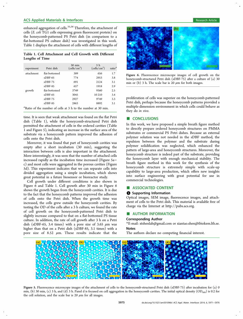
Figure 6 and Table 1. Cell growth after 30 min in Figure 6shows the growth began from the honeycomb cavities. It is dueto the fact that the honeycomb patterns improved the adhesionof cells onto the Petri dish. When the growth time wasincreased, the cells grew outside the honeycomb cavities. Bytesting the OD of the cells after a 3 h culture, we found the rateof cell growth on the honeycomb-patterned Petri dish toslightly increase compared to that on a flat-bottomed PS tissueculture. In addition, the rate of cell growth after 3 h on a Petridish (sDBF-65, 3.4 times) with a pore size of 3.65 μm washigher than that on a Petri dish (sDBF-85, 3.1 times) with apore size of 8.52 μm. These results indicate that the
proliferation of cells was superior on the honeycomb-patternedPetri dish, perhaps because the honeycomb patterns provided amultiple-dimension environment in which cells could behave asthey do in vivo.
■ CONCLUSIONSIn this work, we have proposed a simple breath figure methodto directly prepare ordered honeycomb structures on PMMAsubstrates or commercial PS Petri dishes. Because an externalpolymer solution was not needed in the sDBF method, therepulsion between the polymer and the substrate duringpolymer solidification was neglected, which enhanced thepattern of large-area and honeycomb structures. Moreover, thehoneycomb structure is indeed part of the substrate, providingthe honeycomb layer with enough mechanical stability. Thebreath figure method in this work for the synthesis of thehoneycomb structure is extremely simple with scale-upcapability to large-area production, which offers new insightsinto surface engineering with great potential for use incommercial technologies.
■ ASSOCIATED CONTENT*S Supporting InformationOptical images, SEM image, fluorescence images, and attach-ment of cells to the Petri dish. This material is available free ofcharge via the Internet at http://pubs.acs.org.
■ AUTHOR INFORMATIONCorresponding Author*E-mail: [email protected] or [email protected] authors declare no competing financial interest.
Table 1. Cell Attachment and Cell Growth with DifferentLengths of Time
experiment Petri dish30 min
(cells/cm2)3 h
(cells/cm2) ratioa
attachment flat-bottomed 389 656 1.7sDBF-65 774 2953 3.8sDBF-75 691 2124 3.1sDBF-85 657 1918 2.9
growth flat-bottomed 3749 9360 2.5sDBF-65 3045 10 210 3.4sDBF-75 2927 9749 3.3sDBF-85 2863 8892 3.1
aRatio of the number of cells at 3 h to the number at 30 min.
Figure 5. Fluorescence microscope images of the attachment of cells to the honeycomb-structured Petri dish (sDBF-75) after incubation for (a) 0min, (b) 30 min, (c) 3 h, and (d) 3 h. Panel d is focused on cell aggregation in the honeycomb cavities. The initial optical density (OD600) is 0.2 forthe cell solution, and the scale bar is 20 μm for all images.
Figure 6. Fluorescence microscope images of cell growth on thehoneycomb-structured Petri dish (sDBF-75) after a culture of (a) 30min or (b) 3 h. The scale bar is 20 μm for both images.
ACS Applied Materials & Interfaces Research Article
dx.doi.org/10.1021/am501096k | ACS Appl. Mater. Interfaces 2014, 6, 5971−59765975

■ REFERENCES(1) Davis, M. E. Ordered Porous Materials for EmergingApplications. Nature 2002, 417, 813−821.(2) Zhang, H. Controlled/“Living” Radical Precipitation Polymer-ization: A Versatile Polymerization Technique for Advanced Func-tional Polymers. Eur. Polym. J. 2013, 49, 579−600.(3) Nie, Z.; Kumacheva, E. Patterning Surfaces with FunctionalPolymers. Nat. Mater. 2008, 7, 277−290.(4) Wu, D.; Xu, F.; Sun, B.; Fu, R.; He, H.; Matyjaszewski, K. Designand Preparation of Porous Polymers. Chem. Rev. 2012, 112, 3959−4015.(5) Park, J. S.; Lee, S. H.; Han, T. H.; Kim, S. O. HierarchicallyOrdered Polymer Films by Templated Organization of AqueousDroplets. Adv. Funct. Mater. 2007, 17, 2315−2320.(6) Lee, S. H.; Park, J. S.; Lim, B. K.; Mo, C. B.; Lee, W. J.; Lee, J. M.;Hong, S. H.; Kim, S. O. Highly Entangled Carbon Nanotube Scaffoldsby Self-Organized Aqueous Droplets. Soft Matter 2009, 5, 2343−2346.(7) Lee, S. H.; Lee, D. H.; Lee, W. J.; Kim, S. O. Tailored Assemblyof Carbon Nanotubes and Graphene. Adv. Funct. Mater. 2011, 21,1338−1354.(8) Bunz, U. H. F. Breath Figures as a Dynamic Templating Methodfor Polymers and Nanomaterials. Adv. Mater. 2006, 18, 973−989.(9) Bai, H.; Du, C.; Zhang, A.; Li, L. Breath Figure Arrays:Unconventional Fabrications, Functionalizations, and Applications.Angew. Chem., Int. Ed. 2013, 52, 12240−12255.(10) Yu, Y.; Ma, Y. Breath Figure Fabrication of Honeycomb Filmswith Small Molecules through Hydrogen Bond Mediated Self-Assembly. Soft Matter 2011, 7, 884−886.(11) Srinivasarao, M.; Collings, D.; Philips, A.; Patel, S. Three-Dimensionally Ordered Array of Air Bubbles in a Polymer Film.Science 2001, 292, 79−83.(12) Stenzel-Rosenbaum, M. H.; Davis, T. P.; Fane, A. G.; Chen, V.Porous Polymer Films and Honeycomb Structures Made by the Self-Organization of Well-Defined Macromolecular Structures Created byLiving Radical Polymerization Techniques. Angew. Chem., Int. Ed.2001, 40, 3428−3432.(13) Hoa, M. L. K.; Lu, M.; Zhang, Y. Preparation of porousmaterials with ordered hole structure. Adv. Colloid Interface Sci. 2006,121, 9−23.(14) Gau, H.; Herminghaus, S. Ripening of Ordered Breath Figures.Phys. Rev. Lett. 2000, 84, 4156−4159.(15) Boker, A.; Lin, Y.; Chiapperini, K.; Horowitz, R.; Thompson,M.; Carreon, V.; Xu, T.; Abetz, C.; Skaff, H.; Dinsmore, A. D.; Emrick,T.; Russell, T. P. Hierarchical Nanoparticle Assemblies Formed byDecorating Breath Figures. Nat. Mater. 2004, 3, 302−306.(16) Hernandez-Guerrero, M.; Stenzel, M. H. HoneycombStructured Polymer Films via Breath Figures. Polym. Chem. 2012, 3,563−577.(17) Farbod, F.; Pourabbas, B.; Sharif, M. Direct Breath FigureFormation on PMMA and Superhydrophobic Surface Using In SituPerfluoro-Modified Silica Nanoparticles. J. Polym. Sci., Part B: Polym.Phys. 2013, 51, 441−451.(18) Wan, L.; Ke, B.; Li, X.; Meng, X.; Zhang, L.; Xu, Z. Honeycomb-Patterned Films of Polystyrene/Poly(ethylene Glycol): Preparation,Surface Aggregation and Protein Adsorption. Sci. China, Ser. B: Chem.2009, 52, 969−974.(19) Park, M. S.; Kim, J. K. Breath Figure Patterns Prepared by SpinCoating in a Dry Environment. Langmuir 2004, 20, 5347−5352.(20) Megelski, S.; Stephens, J. S.; Chase, D. B.; Rabolt, J. F. Micro-and Nanostructured Surface Morphology on Electrospun PolymerFibers. Macromolecules 2002, 35, 8456−8466.(21) Stenzel, M. H.; Barner-Kowollik, C.; Davis, T. P. Formation ofHoneycomb-Structured, Porous Films via Breath Figures withDifferent Polymer Architectures. J. Polym. Sci., Part A: Polym. Chem.2006, 44, 2363−2375.(22) Widawski, G.; Rawiso, M.; Francois, B. Self-OrganizedHoneycomb Morphology of Star-Polymer Polystyrene Films. Nature1994, 369, 387−389.
(23) Park, M. S.; Kim, J. K. Broad-Band Antireflection Coating atNear-Infrared Wavelengths by a Breath Figure. Langmuir 2005, 21,11404−11408.(24) Chen, P.-C.; Wan, L.-S.; Ke, B.-B.; Xu, Z.-K. Honeycomb-Patterned Film Segregated with Phenylboronic Acid for GlucoseSensing. Langmuir 2011, 27, 12597−12605.(25) Lomoschitz, M.; Edinger, S.; Bauer, G.; Friedbacher, G.;Schubert, U. Sol−Gel Films with Polymodal Porosity by Surfactant-Assisted Breath Figure Templating. J. Mater. Chem. 2010, 20, 2075−2078.(26) Deans, T. L.; Singh, A.; Gibson, M.; Elisseeff, J. H. RegulatingSynthetic Gene Networks in 3D Materials. Proc. Natl. Acad. Sci. U.S.A.2012, 109, 15217−15222.(27) Zhang, S. Beyond the Petri Dish. Nat. Biotechnol. 2004, 22,151−152.(28) Napolitano, A. P.; Dean, D. M.; Man, A. J.; Youssef, J.; Ho, D.N.; Rago, A. P.; Lech, M. P.; Morgan, J. R. Scaffold-Free Three-Dimensional Cell Culture Utilizing Micromolded NonadhesiveHydrogels. BioTechniques 2007, 43, 494−500.(29) Krotz, S. P.; Robins, J. C.; Ferruccio, T.-M.; Moore, R.;Steinhoff, M. M.; Morgan, J. R.; Carson, S. In Vitro Maturation ofOocytes via the Pre-Fabricated Self-Assembled Artificial HumanOvary. J. Assisted Reprod. Genet. 2010, 27, 743−750.(30) http://www.time.com/time/specials/packages/article/0,28804,2035319_2034529_2034518,00.html.(31) Beattie, D.; Wong, K. H.; Williams, C.; Poole-Warren, L. A.;Davis, T. P.; Barner-Kowollik, C.; Stenzel, M. H. Honeycomb-Structured Porous Films from Polypyrrole-Containing Block Copoly-mers Prepared via RAFT Polymerization as a Scaffold for Cell Growth.Biomacromolecules 2006, 7, 1072−1082.(32) Wu, X.; Wang, S. Regulating MC3T3-E1 Cells on DeformablePoly(ε-caprolactone) Honeycomb Films Prepared Using a Surfactant-Free Breath Figure Method in a Water-Miscible Solvent. ACS Appl.Mater. Interfaces 2012, 4, 4966−4975.(33) Nishikawa, T.; Nishida, J.; Ookura, R.; Nishimura, S. I.; Wada,S.; Karino, T.; Shimomura, M. Honeycomb-Patterned Tthin Films ofAmphiphilic Polymers as Cell Culture Substrates. Mater. Sci. Eng., C1999, 8−9, 495−500.(34) Zhao, W.; Lang, M.; Li, Y.; Li, L.; Shi, J. Robust and HydrophilicPolymeric Films with Honeycomb Pattern and Their Cell ScaffoldApplications. J. Mater. Chem. 2009, 19, 2789−2796.(35) Lee, J.; Cuddihy, M. J.; Kotov, N. A. Three-Dimensional CellCulture Matrices: State of the Art. Tissue Eng., Part B 2008, 14, 61−86.(36) Zhang, S.; Gelain, F.; Zhao, X. Designer Self-AssemblingPeptide Nanofiber Scaffolds for 3D Tissue Cell Cultures. Semin.Cancer Biol. 2005, 15, 413−420.
ACS Applied Materials & Interfaces Research Article
dx.doi.org/10.1021/am501096k | ACS Appl. Mater. Interfaces 2014, 6, 5971−59765976