breeding short-muzzled dogs · 2020-02-05 · 1 breeding short-muzzled dogs criteria for the...
TRANSCRIPT

1
BREEDING SHORT-MUZZLED DOGS
Criteria for the enforcement of Article 3.4. of the Animal Keepers Decree
(Besluit Houders van dieren) – Breeding Companion Animals
Dr Marjan AE van Hagen
Department of Animals in Science and Society
and the Expertise Centre Genetics of Companion Animals
Commissioned by the Ministry of Agriculture, Nature and Food Quality
21 01 2019
NOTE: In the translation we have done our utmost to uphold the content of the original text in Dutch. However, for proper reading in some cases scientific English terms where used, instead of layman´s language and where
necessary a sentence was rephrased to increase readability.

2
Contents
Introduction……………………………………………………………………………………………………………………….. 4
Part I: Brachycephaly – dysmorphology of the skull and muzzle ………………………………………... 5
1. Morphology: skull and muzzle shape ……………………………………………………………………………... 5
2. Health and welfare risks for short-muzzled dogs……………………………………………………………… 7
2.1. Brachycephalic Ocular syndrome (BOS) ……………………………………………………………………….. 8
2.3. Brachycephalic Obstructive Airway Syndrome (BOAS) ………………………………………………….. 9
Part II: List of existing measuring methods ………………………………………………………………………….. 11
1. Skull conformation …………………………………………………………………………………………………………… 11
1.1. post mortem ……………………………………………………………………………………………………. 11
1.2. in vivo ……………………………………………………………………………………………………………….. 12
2. Relative muzzle length …………………………………………………………………………………………………… 13
3. Nostril stenosis ………………………………………………………………………………………………………………… 14
3.1.: nares/cartilage ratio
3.2.: The 'Liu degrees of nostril stenosis' ………………………………………………………………… 15
4. Presence of a nasal skin fold ……………………………………………………………………………………………. 16
5. Exposed sclera ………………………………………………………………………………………………………….. 17
6. Relative palpebral fissure length ………………………………………………………………………………………. 17
7. Exercise tolerance tests ……………………………………………………………………………………………………. 19
7.1. The 6-minute walk test ……………………………………………………………………………………… 19
7.2. The 1000-metre walk test ………………………………………………………………………………… 19
8. Additional measurements ………………………………………………………………………………………………… 21
8.1. Breed-defining measurement protocols ……………………………………………………………… 21
8.2. Body condition score (excess weight) …………………………………………………………………. 21
3
Part III. Proposed criteria for the enforcement of Article 3.4. Breeding Companion Animals …… 23
1. Traffic-light system: Moving towards a healthy dog population …………………………………………… 24
2. Widely endorsed movement of breeders, veterinary specialists and enforcement officers ……24
I. Criteria verified by the Netherlands Food and Consumer Product Safety Authority (NVWA) and
Inspectorate of the Dutch Society for the Protection of Animals (LID) inspectors ……………….. 26
II. Additional criteria to check by primary veterinary surgeons and/or specialists
A. Brachycephalic Ocular Syndrome ………………………………………………………………………… 27
A.1. By a primary veterinary surgeon (or Ophthalmology specialist) …………… 27
A.2. By a veterinary surgeon specialising in Ophthalmology …………………………. 27
B. Brachycephalic Obstructive Airway Syndrome ………………………………………………………. 29
B.1. By a primary veterinary surgeon (or ENT specialist) ………………………………… 29
B.2. Exercise tolerance test …………………………………………………………………………… 30

4
Introduction
A number of changes in the Animal Keepers Decree entered into effect on 1 July 2014. Those changes
were introduced in order to improve the welfare of animals involved in the commercial breeding of
companion animals.
Article 3.4. of the Decree contains rules in respect of the breeding of companion animals. The general
rule is that it is prohibited to breed companion animals in a manner that is detrimental to the welfare
and health of the parent animal or its offspring. This means, among other things, ensuring, where
possible, that when breeding dogs no serious abnormalities and diseases are passed on to or could
be acquired by the offspring. In addition, where possible, no conformational features that may
compromise the welfare and health of the animal must be passed on. The same applies to serious
behavioural abnormalities. However, the Decree is limited to target requirements, making the
standards open to interpretation and in need of further elaboration. To date, the sector has been
slow in taking the action necessary to achieve this, making it difficult for the Inspectorate and
judiciary to enforce the provisions laid down in the Article.
To enforce Article 3.4. it has to be brought to light that a breeder has made insufficient effort to
prevent harmful conformational features, serious diseases or behavioural abnormalities from being
passed on to offspring. A genetic predisposition to serious diseases or behavioural abnormalities is
not always noticeable externally and this complicates enforcement based on prevention. In this
project we have focused on clearly visible harmful conformational features of animals used for
breeding related to brachycephaly which are passed on to offspring and represent a serious welfare
risk.
The popularity of brachycephalic dogs (brachy = short and cephalic = relating to the head) is
increasing throughout the world. Breeding selection has resulted in an ever increasing
dysmorphology of dogs' skulls. Breeding dogs with this kind of serious skull and muzzle abnormalities
results in physical and psychological distress and restrictions in their natural behaviour, which both
infringes the animals' integrity and poses a great risk to their welfare. This is contrary to the
provisions of Dutch legislation (the Animals Act (Wet Dieren)).i Breeding selection towards
increasingly less extreme dysmorphology could remedy infringements of integrity and reduce welfare
risks. The results of this project will therefore form an important prelude to the further extension of
enforcement criteria for breeding dogs (and other animals) in terms of a wider range of health and
welfare risks.
Composition of the project group
• Dr Hille Fieten, veterinary surgeon, Specialist in Internal Medicine for Companion Animals,
coordinator of the Expertise Centre Genetics of Companion Animals, Faculty of Veterinary
Medicine
• Dr Franck L.B. Meijboom, associate professor, Department of Animals in Science and Society
(DWM), Faculty of Veterinary Medicine
• Dr Marjan AE van Hagen, veterinary surgeon, Animal Welfare Specialist, Ethics & Law,
Associate Professor and staff member of the Animal Behaviour Clinic, DWM, Faculty of
Veterinary Medicine
5
Part I:
Brachycephaly – dysmorphology of the skull and muzzle
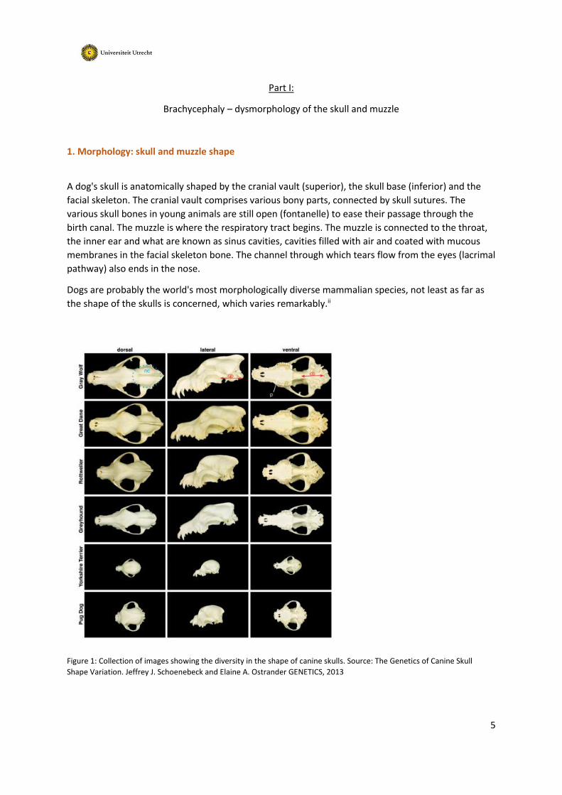
1. Morphology: skull and muzzle shape
A dog's skull is anatomically shaped by the cranial vault (superior), the skull base (inferior) and the
facial skeleton. The cranial vault comprises various bony parts, connected by skull sutures. The
various skull bones in young animals are still open (fontanelle) to ease their passage through the
birth canal. The muzzle is where the respiratory tract begins. The muzzle is connected to the throat,
the inner ear and what are known as sinus cavities, cavities filled with air and coated with mucous
membranes in the facial skeleton bone. The channel through which tears flow from the eyes (lacrimal
pathway) also ends in the nose.
Dogs are probably the world's most morphologically diverse mammalian species, not least as far as
the shape of the skulls is concerned, which varies remarkably.ii
Figure 1: Collection of images showing the diversity in the shape of canine skulls. Source: The Genetics of Canine Skull
Shape Variation. Jeffrey J. Schoenebeck and Elaine A. Ostrander GENETICS, 2013

6
In humans, brachycephaly is defined as a developmental disorder affecting the face and mandible
and maxilla including, inter alia, facial retrusion (flattening of the facial skeleton), proximodistal
shortness of the nose and widening of the hard palate. Various genetic backgrounds lie behind the
developmental disorder. By comparing the genetic information of (374) dogs (various breeds and
cross-breeds) with measurements of their skulls, American researchers were able to pinpoint DNA
variations that are associated with different head shapes. One variation – found to disrupt the
activity of a gene called SMOC2 – was strongly linked to the length of the dog’s face. iii
The appearance of brachycephalic dogs is characterised by a short, convex skull shape with a marked
fold (stop) between the cranium and the nasal bridge.1 In the past, dogs with this developmental
disorder were selected for breeding because certain dysmorphic characteristics benefited them
when carrying out the work for which they were used. This was because the dysmorphia of the skull
is often accompanied by an underbite, enabling the dog to breathe through its nose when it grips
something with its teeth. During the last century, the breeding selection of dogs with this type of
developmental disorder assumed such extreme proportions that the functionality of the dogs was
impaired.
Figure 2: Top: 1. from right to left – brachycephalic skulls developing an increasingly extreme shape Bottom: 2. a
brachycephalic French Bulldog (left) and an extremely brachycephalic dog (right), 3. On the left, an extremely
brachycephalic Pug (left) and a (far less) brachycephalic ancestor (right).
1 Source: https://upload.wikimedia.org/wikipedia/commons/7/7b/Brachycephalie

7
2. Health and welfare problems in short-muzzled dogs
As a dog's skull develops, there are four bony plates which meet in the upper middle of the head and
are the last bony structures to fuse or ossify. Skull sutures usually close about 4-5 weeks after birth.
There are cases where the opening is fairly slow to close and the process is not completed until the
dog is six months old. In extreme cases the fontanelle remains open.
Figure 3. On the left is a normal dog skull and on the right a hydrocephalic skull with an open fontanelle.2
An open fontanelle is related to the development of water in the brain, or hydrocephalus ("hydro"
(water) and "cephalus" (head)) – Fig. 3. Water in the brain occurs when the drainage system in the
skull does not work properly, obstructing the absorption and elimination of cerebrospinal fluid from
the brain and causing a build-up. The increased intracranial pressure means that the frontal and
parietal bones do not close (Fig. 3.) In principle, an open fontanelle can occur in any breed of dog, but
the condition is most commonly found in small, brachycephalic dog breeds such as the Maltese,
Chihuahua, Boston Terrier and Pomeranian. An open fontanelle not only leaves the brain vulnerable
to injury, but can also result in neurological abnormalities should water in the brain occur. Since the
condition can be hereditary, a dog with an open fontanelle should not be used for breeding.
Not only do brachycephalic dogs (hereinafter called 'short-muzzled dogs') suffer from health and
welfare problems related to the abnormal shape of their skulls, e.g. an open fontanelle and Chiari-
like malformation – Syringomyelia – we have also observed other hereditary skeletal conditions
within this population, such as patella luxation, in growing corkscrew tails and congenital
malformations of vertebrae associated with neurological deficiencies.
In this project we have focused on a key group of harmful health and welfare problems related to the
abnormal shape of skulls and muzzles, namely: Brachycephalic Ocular Syndrome (BOS) and
Brachycephalic Obstructive Airway Syndrome (BOAS). The background to these harmful effects are
described in more detail below.
2 http://vanat.cvm.umn.edu/vetAnomal/sysNV/NV2.html
8
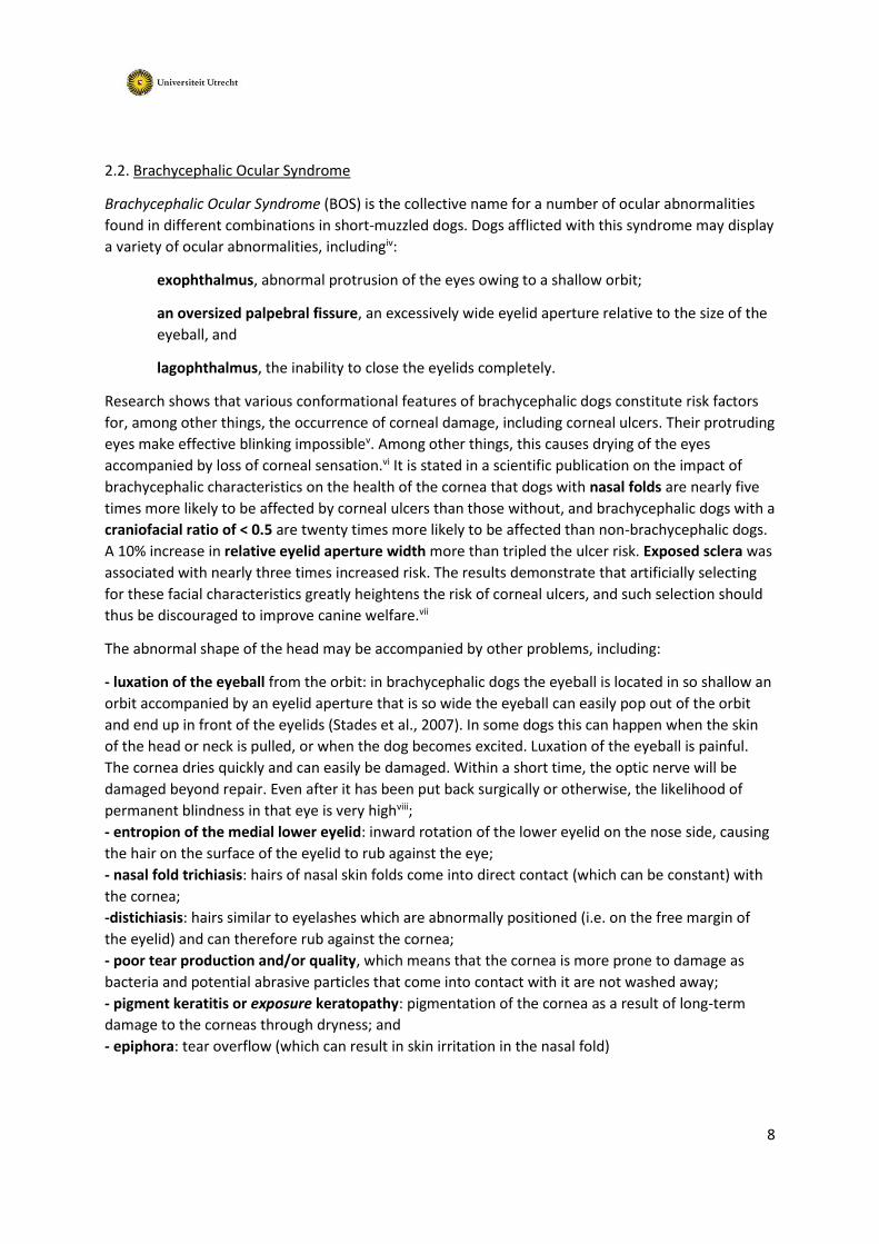
2.2. Brachycephalic Ocular Syndrome
Brachycephalic Ocular Syndrome (BOS) is the collective name for a number of ocular abnormalities
found in different combinations in short-muzzled dogs. Dogs afflicted with this syndrome may display
a variety of ocular abnormalities, includingiv:
exophthalmus, abnormal protrusion of the eyes owing to a shallow orbit;
an oversized palpebral fissure, an excessively wide eyelid aperture relative to the size of the
eyeball, and
lagophthalmus, the inability to close the eyelids completely.
Research shows that various conformational features of brachycephalic dogs constitute risk factors
for, among other things, the occurrence of corneal damage, including corneal ulcers. Their protruding
eyes make effective blinking impossiblev. Among other things, this causes drying of the eyes
accompanied by loss of corneal sensation.vi It is stated in a scientific publication on the impact of
brachycephalic characteristics on the health of the cornea that dogs with nasal folds are nearly five
times more likely to be affected by corneal ulcers than those without, and brachycephalic dogs with a
craniofacial ratio of < 0.5 are twenty times more likely to be affected than non-brachycephalic dogs.
A 10% increase in relative eyelid aperture width more than tripled the ulcer risk. Exposed sclera was
associated with nearly three times increased risk. The results demonstrate that artificially selecting
for these facial characteristics greatly heightens the risk of corneal ulcers, and such selection should
thus be discouraged to improve canine welfare.vii
The abnormal shape of the head may be accompanied by other problems, including:
- luxation of the eyeball from the orbit: in brachycephalic dogs the eyeball is located in so shallow an
orbit accompanied by an eyelid aperture that is so wide the eyeball can easily pop out of the orbit
and end up in front of the eyelids (Stades et al., 2007). In some dogs this can happen when the skin
of the head or neck is pulled, or when the dog becomes excited. Luxation of the eyeball is painful.
The cornea dries quickly and can easily be damaged. Within a short time, the optic nerve will be
damaged beyond repair. Even after it has been put back surgically or otherwise, the likelihood of
permanent blindness in that eye is very highviii;
- entropion of the medial lower eyelid: inward rotation of the lower eyelid on the nose side, causing
the hair on the surface of the eyelid to rub against the eye;
- nasal fold trichiasis: hairs of nasal skin folds come into direct contact (which can be constant) with
the cornea;
-distichiasis: hairs similar to eyelashes which are abnormally positioned (i.e. on the free margin of
the eyelid) and can therefore rub against the cornea;
- poor tear production and/or quality, which means that the cornea is more prone to damage as
bacteria and potential abrasive particles that come into contact with it are not washed away;
- pigment keratitis or exposure keratopathy: pigmentation of the cornea as a result of long-term
damage to the corneas through dryness; and
- epiphora: tear overflow (which can result in skin irritation in the nasal fold)

9
The underdeveloped muzzle is out of proportion with the normally developed scalp, resulting in deep
nasal folds. In addition to the nasal fold trichiasis risk, the animals are also at high risk of developing
dermatitis, inflammation of the skin in the nasal fold caused by rubbing and microbial overgrowth
(bacteria and yeast), promoted by the elevated humidity and build-up of secretions in this
environment. Skin fold dermatitis gives rise to an unpleasant odour and causes the dog discomfort; in
some cases it can trigger self-injury through rubbing and/or scratching.
Daily cleaning of the folds is widely recommended as standard care for dogs of this type, but is often
not enough to manage the problem. French Bulldogs have a nasal fold on the dorsal surface of the
muzzle, which is so close the tip of the nose that it is completely impossible to clean this deep fold
(verbal expert opinion).
2.3. Brachycephalic Obstructive Airway Syndrome
Canine brachycephalic obstructive airway syndrome (BOAS) is associated with a collection of
respiratory conditions affecting short-muzzled dogs. They can occur in an individual dog all at the
same time, but this will not necessarily always be the case. The various conditions include:
• Stenosis and narrowed nostrils
• Excessively long and/or thickened palatum molle (soft palate)
• Enlarged tonsils
• Dysmorphia of the larynx or an abnormally shaped larynx
• Trachea hypoplasia or an abnormally narrow windpipe
The clinical signs of BOAS present as a result of obstruction of the upper airways caused by
anatomical abnormalities which narrow the airways and increase airway resistance. The abnormally
short muzzle means that the mouth is unable to accommodate either the soft palate or the tongue.
The tongue and the soft palate are too large and too long relative to the muzzle. Consequently,
short-muzzled dogs will often be seen with their tongues hanging out and most make a snoring noise
when breathing. This snoring noise is produced by the excessively long soft palate, which is
suspended in the pharynx and caused to vibrate by the flow of air. The snoring can range from a mild
form that occurs only when the dog exerts itself to a permanent noise audible even when the dog is
resting. The increased resistance in the airways leads to an increased risk of secondary laryngeal
collapse.
Figure 4. A scan of a canine skull of a normal length (left) and one of a short-muzzled dog; the air-filled frontal sinus and
nasal cavity in the normal skull are coloured black. Source:http://brachycephalia.com/

10
Dogs at rest mainly breathe through their noses. Since dogs have no sweat glands, panting is their
only means of expelling excess body heat. Breathing problems in dogs with BOAS therefore result not
only in breathlessness, but also compromised thermoregulation because of their inability to expel
body heat effectively. Complaints range from mild snoring to severe breathlessness (respiratory
distress), exercise intolerance, loss of consciousness and death through overheating (heat stroke).
The impact of BOAS is not limited to the respiratory system, but can also lead to secondary problems
in the oesophagus and stomach. Excessive salivation (hypersalivation) and retching can occur. The
latter is caused by the negative pressure during inspiration which, as a secondary problem, can cause
a widening of the opening where the oesophagus passes through the diaphragm (hiatus hernia),
causing a reflux of gastric acid and, potentially, inflammation of the oesophagus. (gastro-oesophageal
reflux and oesophagitis).
The main risk factors for BOAS are: a flat muzzle, thick neck and excess weight.ix Short-muzzled dogs
are generally at very high risk of developing BOAS.x With a mild form of BOAS the dog's welfare is
reduced owing to its difficulty in breathing and, with a severe form, limited mobility, poor appetite
and reduced playfulness will seriously diminish its quality of life. Research shows that the risk of
BOAS increases as the short-muzzle morphology becomes more exaggerated.xi Careful selection of
parent animals with far less extreme dysmorphic characteristics can gradually reduce the risk of BOS
and BOAS in short-muzzled dogs. Although this has been known for decades, we can see barely any
progress being made by breeders in this direction. Establishing standards to enable the enforcement
of Article 3.4 will help to encourage breeders to start selecting parent animals with a greater
emphasis on functionality.

11
Part II:
List of existing measuring methods
1. Skull shape measurements
1.1. Post mortem
In the past, craniofacial morphometric parameters were measured (postmortem) on a dry canine
skull. xii xiii The mean skull length of the lion, dog and cat were found to be 39.7±1.04 cm, 20.02±1.4
cm and 8.4±1.5 cm respectively, with an average skull width of 28±2.3 cm, 10.04±0.5 cm and 6.8±1.4
cm, respectively.xiv For a long time, these were the only official measurement data for the average
canine skull length.
Based on this postmortem measuring method, canine skulls are divided into dolicocephalic (long
skull), mesocephalic and brachycephalic (short skull) skull shapes. Moreover, a ratio between the
short, broad muzzle (facial length) and the skull width of 0.81 or greater is regarded as
brachycephalic. However, German authors based their classification on the relationship between the
cranial length and the skull length, where a skull length/cranial length ratio of 1.6 to 3.4 is deemed
brachycephalic.3 (Figure 5).
Figure 5. Post-mortem measuring methods used to define a brachycephalic (short skull) skull shape.
3 Brachycephalicsyndrome Compendium Koch, 2003

12
1.2. In vivo
In 2008, Sutter et al. examined measurements in vivo, i.e. on live animals, in relation to the
classifications in dog breeds.xv In total, thirteen conformational features were shown to be breed-
defining. They are known as the ‘Breed-defining measurement protocols’. The following variables
were defined in respect of the skull:
1.Snout length – SL
2. Cranial length – CL
3. Skull width – SW
4. Eye width – EW
Figure 6: Illustration of 'in vivo' measurements (French Bulldog): Left: snout length (SL) from P1 to P2, cranial length (CL)
from P2 to P3, skull length P1 to P3, Right: Eye width (EW), Skull Width (SW)
A skull width of at least 80% of the length is a characteristic feature of brachycephalic dogs. A skull
width of more than 80% of the length is therefore deemed extreme brachycephalic.4 This provides
guidance for a standard that could be used to establish an enforcement criterion.
4 https://www.petinsurancequotes.com/petinsurance/brachycephalic-skull.html

13
2. Relative muzzle length
The craniofacial ratio (CFR), or relative muzzle length is calculated by dividing the muzzle length (cm)
by the cranial length (cm). Relative muzzle length follows from:
Figure 7: Illustration of the determination of relative muzzle length by dividing the nose length from A to B and the cranial
length from B to C. The relative muzzle length of the Cavalier King Charles Spaniel depicted is 0.27: nose length 28
mm/cranial length 102 mm.
This ratio proves a good gauge for measuring the severity of BOAS. (Fig. 7)
Figure 8.: This figure illustrates the relationship between the external craniofacial factor (CRF) and the corresponding
internal anatomical structure of the upper airways
Figure 9. Relative muzzle length: muzzle length from A to B and cranial length from B to C. From left to right, the relative
muzzle length is: 0.08, 0.23 and 0.35.
Muzzle length (A-B)
cranial length (B-C)

14
3. Nostril stenosis
Determining the width of the nostril as a measure of the severity of the restriction of airflow through
the nose is another way of assessing the severity of the anatomical malformation of the airway. Two
methods for quantifying the degree of narrowing of the external nares are described.
3.1. nares -catilage ratio
With this measuring method, a calliper is used to take four measures of the width of the wing of the
nostril – cartilage (‘a’), along with the width of the adjacent airspace (‘b’). xvi 1. The lowest measure
of the lower edge of the nostril wing, 2. the highest measure of the upper edge of the nostril wing
and 3. a further two measures of equal distance between the highest and lowest point. The calliper
must be placed directly on the nostril so that the dog's head can be kept still to ensure that the
measuring instrument does not injure the animal.
Figure 10.: Measuring the nostril using the four-part measuring method
The ratio of the mean nostril thickness to the mean nostril diameter (b/a) is referred to as the nares
ratio. The greater the ratio, the greater the air opening.
Figure 11. Examples of nostril air opening expressed as the ' nares ratio' ranging from a ratio of 0.06 (extremely nostril
stenosis) to 0.06 – wide nostrils. Source: Building better brachycephalics, Packer et al. (2012) Animal Welfare, 21, 81-93
https://www.ufaw.org.uk/downloads/welfare-downloads/building-better-brachycephalics-2013-report.pdf
'a' (dotted lines) indicates the width of the alae nasi (nostril cartilage)
'b' (solid lines) indicates the width of the adjacent external airway
The central line indicates the philtrum, delineating the left and right sides of
the nasal planum.

15
3.2. The 'Liu degrees of nostril stenosis
With this method, developed by Liu et al. (2016), the degree of nostril stenosis is divided into four
grades, based on the extent to which the lateral structure makes contact with the medial septum.
Figure 12. Grading the severity of nostril narrowing as in Liu, 2016. 5
Examples of different degrees of nostril stenosis in pugs, French bulldogs, and bulldogs.
The following descriptions were adapted from a published figure by the authors (Fig 1 in Liu et al. 2016):
“Open nostrils: nostrils are wide open; mildly stenotic nostrils: slightly narrowed nostrils where the
lateral nostril wall does not touch the medial nostril wall. Immediately after the exercise tolerance test
(ETT), the nostril wings should move dorsolaterally to open on inspiration; moderately stenotic nostrils:
the lateral nostril wall touches the medial nostril wall at the dorsal part of the nostrils and the nostrils are
only open at the bottom. Immediately after the ETT, the nostril wings are not able to move dorsolaterally
and there may be nasal flaring (ie, muscle contraction around the nose trying to enlarge the nostrils;
severely stenotic nostrils: nostrils are almost closed. The dog may switch to oral breathing from nasal
breathing with stress or very gentle exercise such as playing.” (Liu et al. 2016).
https://doi.org/10.1371/journal.pone.0181928.g001
5 https://www.researchgate.net/publication/318842582_Conformational_risk_factors_of_brachycephalic_obstructive_airway_syndrome_B
OAS_in_pugs_French_bulldogs_and_bulldogs/figures

16
4. Presence of a nasal skin fold
Alongside the craniofacial ratio, the presence of a nasal fold proves to be a good gauge for measuring
the severity of muzzle shortness and the accompanying risk of eye problems. The skin of
mesocephalic or dolichocephalic dogs generally covers the facial bones without any folds being
created. A nasal fold will usually be present in short-muzzled dogs. The nasal fold is described in the
breed standards for certain short-muzzled dog breeds (Table 1).
A nasal fold is defined as a discernible fold of skin on the dorsal surface of the muzzle that was
present without manipulation of the skin, and could be easily grasped between vernier callipers
Table 12. Nasal folds in breed standards and nasal fold related statements from The Kennel Club (UK) ‘Breed Watch’
initiative. Source: Impact of Facial Conformation on Canine Health: Corneal Ulceration, Packer et all. Plos One, 2015
.

17
5. Exposed sclera
Exposed sclera is a guide used to establish the anatomical ratio between the orbit (skull) and eyeball
size. There is relatively little variation in eyeball size between dog breeds [16], but in short-muzzled
dogs the eye orbit is too flat and shallow to be able to accommodate the eyeball fully (and/or the
palpebral fissure is too wide). This causes the eyes to protrude, exposing more white of the eye than
would be visible with a healthy ratio.
Figure 13: Exposed sclera due to the protruding eyes (exophthalmus) in a pug. Source: http://pug.at/
Dogs are examined for the visibility of 'white of the eye' (sclera) when looking directly forwards. This
is carried out by gaining the dog’s attention (using a toy or treat, for example) and taking a
photograph using a digital camera. The overall presence of visible sclera is recorded and further
broken down into whether this was visible dorsally, ventrally or laterally to the iris of the eye. This
translates to a score of 0-4 dependent on how many quadrants of sclera are visible (max. 4/4).
6. Relative palpebral fissure width
The relative palpebral fissure width (%) is an additional measure used to determine the
disproportionate construction of the orbit in short-muzzled dogs. A soft tape measure (or callipers)
are used to measure the unstretched palpebral fissure width (mm) in conscious dogs by pulling the
tape measure taut between the outer and inner corners of the eye (from D-E).
Figure 14: Left: Quantitative determination of palpebral fissure width (D-E). Here, the palpebral fissure width is defined as
the straight-line distance between the medial and lateral canthus. Right: Quantitative determination of cranial length (B-C)
Relative palpebral fissure width = (palpebral fissure length (mm)/ cranial length (mm) x 100

18
Unlike absolute palpebral fissure width, relative palpebral fissure width (relative to skull length) turns
out to be highly relevant as regards the increased risk of corneal damage. Dogs with corneal ulcers
had a wider palpebral fissure relative to the length of their skulls than dogs without corneal ulcers.
Relative palpebral fissure width was found to increase the risk of ulcers by 1.12. As an illustrative
example, the mean relative palpebral fissure width for a Labrador Retriever was 19.0 and for a
Pekingese was 34.2, a difference of over 15%. This 5% increase equates to increased odds of 5.47 of
corneal ulcers.

19
7. Exercise intolerance test
Since exercise intolerance and impaired recovery after exercise are major signs of BOAS, an exercise
test, such as the 6-minute walk testxvii or the timed 1000-metre walk test are used to assess BOAS
severity.
7.1. The 6-minute walk test
In this test, dogs walk on a lead for six minutes at a reasonable pace (at least 5 km/hour) on an
unimpeded pathway or hallway measuring approx. 25 meters. No other people or animals with the
potential to distract the animal should be present during the walk test. Each dog was walked for six
minutes, timed with a stopwatch.
In research settings where the 6-minute walk test is used heart rate, blood pressure (mean systemic
arterial pressure, MAP) and blood oxygen levels are also measured (Swimmer, 2011). In that case
the dogs' heart rates and blood pressures are measured before and after they have walked for six
minutes. Heart rate is obtained by auscultation and MAP with the use of a blood pressure monitor.
The blood oxygen level is determined with the aid of a pulse oximeter.
The average dog's normal blood pressure is approx. 133/76 mm Hg (systolic/diastolic). This normal
value can vary slightly from breed to breed. At rest, a value of 160 mm Hg or higher is considered
abnormal. When the animal is subject to stress, its blood pressure will rise and may exceed 160 mm
Hg.
The equipment used to measure a dog's and/or a cat's blood pressure is generally the same as that
used for humans. The inflatable armband is placed around the tail or a fore limb. It takes a few
minutes to record the animal's blood pressure. The conclusion from this research was that the 6-
minute walk test is simple to carry out and shows the difference between healthy dogs and dogs with
an airway disorder. xviii
The 6-minute walk test has also been used in research into pulmonary fibrosis in West Highland
White Terriers to assess cardiopulmonary function.xix
7.2. The timed 1000-meter walk test
In this fitness test, dogs cover 1000 meters in 12 minutes and be able to recover in 15 minutes. The
test is conducted under the supervision of two veterinary surgeons. They establish the dogs' heart
rate and temperature. This is done before the start of the test and five, ten and fifteen minutes after
its completion. The breathing noises made by the dogs are also assessed at those points (no noise is
made, the dog is panting, or the panting is accompanied by a high-pitched sound) If the temperature
and heart rate drop and breathing returns to a normal type and frequency, the dog passes the
walking test.
The rules are laid down in an agreement between the Dutch Kennel Club and two English Bulldog
breed clubs.68 Special days are organised during which a breed suitability inspection takes place. The
Bulldogs are subjected to a fitness test on those days. (They are also tested for patellar luxation and
receive a conformation score.)
6 The rules of the agreement and the breeding rules of the breed clubs apply to members of the breed club. EBCN, 2014

20
A Finnish study evaluated the severity of respiratory symptoms and anatomic components of BOAS in
a group of prospectively recruited young adult English Bulldogs (n = 28) and investigated the
correlations of the 6-minute walk test or the 1000-meter walk test with a veterinary assessment of
BOAS severity. Based on veterinary clinical examination findings, the severity of the symptoms was
scored using an ordinal 4-level scale for: 1. the presence or absence of upper respiratory noise at rest
and 2. after exercise, 3. respiratory type at rest and 4. resting dyspnoea or cyanosis.
English Bulldogs with more severe BOAS walked a shorter distance, more slowly and their recovery
from exercise took longer than those with only mild symptoms of BOAS. Control dogs of different
breeds (n = 10) performed the exercise tests significantly better (i.e. longer distance, faster time and
recovery) than English Bulldogs. Increases in body temperature during exercise were significantly
higher in English Bulldogs than in controls. The results of this study support the use of exercise tests
for objective evaluation of the severity of BOAS in English Bulldogs.xx

21
8. Additional measuring methods for determining skeletal shape
8.1. Breed-defining measurement protocols
In addition to skull measurements, in 2008 measurements of other parts of the body of live animals
were described in relation to classification into dog breeds.xxi These were the thirteen breed-defining
metrics, known as 'Breed-defining measurement protocols, namely:
1. Snout length – SL 2. Cranial length – CL 3. Skull width – SW 4. Eye width – EW 5. Neck length (NL) 6. Neck girth 7. Chest girth 8. Body length - BL 9. Height of the withers 10. Height of the base of the tail 11. Fore limb circumference 12. Hind limb circumference
Researchers also identified a thick neck as a risk factor for the development of BOAS. What is known
as ‘relative neck girth’, which is established by dividing the circumference of the neck by the
circumference of the chest (neck girth/chest girth), is a good indicator of a high BOAS risk. The
circumference of the neck is measured in the centre of the distance between the two occipital bones
and the point between the cranial borders of the right and left scapulae. The circumference of the
chest is measured at the deepest point of the chest cavity.
Obesity is another important risk factor for the potential exacerbating effects.
8.2. Body condition score (overweight):
Among other things, overweight increases the risk of respiratory problems, heart disorders, and also
joint and bone problems. Incidence of overweight and obesity in dogs exceeds 30%, and several
breeds are predisposed to this heritable phenotype.xxii Genetic predisposition to excess weight is
demonstrated in, among other breeds, the Labrador Retriever, but to date insufficient research has
been conducted within the short-muzzled dog population.xxiii However, since short-muzzled purebred
dogs are genetically predisposed to airway problems, resulting in breathlessness, they should never
be allowed to become overweight.
Various scales have been developed to score excess weight objectively. The scoring system (1-9)
promoted by the World Small Animal Veterinary Association is a frequently used scale.

22

23
Part III:
Proposed criteria for the enforcement of Article 3.4. Breeding Companion Animals
There are various scientifically substantiated measuring methods that can be used to quantify
objectively the morphological characteristics of short-muzzled dogs related to the risks of BOS and
BOAS developing (see the list in part II of this report). Choosing breeding dogs with mild dysmorphia
can have a positive impact on the welfare risks faced by offspring. Unfortunately, few people
involved in dog breeding today are making such choices.
Article 3.4. The purpose of the Breeding Companion Animals section of the Animal Keepers Decree is
to ensure that no conformation features which could have a harmful effect on the welfare and health
of the animal are passed on. The complexity of the pure-bred dog world, inhabited by hobby and
commercial breeders and dealers, combined with standards that are open to interpretation has so
far resulted in an inability to take adequate action when it comes to enforcing this Article. This
inability to take adequate action can be remedied only through the adherence to enforcement
criteria which are not only substantiated by science but are also monitored in the field by NVWA and
LID inspectors for large groups of dogs.
To facilitate this transition to practice, the available measuring methods have been presented to a
panel of veterinary specialists in the areas of ENT, ophthalmology, dermatology and animal welfare.
Membership of the advisory group:
• Dr Sylvia C. Djajadiningrat-Laanen – Veterinary Surgeon, Ophthalmology Specialist, University
Clinic for Companion Animals, Faculty of Veterinary Medicine, Utrecht
• Dr Jeffrey de Gier – Veterinary Surgeon, Reproduction Specialist, Companion Animal sub-
specialisation, University Clinic for Companion Animals, Faculty of Veterinary Medicine,
Utrecht
• Dr Gert ter Haar – Veterinary Surgeon, Companion Animal Surgery Specialist (ENT), Specialist
Animal Clinic, Utrecht
• Dr Paul J. J. Mandigers – Veterinary Surgeon, Neurology and Internal Medicine of Companion
Animals Specialist, University Clinic for Companion Animals, Faculty of Veterinary Medicine,
Utrecht
• Dr Yvette Schlotter – Veterinary Surgeon, Dermatology Specialist, University Clinic for
Companion Animals, Faculty of Veterinary Medicine, Utrecht
The group was asked to identify the minimum number of criteria and the standard that could be used
to ensure enforcement takes place in order to achieve the intended objective. Enforcement officers
and specialists then entered into consultation. The outcome of that consultation is described in this
section.

24
1. Traffic-light system: Moving towards a health dog population
In the workfield of dog breeding it is often stated that the ideal standard, which should be uphold
concerning the animal health and welfare, is not suitable for certain short-muzzled (pure-bred) dog
populations because hardly any of them would fall within the standard. While very worrisome in
itself, this observation does require a practical approach. Hence the decision is made to opt for a
transitional phase during which breeders are given the opportunity to shift – within two to three
generations – in the direction of the minimum standard and a low-risk dog population by means of
breed selection. Therefore the TRAFFIC LIGHT principle will apply, meaning the first (practicable)
step of which is to set dogs with the most extreme conformational features as the limit and to
continue to tolerate dogs with less extreme conformational features after which (in two generations'
time, for example) the limits will be tightened so that ultimately all future breeding animals will meet
or will have to meet the ideal standard.
In populations where virtually every dog turns out to exceed the limits of the standard, leaving little
to no suitable dogs for breeding, a mandatory breeding programme using, for example, out-cross
animals, could be a solution. Such a breeding programme would require not only an assessment of
BOS and BOAS risks but also other desired health and behavioural characteristics.
2. Widely endorsed movement of breeders, veterinary specialists and enforcement officers
Given the circumstances in which enforcement officers sometimes have to work, a lot of
measurements or investigations are impracticable for them to carry out. Enforcement will therefore
have to be focused on criteria which the inspectors can determine themselves in situ. In cases of
doubt or where the dog owner objects to findings made during an inspection, an assessment or 're-
assessment' could be carried out by a designated and skilled primary veterinary surgeon and/or a
veterinary surgeon specialising in ophthalmology and/or one specialising in internal medicine,
surgery or ENT.
Centralised record-keeping is recommended to enable monitoring of the impact, positive or
otherwise, of enforcement on the dogs. Keeping central records of all measurement results will show
whether implementation of the enforcement and other criteria is having the desired effect on the
dog population. Centralised record-keeping will provide an insight into the current state of affairs –
baseline measurement – within different populations and also the speed at which breeding selection
is bringing the risk level down to the ideal standard. This is a key evaluation step for the policy
applied. The Expertise Centre Genetics of Companion Animals (ECCG) can analyse these data and,
based on the findings, advise the NVWA and LID whether and when the standard should be adjusted
from red to amber and green. In future, the database can be expanded to include additional
measurements of other breed characteristics and/or hereditary disorders.
In addition to the minimum number of enforceable criteria, veterinary surgeons (specialists) can also
perform supplementary measures that could help breeders to make better choices and select
healthy breeding dogs at limited risk of acquiring BOAS or BOS.

25
The boundaries between green, amber and red are described in the tables in the following pages,
accompanied by the score given when the limits of the standard are exceeded and breeding with the
dog is not permitted.
This involves:
1. The criteria and standards for short-muzzled dogs with which NVWA and LID inspectors can
arm themselves and use to select animals which are unhealthy and/or unsuitable ('high risk')
for breeding and therefore need to be excluded.
2. Other, additional criteria which only a practitioner or specialist can verify and may be used as
a supplement, for instance to supplement evidence in legal proceedings, but also with a view
to careful breeding selection.
3. Elaboration of the action primary veterinary surgeons can take in accordance with the Royal
Netherlands Veterinary Association Guideline on Veterinary action in respect of welfare risks
affecting dogs and cats with hereditary disorders and harmful breed characteristics to assist
with accurate breeding selection.
26
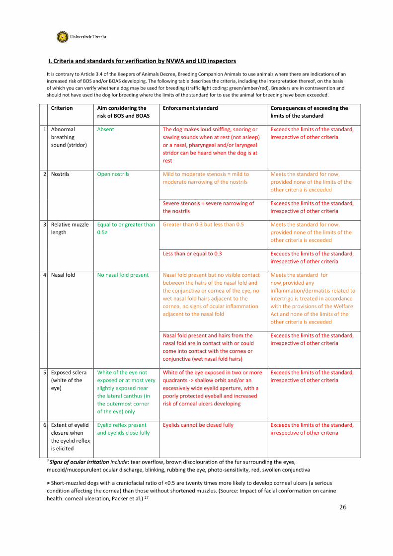
I. Criteria and standards for verification by NVWA and LID inspectors
It is contrary to Article 3.4 of the Keepers of Animals Decree, Breeding Companion Animals to use animals where there are indications of an
increased risk of BOS and/or BOAS developing. The following table describes the criteria, including the interpretation thereof, on the basis
of which you can verify whether a dog may be used for breeding (traffic light coding: green/amber/red). Breeders are in contravention and
should not have used the dog for breeding where the limits of the standard for to use the animal for breeding have been exceeded.
Criterion Aim considering the
risk of BOS and BOAS
Enforcement standard Consequences of exceeding the
limits of the standard
1
Abnormal
breathing
sound (stridor)
Absent The dog makes loud sniffing, snoring or
sawing sounds when at rest (not asleep)
or a nasal, pharyngeal and/or laryngeal
stridor can be heard when the dog is at
rest
Exceeds the limits of the standard,
irrespective of other criteria
2
Nostrils Open nostrils Mild to moderate stenosis = mild to
moderate narrowing of the nostrils
Meets the standard for now,
provided none of the limits of the
other criteria is exceeded
Severe stenosis = severe narrowing of
the nostrils
Exceeds the limits of the standard,
irrespective of other criteria
3
Relative muzzle
length
Equal to or greater than
0.5≠
Greater than 0.3 but less than 0.5 Meets the standard for now,
provided none of the limits of the
other criteria is exceeded
Less than or equal to 0.3 Exceeds the limits of the standard,
irrespective of other criteria
4
Nasal fold No nasal fold present Nasal fold present but no visible contact
between the hairs of the nasal fold and
the conjunctiva or cornea of the eye, no
wet nasal fold hairs adjacent to the
cornea, no signs of ocular inflammation
adjacent to the nasal fold
Meets the standard for
now,provided any
inflammation/dermatitis related to
intertrigo is treated in accordance
with the provisions of the Welfare
Act and none of the limits of the
other criteria is exceeded
Nasal fold present and hairs from the
nasal fold are in contact with or could
come into contact with the cornea or
conjunctiva (wet nasal fold hairs)
Exceeds the limits of the standard,
irrespective of other criteria
5
Exposed sclera
(white of the
eye)
White of the eye not
exposed or at most very
slightly exposed near
the lateral canthus (in
the outermost corner
of the eye) only
White of the eye exposed in two or more
quadrants -> shallow orbit and/or an
excessively wide eyelid aperture, with a
poorly protected eyeball and increased
risk of corneal ulcers developing
Exceeds the limits of the standard,
irrespective of other criteria
6
Extent of eyelid
closure when
the eyelid reflex
is elicited
Eyelid reflex present
and eyelids close fully
Eyelids cannot be closed fully Exceeds the limits of the standard,
irrespective of other criteria
¹ Signs of ocular irritation include: tear overflow, brown discolouration of the fur surrounding the eyes,
mucoid/mucopurulent ocular discharge, blinking, rubbing the eye, photo-sensitivity, red, swollen conjunctiva
≠ Short-muzzled dogs with a craniofacial ratio of <0.5 are twenty times more likely to develop corneal ulcers (a serious
condition affecting the cornea) than those without shortened muzzles. (Source: Impact of facial conformation on canine
health: corneal ulceration, Packer et al.) 27

27
II. Additional criteria for testing by primary veterinary surgeons and/or specialist veterinary surgeons To supplement the above criteria with a view to careful breeding selection where there are doubts regarding potential or
current parent animals, or to be used as evidence in legal proceedings (objection/appeal).
A. Brachycephalic Ocular Syndrome (BOS):
A.1. By a primary veterinary surgeon (or veterinary surgeon, specialised in ophthalmology)
Diagnosis Aim Enforcement standard To be factored in or conclusive?
1
Tear production
(Schirmer tear test)
13-25 mm in one minute (in the
absence of corneal defects
and/or sources or signs of eye
irritation; in the presence
thereof: see "9-12 mm of tear
fluid in one minute")
9-12 mm in one minute (in the
absence of corneal defects and/or
sources or signs of eye irritation; in
the presence thereof: see "less than
9 mm of tear fluid in one minute")
Meets the standard, provided none
of the limits of the other criteria is
exceeded
Less than 9 mm in one minute and
signs of keratoconjunctivitis sicca
Exceeds the limits of the standard,
irrespective of other criteria
2
Trichiasis of the nasal fold
(nasal fold hairs make
contact with the margin
of the eyelid, conjunctiva
or eyeball; tell-tale sign:
wet hairs)
No visible wet hairs in the nasal
fold and no hairs visibly in
contact with the eye lid margin,
conjunctiva or eyeball (N.B. the
nasal fold hair may not be
trimmed)
Nasal fold hairs are in contact with
the eyelid margin(s), conjunctiva or
eyeball, but there are no visible signs
of ocular inflammation adjacent to
those hairs
To be factored in
Nasal fold hairs are in contact with
the eyelid margin(s), conjunctiva or
eyeball and there are signs of ocular
inflammation at the site of those
hairs, such as detritus on the nasal
fold hairs, oedema, pigmentation or
defects of the cornea or corneal
neovascularisation.
Exceeds the limits of the standard,
irrespective of other criteria
3
Distichiasis (hair growth
from the free margin of
the eyelid)
No hair growth from the free
margin of the eyelid
Hair growth from the free margin of
the eyelid with no clinical signs of
corneal irritation
To be factored in
Hair growth from the free margin of
the eyelid, with one or more of the
following symptoms: stiff hairs;
mucus plug surrounding the hairs;
oedema and/or pigmentation and/or
a defect of the adjacent cornea;
corneal neovascularisation
Exceeds the limits of the standard,
irrespective of other criteria
4
Ectopic cilia (hairs that
grow through the
conjunctiva from the
eyelid next to the free
margin of the eyelid)
No ectopic cilia One or more ectopic cilia Exceeds the limits of the standard,
irrespective of other criteria
5
Pigmentation of the
cornea
No corneal pigmentation Pigmentation extends to or past the
centre of the cornea
Exceeds the limits of the standard,
irrespective of other criteria
6
Entropion of the medial
lower eyelid (in the
region of the nasal fold,
often difficult to see)
No entropion of the medial
lower eyelid
Entropion of the medial lower eyelid,
but no signs of corneal irritation
visible adjacent to the inverted part
of the lower eyelid
To be factored in

28
Entropion of the medial lower eyelid,
including signs of irritation of the
adjacent cornea, such as detritus on
the hairs of the eyelid, oedema,
pigmentation or defects of the
cornea, or corneal neovascularisation
Exceeds the limits of the standard,
irrespective of other criteria
7
Scars from surgical
correction of medial
entropion, entropion of
the lower eyelid, nasal
fold trichiasis, trichiasis of
the upper eyelid,
distichiasis or ectopic cilia
No scars Scar or scars indisputably present Exceeds the limits of the standard,
irrespective of other criteria
8
Corneal ulcer (retention
of fluorescein stain
indicates the presence of
a defect)
No corneal ulcer
Superficial corneal ulcer (epithelial) To be factored in
Deep corneal ulcer (stromal) Exceeds the limits of the standard,
irrespective of other criteria
9
Residual symptoms of
luxated eyeball
No residual symptoms Residual symptoms present
(strabismus divergens and/or
blindness owing to damage to the
optic nerve)
Exceeds the limits of the standard,
irrespective of other criteria
A.2. By a veterinary surgeon specialised in ophthalmology
Diagnosis Aim Enforcement standard To be factored in or conclusive?
1
Caruncle trichiasis Only soft, short, hairs
oriented towards the
centre grow from the
caruncle
Long hairs are growing from the
caruncle and are in contact with the
cornea and/or conjunctiva, but there
are no signs of corneal irritation.
To be factored in
Long hairs are growing from the
caruncle and are in contact with the
cornea and/or conjunctiva and there
are signs of corneal irritation at the
site of those hairs, such as tear
overflow, detritus on the hairs,
oedema, pigmentation or defects of
the cornea or corneal
neovascularisation
Exceeds the limits of the
standard, irrespective of other
criteria
2
Scars from a previous
deep corneal ulcer
Not present Present To be factored in (as being of
significant importance)
3
Corneal sensation
(esthesiometry)xxiv
Normal Reduced To be factored in
4
Indications pointing to
qualitative tear film
deficiency (Bengal pink
staining)
Bengal pink staining
negative
Bengal pink staining positive Meets the standard, provided
none of the limits of the other
criteria is exceeded
» The caruncula lacrimalis is the small, pink, globular nodule at the inner corner (the medial canthus) of the eye. It is made of skin covering
sebaceous and sweat glands.
29
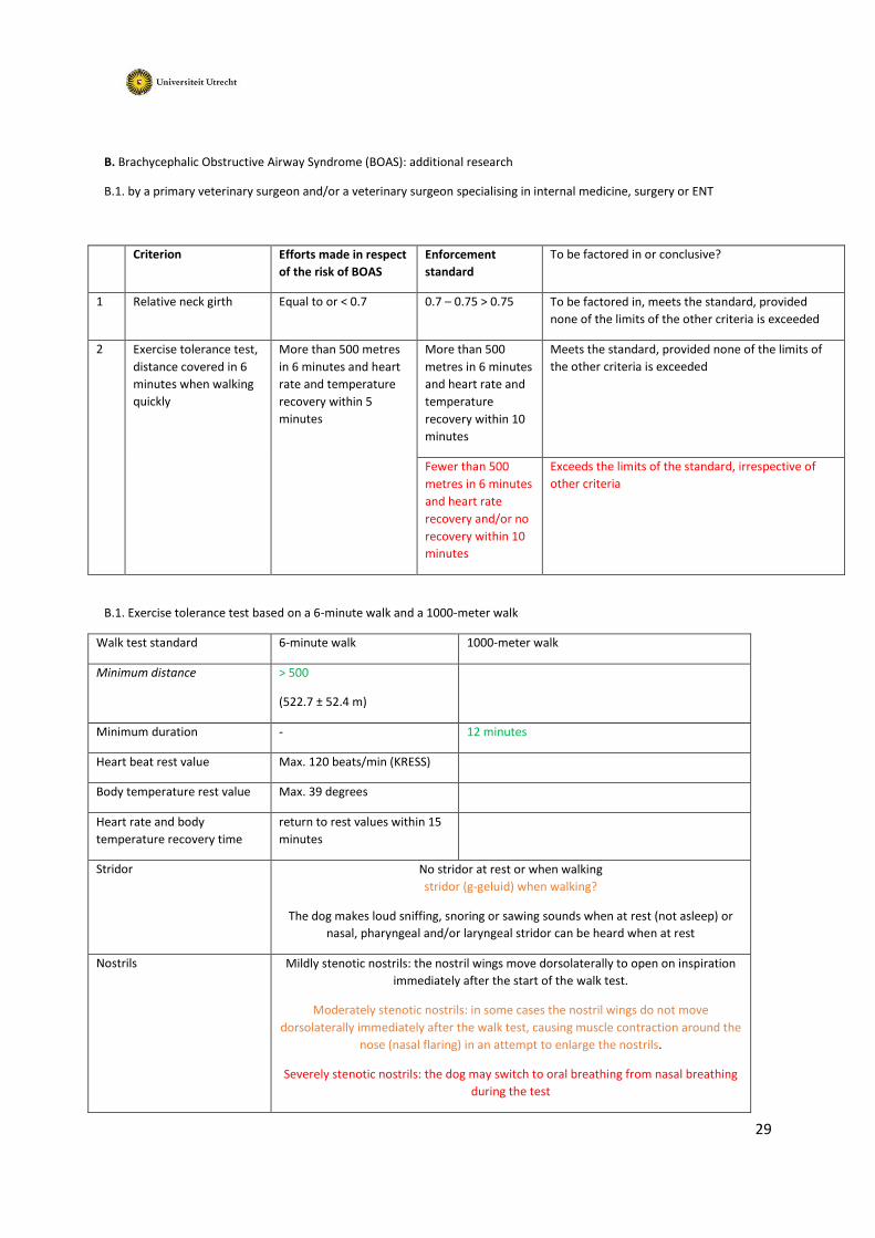
B. Brachycephalic Obstructive Airway Syndrome (BOAS): additional research
B.1. by a primary veterinary surgeon and/or a veterinary surgeon specialising in internal medicine, surgery or ENT
Criterion Efforts made in respect
of the risk of BOAS
Enforcement
standard
To be factored in or conclusive?
1 Relative neck girth Equal to or < 0.7 0.7 – 0.75 > 0.75 To be factored in, meets the standard, provided
none of the limits of the other criteria is exceeded
2 Exercise tolerance test,
distance covered in 6
minutes when walking
quickly
More than 500 metres
in 6 minutes and heart
rate and temperature
recovery within 5
minutes
More than 500
metres in 6 minutes
and heart rate and
temperature
recovery within 10
minutes
Meets the standard, provided none of the limits of
the other criteria is exceeded
Fewer than 500
metres in 6 minutes
and heart rate
recovery and/or no
recovery within 10
minutes
Exceeds the limits of the standard, irrespective of
other criteria
B.1. Exercise tolerance test based on a 6-minute walk and a 1000-meter walk
Walk test standard 6-minute walk 1000-meter walk
Minimum distance > 500
(522.7 ± 52.4 m)
Minimum duration - 12 minutes
Heart beat rest value Max. 120 beats/min (KRESS)
Body temperature rest value Max. 39 degrees
Heart rate and body
temperature recovery time
return to rest values within 15
minutes
Stridor No stridor at rest or when walking
stridor (g-geluid) when walking?
The dog makes loud sniffing, snoring or sawing sounds when at rest (not asleep) or
nasal, pharyngeal and/or laryngeal stridor can be heard when at rest
Nostrils Mildly stenotic nostrils: the nostril wings move dorsolaterally to open on inspiration
immediately after the start of the walk test.
Moderately stenotic nostrils: in some cases the nostril wings do not move
dorsolaterally immediately after the walk test, causing muscle contraction around the
nose (nasal flaring) in an attempt to enlarge the nostrils.
Severely stenotic nostrils: the dog may switch to oral breathing from nasal breathing
during the test

30
Article 1.3. Intrinsic value
1 The intrinsic value of the animal is recognised.
2. Recognition of intrinsic value as referred to in Subsection 1 is understood to mean recognition of
the value that animals possess in their own right as sentient beings. In drawing up rules under or
pursuant to this Act, and in taking decisions on the basis of these rules, due consideration shall be
given to the impact of these rules or decisions on the intrinsic value of the animal, notwithstanding
other legitimate interests. In all cases, any violation of the integrity or well-being of animals, beyond
what is reasonably necessary, shall be avoided and the care reasonably required by the animals
guaranteed.
3 For the purpose of Subsection 2, the care reasonably required by animals shall in any event include
safeguarding the animals against:
a. thirst, hunger and malnutrition;
b. physical and physiological discomfort;
c. pain, injury and diseases;
d. fear, distress, and chronic stress;
e. limitation of their natural behaviour; insofar as can be reasonably required.
ii The Genetics of Canine Skull Shape Variation. Jeffrey J. Schoenebeck and Elaine A. Ostrander
GENETICS 2013, 193, 2; 317-325
iii Canine Brachycephaly Is Associated with a Retrotransposon-Mediated Missplicing of SMOC2.
Marchant et al. Current Biology, 2017, 27; 1573-1584
iv Slatter’s Fundamentals of Veterinary Opthamology. Maggs D, Miller P, Ofri R, Slatter D, 2008, 4th
Ed. Elsevier Health Sciences: Edinburgh, UK
v A Retrospective Study of Ulcerative Keratitis in 32 Dogs. Kim JY, Won HJ, Jeong SW. International
Journal of Applied Research in Veterinary Medicine, 2009, 7, 27-31
vi Corneal innervation in mesocephalic and brachycephalic dogs and cats: assessment using in vivo
confocal microscopy. Christiane Kafarnik C et al. Veterinary Opthalmology, 11, 2008
vii Impact of Facial Conformation on Canine Health: Corneal Ulceration. Packer RMA, Hendricks A,
Burn CC (2015) PLoS ONE 10(5): e0123827. doi:10.1371/journal.pone.0123827
viii Prolapsus bulbi in small animals. A retrospective study of 36 cases. Fritsche, J., Spiess, B. M., Ruhli,
M. B., and Bollinger, J., 1996 Tierarztl Prax. 24: 55-61
ix Conformational risk factors of brachycephalic obstructive airway syndrome (BOAS) in pugs, French
bulldogs, and bulldogs, 2017, PLoS ONE 12(8):e0181928, DOI: 10.1371/journal.pone.0181928
x Surgical correction of brachycephalic syndrome in dogs: 62 cases (1991- 2004). Riecks TW, Birchard
SJ and Stephens JA. Journal of the American Veterinary Medical Association, 2007, 230: 1324-1328 –
Bronchial abnormalities found in a consecutive series of 40 brachycephalic dogs. De Lorenzi D,
Bertoncello D and Drigo M. Journal of the American Veterinary Medical Association, 2009 235: 835-
840 – Prevalence of gastrointestinal tract lesions in 73 brachycephalic dogs with upper respiratory

31
syndrome. Poncet CM, Dupre GP, Freiche VG, Estrada MM, Poubanne YA and Bouvy BM . Journal of
Small Animal Practice, 2005,46: 273-279 32
- Results of surgical correction of abnormalities associated with brachycephalic airway obstruction
syndrome in dogs in Australia. Torrez CV and Hunt GB. Journal of Small Animal Practice, 2006 .47:
150-154
- Nasopharyngeal turbinates in brachycephalic dogs and cats. Ginn JA, Kumar MSA, McKiernan BC
and Powers BE. Journal of the American Animal Hospital Association, 2008, 44: 243-249 – The
influence of phylogenic origin on the occurrence of brachycephalic airway obstruction syndrome in a
large retrospective study. Njikam IN, Huault M, Pirson V and Detilleux J. The International Journal of
Applied Research in Veterinary Medicine, 2009, 7: 138-143
xi Impact of Facial Conformation on Canine Health: Brachycephalic Obstructive Airway Syndrome.
Packer RMA et a. l PLoS One. 2015; 10(10): e0137496. doi: 10.1371/journal.pone.0137496
xii Craniofacial angle in dolicho-,meso- and brachycephalic dogs: radiological determination and
application. Annals of Anatomy- Anatomischer Anzeiger 175, 4, 1993;361-363
xiii Miller’s Anatomy of the Dog: Saunders Evans HE, 1993
xiv Skull morphometric lion, dog and cat, Saber and Gummow. J. Vet. Anat. 2015
xv Morphometrics within dog breeds are highly reproducible and dispute Rensch’s rule. Sutter NB et
al. Mammalian Genome 19;713-723, 2008
xvi Building better brachycephalics, Packer et al. (2012) Animal Welfare, 21, 81-93
https://www.ufaw.org.uk/downloads/welfare-downloads/building-better-brachycephalics-2013-
report.pdf
xvii Evaluation of the 6-minute walk test in dogs. Boddy KN et al. American Journal of Veterinary
Research, 2004, Vol. 65, No. 3; 311-313, https://doi.org/10.2460/ajvr.2004.65.311
xviii Evaluation of the 6-minute walk test in pet dogs, Swimmer RA and. Rozanski EA. Journal of
Veterinary Internal Medicine, 2011
xix Long‐Term Outcome and Use of 6‐Minute Walk Test in West Highland White Terriers with
Idiopathic Pulmonary Fibrosis. Lilja Maula LIO et al. Journal of Veterinary Internal Medicine, 2014
xx Comparison of submaximal exercise test results and severity of brachycephalic obstructive airway
syndrome in English bulldogs. Lilja-Maula LIO et al. The Veterinary Journal, 219, 2017: 22-26
xxi Morphometrics within dog breeds are highly reproducible and dispute Rensch’s rule. Sutter NB et
al, Mammalian Genome 19;713-723, 2008
xxii Dog obesity--the need for identifying predisposing genetic markers. Switonski M1, Mankowska
M. Res Vet Sci. 2013 Dec;95(3):831-6. doi: 10.1016/j.rvsc.2013.08.015.
xxiii A deletion in the pro‐opiomelanocortin (POMC) gene in Labrador retrievers is associated with
increased appetite and risk of obesity. Davison L.J. et al. 2017 Mar-Apr; 31(2): 343–348
xxiv Absolute corneal sensitivity and corneal trigeminal nerve anatomy in normal dogs. Barrett PM,
Scagliotti RH, Merideth RE. Progress in Veterinary & Comparative Ophthalmology, 1991; 1:245–254