bric kit - mbl · for analysis of rna metabolism which will lead us to the discovery of novel ncrna...
TRANSCRIPT

MEDICAL & BIOLOGICAL LABORATORIES CO., LTD. TEL: (052) 238-1904, E-mail: [email protected]
URL: http://ruo.mbl.co.jp/je/rip-assay/
Printed June. 26, 2014
Version 1.0
BRIC Kit
20 assays
CODE No. RN1007/RN1008
For Research Use Only. Not for use in diagnostic procedures.

-1-
Contents
I. Introduction…………………………………………………………………. 2
1. Background and Introduction
2. Product Description
3. Kit Components
4. Storage and Stability
5. Materials Required but Not Provided
II. BRIC Kit Procedure…………………………………………………………6 1. Procedure Summary
2. Buffer Preparation
3. Protocols for 5-Bromouridine Immunoprecipitation Chase (BRIC)
Metabolic labeling of RNA with BrU
Immunoprecipitation of BrU-labeled RNA (RNA Immunoprecipitation)
RNA Isolation
III. Examples of BRIC Analysis………………………………………………. 16 1. Loss of BrU-labeled RNA in 24 hours
2. Target RNA decay in 24 hours
3. Time-course analysis of BrU-labeled RNA with NanoDrop
4. Time-course analysis of BrU-labeled RNA with Bioanalyzer
5. Measurement of half-life of target RNA by RT-qPCR
6. Comparison of BrU- and EU-labeled RNA recovery
IV. Related Products……………………………………………………………20
V. Appendix…………………………………………………………………… 20

-2-
I. Introduction
Please read these instructions carefully before beginning the assay.
It is very important to isolate “high-quality BrU-labeled RNA” from various
materials to validate experiments such as reverse transcription quantitative
polymerase chain reaction (RT-qPCR) and gene expression analysis based on deep
sequencing technology because experimental results may be sensitive to RNA
quality. In order to obtain “high-quality BrU-labeled RNA”, and reduce the chance
of RNase contamination, gloves should be worn when proceeding BRIC, and
RNase-free microcentrifuge tubes and pipette tips should be used for the assay.
1. Background and Introduction Regulation of gene expression by RNA degradation is a critical step for the coordination of
various physiological processes. Also, RNA degradation pathways play important roles in mRNA
surveillance systems: nonsense-mediated mRNA decay (NMD), nonstop mRNA decay (NSD) and no-go
decay (NGD). In order to analyze RNA degradation, transcriptional inhibitor such as Actinomycin D
(ActD) has been widely used so far. However, the transcriptional inhibitors are known to influence cellular
physiology, including splicing, polyadenylation and other mRNA processing events. Furthermore, ActD
alters the localization and stability of RNAs, especially in the case of long noncoding RNA (lncRNA).
Recently, genome-wide RNA decay analysis in mammalian cells under inhibitor-free condition
has been developed. To perform the analysis, endogenous RNA should be transcriptionally pulse-labeled
with modified nucleosides, such as 4-thiouridine (4sU), 5-ethynyl uridine (EU) and 5-bromouridine (BrU),
followed by purification of newly labeled RNA. Each modified uridine has both advantages and
disadvantages in this analysis. BrU is less harmful for cells than other modified uridines and BrU-labeled
RNA can be easily immunoprecipitated by anti-bromodeoxyuridine (BrdU) antibody through its
cross-reactivity, while BrU-labeling takes 24 hours for effective cellular incorporation. In contrast to
immunopurification by anti-BrdU antibody, another method, which depends on “click chemistry” utilizing
EU-labeled RNA, requires biotinylation of the labeled RNA to specifically collect the biotin-labeled RNA
by streptavidin beads. There is a crucial problem that the biotinylation step via click reaction can be a
trigger for RNA degradation. In the case of 4sU, 4sU permits base-pairing with guanine instead of adenine,
which causes base-changes in the RNA sequence. Unlike EU- and 4sU-based analyses, 5-bromouridine
immunoprecipitation chase (BRIC) method has no undesirable effects for downstream applications. Thus,
BRIC will be the most suitable method for the analysis of RNA decay.
It is thought that the half-life of RNA closely relates to its own physiological function. mRNAs
derived from major housekeeping genes generally have long half-lives, while mRNAs of many regulatory
genes, which encode functional proteins required for only a limited time in cell – such as cell cycle
regulators, cytokines, transcriptional factors and oncoproteins – typically have short half-lives. In addition,
the genome-wide analysis by BRIC-deep sequencing (BRIC-seq) has lately revealed that majority of
non-coding RNAs (ncRNAs) with long half-lives involved in housekeeping functions, whereas the subsets
of ncRNAs with short half-lives included known regulatory ncRNAs. The result implies that the subsets of

-3-
ncRNAs may play central roles in the important biological processes. Thus, BRIC can be a powerful tool
for analysis of RNA metabolism which will lead us to the discovery of novel ncRNA biomarkers and drug
targets.
2. Product Description
BRIC Kit enables determination of RNA stability by chasing chronological decreases of
BrU-labeled RNA under physiologically undisturbed conditions. In the BRIC protocol, cells are
pulse-labeled with BrU for constant period and washed with PBS to remove the BrU-containing medium,
and then cells are chronologically harvested, followed by preparation of total RNA including newly
BrU-labeled RNA. The BrU-labeled RNA can be specifically immunoprecipitated with Anti-BrdU mAb
provided by MBL, followed by isolation of BrU-labeled RNA from immunocomplex on carrier material,
such as protein G magnetic beads. The isolated BrU-labeled RNA can be analyzed to determine its own
stability and half-life by various methods in molecular biology – RT-qPCR, deep sequencing or
microarray.
It is reported that the RNA labeling efficiency of BrU is less effective than that of EU in HeLa
cells under serum-starved condition. On the other hand, we confirmed that Anti-BrdU mAb (MBL) can
immunoprecipitate both BrU- and EU-labeled RNAs because of its cross-reactivity. Thus, RNA stability
can be analyzed by both BrU and EU with BRIC Kit.
[References]
1) Tani, H., et al., Genome Res. 22, 947-956 (2012)
2) Tani, H. and Akimitsu, N., RNA Biol. 9, 1233-1238 (2012)
3) Imamachi, N., et al., Methods 67, 55-63 (2014)
4) Tani, H., et al., PLoS One 8, e55684 (2013)
5) Tani, H., et al., RNA Biol. 9, 1370-1379 (2012)
3. Kit Components 20 assays
[RN1008 / Store at 2-8ºC]
1. RNA-IP buffer 18 mL × 2 bottles
2. Wash buffer 41 mL × 3 bottles
3. mi-Solution I 240 L × 1 vial: enzyme solution
4. mi-Solution II† 5.8 mL × 1 vial: diluent for Solution I
5. mi-Solution III‡ 3.6 mL × 1 vial: protein dissolvent
Solution III can dissolve proteins and dissociate immunocomplex.
6. mi-Solution IV 90 L × 1 vial: co-precipitator
Solution IV can increase RNA precipitation efficiently.
7. Protein G-Magnetic beads* 1.5 mL × 4 vial
1% beads slurry (mouse IgG binding capacity: 7 μg/mg beads)

-4-
[RN1007 / Store at -20ºC]
8. BrU solution (100 mM) 1.1 mL × 2 vials
9. Anti-BrdU mAb 450 L × 1 vial
Cross-reacts with 5-bromouridine. BrU-labeled RNA is usually
captured by this antibody.
10. Spike-in control** 80 L × 1 vial
Spike-in control is a BrU-labeled RNA synthesized by in vitro
transcription from lacZ-encoding cDNA, the reference sequence
(RefSeq) accession number NC_007779.1, region 363130-364149,
with 3ʹ-terminal poly(A) tail.
Note: † mi-Solution II may become turbid when stored for long-term at 2-8ºC. Turbidity does not affect
performance. If mi-Solution II is turbid, equilibrate to room temperature (15-25ºC) and mix well
before use.
‡ Precipitates may appear when mi-Solution III is stored for long-term at 2-8ºC. If mi-Solution
III contains precipitates, dissolve them by equilibrating the solution to room temperature
(15-25ºC) and mix well before use.
This reagent contains guanidine hydrochloride; this is a potentially hazardous substance and
should be used with appropriate caution.
* We recommend using Protein G-Magnetic beads in BRIC Kit, but other protein G beads can
be available for BRIC, if desired. Please note that protein G is more suitable than protein A
because the subclass of Anti-BrdU mAb, one of the components of BRIC Kit, is mouse IgG1.
The commercially available products related to protein G beads, which are confirmed to work
well with BRIC Kit, are shown in Appendix.
** Spike-in control should be divided into appropriate aliquots to avoid freeze-thaw cycles when
you receive, and stored at -80ºC or below until just before use.
4. Storage and Stability
The component 10 of BRIC Kit (Spike-in control) should be divided into appropriate aliquots to avoid
freeze-thaw cycles when you receive, and stored at -80ºC or below until just before use because the
component 10 is an RNA synthesized by in vitro transcription. The components 8 (BrU solution) and 9
(Anti-BrdU mAb) should be stored at -20ºC or below. Other components should be stored at 4ºC (Do not
freeze). These components are stable for one and a half years from the date of manufacture when stored at
the indicated conditions.
5. Materials Required but Not Provided
Equipments
1. Microcentrifuge capable of 15,000 × g
2. Microcentrifuge tubes (1.5 mL or 2 mL) (Nuclease-free)
(Recommendation; use low-adhesion tube for RNA-IP)
3. Centrifuge capable of 2,000 × g
4. Centrifuge tubes (15 mL or 50 mL)
5. Pipettes (5 mL, 10 mL or 25 mL) (Nuclease-free)

-5-
6. Pipette tips (10 L, 20-100 L, 200 L, and 1,000 L) (Nuclease-free)
(Recommendation; use low-adhesion pipette tip for RNA-IP)
7. Ultra low temperature freezer (-80ºC)
8. Freezer (below -20ºC)
9. End-over-end rotator
10. Vortex mixer
11. Magnetic stand
12. Gloves
Reagents
13. RNase inhibitor*
14. Dithiothreitol (DTT)*
15. 100% Ethanol (molecular biology grade)
16. 100% 2-Propanol (molecular biology grade)
17. Nuclease-free PBS
18. Nuclease-free water
19. Reagents for reverse transcription**
20. Primer set for Spike-in control***
21. Reagents for qPCR****
Note: * Recommended concentration of each reagent is shown in Appendix.
** Commercially available reagents confirmed to work with BRIC Kit are shown in
Appendix.
*** The recommended primer sequences to analyze the Spike-in control levels by RT-qPCR
are described below:
Forward primer : 5ʹ-AACTCTGGCTCACAGTACGC-3ʹ
Reverse primer : 5ʹ-TGGCGGTTAAATTGCCAACG-3ʹ
**** Commercially available reagents confirmed to work with BRIC Kit are shown in
Related Products.

-6-
II. BRIC Kit Procedure
1. Procedure Summary
-7-

-8-

-9-
-10-
2. Buffer Preparation
1. RNA-IP buffer
Add appropriate concentrations of RNase inhibitor and dithiothreitol (DTT) to RNA-IP
buffer just before use. RNA-IP buffer containing these reagents is described as RNA-IP
buffer (+) in the following protocols. The optimal concentration of each reagent for BRIC is
shown in Appendix.
2. Wash buffer
Add appropriate concentration of dithiothreitol (DTT) to Wash buffer just before use. Wash
buffer containing DTT is described as Wash buffer (+) in the following protocols. The
optimal concentration of the reagent for BRIC is shown in Appendix.
3. Protocols for 5-Bromouridine Immunoprecipitation Chase (BRIC) The following protocol is for the RNA decay analysis by BRIC: labeling various cell lines with BrU,
extracting total RNA from BrU-labeled cells, immunoprecipitating BrU-labeled RNA using Anti-BrdU
mAb and isolating BrU-labeled RNA. The cell harvest should be performed at several time points after
changing medium to analyze the RNA half-lives [e.g., 0, 4, 8, 12 and 24 hours]. The quantity and quality
of isolated BrU-labeled RNAs can be checked by a spectrophotometer and/or Bioanalyzer. Then, the
stabilities and half-lives of RNAs can be analyzed by deep sequencing, RT-qPCR and microarray.
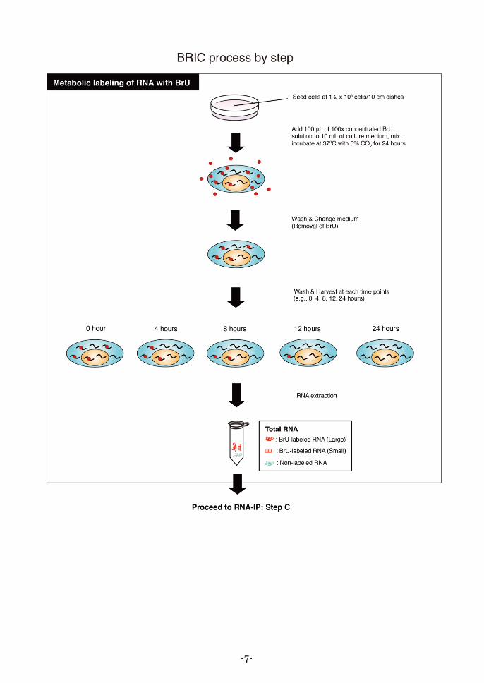
Metabolic labeling of RNA with BrU
Note: In some cell lines, BrU-labeling efficiency into newly synthesized RNA depends on the cell
conditions. In order to obtain reproducible results, prior to BRIC procedure, continue to
culture the cryopreserved cells for about 1 week until the viability is recovered and pay
much attention on the overgrowth of the cells. Labeling efficiencies of several cell lines are
described in the table below.
Note: * By labelling with 500 M BrU, BrU-labeled RNA can be quantified with NanoDrop.
** It may be difficult to analyze the RNA stability in these cell lines because BrU-labeled
RNAs prepared from these cell lines pulse-labeled with 500 M BrU could not be
quantified with NanoDrop.
1. Make 100 concentrated BrU solution by diluting 100 mM BrU stock solution (component 8) in
nuclease free PBS (e.g., dilute 100 mM BrU stock solution to 15 mM with nuclease-free PBS).
Note: For pulse-labeling the cell, the recommended final concentration of BrU in culture media
is 150 M. If desired, higher concentrations of BrU are also available for BRIC, but please
note that higher concentrations of BrU can be toxic to cell. Pulse-labeling with 500 M of
BrU is more efficient, but this concentration may affect growth rate of cells and/or other
biological processes.
Labelling efficiency Cell lines
High HeLa, K562
Moderate 293T, HT29
Low* Jurkat, HEK293, T-47D, PC3
Very Low** MCF-7, ZR-75-1, KATO III, MEG-01, WERI-Rb-1

-11-
2. Seed cells at 1–2 × 10⁶ cells/10 cm dishes.
Note: Timing of pulse-labeling might affect the BrU-labeling efficiency of RNA in some cell
lines. If desired, pre-culture the cells in a humidified incubator at 37ºC with 5% CO2 for 24
hours in order to more stabilize the cellular conditions. Please pay much attention on the
overgrowth of the cells.
3. Add 100 L of 100 concentrated BrU solution to 10 mL of culture medium, then gently mix.
Note: The recommended final BrU concentration, 150 M, is mild condition for the cells and
exhibits moderate pulse-labeling efficiency.
4. Incubate the cells in a humidified incubator at 37ºC with 5% CO2 for 24 hours. Protect from
intense light exposure.
5. Remove BrU-containing medium (pulse media), and gently wash the cells 3 times with 5 mL of
PBS.
6. Remove PBS, and add 10 mL of fresh medium (chase media).
7. Incubate the cells at 37ºC with 5% CO2 for appropriate hours [e.g., 0, 4, 8, 12 and 24 hours].
8. Wash the cells 3 times with 5 mL of PBS at each time point.
9. Detach the cells from the culture dish by pipetting or using a cell scraper.
10. Centrifuge the cell suspension at 300 × g for 5 minutes at 4ºC to pellet the cells. Carefully remove
and discard the supernatant.
11. Prepare the total RNA using commercially available RNA extraction reagent [e.g., RNAiso Plus,
Takara Bio]. Finally, reconstitute the pellet at each time point in 50 L of nuclease-free water.
Note: If desired, reconstitution volume can be changed from 50 L, but it is important to fix the
reconstitution volume at each time point. The details are described in the Step C in next
section (RNA Immunoprecipitation).
12. Quantify the extracted total RNA with NanoDrop (Thermo Fisher Scientific Inc.), and check the
quality and quantity of total RNA.
13. Store at -80ºC or below until starting RNA immunoprecipitation. For the preparation of input total
RNA, please check the Step C in next section (RNA Immunoprecipitation).
Immunoprecipitation of BrU-labeled RNA (RNA Immunoprecipitation)
(A. Preparation of Anti-BrdU mAb-immobilized protein G magnetic beads)
Note: Immunoprecipitation protocol described below is designed only for the Protein G
-Magnetic beads (provided by MBL). In case of using protein G agarose beads,
centrifuge the tube (2,000 × g for 1 minute at 4ºC) during washing step instead of
magnetic separation with magnetic stand.
1. Suspend the Protein G-Magnetic beads slurry by vortex thoroughly, then spin-down briefly.
2. Resuspend the Protein-G Magnetic beads slurry by pipetting, Aliquot 300 L of the beads slurry to
each new microcentrifuge tube.
3. Place the tube on the magnetic stand to separate the beads from the solution.
4. After the solution becomes clear (about 1 minute), aspirate the supernatant carefully and remove
the tube from the magnetic stand.

-12-
5. Add 1 mL of Wash buffer (+) to each tube and vortex thoroughly, then spin-down briefly.
6. Place the tube on the magnetic stand to separate the beads from the solution.
7. After the solution becomes clear (about 1 minute), aspirate the supernatant carefully.
8. Add 500 L of Wash buffer (+) to each tube.
9. Add 20 L of Anti-BrdU mAb to each tube, then vortex briefly but thoroughly.
10. Incubate the tube with constant rotation for at least 30 minutes at 4ºC. If necessary, this incubation
can be extended to overnight.
(B. Washing the antibody-immobilized protein G magnetic beads)
11. Spin-down briefly, and then place the tube on the magnetic stand to separate the beads from the
solution.
12. After the solution becomes clear (about 1 minute), aspirate the supernatant carefully and remove
the tube from the magnetic stand.
13. Add 1 mL of RNA-IP buffer (+) to each tube and vortex thoroughly, then spin-down briefly.
14. Place the tube on the magnetic stand to separate the beads from the solution.
15. After the solution becomes clear (about 1 minute), aspirate the supernatant carefully, and then
remove the tube from the magnetic stand.
16. Add 500 L of RNA-IP buffer (+) to each tube.
17. Leave the beads at 4ºC or on ice and use the beads in Step 21.
(C. Preparation of antibody-immobilized protein G magnetic beads-BrU-labeled RNA complex)
Note: When performing immunoprecipitations with constant quantity (g) of total RNA at several time
points, the percentage of BrU-labeled RNA to input total RNA gradually diminishes just after
removal of pulse-labeling medium. This is due to both the decrease of BrU-labeled RNA and the
increase of non-labeled total RNA, which are caused by cell proliferation during chase period. In
order to accomplish more precise analysis of RNA half-life, please select one of the two
“Normalization methods” described below:
A. Normalization by the quantity of stable transcripts
In case of RT-qPCR or deep sequencing (RNA-seq) analysis, the expression level of each
transcript at each time point can be normalized with several stable transcripts such as GAPDH.
The analyses might be more accurate if the quantities of BrU-labeled RNA among the time
points are normalized by the geometric mean of several transcripts (e.g., GAPDH, PGK1, GRN
and TRAP1). When choosing this normalization method, prepare input total RNA of each time
point to be 40 g.
B. Normalization by Spike-in control
The proportions of BrU-labeled RNA to total RNA at each time point can be normalized by
fixing input total RNA volume (L) for immunoprecipitation and utilizing the result of
Spike-in control. In this case, each total RNA including BrU-labeled RNA, extracted from each
time point sample, should be reconstituted in the equal volume of nuclease-free water (e.g., 50
L each). The constant volume to all input total RNA samples should be calculated based on
quantity of chase time 0 hour sample which is recommended to be 40 µg. Normalize the
expression levels of various transcripts by expression data of Spike-in control.

-13-
18. According to “Normalization methods” above, prepare appropriate quantity (or volume) of input
total RNA. Then add 2 L of Spike-in control to the tube.
Note: Excess volume of input total RNA could affect immunopurification efficiency. The
volume of input total RNA should be less than 100 L.
19. Denature the input RNA by heating for 2 minutes at 80ºC.
20. Quench the RNA 4ºC or on ice at least 5 minutes.
21. Spin-down briefly, and then transfer the denatured input RNA into the tube prepared in step 17.
22. Incubate the tube with rotation for 3 hours at 4ºC.
(D. Washing antibody-immobilized protein G magnetic beads-RNA complex)
23. Spin-down the tube (prepared in step 22) containing antibody-immobilized protein G magnetic
beads-RNA complex, and place the tube on the magnetic stand to separate the beads from the
solution.
24. After the solution becomes clear (about 1 minute), aspirate the supernatant carefully.
25. Add 1 mL of Wash buffer (+) and vortex briefly but thoroughly.
26. Spin-down briefly, and place the tube on the magnetic stand to separate the beads from the
solution.
27. After the solution becomes clear (about 1 minute), aspirate the supernatant carefully.
28. Wash the antibody-immobilized beads-RNA complex; repeat the steps 25-27 three times.
RNA Isolation
(from antibody-immobilized protein G magnetic beads-RNA complex)
mi-Solution II and mi-Solution III should be equilibrated to room temperature before use.
The reagents should be briefly but thoroughly mixed before use.
Note: The following RNA isolation protocol in this section is almost identical to “Separation method”
used in RIP-Assay kit for microRNA described at http://ruo.mbl.co.jp/gtf/1/1/RN1005.pdf. In this
method, large RNA and small RNA are divided into individual microcentrifuge tubes. If you do
not intend to analyze small RNA, large RNA fraction prepared by Separation method is
sufficient for analysis of large RNA. If simultaneous analysis of small and large RNAs is required,
2-step method in RIP-Assay kit for microRNA can be available, although 2-step method has a
little higher background than Separation method.
§ Comparative table of RNA isolation methods
Separation method 2-step method 1-step method
Collectable RNA species
large RNA
small RNA
(in individual tubes)
large RNA
small RNA
(in one tube)
small RNA
(a small amount of large RNA)
Recovery rate for large RNA >90% >90% <40%
Recovery rate for small RNA >80% >90% >90%
Classification by nucleotide length
Yes
(large RNA: >60-80 nt)
(small RNA: <60-80 nt)
No No
Assay time 75 min. 75 min. 45 min.
Background (silver staining) Low High Moderate
Advantage Available for multiple applications High-recovery rate for large/small RNA Short assay time
DisadvantageA little loss in recovery of small RNA
compared to the other 2 methods
Not suitable for visualization by silver
staining following denaturing PAGE
Low-recovery rate for large RNA
(~40% of large RNAs)
-14-
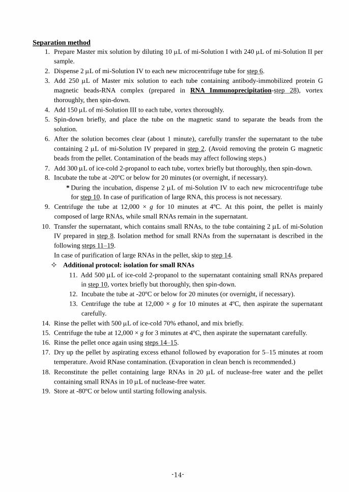
Separation method
1. Prepare Master mix solution by diluting 10 L of mi-Solution I with 240 L of mi-Solution II per
sample.
2. Dispense 2 L of mi-Solution IV to each new microcentrifuge tube for step 6.
3. Add 250 L of Master mix solution to each tube containing antibody-immobilized protein G
magnetic beads-RNA complex (prepared in RNA Immunoprecipitation-step 28), vortex
thoroughly, then spin-down.
4. Add 150 L of mi-Solution III to each tube, vortex thoroughly.
5. Spin-down briefly, and place the tube on the magnetic stand to separate the beads from the
solution.
6. After the solution becomes clear (about 1 minute), carefully transfer the supernatant to the tube
containing 2 L of mi-Solution IV prepared in step 2. (Avoid removing the protein G magnetic
beads from the pellet. Contamination of the beads may affect following steps.)
7. Add 300 L of ice-cold 2-propanol to each tube, vortex briefly but thoroughly, then spin-down.
8. Incubate the tube at -20ºC or below for 20 minutes (or overnight, if necessary).
* During the incubation, dispense 2 L of mi-Solution IV to each new microcentrifuge tube
for step 10. In case of purification of large RNA, this process is not necessary.
9. Centrifuge the tube at 12,000 × g for 10 minutes at 4ºC. At this point, the pellet is mainly
composed of large RNAs, while small RNAs remain in the supernatant.
10. Transfer the supernatant, which contains small RNAs, to the tube containing 2 L of mi-Solution
IV prepared in step 8. Isolation method for small RNAs from the supernatant is described in the
following steps 11–19.
In case of purification of large RNAs in the pellet, skip to step 14.
Additional protocol: isolation for small RNAs
11. Add 500 L of ice-cold 2-propanol to the supernatant containing small RNAs prepared
in step 10, vortex briefly but thoroughly, then spin-down.
12. Incubate the tube at -20ºC or below for 20 minutes (or overnight, if necessary).
13. Centrifuge the tube at 12,000 × g for 10 minutes at 4ºC, then aspirate the supernatant
carefully.
14. Rinse the pellet with 500 L of ice-cold 70% ethanol, and mix briefly.
15. Centrifuge the tube at 12,000 × g for 3 minutes at 4ºC, then aspirate the supernatant carefully.
16. Rinse the pellet once again using steps 14–15.
17. Dry up the pellet by aspirating excess ethanol followed by evaporation for 5–15 minutes at room
temperature. Avoid RNase contamination. (Evaporation in clean bench is recommended.)
18. Reconstitute the pellet containing large RNAs in 20 L of nuclease-free water and the pellet
containing small RNAs in 10 L of nuclease-free water.
19. Store at -80ºC or below until starting following analysis.

-15-
Additional Procedure: Analysis of isolated RNA
We recommend qualitative and quantitative analysis of isolated RNAs prior to downstream analysis
such as RT-qPCR, deep sequencing and microarray. These technologies may be useful for profiling
RNAs.
Quality control for isolated RNAs
It is very important for comprehensive analysis such as deep sequencing or RT-qPCR to retain
high-quality RNA because experimental results may be sensitive to RNA quality. Please confirm
the quality of RNAs with Bioanalyzer (Agilent Technologies, Inc.) after the quantification of the
isolated RNAs with NanoDrop (Thermo Fisher Scientific Inc.).

-16-
III. Examples of BRIC Analysis
1. Loss of BrU-labeled RNA in 24 hours
HeLa, HEK293T (293T), Jurkat, K562, HT29, T-47D and HEK293 cells were pulse-labeled with 150 M
BrU for 24 hours. Then, the cells were washed and harvested at chase time 0 and 24 hours. After RNA
extraction using RNAiso Plus (Takara Bio), BrU-labeled RNA was isolated by BRIC Kit. The isolated
BrU-labeled RNA was quantified with a spectrophotometer (NanoDrop) according to manufacturer’s
instructions (Thermo Fisher Scientific Inc.).
HeLa cells were most effectively labeled with 150 M BrU in 7 cell lines, while BrU-labeled RNA from
Jurkat, T-47D and HEK293 were almost not detected by NanoDrop. Quantity of BrU-labeled RNA in
HeLa, K562, and 293T drastically decreased in 24 hours.
-17-
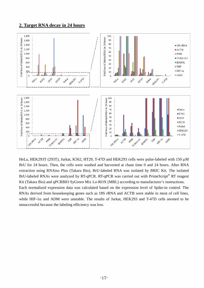
2. Target RNA decay in 24 hours
HeLa, HEK293T (293T), Jurkat, K562, HT29, T-47D and HEK293 cells were pulse-labeled with 150 M
BrU for 24 hours. Then, the cells were washed and harvested at chase time 0 and 24 hours. After RNA
extraction using RNAiso Plus (Takara Bio), BrU-labeled RNA was isolated by BRIC Kit. The isolated
BrU-labeled RNAs were analyzed by RT-qPCR. RT-qPCR was carried out with PrimeScript® RT reagent
Kit (Takara Bio) and qPCRBIO SyGreen Mix Lo-ROX (MBL) according to manufacturer’s instructions.
Each normalized expression data was calculated based on the expression level of Spike-in control. The
RNAs derived from housekeeping genes such as 18S rRNA and ACTB were stable in most of cell lines,
while HIF-1 and ADM were unstable. The results of Jurkat, HEK293 and T-47D cells seemed to be
unsuccessful because the labeling efficiency was low.

-18-
3. Time-course analysis of BrU-labeled RNA with NanoDrop
HeLa cells were pulse-labeled with 150 M BrU for 24
hours. Then, cells were washed and harvested at chase time
0, 4, 8, 12 and 24 hours. After RNA extraction by RNAiso
Plus (Takara Bio), BrU-labeled RNA was isolated by BRIC
Kit and quantified with a spectrophotometer (NanoDrop)
according to manufacturer’s instructions (Thermo Fisher
Scientific Inc.).
The percentage of isolated BrU-labeled RNA rapidly
decreased to 60% in the first 4 hours and then gradually decreased in the next 20 hours. This result
suggests that a lot of unknown functional ncRNAs may be buried in short-lived RNA population (T1/2 < 4
hours).
4. Time-course analysis of BrU-labeled RNA with Bioanalyzer
HeLa cells were pulse-labeled with 150 M BrU for 24 hours. Then, cells were washed and harvested at
chase time 0, 4, 8, 12 and 24 hours. After RNA extraction by RNAiso Plus (Takara Bio), BrU-labeled RNA
was isolated by BRIC Kit (RNA Isolation: Separation method) and analyzed on a Bioanalyzer RNA pico
chip (Agilent Technologies, Inc.) according to manufacturer’s instructions.
The migration profile of the 0 hour showed several minor peaks with 2 main peaks around 2,000 and 4,000
nucleotides, corresponding to 18S and 28S ribosomal RNA, respectively. Several minor peaks
corresponding to mRNA and rRNA precursor faded out over time. The low peak (indicated by arrowhead),
probably corresponding to rRNA precursor, around the 28S rRNA, was not detected at time points after 8
hours. In contrast, the peak of 28S rRNA increased during the time. Furthermore, the heights of 2 main
peaks were observed even at 24 hours. These results reveal the stability of processed rRNA and suggest
that rRNA processing can be monitored by BRIC and Bioanalyzer.
Nucleotide lengthNucleotide length
RN
A i
nte
nsi
tyR
NA
in
ten
sity

-19-
5. Measurement of half-life of target RNA by RT-qPCR
HeLa cells were pulse-labeled with 150 M BrU for 24 hours. Then, cells were washed and harvested at
chase time 0, 4, 8, 12 and 24 hours. After RNA extraction by RNAiso Plus (Takara Bio), BrU-labeled RNA
was isolated by BRIC Kit. Isolated BrU-labeled RNA was analyzed by RT-qPCR. RT-qPCR was carried
out with PrimeScript® RT reagent Kit (Takara Bio) and qPCRBIO SyGreen Mix Lo-ROX (MBL)
according to manufacturer’s instructions.
As expected, the transcripts derived from housekeeping genes, such as 18S rRNA and ACTB, showed
relatively long half-lives, while HIF-1 and ADM* showed much shorter half-lives.
*ADM gene encodes a potent hypotensive peptide which plays important roles in both normal and disease conditions.
6. Comparison of BrU- and EU-labeled RNA recovery
HeLa cells were pulse-labeled with 150 M of BrU or EU (Life
Technologies Inc.) for 24 hours. Then, cells were washed and
harvested without additional chase time. After RNA extraction
by RNAiso Plus (Takara Bio), BrU- and EU-labeled RNA were
isolated by BRIC Kit. These RNAs were quantified with a
spectrophotometer (NanoDrop) according to manufacturer’s
instructions (Thermo Fisher Scientific Inc.).
As mentioned above, Anti-BrdU mAb cross-reacted with
EU-labeled RNA. This result indicates that the recovery of
EU-labeled RNA was almost equal to BrU-labeled RNA.

-20-
IV. Related Products RIP-Assay Kit
RN1001 RIP-Assay Kit (10 assays)
RIP-Assay Kit for microRNA RN1005 RIP-Assay Kit for microRNA (10 assays)
RiboTrap Kit RN1011/RN1012 RiboTrap Kit (10 assays)
Protein G magnetic beads
MJS002 Protein G-Magnetic beads
Magnetic stand
3190 Magnetic Rack
qPCRBIO SyGreen Mix
PB20-11-01 qPCRBIO SyGreen Mix Lo-ROX
PB20-11-05 qPCRBIO SyGreen Mix Lo-ROX
PB20-12-01 qPCRBIO SyGreen Mix Hi-ROX
PB20-12-05 qPCRBIO SyGreen Mix Hi-ROX
Anti-BrdU antibody
MI-11-3 Anti-Bromodeoxyuridine mAb
Isotype Control Antibody
M075-3 Mouse IgG1 (isotype control)
RIP-Certified Antibody and RBP Antibody
Various antibodies in “RNA-RNP network” are also available from MBL.
Please visit our website at http://ruo.mbl.co.jp/je/rip-assay/
V. Appendix The following commercially available reagents have been confirmed to work with BRIC Kit at the
indicated final concentration.
140626-1.0
Distribution source
Protein G-Magnetic beads MBL Dynabeads protein G Life Technologies
Final concentration
DTT 1.5 mM
RNase inhibitor Distribution source Code No. Final concentration RNase OUT 10777-019 50 - 200 U/mL
Distribution source Code No. PrimeScript® RT reagent Kit Takara Bio RR037A
Reagents for reverse transcription
Protein G beads
Reducing agent
Code No. MJS002 DB10004 22852 Immobilized Protein G Plus
5-Ethynyl uridine
5-Ethynyl uridine (EU) Life Technologies E10345
Distribution source Code No.
RNA Extraction reagent
RNAiso Plus Takara Bio
Distribution source Code No.
9109
Pierce
Life Technologies