brief review - media.tghn.org
TRANSCRIPT

1
From the Faculty of Medicine, University of Oslo, Oslo, Norway and Departments of Obstetrics and Gynaecology, Oslo University Hospital, Oslo, Norway (A.C.S., K.M.); Nuffield Department of Obstetrics and Gynaecology (C.W.G.R), Radcliffe Department of Medicine (P.L.), University of Oxford, Oxford, United Kingdom; Department of Maternal Medicine, Institute for Women’s Health, University College London, London, United Kingdom (D.W.); Department of Obstetrics and Gynaecology, St. George’s Hospital, London, United Kingdom (B.T.); Division of Epidemiology, Norwegian Institute of Public Health, Oslo, Norway (P.M.); Department of Obstetrics and Gynecology, Erasmus Medical Centre, Rotterdam, The Netherlands (E.A.P.S.); Preeclampsia Foundation, Melbourne, FL (E.Z.T.); Division of Epidemiology and Disease Control, University of Texas School of Public Health, Houston (R.B.N.); University of Texas Health Science Center San Antonio (L.M.); Department of Women’s Health, King’s College London, London, United Kingdom (L.P.); and Department of Obstetrics Gynecology and Reproductive Sciences, Epidemiology and Clinical and Translational Research, Magee-Womens Institute, University of Pittsburgh, PA (J.M.R.).
*A list of the CoLab consortium participants among the study authors is given in the Appendix.The online-only Data Supplement is available with this article at http://hyper.ahajournals.org/lookup/suppl/doi:10.1161/HYPERTENSIONAHA.
115.06357/-/DC1.Correspondence to Anne Cathrine Staff, Department of Obstetrics and Department of Gynaecology, Oslo University Hospital, location Ullevål,
Kirkeveien 166, Post Box 4956, Nydalen, 0424 Oslo, Norway. E-mail [email protected]
Pregnancy and Long-Term Maternal Cardiovascular HealthProgress Through Harmonization of Research Cohorts and Biobanks
Anne Cathrine Staff, Christopher W.G. Redman, David Williams, Paul Leeson, Kjartan Moe, Basky Thilaganathan, Per Magnus, Eric A.P. Steegers, Eleni Z. Tsigas, Roberta B. Ness,
Leslie Myatt, Lucilla Poston, James M. Roberts; for the Global Pregnancy Collaboration (CoLab)*
(Hypertension. 2016;67:00-00. DOI: 10.1161/HYPERTENSIONAHA.115.06357.)© 2015 American Heart Association, Inc.
Hypertension is available at http://hyper.ahajournals.org DOI: 10.1161/HYPERTENSIONAHA.115.06357
Background: Why We Need to Better Understand the Associations Between
Pregnancy and Future Cardiovascular HealthIn 2011, the American Heart Association added preeclamp-sia, gestational diabetes mellitus (GDM), and delivery of a growth-restricted child as pregnancy-related risk factors for cardiovascular disease (CVD).1 This move was applauded by the obstetric research community, which for some years had emphasized the importance of pregnancy as a stress test for detecting women at excessive risk for premature CVD.2–4
CVD is the leading cause of death for men and women in high-income and most low-to-middle–income countries.5 Globally, coronary artery disease kills more women than men,6 although women develop CVD 10 to 15 years later than men. Women frequently present with unrecognized CVD symptoms and are twice as likely as men to die of a first acute myocardial infarction if <50 years old.7 The preclinical stages of CVD are evident from a young age and are modifiable through control of classic risk factors (insulin resistance/diabetes mellitus, obesity, lack of exercise, tobacco smoking, hypertension, and hyperlip-idemia).8,9 In this regard, pregnancy is a window of opportunity for identifying those women with perinatal complications who may benefit from early risk detection and early CVD prevention.
In this article, we summarize the associations between pregnancy, placenta-related pregnancy complications, and future maternal CVD. We present established as well as more novel hypotheses, which may explain these epidemiological associations. The interventions that potentially could reduce risks of future CVD are enumerated. To facilitate progress, we suggest methods of harmonizing study designs, long-term follow-ups of pregnancy cohorts and biobanks, and pooling of the world’s data in ways that can enhance the power of current and future research.
Associations Between Pregnancy Complications and Future CVD Risk
Preeclampsia and Fetal Growth Restriction and Future CVD RiskPreeclampsia is a pregnancy-specific multisystem disorder defined by new-onset hypertension and proteinuria after ges-tational week 20, or new onset preeclampsia-associated signs in the absence of proteinuria.8 Preeclampsia requires the pres-ence of a placenta or residual placental components (postpar-tum preeclampsia), but the relative contributions of maternal predisposing factors versus placental factors to its patho-physiology are not well delineated.9 Women with essential hypertension, obesity, pregestational diabetes mellitus, and renal disease are at elevated risk for developing preeclamp-sia. Several large-population–based studies demonstrate that women who have had preeclampsia are at increased risk for later CVD and premature death compared with women with healthy pregnancies.10–14 Women who have experienced either preeclampsia or fetal growth restriction have a 2-fold increased risk compared with pregnancies with a normal outcome. When a woman has both preeclampsia and fetal growth restriction, the likelihood of CVD may be as much as 8-fold higher.15,16 Recurrent,12,14,17 more severe, and early-onset preeclampsia, as well as preeclampsia with concurrent neonatal morbidity, increases the risk of later life CVD,14,16,18,19 much more than gestational hypertension (without protein-uria or other preeclampsia-associated features) or late-onset preeclampsia.13,20–22
Prematurity, Miscarriage, and Future CVD RiskWomen with a history of preterm birth (<37 weeks gestation), even without pregnancy-induced hypertension23 or preeclamp-sia,10,11,24 are twice as likely to die from CVD compared with
Brief Review
at MERCY HOSP OF PITTSBURGH on January 5, 2016http://hyper.ahajournals.org/Downloaded from at MERCY HOSP OF PITTSBURGH on January 5, 2016http://hyper.ahajournals.org/Downloaded from at MERCY HOSP OF PITTSBURGH on January 5, 2016http://hyper.ahajournals.org/Downloaded from at MERCY HOSP OF PITTSBURGH on January 5, 2016http://hyper.ahajournals.org/Downloaded from at MERCY HOSP OF PITTSBURGH on January 5, 2016http://hyper.ahajournals.org/Downloaded from at MERCY HOSP OF PITTSBURGH on January 5, 2016http://hyper.ahajournals.org/Downloaded from at MERCY HOSP OF PITTSBURGH on January 5, 2016http://hyper.ahajournals.org/Downloaded from at MERCY HOSP OF PITTSBURGH on January 5, 2016http://hyper.ahajournals.org/Downloaded from at MERCY HOSP OF PITTSBURGH on January 5, 2016http://hyper.ahajournals.org/Downloaded from at MERCY HOSP OF PITTSBURGH on January 5, 2016http://hyper.ahajournals.org/Downloaded from

2 Hypertension February 2016
women who delivered at term. Spontaneous preterm labor is caused by multiple pathological processes25; nonetheless, delivery of a preterm or small for gestational age infant over-all, independent of smoking and other risk factors, has been shown to increase the risk of CVD death and hospitalization later in life.26 Although less frequently investigated, recurrent miscarriages have also been linked to future CVD27 and to endothelial dysfunction.28 Additionally, recurrent pregnancy loss is associated with pregnancy complications, such as pla-cental abruption and hypertensive pregnancy disorders, which are independently associated with markers of cardiovascular dysfunction, at least in the short term.29,30
Diabetes Mellitus in Pregnancy and Future CVD RiskWomen who develop GDM have a 70% higher risk for future CVD than those with no history of the disorder, mostly attrib-uted to an increased risk for developing type 2 diabetes melli-tus.31 As many as half of women with a pregnancy complicated by GDM develop type 2 diabetes mellitus within 5 years,32 and the diabetes mellitus risk is reported as 7-fold when compared with normoglycemic pregnancies.33 Whether pregnancy per se exacerbates the increased risk for later life CVD associated with pre-existing diabetes mellitus is not known. Women with type 1 diabetes mellitus seem, however, to be more at risk for developing retinopathy and nephropathy later in life if they had preeclampsia.34 Chronic kidney disease is considered an independent CVD risk factor,35 and nephropathy may, there-fore, add to the overall CVD risk after a preeclamptic preg-nancy in women with pregestational diabetes mellitus.
Normal Pregnancy and Future Risk for CVDSeveral studies report an association between the number of a woman’s pregnancies, even without adverse outcomes and maternal CVD risk,36–40 whereas others fail to find such an association.41,42 For men, a high number of children does not associate with increased CVD risk.43 In a large Swedish population-based registry study, parity was independently associated with future maternal CVD in a J-shaped fashion (where 2 births represented the lowest risk) after adjustment for socioeconomic factors and pregnancy-related complica-tions. The highest risk was among women with >5 births.40 The same J-shaped trend between number of births and maternal cardiovascular mortality was found in a recent study from the Norwegian Birth Registry, but only in women with <10 years of education.44 The number of offspring does not seem to increase the CVD risk for the male part-ners, after correcting for obesity and metabolic risks,38 sug-gesting a pathophysiologic effect from pregnancy, but this finding requires replication. Not only nulliparity per se has been associated with increased CVD risk40 but also subfer-tile women who eventually conceive and have a child are at increased risk for CVD, even after adjusting for CVD risk factors and adverse pregnancy outcomes, suggesting shared risk factors for CVD and infertility.45
Women who deliver either large or small birth weight for gestational age infants have been identified as being at increased risk for future CVD.46 However, study results are inconsistent,46 and the association may be influenced by the
population prevalence of gestational and pregestational diabe-tes mellitus, as these conditions lead to large gestational age babies. The association of both large gestational age and small gestational age with preeclampsia47 further confounds the birth weight and CVD relationship. Perhaps, because placen-tal weight and newborn weight are highly correlated, low pla-cental weight also seems to increase maternal risk for future CVD.48 The impact of breast feeding on long-term maternal CVD seems also to be protective.49
Pregnancy: Mechanistic Associations to Future CVD
During pregnancy, the maternal cardiovascular system under-goes substantial physiological adaptive changes,50 which may also differ according to fetal sex and pregnancy outcome.51 Repetitive cardiac stress could underlie a report of an asso-ciation between the number of live births with a small, but significant, increase in left ventricular mass and a small reduc-tion in left ventricular ejection fraction from middle age.52 In addition, the metabolic consequences of uncomplicated pregnancies could be potentially atherogenic,53 which could be exaggerated in those with pre-existing dyslipidemia, for example, in obese women or diabetics.
Preeclampsia/Placental Dysfunction and Mechanisms for Increased Maternal CVD RiskThe most widely held hypothesis to explain the link between preeclampsia and CVD focuses on common risk factors.54 Preeclampsia and CVD may share common genetic risk fac-tors,55,56 although specific genetic origins of preeclampsia and placental dysfunction remain ill defined. Both preeclampsia57 and atherosclerosis58,59 arise from vascular inflammation with its associated endothelial dysfunction. Common risks include obesity, diabetes mellitus, insulin resistance and hyperglyce-mia, dyslipidemia (including hypertriglyceridemia and small, dense low-density lipoprotein particles),60–64 hypertension, a family history of CVD,65 and the metabolic syndrome.18,66 Paradoxically, cigarette smoking, which augments the risk for atherosclerosis and CVD, reduces the risk for preeclampsia in women who smoke in middle and late pregnancy.67 The latter may be mediated by a modulatory effect of carbon monox-ide on placental production of angiogenic and antiangiogenic factors.68 The antiangiogenic factor soluble fms-like tyrosine kinase 1 (sFlt1; reviewed below as an important biomarker for early-onset preeclampsia) is lower in smokers than in non-smokers during pregnancy.69
An alternative hypothesis suggests that pregnancy in general, and preeclampsia (and other placental disorders) in particular, worsen pre-existing, subclinical CVD risk fac-tors or even induce de novo risk as reviewed above. A large Norwegian population-based study, while proposing that pre-pregnancy risk factors are more important,70–72 also showed that most CVD risk factors remained significantly higher after preeclampsia following adjustment for prepregnancy values. Possibly, the dyslipidemia of preeclampsia could accelerate progression toward clinical and more advanced atheroscle-rotic lesions and hypertension.73
It is possible that products of the dysfunctional pla-centa in preeclampsia could permanently compromise the
at MERCY HOSP OF PITTSBURGH on January 5, 2016http://hyper.ahajournals.org/Downloaded from

Staff et al Pregnancy and Long-Term CVD Research Harmonization 3
maternal cardiovasculature.73,74 These could include inflam-matory molecules in general as well as factors that perturb maternal angiogenic balance: increased circulating sFlt1 and soluble endoglin and reduced placental growth factor (PlGF), as well as unmeasurable low levels of free vascu-lar endothelial growth factor during pregnancy.75 Although sFlt1 falls rapidly after delivery, a modest dysregulation several months and years after a preeclamptic pregnancy has been described.76–78 Increased angiotensin II sensitivity and sFlt1 response to angiotensin II infusion in women with previous preeclampsia has been reported, supporting lasting dysfunctional angiogenic responses.79 Interestingly, agonis-tic autoantibodies against the angiotensin II type 1 receptor are present in many preeclamptic pregnancies and may also persist postpartum in some cases and seem to correlate with dysregulated angiogenic biomarkers,80 suggesting another potential molecular link between pregnancy and future CVD that merits further research. Studies before conception are needed to determine whether these pregnancy and postpar-tum findings reflect a pre-existing profile or placental dys-function. Of relevance, a precipitating role for preeclampsia per se has been implicated in the study of the serum pro-teome of an experimental mouse model in which preeclamp-sia was induced by adenovirus delivery of sFlt1. At 6 months postpartum, increased expressions of proteins related to CVD were found in comparison with the postpartum profile of normally pregnant mice.81
Stem cells, either maternal mesenchymal stem cells or endothelial progenitor cells (EPCs), offer intriguing potential as mediators of persistent cardiovascular dysregulation caused by a dysfunctional placenta. Circulating EPCs are reportedly reduced in preeclampsia,82 but prepregnancy studies of EPC are lacking. EPCs, markers of endothelial health, are simi-larly reduced in patients with essential hypertension, in whom EPC senescence is accelerated. It is possible, although not established, that the extremely low free vascular endothelial growth factor concentrations associated with any pregnancy, and possibly even lower in early-onset preeclampsia or fetal growth restriction, could reduce EPCs. Both vascular endothe-lial growth factor and PlGF increase EPC recruitment, mobi-lization, and survival outside of pregnancy.83,84 A reduction in EPC in pregnancy, such as observed in preeclampsia, could potentially affect long-term endothelial function.
The influence of pregnancy on the maternal heart,52 and effects of preeclamptic pregnancies in particular, has recently been strongly implicated in long-term cardiovascular risk.85 Eighty percent of women with preeclampsia show an adap-tive response to the increased afterload of preeclampsia by left ventricular remodeling. One year postpartum, even in the absence of hypertension, one third of previously preeclamptic women presented global diastolic and regional longitudinal systolic dysfunction with septal bulging, indicative of myo-cardial damage, possibly as a consequence of ischemia or fibrosis. These changes were more severe and more frequent when associated with preterm, rather than term preeclamp-sia.85 The long-term CVD outcome remains unknown,74 but as diastolic dysfunction is recognized to predate heart failure and increased mortality,86,87 poor long-term cardiovascular health is likely.
Important Research QuestionsOne of the most important questions is whether pregnancy causes or reveals an increased risk for CVD problems. The primary issues are of prepregnancy predisposition, the effect of pregnancy itself, and exaggeration of risk by pregnancy complications. These can only be resolved by new and nec-essarily expensive prospective longitudinal cohort studies of women prepregnancy and postpregnancy. Medical manage-ment will be much better targeted and evidence based once these issues have been clarified.
Optimal Long-Term Medical Supervision of Women After Pregnancies Associated With Increased CVD RiskIn general, it is recommended that after pregnancy, women with pre-existing renal or cardiac complications or who had diabetes mellitus should be offered appropriate specialist follow-up to assess CVD risk and reduce ultimate CVD morbidity. The advice for clinical follow-up of an other-wise healthy woman after complication in pregnancy asso-ciated with higher CVD risk is, however, fragmentary, and there is no global consensus (Table S1 in the online-only Data Supplement). Furthermore, many of the recommenda-tions recognize the inadequacy of informative data,88 and in the case of the American College of Obstetricians and Gynecologists, the recommendations are presented only as suggestions.8 For women with GDM, several guidelines (Table S1) recommend routine oral glucose tolerance test-ing postpartum, or measurements of fasting glucose and hemoglobin A1c (HbA1c).32 Adherence to these postpar-tum recommendations is generally unknown, and long-term follow-up recommendations after GDM are lacking in most guidelines. Currently, there are no recommendations for maternal follow-up after premature delivery, fetal growth restriction, small gestational age, or recurrent pregnancy loss in relation to future CVD.
A much better understanding of the natural history and time course of progression toward CVD after at-risk pregnan-cies is needed if evidence-based strategies for follow-up are to be more widely adopted. Demonstration of cost benefit is essential to convince policy makers and payers. We also need to know if early intervention would be more effective than current ad hoc and unsystematic follow-up. An established risk score for CVD, the Framingham score, calculates the 10-year sex-specific risk for cardiovascular events. A young population is in general unlikely to have CVD in the next 10 years. Framingham risk score is, therefore, low for young women, even for those with classical and sex-independent risk factors for CVD, such as diabetes mellitus and obesity.89 This present risk score, therefore, seems inapplicable for young women, especially because the CVD risk associated with pregnancy disorders is not included. Indeed, the American Heart Association emphasizes that a low Framingham risk score is not sufficiently exclusive of risk for CVD in young women5 and have implemented lifestyle advice independent of this scoring system for women whose pregnancies were complicated by preeclampsia, fetal growth restriction, GDM, or a premature delivery (Table S1).
at MERCY HOSP OF PITTSBURGH on January 5, 2016http://hyper.ahajournals.org/Downloaded from

4 Hypertension February 2016
Pregnancy Biomarkers and Improved Risk Stratification for CVD and Targeted InterventionPreeclampsia and preterm birth are associated with increased insulin resistance, dyslipidemia, and inflammatory activa-tion all relevant in the nonpregnant setting to CVD. It is possible that the degree of abnormality could be relevant to later life CVD. Specific to pregnancy, maternal circulating angiogenic and antiangiogenic biomarkers are dysregulated in placenta-related pregnancy disorders.76 Elevated circu-lating sFlt1 and low PlGF in pregnancy may have poten-tial as predictors also of long-term CVD many years after pregnancy; a high sFlt1:PlGF ratio might direct postpartum interventions to those with greatest need. This hypothesis is readily testable in cohorts with postpartum clinical cardio-vascular follow-up data.
Outside pregnancy, a high circulating PlGF (assumed to be endothelial derived) is related to an increase in CVD events but has only been investigated in elderly women with a previous CVD event.90 There is little data on the associations between pregnancy and postpartum sFlt1 or PlGF levels and not known if they play a role as potential biomarkers of future CVD risk. A continuing search for guidance of stratification by preeclampsia biomarkers is an important target for future research.
Therapeutic Strategies to Reduce Long-Term Risk for CVDBoth the American College of Obstetricians and Gynecologists8 and the UK National Institute for Health and Clinical Excellence91 guidelines include advice for women after preg-nancy complications associated with increased CVD risk to keep a healthy weight, engage in increased physical activity, and refrain from smoking (Table S1). The impact of short-term prolongation of a severely preeclamptic pregnancy, or of more aggressive antihypertensive therapy during pregnancy, on the risk for future maternal CVD is uncertain. Also, the indepen-dent effect of a further pregnancy is not known, either if it is normal or complicated by recurrent preeclampsia. Because the early stages of atherosclerosis are reversible, it is possible that prompt postpartum intervention (eg, with statins, metformin, platelet inhibitors/anti-inflammatory drugs, such as low-dose aspirin, angiotensin-converting–enzyme inhibitors, or angio-tensin receptor blockers) could reduce CVD risk. But, cur-rently, there is no evidence in favor of any intervention whether reserved for the highest risk groups or more widely applied.
Use of Existing Pregnancy Cohorts and Research Biobanks
Collaboration between researchers who have existing preg-nancy cohorts and biobanks across the world is a necessary prerequisite to solving the problems identified. By prolonging follow-up, using standardized protocols and combining data, it should be possible to establish if pregnancy or postpartum biomarker measurement can help stratify risk in seemingly healthy parous women whose pregnancy outcomes identify them as at risk for CVD. Angiogenic factors measured in preg-nancy and postpartum78 and related factors should be evalu-ated as CVD risk assessment tools, and -omics strategies could be used to identify new candidate risk or pathophysiological
factors. Such large data sets, with data collected in a unified way across multiple institutions and nations, could be a pow-erful guide to future intervention trials aimed at reducing the global burden of CVD.
Potential Pregnancy Biobanks for Long-Term Cardiovascular Follow-UpThe Global Pregnancy Collaboration (CoLab) (https://pre-empt.cfri.ca/colaboratory) includes 30 international member centers, with data from >300 000 pregnancies and biological materials from 20 000 pregnancies. The goal is to provide conclusive and globally generalizable insight into disorders of pregnancy. The CoLab initiative has published recommenda-tions for standardizing clinical data and sample collection in studies of preeclampsia.92,93 It is also undertaking a pooling of individual measurements of placentally associated biomarkers analyzed in 28 different cohorts worldwide.94
Several of the contributing pregnancy registries and bio-banks within the CoLab network are undertaking or planning long-term follow-up of maternal disease remote from preg-nancy. Data and samples have been usually acquired dur-ing pregnancy, and only rarely before pregnancy. The Dutch Generation R study has followed a large population-based cohort of women and their offspring after pregnancy.95–97 Another, the Norwegian MoBa study98,99 including >70 000 women and 100 000 pregnancies also has a long-term follow-up goal. One longitudinal United Kingdom study, OxWatch,100 is studying women from before pregnancy, during pregnancy, and beyond. The Preeclampsia Registry, developed and man-aged by the Preeclampsia Foundation and associated with CoLab, is accepting participants worldwide and has currently enrolled 2000 women, most of whom have had preeclamp-sia. The database also includes nulliparous and parous sisters, other family members, and controls.
Harmonization of DatabasesThe ability to merge the data or samples from different stud-ies is limited by the heterogeneity in how and which data are collected, as well as in the frequencies and intervals of clinical follow-up. CoLab is developing an online clinical database, originally for prospective studies of preeclampsia, which will standardize collection of appropriate data for pregnancy research and will be available for general use in 2016. It follows up on previously recommended minimal and optimal data sets for preeclampsia research92 and will facilitate pool-ing of data from such new prospective studies. The principle of harmonization of study data can be extended to all other long-term health outcomes after pregnancy, uncomplicated or complicated. Any research study related to human pregnancy information (both within and outside the CoLab organization) is encouraged to register their pregnancy and long-term follow-up research study at an open web platform (www.linkregistry.org), to promote research collaboration across studies.
Ideal Cohort to Study Remote CVD After PregnanciesAn option, although costly, is to construct a new international, prospective, longitudinal, research cohort, which would com-mence before pregnancy, and follow women longitudinally
at MERCY HOSP OF PITTSBURGH on January 5, 2016http://hyper.ahajournals.org/Downloaded from

Staff et al Pregnancy and Long-Term CVD Research Harmonization 5
Table. Harmonization of Research Studies for Future CVD Follow-Up in Low-Risk Young Women
Visit Clinical InformationBiological Samples for Research Biobanking CV Risk Phenotyping
Pregestational Family history of CVD: CVD/CVD death in firstst-degree relative, type of CVD, age at time of diagnosis or death*
Blood (plasma and serum) and urine sampling†
BP, measured and reported according to accepted guidelines*
Basics (physical, anthropological and ethnographic data): age, height, and weight (body mass index) and waist/hip ratio*
Blood screen for dyslipidemia (total cholesterol, LDL cholesterol, and HDL cholesterol) and diabetes mellitus (fasting blood glucose or HbA1c)*
Smoking history (never, irregularly, regularly use, current use): cigarette/cigar or snuff or chews tobacco/nicotine*
Urine screen: protein (and hematuria/glucosuria)*
Medical history: hypertension, cardiac disease, stroke, renal disease, pregestational diabetes mellitus (type and treatment), collagen vascular disease, systemic lupus erythematosus, obstructive sleep apnea*
Obstetric history (gravidity, parity: indicate numbers and gestational age at deliveries): miscarriage, stillbirth, abortions (induced/spontaneous); pregnancy-induced hypertension, preeclampsia, eclampsia, HELLP, small for gestational age/fetal growth restriction, GDM (treatment type), preterm delivery (<37 wk), neonatal death, placental weights*
Self-described ethnicity (white, black, Asian, Hispanic, unknown, or other [mixed])*
Cardiovascular phenotyping 1: Macrovasculature function (endothelial function and arterial stiffness) and structure (carotid imaging)†
Years of schooling/other socioeconomic indicator† Cardiovascular phenotyping 2: microvasculature (rarefaction)†
Maternal/paternal (and grandparent) country of birth†
Cardiovascular phenotyping 3: echocardiography†
Physical activity (IPAQ) and diet questionnaires† Oral glucose tolerance test (OGT)/HOMA score†
Breast feeding history (duration and after how many pregnancies?)†
Pregnancies (all trimesters preferably)
Updated family history of CVD, basics, smoking, medical/obstetric history (as in pregestational visit above)*:
Longitudinal blood (plasma and serum) and urine sampling, according to Myatt et al92†
BP, measured and reported according to accepted guidelines (Supplemental references in the online-only Data Supplement for pregnancy BP)*
Pregnancy clinical information, including maternal/fetal outcome and placenta variables92*
Placental sampling, according to Burton et al93†
Urine screen: protein (and hematuria/glucosuria and UTI screen). Albumin/creatinine ratio (longitudinal, until positive diagnosis of proteniuria/preeclampsia)*
OGT/HOMA score*
US registrations from pregnancy: uteroplacental Doppler blood flow findings and fetal growth measurements†
Cardiovascular phenotyping:1–3 (as above)†
Postpartum (6–12 wk, 6 mo, and 1 y after index pregnancy, then every 5th year)
Update of family history of CVD, basics, smoking, medical/obstetric history (as in pregestational visit above)*
Blood (plasma and serum) and urine sampling†
BP, measured and reported according to accepted guidelines*
Physical activity (IPAQ) and diet questionnaires† Urine screen: protein (and hematuria/glucosuria)*
Breast feeding history (duration)† Blood screen for dyslipidemia (total cholesterol, LDL cholesterol, HDL cholesterol), and diabetes mellitus (fasting blood glucose or HbA1c)*
Oral glucose tolerance test (3–6 mo postpartum after GDM is recommended clinically)†
Cardiovascular phenotyping: 1–3 (as above)†
BP indicates blood pressure; CVD, cardiovascular disease, GDM, gestational diabetes mellitus; HbA1c, hemoglobin A1c; HDL, high-density lipoprotein; HELLP, hemolysis, elevated liver enzymes, low platelets; HOMA, homeostasis model assessment; IPAQ, International Physical Activity Questionnaire; LDL, low-density lipoprotein; and UTI, urinary tract infection.
*Minimal data set.†Extended data set.
at MERCY HOSP OF PITTSBURGH on January 5, 2016http://hyper.ahajournals.org/Downloaded from

6 Hypertension February 2016
over many years to include the hard end points of CVD (eg, death, stroke, and myocardial infarct). Such a cohort should be global in every sense. This International Longitudinal Women’s Health Cohort will be challenging to fund and administer. Even without such a large formal cohort, we encourage that our recommendations of data storage harmo-nization are adopted for smaller individual studies to facilitate study linkages at a later date.
Minimal and Extended Follow-Up Research Data Set for Future CVD After PregnancyThe Table summarizes our suggestions for a minimal and extended research data set for studying long-term cardio-vascular health after pregnancy, including suggestions for cardiovascular phenotyping, collection of general health assessments, and pregnancy information. Recruitment should not only be limited to women at elevated risk for CVD but also to uncomplicated pregnancies. Ideally, recruitment should be population based. The suggested follow-up in the Table focuses specifically on CVD but could be modified to meet the needs of different health outcomes after pregnancy, such as renal, thyroid, neurodegenerative, or psychiatric disease. Such studies could hopefully identify suitable time points for cost-efficient analyses of the follow-up of (apparently) healthy par-ous women after pregnancy complications.
In contrast, women in high-income countries who have clinical evidence of CVD, either prepregnancy, during preg-nancy, or postpartum, would be followed up by a specialist (eg, a cardiologist), with clinical strategies that need individu-alization and may differ from the suggested research oriented suggestion of the Table. Women with prepregnancy diabetes mellitus or renal disease should be offered appropriate spe-cialist follow-up postpartum, whatever pregnancy complica-tions, to reduce the risk for end-stage organ damage.
It is important that the data collected and the timing of col-lection in follow-up studies are similar across different stud-ies (Table). We suggest that follow-up research studies after pregnancy obtain a clinical history, including data on smok-ing, hypertension and diabetes mellitus, obstetric history and length of breast feeding from all pregnancies,92 and family his-tory of CVD risk factors. In addition, we suggest a minimal clinical assessment including blood pressure measurement and testing for insulin resistance (with fasting blood glucose as a first screening1 or HbA1c or the more labor extensive oral glucose testing or homeostasis model assessment score for the extended data set). Our suggested testing for other risk fac-tors for CVD, such as renal disease (urine dipstick for pro-teinuria as a first screening or albumin/creatinine ratio), body mass index, total cholesterol, low-density lipoprotein choles-terol, high-density lipoprotein -cholesterol, is consistent with CVD risk screening recommended by the American Heart Association.1 The Table presents a minimal data set and an extended data set for research follow-up. The minimal data set is chosen as information that can be collected also in low-resource setting, in recognition of the necessity of information specific to settings with the highest rates of pregnancy compli-cations and deaths from these conditions.
An extended follow-up research plan for CVD includes more detailed cardiovascular phenotyping as well as blood
sampling for research purposes. Currently, there is no bio-marker that is known to precisely predict future CVD in young and symptom-free women, who have normal kidney function, blood sugar, lipids, and blood pressure, and therefore, ade-quate samples for various analytic options should be collected. Sampling and storage of biological material (blood, placenta, possibly urine and feces, and other material) would cover a broad range of analytic options and biomarker discovery, including options for -omics (metabolomics, etc.). 1H nuclear magnetic resonance metabonomics (a form of metabolomics related to nutrition) could, for example, explore atherosclerotic and CVD pathophysiology.101 Supplemental Data S2 in the online-only Data Supplement details the current most sensible options for extended cardiovascular phenotyping in a long-term follow-up clinical research setting, provided the necessary skill base is available. Linking imaging and physiological pheno-types with later health outcomes, similar to approaches being used in large-scale longitudinal cohorts, such as UK Biobank,102 may also identify novel vascular or cardiac risk markers that predict which women are at greatest risk for later CVD.5 Such studies typically use a comprehensive approach that captures data on a broad range of cardiovascular parameters and often include assessment of other related systems through metabolic, bone, cerebral, or renal imaging and assessment. We recognize that many pregnancy-associated research centers may have specialist experience or equipment for evaluation of only one, or a few, of these different areas but, through a collaborative approach, centers with similar data could be linked to generate combined data sets. In addition, there are some noninvasive techniques that do not require major infrastructure and so are widely available across multiple sites. Supplemental File S2 considers these common noninvasive techniques that could be incorporated into an extended follow-up data set for research purpose and would allow comprehensive assessment across the woman’s macrovasculature (both functional and structural investigations), microvasculature, and heart.
Time Points for Measurement and Frequency of Postpartum Research Follow-Up VisitsThe optimal time points for measurements of cardiovascu-lar variables in longitudinal follow-up cohorts are unknown. Prepregnancy measures will be invaluable to discriminate those cardiovascular changes that predispose to pregnancy outcomes as opposed to those developing or aggravated by pregnancy itself. However, the challenges of a prepregnancy cohort include that women do not always plan their pregnan-cies, nor do pregnancies necessarily occur when planned. Thus, the time between prepregnancy testing and the subse-quent pregnancy will vary. Because vascular measures change with age, the gap between the time of measurement and the index pregnancy may reduce the value of the prepregnancy measure. It is not known whether or when in pregnancy vas-cular phenotyping would be most relevant for unmasking the most reliable risk for long-term CVD. Until this question is resolved, testing could ideally be done in early, mid, and late pregnancy and, if possible, in the case of preeclampsia, when the woman manifests the clinical signs.
The frequency of the suggested clinical research follow-up after pregnancy should be standardized to allow determination
at MERCY HOSP OF PITTSBURGH on January 5, 2016http://hyper.ahajournals.org/Downloaded from

Staff et al Pregnancy and Long-Term CVD Research Harmonization 7
of the natural history of the progression to CVD. Potential clini-cal findings at the follow-up will likely vary between premeno-pausal and postmenopausal women. Postmenopausal women have a higher short-time CVD risk, with greater disease preva-lence. However, identifying increased risk in younger women would be optimal to prevent CVD, favoring more frequent and regular examinations of younger and clinically healthy women. For harmonizing purposes, we suggest a first 6 to 12 weeks postpartum follow-up after pregnancy, as this timing is used clinically today as a routine check-up after pregnancy in many countries. Thereafter, we suggest a 6-month and 1-year follow-up, with subsequent follow-up at least every 5 years for the clinically healthy women. If evidence of clinical CVD is found in this research setting, appropriate clinical and special-ist follow-up is, of course, recommended. Importantly, even if the resources are not available for such work-intensive follow-up, we strongly recommend studies be designed to allow cou-pling of pregnancy data from the recruited women to other potential registries or patient databases documenting clinical (including hard) CVD end points.
Several countries currently offer population-based health screening programs, such as screening for cervical and breast cancer, which are assumed to be cost-effective. However, CVD is the greatest cause of death and years of life lost in the world,103 yet no screening is offered due to lack of evi-dence for its efficiency. To increase patient compliance and to secure a cost-efficient follow-up of CVD screening, our sug-gested minimal data set follow-up could be adapted to such pre-existing screening programs, providing added value with-out much extra cost. This may be a payer issue in countries without government-provided health care, which needs to be taken on by professional and consumer organizations as an advocacy initiative.
Women’s Involvement in Long-Term Follow-Up for CVD
The involvement of women with relevant pregnancy experience in the development of these research programs is underappreci-ated, but important and enlightening.104 We, therefore, recom-mend that this research initiative is developed with continuing discussion with appropriate patient groups to ensure that their views are fully expressed and incorporated into research plan-ning. Patient-run organizations, such as in the United States (www.preeclampsia.org), United Kingdom (www.action-on-pre.eclampsia.org.uk), and Australia (www.aapec.org.au), actively support preeclampsia research and long-term follow-up for CVD. These organizations have a key role in updating women about the evidence for risks of CVD in relation to their pregnancy histories, even though women cannot expect routine follow-up without sound evidence that this is beneficial. But women can strengthen the call for more and better research to gain the evidence. They can also help to update health personnel who are still relatively unfamiliar with the association of future CVD with pregnancy complications.105 They can demand that hospitals provide more and better patient-oriented information regarding long-term CVD risk after pregnancy complications (eg, www.themothersprogram.ca).
Supporting patient involvement should increase compli-ance with current (well-meant but often ignored) advice on
weight control, smoking cessation, and management of addi-tional CVD risk factors (eg, diabetes mellitus and hyperlip-idemia). The current United States8 and United Kingdom106 advice on patient-oriented recommendations is general. More CVD follow-up research is needed to enable better and more specific recommendations.
Translation into Clinical PracticeWe hope that, in the future, the longitudinal research stud-ies, described here, will be translated into widely practiced, evidence-based routines of clinical follow-up, for selected women who have had complicated pregnancies. A sug-gested template is given in Figure S1 in the online-only Data Supplement. Resolution of the outstanding research issues that have been identified in this article could identify which biomarkers would help to refine recommendations for, and timing of, the follow-ups and maximize health outcomes cost effectively. This or a similar template could be adapted to differing health systems and even linked to other established screening programs, for example, those for cervical and breast cancer.
PerspectivesDespite a clearly documented increased risk for CVD after pregnancy complicated by placental dysfunction or GDM, our understanding of the underlying mechanisms is poor. It is also not clear how to appropriately target preventive strategies to the women at highest risk and what interventions are likely to confer benefit. More long-term research programs are needed particularly to discriminate between the specific effects of pregnancy and prepregnancy risk factors on future maternal CVD.
The need for adequately powered, large, longitudinal studies is identified as a critical issue. These are expensive and difficult to achieve in isolation. Progress will be faster if data and samples are collected in such a way that separate studies can be combined to achieve collaboratively deter-mined goals that are otherwise unattainable. This would be powerfully facilitated by preagreed harmonization of research protocols to ensure that important data and sam-ples can be readily pooled. We suggest a provisional format for such harmonization and encourage discussion, between those involved, to refine its design. In addition, to address the crucial question of the role of prepregnancy risk factors, we promote the concept of a new International Longitudinal Women’s Health Cohort. It should then become possible to validate markers of long-term CVD in young women and identify new therapeutic targets for intervention, in collabo-ration with clinical experts on CVD. Better surrogate mark-ers, singly or in combination, for long-term CVD in young women will enable targeted testing of primary prophylactic agents many decades before the first, and possibly lethal, evidence of atherosclerosis and CVD.
AppendixAnne Cathrine Staff, Christopher W.G. Redman, Per Magnus, Eric A.P. Steegers, Eleni Z. Tsigas, Leslie Myatt, Lucilla Poston, and James M. Roberts are members of the Global Pregnancy Collaboration (CoLab) consortium.
at MERCY HOSP OF PITTSBURGH on January 5, 2016http://hyper.ahajournals.org/Downloaded from

8 Hypertension February 2016
AcknowledgmentWe are grateful for support of the CoLab organization by The Melinda and Bill Gates Foundation.
Sources of FundingThis study was supported by the Melinda and Bill Gates Foundation.
DisclosuresNone.
References 1. Mosca L, Benjamin EJ, Berra K, et al. Effectiveness-based guidelines for
the prevention of cardiovascular disease in women–2011 update: a guide-line from the American Heart Association. Circulation. 2011;123:1243–1262. doi: 10.1161/CIR.0b013e31820faaf8.
2. Sattar N, Greer IA. Pregnancy complications and maternal cardio-vascular risk: opportunities for intervention and screening? BMJ. 2002;325:157–160.
3. Rich-Edwards JW. Reproductive health as a sentinel of chronic dis-ease in women. Womens Health (Lond Engl). 2009;5:101–105. doi: 10.2217/17455057.5.2.101.
4. Roberts JM, Hubel CA. Pregnancy: a screening test for later life car-diovascular disease. Womens Health Issues. 2010;20:304–307. doi: 10.1016/j.whi.2010.05.004.
5. Mosca L, Benjamin EJ, Berra K, et al; American Heart Association. Effectiveness-based guidelines for the prevention of cardiovascular disease in women–2011 update: a guideline from the American Heart Association. J Am Coll Cardiol. 2011;57:1404–1423. doi: 10.1016/j.jacc.2011.02.005.
6. Lawton JS. Sex and gender differences in coronary artery disease. Semin Thorac Cardiovasc Surg. 2011;23:126–130. doi: 10.1053/j.semtcvs.2011.07.006.
7. Vaccarino V, Parsons L, Every NR, Barron HV, Krumholz HM. Sex-based differences in early mortality after myocardial infarction. National Registry of Myocardial Infarction 2 Participants. N Engl J Med. 1999;341:217–225. doi: 10.1056/NEJM199907223410401.
8. American College of Obstetricians and Gynecologists; Task Force on Hypertension in Pregnancy. Hypertension in pregnancy. Report of the American College of Obstetricians and Gynecologists’ task force on hypertension in pregnancy. Obstet Gynecol. 2013;122:1122–1131. doi: 10.1097/01.AOG.0000437382.03963.88.
9. Redman CW, Sargent IL, Staff AC. IFPA Senior Award Lecture: making sense of pre-eclampsia—two placental causes of preeclampsia? Placenta. 2014;35:S20–S25. doi: 10.1016/j.placenta.2013.12.008.
10. Irgens HU, Reisaeter L, Irgens LM, Lie RT. Long term mortality of moth-ers and fathers after pre-eclampsia: population based cohort study. BMJ. 2001;323:1213–1217.
11. Smith GC, Pell JP, Walsh D. Pregnancy complications and maternal risk of ischaemic heart disease: a retrospective cohort study of 129,290 births. Lancet. 2001;357:2002–2006. doi: 10.1016/S0140-6736(00)05112-6.
12. Lykke JA, Langhoff-Roos J, Sibai BM, Funai EF, Triche EW, Paidas MJ. Hypertensive pregnancy disorders and subsequent cardiovascu-lar morbidity and type 2 diabetes mellitus in the mother. Hypertension. 2009;53:944–951. doi: 10.1161/HYPERTENSIONAHA.109.130765.
13. Bellamy L, Casas JP, Hingorani AD, Williams DJ. Pre-eclampsia and risk of cardiovascular disease and cancer in later life: systematic review and meta-analysis. BMJ. 2007;335:974. doi: 10.1136/bmj.39335.385301.BE.
14. Wikström AK, Haglund B, Olovsson M, Lindeberg SN. The risk of mater-nal ischaemic heart disease after gestational hypertensive disease. BJOG. 2005;112:1486–1491. doi: 10.1111/j.1471-0528.2005.00733.x.
15. Ness RB, Sibai BM. Shared and disparate components of the pathophysi-ologies of fetal growth restriction and preeclampsia. Am J Obstet Gynecol. 2006;195:40–49. doi: 10.1016/j.ajog.2005.07.049.
16. Newstead J, von Dadelszen P, Magee LA. Preeclampsia and future car-diovascular risk. Expert Rev Cardiovasc Ther. 2007;5:283–294. doi: 10.1586/14779072.5.2.283.
17. Mongraw-Chaffin ML, Cirillo PM, Cohn BA. Preeclampsia and car-diovascular disease death: prospective evidence from the child health and development studies cohort. Hypertension. 2010;56:166–171. doi: 10.1161/HYPERTENSIONAHA.110.150078.
18. Ness RB, Hubel CA. Risk for coronary artery disease and morbid pre-eclampsia: a commentary. Ann Epidemiol. 2005;15:726–733. doi: 10.1016/j.annepidem.2005.02.007.
19. Harskamp RE, Zeeman GG. Preeclampsia: at risk for remote cardio-vascular disease. Am J Med. Sci. 2007;334:291–295. doi: 10.1097/MAJ.0b013e3180a6f094.
20. Ray JG, Vermeulen MJ, Schull MJ, Redelmeier DA. Cardiovascular health after maternal placental syndromes (CHAMPS): population-based retrospective cohort study. Lancet. 2005;366:1797–1803. doi: 10.1016/S0140-6736(05)67726-4.
21. Lazdam M, de la Horra A, Diesch J, Kenworthy Y, Davis E, Lewandowski AJ, Szmigielski C, Shore A, Mackillop L, Kharbanda R, Alp N, Redman C, Kelly B, Leeson P. Unique blood pressure characteristics in mother and offspring after early onset preeclampsia. Hypertension. 2012;60:1338–1345. doi: 10.1161/HYPERTENSIONAHA.112.198366.
22. Veerbeek JH, Hermes W, Breimer AY, van Rijn BB, Koenen SV, Mol BW, Franx A, de Groot CJ, Koster MP. Cardiovascular disease risk factors after early-onset preeclampsia, late-onset preeclampsia, and pregnancy-induced hypertension. Hypertension. 2015;65:600–606. doi: 10.1161/HYPERTENSIONAHA.114.04850.
23. Robbins CL, Hutchings Y, Dietz PM, Kuklina EV, Callaghan WM. History of preterm birth and subsequent cardiovascular disease: a sys-tematic review. Am J Obstet Gynecol. 2014;210:285–297. doi: 10.1016/j.ajog.2013.09.020.
24. Catov JM, Wu CS, Olsen J, Sutton-Tyrrell K, Li J, Nohr EA. Early or recurrent preterm birth and maternal cardiovascular disease risk. Ann Epidemiol. 2010;20:604–609. doi: 10.1016/j.annepidem.2010.05.007.
25. Romero R, Dey SK, Fisher SJ. Preterm labor: one syndrome, many causes. Science. 2014;345:760–765. doi: 10.1126/science.1251816.
26. Bonamy AK, Parikh NI, Cnattingius S, Ludvigsson JF, Ingelsson E. Birth characteristics and subsequent risks of maternal cardiovascular disease: effects of gestational age and fetal growth. Circulation. 2011;124:2839–2846. doi: 10.1161/CIRCULATIONAHA.111.034884.
27. Oliver-Williams CT, Heydon EE, Smith GC, Wood AM. Miscarriage and future maternal cardiovascular disease: a systematic review and meta-analysis. Heart. 2013;99:1636–1644. doi: 10.1136/heartjnl-2012-303237.
28. Germain AM, Romanik MC, Guerra I, Solari S, Reyes MS, Johnson RJ, Price K, Karumanchi SA, Valdés G. Endothelial dysfunction: a link among preeclampsia, recurrent pregnancy loss, and future cardiovascular events? Hypertension. 2007;49:90–95. doi: 10.1161/01.HYP.0000251522.18094.d4.
29. Sheiner E, Levy A, Katz M, Mazor M. Pregnancy outcome following recurrent spontaneous abortions. Eur J Obstet Gynecol Reprod Biol. 2005;118:61–65. doi: 10.1016/j.ejogrb.2004.06.015.
30. Veerbeek JH, Smit JG, Koster MP, Post Uiterweer ED, van Rijn BB, Koenen SV, Franx A. Maternal cardiovascular risk profile after pla-cental abruption. Hypertension. 2013;61:1297–1301. doi: 10.1161/HYPERTENSIONAHA.111.00930.
31. Shah BR, Retnakaran R, Booth GL. Increased risk of cardiovascular dis-ease in young women following gestational diabetes mellitus. Diabetes Care. 2008;31:1668–1669. doi: 10.2337/dc08-0706.
32. Metzger BE, Cho NH, Roston SM, Radvany R. Prepregnancy weight and antepartum insulin secretion predict glucose tolerance five years after ges-tational diabetes mellitus. Diabetes Care. 1993;16:1598–1605.
33. Bellamy L, Casas JP, Hingorani AD, Williams D. Type 2 diabetes mellitus after gestational diabetes: a systematic review and meta-analysis. Lancet. 2009;373:1773–1779. doi: 10.1016/S0140-6736(09)60731-5.
34. Weissgerber TL, Mudd LM. Preeclampsia and diabetes. Curr Diab Rep. 2015;15:9. doi: 10.1007/s11892-015-0579-4.
35. Briasoulis A, Bakris GL. Chronic kidney disease as a coronary artery disease risk equivalent. Curr Cardiol Rep. 2013;15:340. doi: 10.1007/s11886-012-0340-4.
36. Dekker JM, Schouten EG. Number of pregnancies and risk of cardiovas-cular disease. N Engl J Med. 1993;329:1893–1894; author reply 1894.
37. Green A, Beral V, Moser K. Mortality in women in relation to their child-bearing history. BMJ. 1988;297:391–395.
38. Lawlor DA, Emberson JR, Ebrahim S, Whincup PH, Wannamethee SG, Walker M, Smith GD; British Women’s Heart and Health Study; British Regional Heart Study. Is the association between parity and coronary heart disease due to biological effects of pregnancy or adverse lifestyle risk factors associated with child-rearing? Findings from the British Women’s Heart and Health Study and the British Regional Heart Study. Circulation. 2003;107:1260–1264.
39. Ness RB, Harris T, Cobb J, Flegal KM, Kelsey JL, Balanger A, Stunkard AJ, D’Agostino RB. Number of pregnancies and the subsequent risk of cardiovascular disease. N Engl J Med. 1993;328:1528–1533. doi: 10.1056/NEJM199305273282104.
at MERCY HOSP OF PITTSBURGH on January 5, 2016http://hyper.ahajournals.org/Downloaded from

Staff et al Pregnancy and Long-Term CVD Research Harmonization 9
40. Parikh NI, Cnattingius S, Dickman PW, Mittleman MA, Ludvigsson JF, Ingelsson E. Parity and risk of later-life maternal cardiovascular disease. Am Heart J. 2010;159:215–221.e6. doi: 10.1016/j.ahj.2009.11.017.
41. Colditz GA, Willett WC, Stampfer MJ, Rosner B, Speizer FE, Hennekens CH. A prospective study of age at menarche, parity, age at first birth, and coronary heart disease in women. Am J Epidemiol. 1987;126:861–870.
42. Steenland K, Lally C, Thun M. Parity and coronary heart disease among women in the American Cancer Society CPS II population. Epidemiology. 1996;7:641–643.
43. Ness RB, Cobb J, Harris T, D’Agostino RB. Does number of children increase the rate of coronary heart disease in men? Epidemiology. 1995;6:442–445.
44. Halland F, deRoo L, Morken NH, Klungsoyr K, Wilcox AJ, Skjaerven R. Association of women’s reproductive history with long-term mortal-ity and effect of socioeconomic factors. Obstet Gynecol. 2015;126:1181–1187. doi: 10.1097/AOG.0000000000001155.
45. Parikh NI, Cnattingius S, Mittleman MA, Ludvigsson JF, Ingelsson E. Subfertility and risk of later life maternal cardiovascular disease. Hum Reprod. 2012;27:568–575. doi: 10.1093/humrep/der400.
46. Rich-Edwards JW, Fraser A, Lawlor DA, Catov JM. Pregnancy charac-teristics and women’s future cardiovascular health: an underused oppor-tunity to improve women’s health? Epidemiol Rev. 2014;36:57–70. doi: 10.1093/epirev/mxt006.
47. Eskild A, Romundstad PR, Vatten LJ. Placental weight and birth-weight: does the association differ between pregnancies with and with-out preeclampsia? Am J Obstet Gynecol. 2009;201:595.e1–595.e5. doi: 10.1016/j.ajog.2009.06.003.
48. Risnes KR, Romundstad PR, Nilsen TI, Eskild A, Vatten LJ. Placental weight relative to birth weight and long-term cardiovascular mortal-ity: findings from a cohort of 31,307 men and women. Am J Epidemiol. 2009;170:622–631. doi: 10.1093/aje/kwp182.
49. Schwarz EB, Ray RM, Stuebe AM, Allison MA, Ness RB, Freiberg MS, Cauley JA. Duration of lactation and risk factors for maternal cardio-vascular disease. Obstet Gynecol. 2009;113:974–982. doi: 10.1097/01.AOG.0000346884.67796.ca.
50. Liu LX, Arany Z. Maternal cardiac metabolism in pregnancy. Cardiovasc Res. 2014;101:545–553. doi: 10.1093/cvr/cvu009.
51. Broere-Brown ZA, Schalekamp-Timmermans S, Hofman A, Jaddoe V, Steegers E. Fetal sex dependency of maternal vascular adaptation to pregnancy: a prospective population-based cohort study [published online ahead of print July 14, 2015]. BJOG. doi: 10.1111/1471-0528.13519. http://onlinelibrary.wiley.com/doi/10.1111/1471-0528.13519/full. Accessed December 9, 2015.
52. Parikh NI, Lloyd-Jones DM, Ning H, Ouyang P, Polak JF, Lima JA, Bluemke D, Mittleman MA. Association of number of live births with left ventricular structure and function. The Multi-Ethnic Study of Atherosclerosis (MESA). Am Heart J. 2012;163:470–476. doi: 10.1016/j.ahj.2011.12.011.
53. Martin U, Davies C, Hayavi S, Hartland A, Dunne F. Is normal pregnancy atherogenic? Clin Sci (Lond). 1999;96:421–425.
54. Ness RB, Roberts JM. Heterogeneous causes constituting the single syn-drome of preeclampsia: a hypothesis and its implications. Am J Obstet Gynecol. 1996;175:1365–1370.
55. Buurma AJ, Turner RJ, Driessen JH, Mooyaart AL, Schoones JW, Bruijn JA, Bloemenkamp KW, Dekkers OM, Baelde HJ. Genetic variants in pre-eclampsia: a meta-analysis. Hum Reprod Update. 2013;19:289–303. doi: 10.1093/humupd/dms060.
56. Johnson MP, Brennecke SP, East CE, et al; FINNPEC Study Group. Genetic dissection of the pre-eclampsia susceptibility locus on chromo-some 2q22 reveals shared novel risk factors for cardiovascular disease. Mol Hum Reprod. 2013;19:423–437. doi: 10.1093/molehr/gat011.
57. Redman CW, Sacks GP, Sargent IL. Preeclampsia: an excessive maternal inflammatory response to pregnancy. Am J Obstet Gynecol. 1999;180(2 pt 1):499–506.
58. Ross R. Atherosclerosis is an inflammatory disease. Am Heart J. 1999;138(5 pt 2):S419–S420.
59. Ross R. Atherosclerosis–an inflammatory disease. N Engl J Med. 1999;340:115–126. doi: 10.1056/NEJM199901143400207.
60. Rosing U, Samsioe G, Olund A, Johansson B, Kallner A. Serum levels of apolipoprotein A-I, A-II and HDL-cholesterol in second half of normal pregnancy and in pregnancy complicated by pre-eclampsia. Horm Metab Res. 1989;21:376–382. doi: 10.1055/s-2007-1009242.
61. Hubel CA, Lyall F, Weissfeld L, Gandley RE, Roberts JM. Small low-density lipoproteins and vascular cell adhesion molecule-1 are increased in association with hyperlipidemia in preeclampsia. Metabolism. 1998;47:1281–1288.
62. Sattar N, Bendomir A, Berry C, Shepherd J, Greer IA, Packard CJ. Lipoprotein subfraction concentrations in preeclampsia: pathogenic par-allels to atherosclerosis. Obstet Gynecol. 1997;89:403–408. doi: 10.1016/S0029-7844(96)00514-5.
63. Wetzka B, Winkler K, Kinner M, Friedrich I, März W, Zahradnik HP. Altered lipid metabolism in preeclampsia and HELLP syndrome: links to enhanced platelet reactivity and fetal growth. Semin Thromb Hemost. 1999;25:455–462. doi: 10.1055/s-2007-994950.
64. Ware-Jauregui S, Sanchez SE, Zhang C, Laraburre G, King IB, Williams MA. Plasma lipid concentrations in pre-eclamptic and normotensive Peruvian women. Int J Gynaecol Obstet. 1999;67:147–155.
65. Staff AC, Dechend R, Pijnenborg R. Learning from the placenta: acute atherosis and vascular remodeling in preeclampsia-novel aspects for atherosclerosis and future cardiovascular health. Hypertension. 2010;56:1026–1034. doi: 10.1161/HYPERTENSIONAHA.110.157743.
66. Barden A. Pre-eclampsia: contribution of maternal constitutional factors and the consequences for cardiovascular health. Clin Exp Pharmacol Physiol. 2006;33:826–830. doi: 10.1111/j.1440-1681.2006.04448.x.
67. Wikström AK, Stephansson O, Cnattingius S. Tobacco use during pregnancy and preeclampsia risk: effects of cigarette smoking and snuff. Hypertension. 2010;55:1254–1259. doi: 10.1161/HYPERTENSIONAHA.109.147082.
68. Cudmore M, Ahmad S, Al-Ani B, Fujisawa T, Coxall H, Chudasama K, Devey LR, Wigmore SJ, Abbas A, Hewett PW, Ahmed A. Negative regulation of soluble Flt-1 and soluble endoglin release by heme oxygenase-1. Circulation. 2007;115:1789–1797. doi: 10.1161/CIRCULATIONAHA.106.660134.
69. Jeyabalan A, Powers RW, Durica AR, Harger GF, Roberts JM, Ness RB. Cigarette smoke exposure and angiogenic factors in pregnancy and pre-eclampsia. Am J Hypertens. 2008;21:943–947. doi: 10.1038/ajh.2008.219.
70. Magnussen EB, Vatten LJ, Lund-Nilsen TI, Salvesen KA, Davey Smith G, Romundstad PR. Prepregnancy cardiovascular risk factors as predictors of pre-eclampsia: population based cohort study. BMJ. 2007;335:978. doi: 10.1136/bmj.39366.416817.BE.
71. Magnussen EB, Vatten LJ, Smith GD, Romundstad PR. Hypertensive dis-orders in pregnancy and subsequently measured cardiovascular risk factors. Obstet Gynecol. 2009;114:961–970. doi: 10.1097/AOG.0b013e3181bb0dfc.
72. Romundstad PR, Magnussen EB, Smith GD, Vatten LJ. Hypertension in pregnancy and later cardiovascular risk: common antecedents? Circulation. 2010;122:579–584. doi: 10.1161/CIRCULATIONAHA.110.943407.
73. Staff AC, Dechend R, Redman CW. Review: preeclampsia, acute atherosis of the spiral arteries and future cardiovascular disease: two new hypoth-eses. Placenta. 2013;34:S73–S78. doi: 10.1016/j.placenta.2012.11.022.
74. Melchiorre K, Sharma R, Thilaganathan B. Cardiovascular implications in preeclampsia: an overview. Circulation. 2014;130:703–714. doi: 10.1161/CIRCULATIONAHA.113.003664.
75. Staff AC, Benton SJ, von Dadelszen P, Roberts JM, Taylor RN, Powers RW, Charnock-Jones DS, Redman CW. Redefining preeclampsia using placenta-derived biomarkers. Hypertension. 2013;61:932–942. doi: 10.1161/HYPERTENSIONAHA.111.00250.
76. Kvehaugen AS, Dechend R, Ramstad HB, Troisi R, Fugelseth D, Staff AC. Endothelial function and circulating biomarkers are disturbed in women and children after preeclampsia. Hypertension. 2011;58:63–69. doi: 10.1161/HYPERTENSIONAHA.111.172387.
77. Wolf M, Hubel CA, Lam C, Sampson M, Ecker JL, Ness RB, Rajakumar A, Daftary A, Shakir AS, Seely EW, Roberts JM, Sukhatme VP, Karumanchi SA, Thadhani R. Preeclampsia and future cardiovascular dis-ease: potential role of altered angiogenesis and insulin resistance. J Clin Endocrinol Metab. 2004;89:6239–6243. doi: 10.1210/jc.2004-0548.
78. Noori M, Donald AE, Angelakopoulou A, Hingorani AD, Williams DJ. Prospective study of placental angiogenic factors and maternal vascular func-tion before and after preeclampsia and gestational hypertension. Circulation. 2010;122:478–487. doi: 10.1161/CIRCULATIONAHA.109.895458.
79. Saxena AR, Karumanchi SA, Brown NJ, Royle CM, McElrath TF, Seely EW. Increased sensitivity to angiotensin II is present postpartum in women with a history of hypertensive pregnancy. Hypertension. 2010;55:1239–1245. doi: 10.1161/HYPERTENSIONAHA.109.147595.
80. Hubel CA, Wallukat G, Wolf M, Herse F, Rajakumar A, Roberts JM, Markovic N, Thadhani R, Luft FC, Dechend R. Agonistic angioten-sin II type 1 receptor autoantibodies in postpartum women with a his-tory of preeclampsia. Hypertension. 2007;49:612–617. doi: 10.1161/01.HYP.0000256565.20983.d4.
81. Bytautiene E, Bulayeva N, Bhat G, Li L, Rosenblatt KP, Saade GR. Long-term alterations in maternal plasma proteome after sFlt1-induced pre-eclampsia in mice. Am J Obstet Gynecol. 2013;208:388.e1–388.e10. doi: 10.1016/j.ajog.2013.01.042.
at MERCY HOSP OF PITTSBURGH on January 5, 2016http://hyper.ahajournals.org/Downloaded from

10 Hypertension February 2016
82. Lin C, Rajakumar A, Plymire DA, Verma V, Markovic N, Hubel CA. Maternal endothelial progenitor colony-forming units with macro-phage characteristics are reduced in preeclampsia. Am J Hypertens. 2009;22:1014–1019. doi: 10.1038/ajh.2009.101.
83. Autiero M, Waltenberger J, Communi D, et al. Role of PlGF in the intra- and intermolecular cross talk between the VEGF receptors Flt1 and Flk1. Nat Med. 2003;9:936–943. doi: 10.1038/nm884.
84. Li B, Sharpe EE, Maupin AB, Teleron AA, Pyle AL, Carmeliet P, Young PP. VEGF and PlGF promote adult vasculogenesis by enhancing EPC recruitment and vessel formation at the site of tumor neovascularization. FASEB J. 2006;20:1495–1497. doi: 10.1096/fj.05-5137fje.
85. Melchiorre K, Sutherland GR, Liberati M, Thilaganathan B. Preeclampsia is associated with persistent postpartum cardiovascular impairment. Hypertension. 2011;58:709–715. doi: 10.1161/HYPERTENSIONAHA.111.176537.
86. Kane GC, Karon BL, Mahoney DW, Redfield MM, Roger VL, Burnett JC Jr, Jacobsen SJ, Rodeheffer RJ. Progression of left ventricular diastolic dysfunction and risk of heart failure. JAMA. 2011;306:856–863. doi: 10.1001/jama.2011.1201.
87. Redfield MM, Jacobsen SJ, Burnett JC Jr, Mahoney DW, Bailey KR, Rodeheffer RJ. Burden of systolic and diastolic ventricular dysfunction in the community: appreciating the scope of the heart failure epidemic. JAMA. 2003;289:194–202.
88. Roberts JM, Catov JM. Pregnancy is a screening test for later life cardio-vascular disease: now what? Research recommendations. Womens Health Issues. 2012;22:e123–e128. doi: 10.1016/j.whi.2012.01.001.
89. Pinsky JL, Branch LG, Jette AM, Haynes SG, Feinleib M, Cornoni-Huntley JC, Bailey KR. Framingham Disability Study: relationship of disability to cardiovascular risk factors among persons free of diagnosed cardiovascular disease. Am J Epidemiol. 1985;122:644–656.
90. Lenderink T, Heeschen C, Fichtlscherer S, Dimmeler S, Hamm CW, Zeiher AM, Simoons ML, Boersma E; CAPTURE Investigators. Elevated placental growth factor levels are associated with adverse outcomes at four-year follow-up in patients with acute coronary syndromes. J Am Coll Cardiol. 2006;47:307–311. doi: 10.1016/j.jacc.2005.08.063.
91. National Institute for Health and Clinical Excellence. Hypertension in Pregnancy: the Management of Hypertensive Disorders During Pregnancy (Clinical Guideline 107). London: RCOG Press; 2010.
92. Myatt L, Redman CW, Staff AC, Hansson S, Wilson ML, Laivuori H, Poston L, Roberts JM; Global Pregnancy CoLaboratory. Strategy for standardization of preeclampsia research study design. Hypertension. 2014;63:1293–1301. doi: 10.1161/HYPERTENSIONAHA.113.02664.
93. Burton GJ, Sebire NJ, Myatt L, Tannetta D, Wang YL, Sadovsky Y, Staff AC, Redman CW. Optimising sample collection for placental research. Placenta. 2014;35:9–22. doi: 10.1016/j.placenta.2013.11.005.
94. Burke O, Benton S, Szafranski P, et al. [94-OR]: Extending the scope of individual patient data meta-analyses: merging algorithms for biomarker measurements from heterogeneous laboratory platforms. The CoLAB pre-eclampsia angiogenic factor study. Pregnancy Hypertens. 2015;5:50–51.
95. Jaddoe VW, Bakker R, van Duijn CM, van der Heijden AJ, Lindemans J, Mackenbach JP, Moll HA, Steegers EA, Tiemeier H, Uitterlinden AG, Verhulst FC, Hofman A. The Generation R Study Biobank: a resource for epidemiological studies in children and their parents. Eur J Epidemiol. 2007;22:917–923. doi: 10.1007/s10654-007-9209-z.
96. Kruithof CJ, Kooijman MN, van Duijn CM, et al. The Generation R Study: biobank update 2015. Eur J Epidemiol. 2014;29:911–927. doi: 10.1007/s10654-014-9980-6.
97. Gishti O, Jaddoe VW, Felix JF, Reiss I, Hofman A, Ikram MK, Steegers EA, Gaillard R. Influence of maternal angiogenic factors during preg-nancy on microvascular structure in school-age children. Hypertension. 2015;65:722–728. doi: 10.1161/HYPERTENSIONAHA.114.05008.
98. Rønningen KS, Paltiel L, Meltzer HM, Nordhagen R, Lie KK, Hovengen R, Haugen M, Nystad W, Magnus P, Hoppin JA. The bio-bank of the Norwegian Mother and Child Cohort Study: a resource for the next 100 years. Eur J Epidemiol. 2006;21:619–625. doi: 10.1007/s10654-006-9041-x.
99. Magnus P, Irgens LM, Haug K, Nystad W, Skjaerven R, Stoltenberg C; MoBa Study Group. Cohort profile: the Norwegian Mother and Child Cohort Study (MoBa). Int J Epidemiol. 2006;35:1146–1150. doi: 10.1093/ije/dyl170.
100. Harrison S, Petrovic G, Chevassut A, Brook L, Higgins N, Kenworthy Y, Selwood M, Snelgar T, Arnold L, Boardman H, Heneghan C, Leeson P, Redman C, Granne I. Oxfordshire Women and Their Children’s Health (OxWATCH): protocol for a prospective cohort feasibility study. BMJ Open. 2015;5:e009282. doi: 10.1136/bmjopen-2015-009282.
101. Ala-Korpela M. Critical evaluation of 1H NMR metabonomics of serum as a methodology for disease risk assessment and diagnostics. Clin Chem Lab Med. 2008;46:27–42. doi: 10.1515/CCLM.2008.006.
102. Petersen SE, Matthews PM, Bamberg F, et al. Imaging in population science: cardiovascular magnetic resonance in 100,000 participants of UK Biobank - rationale, challenges and approaches. J Cardiovasc Magn Reson. 2013;15:46. doi: 10.1186/1532-429X-15-46.
103. GBD 2013 Mortality and Causes of Death Collaborators. Global, region-al, and national age-sex specific all-cause and cause-specific mortality for 240 causes of death, 1990–2013: a systematic analysis for the Global Burden of Disease Study 2013. Lancet. 2015;385:117–171.
104. Seely EW, Rich-Edwards J, Lui J, Nicklas JM, Saxena A, Tsigas E, Levkoff SE. Risk of future cardiovascular disease in women with prior preeclampsia: a focus group study. BMC Pregnancy Childbirth. 2013;13:240. doi: 10.1186/1471-2393-13-240.
105. Young B, Hacker MR, Rana S. Physicians’ knowledge of future vascular disease in women with preeclampsia. Hypertens Pregnancy. 2012;31:50–58. doi: 10.3109/10641955.2010.544955.
106. Visintin C, Mugglestone MA, Almerie MQ, Nherera LM, James D, Walkinshaw S; Guideline Development Group. Management of hyper-tensive disorders during pregnancy: summary of NICE guidance. BMJ. 2010;341:c2207.
at MERCY HOSP OF PITTSBURGH on January 5, 2016http://hyper.ahajournals.org/Downloaded from

Online Supplemental file Manuscript title: PREGNANCY AND LONG-TERM MATERNAL CARDIOVASCULAR HEALTH: PROGRESS THROUGH HARMONIZATION OF RESEARCH COHORTS AND BIOBANKS
By
Anne Cathrine Staff, Christopher W.G. Redman, David Williams, Paul Leeson, Kjartan Moe, Basky Thilaganathan, Per Magnus, Eric A.P. Steegers, Eleni Z. Tsigas, Roberta B. Ness, Leslie Myatt, Lucilla Poston, James M. Roberts, for the Global Pregnancy Collaboration (CoLab) Corresponding author: Anne Cathrine Staff, Dept. of Obstetrics and Dept. of Gynaecology, Oslo University Hospital, location Ullevål, Kirkeveien 166, Post Box 4956 Nydalen, 0424 Oslo, Norway. Telephone: 0047 41 30 30 81 Fax: 0047 22 11 76 93 E-mail: [email protected] (and [email protected])

Supplemental Tables References: (1) Mosca L, Benjamin EJ, Berra K et al. Effectiveness-based guidelines for the prevention of
cardiovascular disease in women--2011 update: a guideline from the American Heart Association. J Am Coll Cardiol. 2011;57:1404-1423.
(2) Hypertension in pregnancy. Report of the American College of Obstetricians and Gynecologists' Task Force on Hypertension in Pregnancy. Obstet Gynecol. 2013;122:1122-1131.
(3) Management of diabetes in pregnancy. Diabetes Care. 2015;38 Suppl:S77-79. (4) Visintin C, Mugglestone MA, Almerie MQ, Nherera LM, James D, Walkinshaw S.
Management of hypertensive disorders during pregnancy: summary of NICE guidance. BMJ. 2010;341:c2207.
(5) National Institute for Health and Clinical Excellence. Diabetes in pregnancy: management of diabetes and its complications from preconception to the postnatal period. 16 Postnatal care. http://www.nice.org.uk/guidance/ng3/chapter/1-recommendations2015.
(6) The SOMANZ Guideline for the Management of Hypertensive Disorders of Pregnancy, (2014).
(7) Nankervis A, MacIntyre HD, Moses R, Ross GP, Callaway L, Porter C, et al. ADIPS Consensus Guidelines for the Testing and Diagnosis of Hyperglycaemia in Pregnancy in Australia and New Zealand. 2014.
(8) Martin BJ, Anderson TJ. Risk prediction in cardiovascular disease: the prognostic significance of endothelial dysfunction. Can J Cardiol. 2009;25 Suppl A:15A-20A.
(9) Modena MG, Bonetti L, Coppi F, Bursi F, Rossi R. Prognostic role of reversible endothelial dysfunction in hypertensive postmenopausal women. J Am Coll Cardiol. 2002;40:505-510.
(10) Perticone F, Ceravolo R, Pujia A, Ventura G, Iacopino S, Scozzafava A, Ferraro A, Chello M, Mastroroberto P, Verdecchia P, Schillaci G. Prognostic significance of endothelial dysfunction in hypertensive patients. Circulation. 2001;104:191-196.
(11) Xu Y, Arora RC, Hiebert BM, Lerner B, Szwajcer A, McDonald K, Rigatto C, Komenda P, Sood MM, Tangri N. Non-invasive endothelial function testing and the risk of adverse outcomes: a systematic review and meta-analysis. Eur Heart J Cardiovasc Imaging. 2014;15:736-746.
(12) Vlachopoulos C, Aznaouridis K, Stefanadis C. Prediction of cardiovascular events and all-cause mortality with arterial stiffness: a systematic review and meta-analysis. J Am Coll Cardiol. 2010;55:1318-1327.
(13) Mancia G, Fagard R, Narkiewicz K et al. 2013 ESH/ESC guidelines for the management of arterial hypertension: the Task Force for the Management of Arterial Hypertension of the European Society of Hypertension (ESH) and of the European Society of Cardiology (ESC). Eur Heart J. 2013;34:2159-2219.
(14) Van Bortel LM, Laurent S, Boutouyrie P, Chowienczyk P, Cruickshank JK, De BT, Filipovsky J, Huybrechts S, Mattace-Raso FU, Protogerou AD, Schillaci G, Segers P, Vermeersch S, Weber T. Expert consensus document on the measurement of aortic stiffness in daily practice using carotid-femoral pulse wave velocity. J Hypertens. 2012;30:445-448.
(15) Shirwany NA, Zou MH. Arterial stiffness: a brief review. Acta Pharmacol Sin. 2010;31:1267-1276.
(16) Wilkinson IB, Fuchs SA, Jansen IM, Spratt JC, Murray GD, Cockcroft JR, Webb DJ. Reproducibility of pulse wave velocity and augmentation index measured by pulse wave analysis. J Hypertens. 1998;16:2079-2084.
(17) Williams B, Lacy PS, Thom SM, Cruickshank K, Stanton A, Collier D, Hughes AD, Thurston H, O'Rourke M. Differential impact of blood pressure-lowering drugs on central aortic pressure and clinical outcomes: principal results of the Conduit Artery Function Evaluation (CAFE) study. Circulation. 2006;113:1213-1225.
(18) Weber T, Auer J, O'Rourke MF, Kvas E, Lassnig E, Lamm G, Stark N, Rammer M, Eber B.

Increased arterial wave reflections predict severe cardiovascular events in patients undergoing percutaneous coronary interventions. Eur Heart J. 2005;26:2657-2663.
(19) Vanoli D, Wiklund U, Lindqvist P, Henein M, Naslund U. Successful novice's training in obtaining accurate assessment of carotid IMT using an automated ultrasound system. Eur Heart J Cardiovasc Imaging. 2014;15:637-642.
(20) Den Ruijter HM, Peters SA, Anderson TJ, Britton AR, Dekker JM, Eijkemans MJ, Engstrom G, Evans GW, de GJ, Grobbee DE, Hedblad B, Hofman A, Holewijn S, Ikeda A, Kavousi M, Kitagawa K, Kitamura A, Koffijberg H, Lonn EM, Lorenz MW, Mathiesen EB, Nijpels G, Okazaki S, O'leary DH, Polak JF, Price JF, Robertson C, Rembold CM, Rosvall M, Rundek T, Salonen JT, Sitzer M, Stehouwer CD, Witteman JC, Moons KG, Bots ML. Common carotid intima-media thickness measurements in cardiovascular risk prediction: a meta-analysis. JAMA. 2012;308:796-803.
(21) van den Oord SC, Akkus Z, Roeters van Lennep JE, Bosch JG, van der Steen AF, Sijbrands EJ, Schinkel AF. Assessment of subclinical atherosclerosis and intraplaque neovascularization using quantitative contrast-enhanced ultrasound in patients with familial hypercholesterolemia. Atherosclerosis. 2013;231:107-113.
(22) Lewandowski AJ, Davis EF, Yu G, Digby JE, Boardman H, Whitworth P, Singhal A, Lucas A, McCormick K, Shore AC, Leeson P. Elevated blood pressure in preterm-born offspring associates with a distinct antiangiogenic state and microvascular abnormalities in adult life. Hypertension. 2015;65:607-614.
(23) Melchiorre K, Sutherland GR, Liberati M, Thilaganathan B. Preeclampsia is associated with persistent postpartum cardiovascular impairment. Hypertension. 2011;58:709-715.

S1. Examples of current guidelines on clinical follow-up for future cardiovascular disease (CVD) after a pregnancy outcome associated with increased CVD risk . BP: blood pressure; FGR: fetal growth restriction; GDM: gestational diabetes mellitus; OGTT: Oral glucose tolerance test; PE: preeclampsia
Pregnancy outcome
Preeclampsia, FGR, GDM and Premature Delivery
Hypertensive Disorder of Pregnancy
Gestational Diabetes Mellitus
AHA1 Assessments: BP, Lipids, Fasting blood glucose, BMI Lifestyle advice: BMI<25kg/m2 Healthy diet Physical activity No smoking
ACOG2 Assessments (yearly if preterm PE/recurrent PE): BP, Lipids, Fasting blood glucose, BMI Lifestyle advice: Maintain maternal weight Physical activity No smoking
Assessments: OGTT (6 weeks postpartum)
ADA3 Assessments: Screen for diabetes (6-12 weeks postpartum and every 1-3 years)
NICE4;5 Information: Increased risk of gestational hypertension/PE in future pregnancy Increased risk of hypertension and its complications later in life Lifestyle advice: Maintain maternal weight (BMI 18.5-24.9 kg/m2). Healthy diet
Information: GDM risk next pregnancy Symptoms of hyperglycemia Assessments: Fasting plasma glucose before hospital discharge and 6-13 weeks postpartum Test for diabetes when planning next pregnancy Lifestyle advice: Maintain maternal weight Healthy diet Physical activity
SOMANZ6 Assessments: BP (yearly) Lipids (every 5 years) Glucose (every 5 years) Lifestyle advice: Maintain maternal weight Healthy diet Physical activity No smoking
ADIPS7 Assessments: OGTT (6-12 weeks postpartum) Fasting plasma glucose/HbA1C (at least every 1-2 years)

S2. A suggestion of current most sensible options for extended cardiovascular phenotyping in a long-term follow-up clinical research setting after pregnancy complications (also including women with uncomplicated pregnancies), provided local available necessary skills and resources.
Macrovasculature (Function - Endothelial). A plethora of studies have shown that impaired endothelial function is associated with increased risk of CVD,8-10 representing an important factor in the pathogenesis of atherosclerosis, hypertension and heart failure. Furthermore, endothelial function is known to be significantly altered during pregnancy in preeclampsia and in other hypertensive pregnancy disorders as well as for several years after pregnancy. However, studies that have addressed this question are still relatively small and often lack pre-pregnancy testing data. Therefore, we believe there remains a need to assess endothelial function in larger linked datasets to gather definitive information.
Non-invasive endothelial testing methods typically involve measurement of a change in a vascular parameter that is endothelial-dependent, e.g. brachial artery diameter or microvascular blood flow, in response to a reactive hyperemic stimulus. Reported methods include flow-mediated vasodilation (FMD), peripheral artery tonometry (PAT) and other reactive hyperemic index devices.11 All have been validated as being endothelial-dependent and choice should be based on the local expertise of the center in measurement of endothelial function.
Macrovasculature (Function – Arterial Stiffness). Measurement of arterial stiffness provides additional information on large artery structure and function. It is also closely linked to CVD endpoints12 and is recommended in the evaluation of hypertension, according to European guidelines.13 Typically arterial stiffness is measured as carotid-femoral pulse wave velocity (PWV) by applanation tonometry and extensive guidelines for measuring PWV have been published by van Bortel et al.14 Most techniques for assessment of arterial stiffness based on applanation tonometry also apply pulse wave analysis (PWA) techniques to generate the augmentation index (AIx) which is related to how the reflected pulse wave alters the pulse profile.15 This measure is reproducible16
and predicts cardiovascular disease in hypertensive patients17 and those undergoing percutaneous coronary intervention (PCI).18 However, the measure does not exclusively characterize central arterial stiffness being altered by heart rate and the peripheral vasculature. Therefore we suggest this measure should not be collected as the only measure of vascular stiffness, but rather as an adjunct to pulse wave velocity.
Macrovasculature (Structure). Information on structural changes of potential relevance to atherosclerotic or hypertensive heart disease can be gathered by measuring carotid vessel changes with ultrasound. These measures include carotid intima-media thickness (CIMT), which is highly reproducible,19 and also presence of plaque. CIMT alone may not predict CVD beyond traditional risk factors for CVD,20;21 but the presence of plaque does appear to allow better risk stratification. Therefore, we suggest carotid imaging should be considered in an extended vascular phenotyping dataset.
Microvasculature. Assessment of capillary rarefaction or microvessel structure22 involves analysis of the microcirculation in the tongue, retina or dorsal finger. Each of these microvascular beds are under different physiological control and also have different embryological origins. However, altered microvascular rarefaction is observed early in the onset of preeclampsia and changes in the microcirculation are seen during the development of several vascular conditions such as hypertension. Therefore analysis of the microcirculation may be of interest to understand the pathway between hypertensive pregnancy disorders and cardiovascular disease. Current techniques based on capillaroscopy of the skin or subungual microvasculature are time-consuming although

retinal imaging is relatively rapid. We suggest, where there is appropriate expertise, microvascular measures in an “extended” dataset follow-up after pregnancy to study how women after a history of pregnancy complications associated with future CVD differ from those without these pregnancy complications.
Cardiac. There is particular interest in the impact of pregnancy on cardiac function and therefore, in centers with appropriate expertise, we suggest to include cardiac echocardiography in longitudinal studies to characterize cardiac structure and function,23 ideally also including pre-pregnancy evaluations.
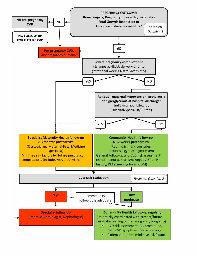
PREGNANCY OUTCOME: Preeclampsia, Pregnancy Induced Hypertension
Fetal Growth Restriction or Gestational diabetes mellitus?
Pre‐pregnancy CVD, Any pregnancy outcome
Severe pregnancy complication? (Eclampsia, HELLP, delivery prior to gestational week 34, fetal death etc.)
NO YES
Community Health follow‐up regularly (Potentially coordinated with present/future cervical screening or mammography programs)
• CVD risk assessment (BP, proteinuria, BMI, CVD symptoms, DM screening)
• Patient education, minimise risk factors
NO
Residual maternal hypertension, proteinuria or hyperglycemia at hospital discharge?
Individualized follow‐up (Hospital/Specialist/GP etc.)
YES
NO YES
Specialist follow‐up (Internist, Cardiologist, Nephrologist)
Specialist Maternity Health follow‐up 2‐3 months postpartum
(Obstetrician, Maternal‐Fetal Medicine specialist)
Minimise risk factors for future pregnancy complications (includes ASA prophylaxis)
Community Health follow‐up 6‐12 weeks postpartum
(Routine in many countries; includes a gynecological exam)
General Follow‐up and CVD risk assessment (BP, proteinuria, BMI, smoking, CVD family history, DM screening for all GDM)
Research Question 1
High Low/ moderate
CVD Risk Evaluation Research Question 2
If community follow‐up is adequate
No pre‐pregnancy CVD
NO FOLLOW‐UP FOR FUTURE CVD

Supplemental Figure S1. An example of follow-up flow chart after 3 categories of complicated pregnancies associated with increased risk for future cardiovascular disease (CVD). ASA: Acetylic Salicylic Acid (low-dose). HELLP: Hemolysis, elevated liver enzymes, low platelets syndrome. Italic Text- Remaining Research Questions:
1. Which biomarkers (before or during pregnancy) can best predict CVD risk in young women?
2. Which biomarkers (postpartum or any time point after pregnancy) can best predict CVD risk in young women?
Green colour: Community Health Level (e.g. General Practitioner: GP) Orange colour: Specialist Maternity Health Care (e.g. Obstetrician) Red colour: Specialist Medical Care (e.g. Internist, Cardiologist, Nephrologist)

Myatt, Lucilla Poston and James M. RobertsBasky Thilaganathan, Per Magnus, Eric A.P. Steegers, Eleni Z. Tsigas, Roberta B. Ness, Leslie Anne Cathrine Staff, Christopher W.G. Redman, David Williams, Paul Leeson, Kjartan Moe,
Harmonization of Research Cohorts and BiobanksPregnancy and Long-Term Maternal Cardiovascular Health: Progress Through
Print ISSN: 0194-911X. Online ISSN: 1524-4563 Copyright © 2015 American Heart Association, Inc. All rights reserved.
is published by the American Heart Association, 7272 Greenville Avenue, Dallas, TX 75231Hypertension published online December 14, 2015;Hypertension.
http://hyper.ahajournals.org/content/early/2015/12/14/HYPERTENSIONAHA.115.06357.citationWorld Wide Web at:
The online version of this article, along with updated information and services, is located on the
http://hyper.ahajournals.org/content/suppl/2015/12/14/HYPERTENSIONAHA.115.06357.DC1.htmlData Supplement (unedited) at:
http://hyper.ahajournals.org//subscriptions/
is online at: Hypertension Information about subscribing to Subscriptions:
http://www.lww.com/reprints Information about reprints can be found online at: Reprints:
document. Permissions and Rights Question and Answer this process is available in the
click Request Permissions in the middle column of the Web page under Services. Further information aboutOffice. Once the online version of the published article for which permission is being requested is located,
can be obtained via RightsLink, a service of the Copyright Clearance Center, not the EditorialHypertensionin Requests for permissions to reproduce figures, tables, or portions of articles originally publishedPermissions:
at MERCY HOSP OF PITTSBURGH on January 5, 2016http://hyper.ahajournals.org/Downloaded from