briefcommunications neuronalnitricoxidesynthase … ·...
TRANSCRIPT

Brief Communications
Neuronal Nitric Oxide Synthase-Dependent S-Nitrosylationof Gephyrin Regulates Gephyrin Clustering at GABAergicSynapses
Borislav Dejanovic1 and Guenter Schwarz1,2,3
1Department of Chemistry, Institute of Biochemistry, 2Cologne Excellence Cluster on Cellular Stress Responses in Aging-Associated Diseases, and 3Centerfor Molecular Medicine, University of Cologne, 50674 Cologne, Germany
Gephyrin, the principal scaffolding protein at inhibitory synapses, is essential for postsynaptic clustering of glycine and GABA type Areceptors (GABAARs). Gephyrin cluster formation, which determines the strength of GABAergic transmission, is modulated by interac-tion with signaling proteins and post-translational modifications. Here, we show that gephyrin was found to be associated with neuronalnitric oxide synthase (nNOS), the major source of the ubiquitous and important signaling molecule NO in brain. Furthermore, weidentified that gephyrin is S-nitrosylated in vivo. Overexpression of nNOS decreased the size of postsynaptic gephyrin clusters in primaryhippocampal neurons. Conversely, inhibition of nNOS resulted in a loss of S-nitrosylation of gephyrin and the formation of largergephyrin clusters at synaptic sites, ultimately increasing the number of cell surface expressed synaptic GABAARs. In conclusion,S-nitrosylation of gephyrin is important for homeostatic assembly and plasticity of GABAergic synapses.
Key words: clustering; GABAA receptor; gephyrin; nNOS; post-translational modification; S-nitrosylation
IntroductionInhibitory signaling in the brain primarily takes place at gamma-aminobutyric acid (GABA)-ergic synapses. Pentameric GABAtype A receptors (GABAARs) are ubiquitously expressed in neu-rons and clustered at postsynaptic sites. The number of postsyn-aptic receptors is dynamic and implicated in the regulation ofvirtually all aspects of brain function (Luscher et al., 2011).
A very crucial factor for clustering and stabilization of synap-tic GABAARs is gephyrin, the principal scaffolding protein atinhibitory postsynapses (Fritschy et al., 2008). Gephyrin directlyinteracts with synaptic GABAAR subunits and is essential for theirclustering and localization (Kneussel et al., 1999; Tretter et al.,2008, 2011; Mukherjee et al., 2011; Kowalczyk et al., 2013). Inaddition to its structural function, gephyrin fulfills importantroles in GABAAR dynamics and synaptic plasticity in vivo (Tya-garajan et al., 2011; Chen et al., 2012; van Versendaal et al., 2012).Gephyrin exerts its physiological functions by interacting with avariety of binding partners, ensuring their close spatial proximityat postsynaptic sites (Fritschy et al., 2008). Accordingly, gephyrin
malfunction and the resulting reduced strength of GABAergicsynaptic transmission is implicated in neurological disorders,such as epilepsy, autism, and schizophrenia (Lionel et al., 2013;Dejanovic et al., 2014). Thus, any signaling that modulatesGABAergic synapses is important for balancing neuronal net-works and synaptic plasticity.
Nitric oxide (NO) is a ubiquitous and important signalingmolecule regulating a wide range of cellular functions in differentorgans (Pacher et al., 2007). In the nervous system, NO has beenfound to modulate both excitation and inhibition. Although ithas been shown to be involved in long-term potentiation (Langeet al., 2012) other studies found a modulation of GABAergictransmission by NO (Li et al., 2004; Zanelli et al., 2009; Cserep etal., 2011). Given that NO is involved in depolarization-inducedsuppression of inhibition (Makara et al., 2007), NO might be animportant regulator of the excitatory/inhibitory balance in thebrain (Le Roux et al., 2009). At the inhibitory synapse, anatomicaland electrophysiological evidence suggest that NO is a retrogradesignaling molecule that can potentiate presynaptic GABA release(Saransaari and Oja, 2006; Szabadits et al., 2007; Zanelli et al.,2009). However, the function of NO at the postsynaptic site ismuch less explored.
In general, the main routes of action of NO include eitherthe production of cGMP and activation of downstream pathwaysor post-translational modification of cysteine residues in pro-teins by the formation of nitrosothiol groups, a process calledS-nitrosylation (Hardingham et al., 2013). Neuronal NO syn-thase (nNOS or NOS1), is the major source of NO in the brainand has been found at GABAergic postsynapses (Szabadits et al.,2007; Cserep et al., 2011). Despite the multiple lines of evidencethat nNOS-produced NO has an impact on GABAergic function,
Received Feb. 7, 2014; revised March 14, 2014; accepted April 16, 2014.Author contributions: G.S. and B.D. designed research; B.D. performed research; B.D. analyzed data; G.S. and B.D.
wrote the paper.This work was supported by the German Science foundation (DFG, SFB635: TPA11), the Center for Molec-
ular Medicine, and the Fonds der Chemischen Industrie. We thank Jean-Marc Fritschy (UZH, Zurich, Switzer-land) for providing GABAAR specific antibodies, and Joana Stegemann and Sabine Rugenberg for excellenttechnical assistance.
The authors declare no competing financial interests.Correspondence should be addressed to Guenter Schwarz, Institute of Biochemistry, University of Cologne, Zul-
picher Street 47, 50674 Cologne, Germany. E-mail: [email protected]:10.1523/JNEUROSCI.0531-14.2014
Copyright © 2014 the authors 0270-6474/14/347763-06$15.00/0
The Journal of Neuroscience, June 4, 2014 • 34(23):7763–7768 • 7763

neuronal proteins that associate with nNOS have not been iden-tified yet.
Here, we show that gephyrin interacts with nNOS and de-tected gephyrin-nNOS complexes in soma and dendrites of pri-mary hippocampal neurons. Additionally, we identified thatgephyrin is physiologically S-nitrosylated, which regulates thesizes of postsynaptic gephyrin clusters and hence modulates cellsurface expression of synaptic GABAARs.
Materials and MethodsExpression constructs and reagents. Enhanced green fluorescent protein-tagged gephyrin has been described previously (Lardi-Studler et al.,2007). nNOS was cloned in frame into the pcDNA3.1(�)myc/His Ausing KpnI and XbaI restriction sites. Where indicated, 100 �M N5-(1-Imino-3-butenyl)-L-ornithine (L-VNIO; from a 1000� stock in DMSO)was added to the growth medium for 14 h. SNOB1 reagent (1 mg/ml)dissolved in DMSO served as a 200� stock and was freshly diluted inprotein lysates.
Cell culture and transfection. HEK293 cells were cultured in DMEM(GE Healthcare) and 10% FCS supplemented with L-glutamine andtransfected with polyethylenimine using standard protocols. Primaryhippocampal neurons were cultured as previously described (Dejanovicet al., 2014). Neurons were usually transfected after 10 days in vitro (DIV)using Lipofectamine 2000 (Invitrogen) according to the manufacturer’smanual and cultured for additional 3 d.
Immunostaining and quantification of cultured cells. Immunostainingand laser scanning microscopy of cultured cells have been describedpreviously (Dejanovic et al., 2014). Antibodies used for the immuno-staining: mouse anti-gephyrin (1:50 cell culture supernatant, clone 3B11,RRID:AB_887719); mouse anti-myc (1:15 cell culture supernatant, clone9E10, RRID:AB_2266850); rabbit anti-vesicular GABA transporter(VGAT; 1:500, Synaptic Systems, AB_887871). Secondary antibodieswere goat raised AlexaFluor 488 and 568 antibodies (1:500, Invitrogen).Images were acquired as a z-stack with three optical sections of 0.5 �m indepth. Maximum intensity projections were created and analyzed usingNIS Elements 3.2 (Nikon) software. After background subtraction,gephyrin clusters and VGAT puncta were quantified within two 20 � 5�m regions-of-interest (ROI) per neuron using the analyze particles op-tion in NIS Elements setting the minimal-threshold of a cluster to �0.08�m 2. Cluster size was averaged per neuron and mean values were com-pared for significance using Student’s t test with the software SigmaPlot(Systat Software). Gephyrin localization in respect to VGAT was ana-lyzed by blinded manual counting of opposed clusters within two ROIper neuron. Images were processed with the software ImageJ (NIH).
Coimmunoprecipitation. Fresh mouse brains were cut into small cubesand homogenized using a Potter S (Sartorius) in lysis buffer (PBS, 0.5%Triton X-100, protease and phosphatase inhibitor cocktail, Roche). Thepostnuclear fraction was incubated with primary antibodies for 2 h atroom temperature before 20 �l of protein-A/G Sepharose beads (SantaCruz Biotechnology) were added for another 2 h. The beads were washedthree times with PBS. Adsorbed proteins were eluted from the beads byboiling in 50 �l SDS-loading buffer containing �-mercatoethanol. Im-munoprecipitated samples were subjected to SDS-PAGE followed byimmunoblotting. Primary antibodies used for immunoprecipitation:anti-nNOS (Santa Cruz Biotechnology, RRID:AB_630935) and anti-S-Nitroso-cysteine (Sigma-Aldrich, RRID:AB_260785).
In situ PLA. Proximity ligation assay (PLA) was performed on primaryhippocampal neurons cultured 14 DIV. The assay was performed usingthe Duolink system and was performed as described by the manufacturer(Olink). Omitting the rabbit-specific secondary probe served as an inter-nal control in the assay. The following antibodies have been used: mouseanti-gephyrin (clone 3B11, RRID:AB_887719), mouse anti-neuroligin 2(Synaptic Systems, 5E6), rabbit anti-nNOS (Santa Cruz Biotechnology,RRID:AB_630935).
Surface biotinylation. Two 6-well dishes with 600,000 neurons cultured14 DIV were used per condition. Neurons were preincubated with 100�M L-VNIO or the solvent for 14 h. Neurons were rinsed twice withice-cold PBS supplemented with 0.5 mM MgCl2 and 1 mM CaCl2. Surface
proteins were biotinylated in a 20 min incubation step at 4°C in 1 ml PBSsupplemented with 1 mg/ml sulfo-NHS-SS-biotin (Thermo Scientific).Unreacted biotin reagent was quenched by washing with 100 mM glycinedissolved in supplemented PBS buffer. Cells were lysed in 500 �l PBScontaining 2% Triton X-100, 0.2% SDS and protease inhibitor cocktail(Roche). After a brief sonication, cells were incubated for 1 h on ice andsubjected to centrifugation (10,000 � g for 2 min). From this, 50 �l weresaved as loading control and supernatants were incubated with Neutra-vidin beads (Thermo Scientific) for 2 h at room temperature or overnightat 4°C.
Biotin switch assay. Cell homogenates were extracted with 2%SDS-buffer (2% SDS, 50 mM Tris-HCl, pH 7.4, 5 mM EDTA, 0.1 mM
neocuproine) and briefly sonicated. Subsequently, 20 mM methylmeth-anethiosulfonate was added to the lysate and rotated head-over-tail for2 h at room temperature. Proteins were precipitated with acetone/meth-anol/H2O (ration 4:1.5:3) three times and 50 mM ascorbate was used toliberate the nitrosylated cysteine residues for 2 h at room temperature.Instead of ascorbate, 50 mM Tris was used as an internal control. Cysteineresidues were labeled with 0.8 mM HPDP-biotin (Pierce) and affinity-purified with Neutravidin-beads (Pierce). Proteins were eluted from thebeads with SDS-loading buffer and boiled for 5 min.
Western blot analysis and antibodies. For immunoblotting, a standardprotocol was followed and detection was performed using chemilumi-nescence and an ECL system with a cooled CCD camera (Decon ScienceTec). The following primary antibodies were used and diluted in Tris-buffered saline/0.5% Tween containing 1% dry milk: anti-gephyrin(3B11, RRID:AB_887719), anti-nNOS (Cell Signaling Technology,C7D7, RRID:AB_2152485), anti-GFP (Santa Cruz Biotechnology,AB_641123), anti-myc (RRID:AB_2266850), anti-GABAAR �1 (giftfrom from Jean-Marc Fritschy, UTH Zurich), anti-GABAAR �2/3 (Mil-lipore, BD17, RRID:AB_2109419), and GABAAR �2 (Synaptic Systems,RRID:AB_2263066).
ResultsnNOS interacts with gephyrinGiven the recently identified impact of nNOS-derived NO onGABAergic sites (Zanelli et al., 2009; Cserep et al., 2011), wewondered whether gephyrin, as major postsynaptic scaffoldingprotein, interacts with nNOS. Following immunoprecipitationswith mouse brain lysates, nNOS specifically coimmunoprecipi-tated gephyrin, suggesting that both proteins are associatedwithin the brain (Fig. 1A). To assess whether nNOS and gephyrincolocalize in cells, we coexpressed GFP-tagged gephyrin andmyc-tagged nNOS in HEK293 cells and neurons. Upon heterol-ogous expression in non-neuronal mammalian cells, gephyrinforms large cytosolic clusters, so-called “blobs” (Lardi-Studler etal., 2007), whereas nNOS was diffusively distributed (Fig. 1B�).When coexpressed, a fraction of nNOS was enriched atgephyrin-blobs, whereas the major part of nNOS remaineddiffuse within the cell (Fig. 1B�). Likewise, nNOS-myc colocal-ized with gephyrin-GFP clusters in primary hippocampal neu-rons cultured 10 � 3 DIV (Fig. 1C). Together, these resultssuggest that gephyrin and nNOS form a complex, whereas theinteraction between the proteins seems rather weak.
Gephyrin presents a modular structure comprising three do-mains: the N-terminal G-domain, C-terminal E-domain, and thecentral C-domain (Fritschy et al., 2008). To identify the domainessential for nNOS interaction, we coexpressed GFP-taggedgephyrin domain variants with nNOS-myc in HEK293 cells.Upon coimmunoprecipitation with nNOS-myc, we could iden-tify the E-domain of gephyrin as potential site of interaction (Fig.1D). To probe whether also endogenous gephyrin and nNOSform a complex in primary hippocampal neurons, we performedan in situ PLA that enables detections of protein-protein interac-tions at single molecular resolution. PLA signals were detected inthe soma and along the dendrites in neurons, confirming the
7764 • J. Neurosci., June 4, 2014 • 34(23):7763–7768 Dejanovic and Schwarz • Gephyrin S-Nitrosylation

complex formation of gephyrin and nNOS (Fig. 1E). Only spo-radic signals were detected for nNOS and the gephyrin-interacting cell adhesion molecule neuroligin 2 (Poulopoulos etal., 2009), whereas no signals were detected without the rabbitspecific secondary probe, both independently proving the speci-ficity of the assay.
Gephyrin is S-nitrosylated in vivoGiven the identified spatial proximity to nNOS, we wonderedwhether gephyrin might be a target for S-nitrosylation by nNOS-produced NO. Therefore, we monitored S-nitrosylation ofendogenous gephyrin in mouse brain lysates using the biotin-switchassay, which relies on the chemical exchange of nitrosylated cysteinesto biotinylated cysteines in an ascorbate-dependent reaction. Weobserved a clear S-nitrosylation signal for gephyrin in brain-extract(Fig. 2A), similarly to the S-nitrosylation of the glutamatergic scaf-folding protein PSD-95 that has been described recently (Ho et al.,2011). In the absence of ascorbate, no signal was detected. Next, we
could also immunoprecipitate gephyrinfrom mouse brain lysates with an S-nitroso-cysteine (SNO-cysteine) specific antibody(Fig. 2B). In a third approach, gephyrin wasaffinity-purified with Neutravidin beadsafter labeling of S-nitrosylated proteinsin brain lysates with the biotin moiety-carrying SNO-binding reagent 1 (SNOB 1;Fig. 2C). Next, we incubated primary hip-pocampal neurons with the nNOS-selectiveinhibitor L-VNIO and, performing a biotin-switch assay, found significantly decreasedlevels of S-nitrosylated gephyrin comparedwith DMSO-treated neurons (Fig. 2D). Inaggregate, these data demonstrate thatgephyrin is physiologically S-nitrosylated inthe brain.
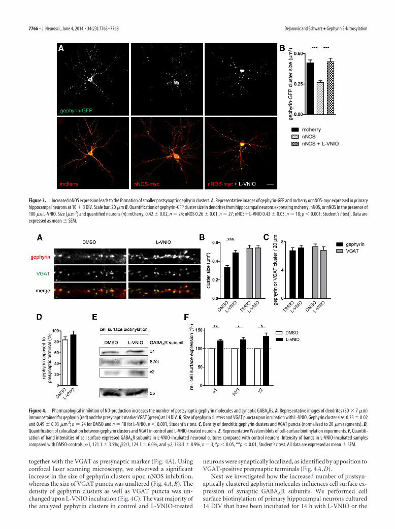
nNOS produced NO decreases the sizeof gephyrin clusters in neuronsWe then asked whether nNOS derived
NO influences postsynaptic clustering of gephyrin. Therefore, wetransiently expressed gephyrin-GFP and nNOS-myc or mCherryin primary hippocampal neurons cultured 10 � 3 DIV and sub-sequently analyzed gephyrin cluster size along dendrites usingconfocal laser scanning microscopy (Fig. 3A). We observed asignificant decrease of gephyrin-GFP cluster area upon coexpres-sion of nNOS as compared with control neurons coexpressingmCherry (Fig. 3B). Interestingly, inhibition of nNOS by applyingL-VNIO for 14 h resulted in gephyrin-GFP cluster sizes compa-rable to control neurons, thus reverting the effect of recombinantnNOS expression. These findings suggest that nNOS-producedNO controls the size of gephyrin clusters.
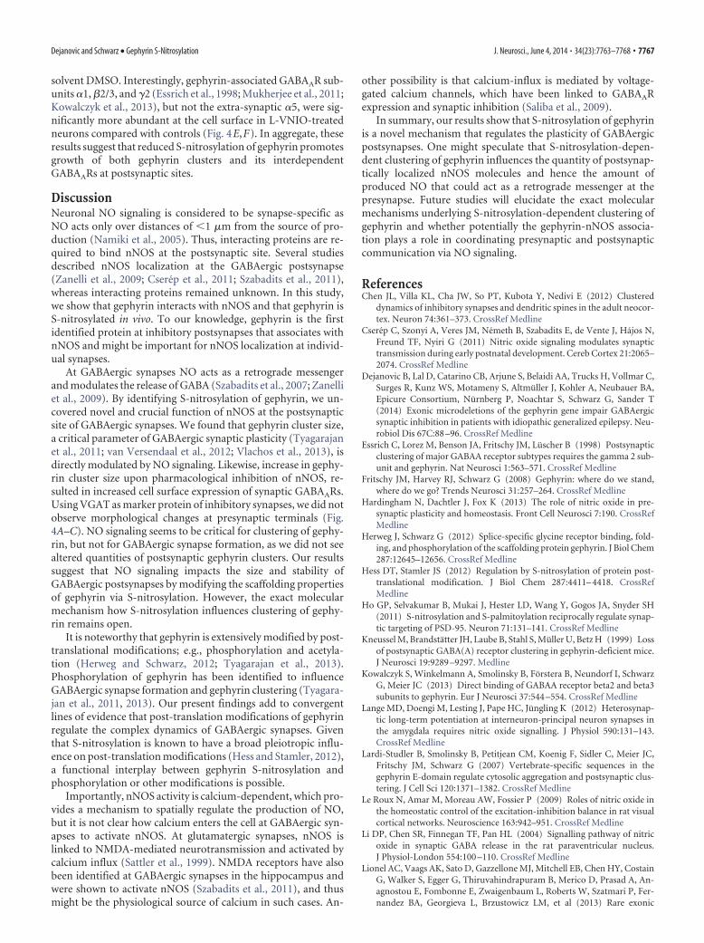
Inhibition of nNOS activity increases gephyrin cluster sizeand cell-surface expression of synaptic GABAARsTo assess whether NO, produced by endogenous nNOS, regulatesclustering of endogenous gephyrin, we incubated primary hip-pocampal neurons with L-VNIO and immunostained gephyrin
Figure 1. Gephyrin interacts with nNOS. A, Coimmunoprecipitation (IP) from brain lysates with nNOS specific or nonspecific control antibodies (control IgG). B�, GFP-tagged gephyrin forms largeintracellular protein aggregates blobs upon expression in HEK293 cells, whereas myc-tagged nNOS is diffusively distributed. There is no bleed-through of the channels. B�, Upon coexpression, afraction of nNOS accumulates with gephyrin blobs (arrows), whereas other blobs show no increased nNOS-immunoreactivity (arrowheads).C, Coexpression of gephyrin-GFP and nNOS-myc inprimary hippocampal neurons at 10 � 3 DIV. Arrows point to colocalized immunoreactivity. D, Coimmunoprecipitation from HEK lysates expressing nNOS-myc and GFP-tagged gephyrin domains(G-, GC-, and E-domain) as well as holo-gephyrin (Geph). E, In situ PLA using antibody-pairs as indicated in red. Scale bars: B�, C, 5 �m; E, 10 �m.
Figure 2. Gephyrin is S-nitrosylated in vivo. A, S-nitrosylation of gephyrin in mouse brain lysate demonstrated by the biotin-switch assay. Ascorbate dependence demonstrates specificity of the signal. B, Immunoprecipitation of gephyrin from brain lysateswith S-Nitroso (SNO)-cysteine specific antibodies. C, Affinity-purification of gephyrin from brain lysates using the SNO-binding(SNOB1) reagent. D, S-nitrosylation of gephyrin in primary hippocampal neurons following the incubation with DMSO or thenNOS-specific inhibitor L-VNIO monitored by the biotin-switch assay.
Dejanovic and Schwarz • Gephyrin S-Nitrosylation J. Neurosci., June 4, 2014 • 34(23):7763–7768 • 7765
together with the VGAT as presynaptic marker (Fig. 4A). Usingconfocal laser scanning microscopy, we observed a significantincrease in the size of gephyrin clusters upon nNOS inhibition,whereas the size of VGAT puncta was unaltered (Fig. 4A,B). Thedensity of gephyrin clusters as well as VGAT puncta was un-changed upon L-VNIO incubation (Fig. 4C). The vast majority ofthe analyzed gephyrin clusters in control and L-VNIO-treated
neurons were synaptically localized, as identified by apposition toVGAT-positive presynaptic terminals (Fig. 4A,D).
Next we investigated how the increased number of postsyn-aptically clustered gephyrin molecules influences cell surface ex-pression of synaptic GABAAR subunits. We performed cellsurface biotinylation of primary hippocampal neurons cultured14 DIV that have been incubated for 14 h with L-VNIO or the
Figure 3. Increased nNOS expression leads to the formation of smaller postsynaptic gephyrin clusters. A, Representative images of gephyrin-GFP and mcherry or nNOS-myc expressed in primaryhippocampal neurons at 10 � 3 DIV. Scale bar, 20 �m B, Quantification of gephyrin-GFP cluster size in dendrites from hippocampal neurons expressing mcherry, nNOS, or nNOS in the presence of100 �M L-VNIO. Size (�m 2) and quantified neurons (n): mCherry, 0.42 � 0.02, n � 24; nNOS 0.26 � 0.01, n � 27; nNOS�L-VNIO 0.43 � 0.03, n � 18; p 0.001; Student’s t test). Data areexpressed as mean � SEM.
Figure 4. Pharmacological inhibition of NO-production increases the number of postsynaptic gephyrin molecules and synaptic GABAARs. A, Representative images of dendrites (30 � 7 �m)immunostained for gephyrin (red) and the presynaptic marker VGAT (green) at 14 DIV. B, Size of gephyrin clusters and VGAT puncta upon incubation with L-VNIO. Gephyrin cluster size: 0.33 � 0.02and 0.49 � 0.03 �m 2; n � 24 for DMSO and n � 18 for L-VNIO, p 0.001, Student’s t test. C, Density of dendritic gephyrin clusters and VGAT puncta (normalized to 20 �m segments). D,Quantification of colocalization between gephyrin clusters and VGAT in control and L-VNIO-treated neurons. E, Representative Western blots of cell-surface biotinylation experiments. F, Quantifi-cation of band intensities of cell surface expressed GABAAR subunits in L-VNIO-incubated neuronal cultures compared with control neurons. Intensity of bands in L-VNIO-incubated samplescompared with DMSO-controls: �1, 121.1 � 3.5%; �2/3, 124.1 � 6.0%, and �2, 133.3 � 8.9%; n � 3, *p 0.05, **p 0.01, Student’s t test. All data are expressed as mean � SEM.
7766 • J. Neurosci., June 4, 2014 • 34(23):7763–7768 Dejanovic and Schwarz • Gephyrin S-Nitrosylation
solvent DMSO. Interestingly, gephyrin-associated GABAAR sub-units �1, �2/3, and �2 (Essrich et al., 1998; Mukherjee et al., 2011;Kowalczyk et al., 2013), but not the extra-synaptic �5, were sig-nificantly more abundant at the cell surface in L-VNIO-treatedneurons compared with controls (Fig. 4E,F). In aggregate, theseresults suggest that reduced S-nitrosylation of gephyrin promotesgrowth of both gephyrin clusters and its interdependentGABAARs at postsynaptic sites.
DiscussionNeuronal NO signaling is considered to be synapse-specific asNO acts only over distances of 1 �m from the source of pro-duction (Namiki et al., 2005). Thus, interacting proteins are re-quired to bind nNOS at the postsynaptic site. Several studiesdescribed nNOS localization at the GABAergic postsynapse(Zanelli et al., 2009; Cserep et al., 2011; Szabadits et al., 2011),whereas interacting proteins remained unknown. In this study,we show that gephyrin interacts with nNOS and that gephyrin isS-nitrosylated in vivo. To our knowledge, gephyrin is the firstidentified protein at inhibitory postsynapses that associates withnNOS and might be important for nNOS localization at individ-ual synapses.
At GABAergic synapses NO acts as a retrograde messengerand modulates the release of GABA (Szabadits et al., 2007; Zanelliet al., 2009). By identifying S-nitrosylation of gephyrin, we un-covered novel and crucial function of nNOS at the postsynapticsite of GABAergic synapses. We found that gephyrin cluster size,a critical parameter of GABAergic synaptic plasticity (Tyagarajanet al., 2011; van Versendaal et al., 2012; Vlachos et al., 2013), isdirectly modulated by NO signaling. Likewise, increase in gephy-rin cluster size upon pharmacological inhibition of nNOS, re-sulted in increased cell surface expression of synaptic GABAARs.Using VGAT as marker protein of inhibitory synapses, we did notobserve morphological changes at presynaptic terminals (Fig.4A–C). NO signaling seems to be critical for clustering of gephy-rin, but not for GABAergic synapse formation, as we did not seealtered quantities of postsynaptic gephyrin clusters. Our resultssuggest that NO signaling impacts the size and stability ofGABAergic postsynapses by modifying the scaffolding propertiesof gephyrin via S-nitrosylation. However, the exact molecularmechanism how S-nitrosylation influences clustering of gephy-rin remains open.
It is noteworthy that gephyrin is extensively modified by post-translational modifications; e.g., phosphorylation and acetyla-tion (Herweg and Schwarz, 2012; Tyagarajan et al., 2013).Phosphorylation of gephyrin has been identified to influenceGABAergic synapse formation and gephyrin clustering (Tyagara-jan et al., 2011, 2013). Our present findings add to convergentlines of evidence that post-translation modifications of gephyrinregulate the complex dynamics of GABAergic synapses. Giventhat S-nitrosylation is known to have a broad pleiotropic influ-ence on post-translation modifications (Hess and Stamler, 2012),a functional interplay between gephyrin S-nitrosylation andphosphorylation or other modifications is possible.
Importantly, nNOS activity is calcium-dependent, which pro-vides a mechanism to spatially regulate the production of NO,but it is not clear how calcium enters the cell at GABAergic syn-apses to activate nNOS. At glutamatergic synapses, nNOS islinked to NMDA-mediated neurotransmission and activated bycalcium influx (Sattler et al., 1999). NMDA receptors have alsobeen identified at GABAergic synapses in the hippocampus andwere shown to activate nNOS (Szabadits et al., 2011), and thusmight be the physiological source of calcium in such cases. An-
other possibility is that calcium-influx is mediated by voltage-gated calcium channels, which have been linked to GABAARexpression and synaptic inhibition (Saliba et al., 2009).
In summary, our results show that S-nitrosylation of gephyrinis a novel mechanism that regulates the plasticity of GABAergicpostsynapses. One might speculate that S-nitrosylation-depen-dent clustering of gephyrin influences the quantity of postsynap-tically localized nNOS molecules and hence the amount ofproduced NO that could act as a retrograde messenger at thepresynapse. Future studies will elucidate the exact molecularmechanisms underlying S-nitrosylation-dependent clustering ofgephyrin and whether potentially the gephyrin-nNOS associa-tion plays a role in coordinating presynaptic and postsynapticcommunication via NO signaling.
ReferencesChen JL, Villa KL, Cha JW, So PT, Kubota Y, Nedivi E (2012) Clustered
dynamics of inhibitory synapses and dendritic spines in the adult neocor-tex. Neuron 74:361–373. CrossRef Medline
Cserep C, Szonyi A, Veres JM, Nemeth B, Szabadits E, de Vente J, Hajos N,Freund TF, Nyiri G (2011) Nitric oxide signaling modulates synaptictransmission during early postnatal development. Cereb Cortex 21:2065–2074. CrossRef Medline
Dejanovic B, Lal D, Catarino CB, Arjune S, Belaidi AA, Trucks H, Vollmar C,Surges R, Kunz WS, Motameny S, Altmuller J, Kohler A, Neubauer BA,Epicure Consortium, Nurnberg P, Noachtar S, Schwarz G, Sander T(2014) Exonic microdeletions of the gephyrin gene impair GABAergicsynaptic inhibition in patients with idiopathic generalized epilepsy. Neu-robiol Dis 67C:88 –96. CrossRef Medline
Essrich C, Lorez M, Benson JA, Fritschy JM, Luscher B (1998) Postsynapticclustering of major GABAA receptor subtypes requires the gamma 2 sub-unit and gephyrin. Nat Neurosci 1:563–571. CrossRef Medline
Fritschy JM, Harvey RJ, Schwarz G (2008) Gephyrin: where do we stand,where do we go? Trends Neurosci 31:257–264. CrossRef Medline
Hardingham N, Dachtler J, Fox K (2013) The role of nitric oxide in pre-synaptic plasticity and homeostasis. Front Cell Neurosci 7:190. CrossRefMedline
Herweg J, Schwarz G (2012) Splice-specific glycine receptor binding, fold-ing, and phosphorylation of the scaffolding protein gephyrin. J Biol Chem287:12645–12656. CrossRef Medline
Hess DT, Stamler JS (2012) Regulation by S-nitrosylation of protein post-translational modification. J Biol Chem 287:4411– 4418. CrossRefMedline
Ho GP, Selvakumar B, Mukai J, Hester LD, Wang Y, Gogos JA, Snyder SH(2011) S-nitrosylation and S-palmitoylation reciprocally regulate synap-tic targeting of PSD-95. Neuron 71:131–141. CrossRef Medline
Kneussel M, Brandstatter JH, Laube B, Stahl S, Muller U, Betz H (1999) Lossof postsynaptic GABA(A) receptor clustering in gephyrin-deficient mice.J Neurosci 19:9289 –9297. Medline
Kowalczyk S, Winkelmann A, Smolinsky B, Forstera B, Neundorf I, SchwarzG, Meier JC (2013) Direct binding of GABAA receptor beta2 and beta3subunits to gephyrin. Eur J Neurosci 37:544 –554. CrossRef Medline
Lange MD, Doengi M, Lesting J, Pape HC, Jungling K (2012) Heterosynap-tic long-term potentiation at interneuron-principal neuron synapses inthe amygdala requires nitric oxide signalling. J Physiol 590:131–143.CrossRef Medline
Lardi-Studler B, Smolinsky B, Petitjean CM, Koenig F, Sidler C, Meier JC,Fritschy JM, Schwarz G (2007) Vertebrate-specific sequences in thegephyrin E-domain regulate cytosolic aggregation and postsynaptic clus-tering. J Cell Sci 120:1371–1382. CrossRef Medline
Le Roux N, Amar M, Moreau AW, Fossier P (2009) Roles of nitric oxide inthe homeostatic control of the excitation-inhibition balance in rat visualcortical networks. Neuroscience 163:942–951. CrossRef Medline
Li DP, Chen SR, Finnegan TF, Pan HL (2004) Signalling pathway of nitricoxide in synaptic GABA release in the rat paraventricular nucleus.J Physiol-London 554:100 –110. CrossRef Medline
Lionel AC, Vaags AK, Sato D, Gazzellone MJ, Mitchell EB, Chen HY, CostainG, Walker S, Egger G, Thiruvahindrapuram B, Merico D, Prasad A, An-agnostou E, Fombonne E, Zwaigenbaum L, Roberts W, Szatmari P, Fer-nandez BA, Georgieva L, Brzustowicz LM, et al (2013) Rare exonic
Dejanovic and Schwarz • Gephyrin S-Nitrosylation J. Neurosci., June 4, 2014 • 34(23):7763–7768 • 7767
deletions implicate the synaptic organizer gephyrin (GPHN) in risk forautism, schizophrenia and seizures. Human molecular genetics 22:2055–2066. CrossRef Medline
Luscher B, Fuchs T, Kilpatrick CL (2011) GABAA receptor trafficking-mediated plasticity of inhibitory synapses. Neuron 70:385– 409. CrossRefMedline
Makara JK, Katona I, Nyíri G, Nemeth B, Ledent C, Watanabe M, de Vente J,Freund TF, Hajos N (2007) Involvement of nitric oxide in depo-larization-induced suppression of inhibition in hippocampal pyramidalcells during activation of cholinergic receptors. J Neurosci 27:10211–10222. CrossRef Medline
Mukherjee J, Kretschmannova K, Gouzer G, Maric HM, Ramsden S, TretterV, Harvey K, Davies PA, Triller A, Schindelin H, Moss SJ (2011) Theresidence time of GABA(A)Rs at inhibitory synapses is determined bydirect binding of the receptor alpha1 subunit to gephyrin. J Neurosci31:14677–14687. CrossRef Medline
Namiki S, Kakizawa S, Hirose K, Iino M (2005) NO signalling decodes fre-quency of neuronal activity and generates synapse-specific plasticity inmouse cerebellum. J Physiol-London 566:849 – 863. CrossRef Medline
Pacher P, Beckman JS, Liaudet L (2007) Nitric oxide and peroxynitrite inhealth and disease. Physiol Rev 87:315– 424. CrossRef Medline
Poulopoulos A, Aramuni G, Meyer G, Soykan T, Hoon M, Papadopoulos T,Zhang M, Paarmann I, Fuchs C, Harvey K, Jedlicka P, Schwarzacher SW,Betz H, Harvey RJ, Brose N, Zhang W, Varoqueaux F (2009) Neuroligin2 drives postsynaptic assembly at perisomatic inhibitory synapses throughgephyrin and collybistin. Neuron 63:628 – 642. CrossRef Medline
Saliba RS, Gu Z, Yan Z, Moss SJ (2009) Blocking L-type voltage-gated Ca2�channels with dihydropyridines reduces gamma-aminobutyric acid typeA receptor expression and synaptic inhibition. J Biol Chem 284:32544 –32550. CrossRef Medline
Saransaari P, Oja SS (2006) Modulation of GABA release by second messen-ger substances and NO in mouse brain stem slices under normal andischemic conditions. Neurochem Res 31:1317–1325. CrossRef Medline
Sattler R, Xiong Z, Lu WY, Hafner M, MacDonald JF, Tymianski M (1999)Specific coupling of NMDA receptor activation to nitric oxide neurotox-icity by PSD-95 protein. Science 284:1845–1848. CrossRef Medline
Szabadits E, Cserep C, Ludanyi A, Katona I, Gracia-Llanes J, Freund TF, Nyíri
G (2007) Hippocampal GABAergic synapses possess the molecular ma-chinery for retrograde nitric oxide signaling. J Neurosci 27:8101– 8111.CrossRef Medline
Szabadits E, Cserep C, Szonyi A, Fukazawa Y, Shigemoto R, Watanabe M,Itohara S, Freund TF, Nyiri G (2011) NMDA receptors in hippocampalGABAergic synapses and their role in nitric oxide signaling. J Neurosci31:5893–5904. CrossRef Medline
Tretter V, Jacob TC, Mukherjee J, Fritschy JM, Pangalos MN, Moss SJ (2008)The clustering of GABA(A) receptor subtypes at inhibitory synapses isfacilitated via the direct binding of receptor alpha 2 subunits to gephyrin.J Neurosci 28:1356 –1365. CrossRef Medline
Tretter V, Kerschner B, Milenkovic I, Ramsden SL, Ramerstorfer J, SaiepourL, Maric HM, Moss SJ, Schindelin H, Harvey RJ, Sieghart W, Harvey K(2011) Molecular basis of the gamma-aminobutyric acid A receptor al-pha3 subunit interaction with the clustering protein gephyrin. J BiolChem 286:37702–37711. CrossRef Medline
Tyagarajan SK, Ghosh H, Yevenes GE, Nikonenko I, Ebeling C, Schwerdel C,Sidler C, Zeilhofer HU, Gerrits B, Muller D, Fritschy JM (2011) Regula-tion of GABAergic synapse formation and plasticity by GSK3beta-dependent phosphorylation of gephyrin. Proc Natl Acad Sci U S A 108:379 –384. CrossRef Medline
Tyagarajan SK, Ghosh H, Yevenes GE, Imanishi SY, Zeilhofer HU, Gerrits B,Fritschy JM (2013) Extracellular signal-regulated kinase and glycogensynthase kinase 3beta regulate gephyrin postsynaptic aggregation andGABAergic synaptic function in a calpain-dependent mechanism. J BiolChem 288:9634 –9647. CrossRef Medline
van Versendaal D, Rajendran R, Saiepour MH, Klooster J, Smit-Rigter L,Sommeijer JP, De Zeeuw CI, Hofer SB, Heimel JA, Levelt CN (2012)Elimination of inhibitory synapses is a major component of adult oculardominance plasticity. Neuron 74:374 –383. CrossRef Medline
Vlachos A, Reddy-Alla S, Papadopoulos T, Deller T, Betz H (2013) Homeo-static regulation of gephyrin scaffolds and synaptic strength at maturehippocampal GABAergic postsynapses. Cereb Cortex 23:2700 –2711.CrossRef Medline
Zanelli S, Naylor M, Kapur J (2009) Nitric oxide alters GABAergic synaptictransmission in cultured hippocampal neurons. Brain Res 1297:23–31.CrossRef Medline
7768 • J. Neurosci., June 4, 2014 • 34(23):7763–7768 Dejanovic and Schwarz • Gephyrin S-Nitrosylation