british thoracic society guideline for diagnostic exible...
TRANSCRIPT
British Thoracic Society guideline for diagnosticflexible bronchoscopy in adultsI A Du Rand,1 J Blaikley,2 R Booton,3 N Chaudhuri,4 V Gupta,2 S Khalid,5 S Mandal,6
J Martin,4 J Mills,7 N Navani,8 N M Rahman,9 J M Wrightson,9 M Munavvar,7
on behalf of the British Thoracic Society Bronchoscopy Guideline Group
▸ Additional material ispublished online only. To viewplease visit the journal online(http://dx.doi.org/10.1136/thoraxjnl-2013-203618).1Worcestershire Royal Hospital,Worcestershire Acute HospitalsNHS Trust, Worcester, UK2The University of Manchester,Manchester, UK3The University of Manchester,Manchester Academic HealthScience Centre, UniversityHospital South ManchesterNHS Foundation Trust,Manchester, UK4University Hospital of SouthManchester, Manchester, UK5Royal Blackburn Hospital,Lancashire, UK6Lane Fox Unit, St Thomas’Hospital, London, UK7Lancashire Teaching HospitalsNHS Trust, Preston, UK8University College LondonHospital and MRC ClinicalTrials Unit, National Institutefor Health Research UniversityCollege London HospitalsBiomedical Research Centre,London, UK9Oxford Centre for RespiratoryMedicine, NIHR OxfordBiomedical Research Centre,Oxford Respiratory Trials Unit,University of Oxford, Oxford,UK
Correspondence toDr Ingrid Du Rand,Worcestershire Royal Hospital,Aconbury East, CharlesHastings Way, Worcester,WR5 1DD, UK;[email protected]
To cite: Du Rand IA,Blaikley J, Booton R, et al.Thorax 2013;68:i1–i44.
SUMMARY OF RECOMMENDATIONSMonitoring, precautions and complications▸ All patients undergoing bronchoscopy should have
heart rate, respiratory rate, blood pressure andoxygen saturation recorded repeatedly, includingbefore, during and after the procedure. (Grade D)
▸ All bronchoscopy units should undertake peri-odic audit of bronchoscopic performance,including efficacy, complications and patient sat-isfaction surveys. (Good practice point (√))
▸ All Trusts should have a ‘safe sedation policy’,and ensure all bronchoscopy unit staff, includingtrainees, receive appropriate training. (√)
Hypoxaemia▸ Patients should be monitored by continuous
pulse oximetry during bronchoscopy. (Grade C)▸ Oxygen supplementation should be used when
desaturation is significant (pulse oximeter oxygensaturation (SpO2)>4% change, or SpO2<90%)and prolonged (>1 min) to reduce the risk ofhypoxaemia-related complications. (Grade D)
▸ The risks of hypoxaemia-related complicationsare associated with baseline arterial oxygen sat-uration (SaO2) and lung function, comorbidity,sedation and procedural sampling. Fitness forbronchoscopy should incorporate an assessmentof these elements, and appropriate monitoringand preprocedure optimisation. (Grade D)
Cardiac arrhythmias▸ Continuous ECG monitoring should be used
when there is a high clinical risk of arrhythmia.(Grade D)
▸ When there is a high risk of arrhythmia, oxygensaturations, pulse rate and blood pressure shouldbe optimised. Appropriate aftercare monitoringand instructions should be given. (Grade D)
▸ Resuscitation equipment should be readily avail-able. (√)
▸ Intravenous access should be established beforesedation is given and maintained until discharge.(√)
Bleeding complications▸ Perform coagulation studies, platelet count and
haemoglobin concentration when there are clinicalrisk factors for abnormal coagulation. (Grade D)
▸ Bronchoscopy with lavage can be performed withplatelet counts >20 000 per μL. Liaise with thelocal haematology team regarding the need forplatelet transfusion before bronchoscopy if
endobronchial biopsy (EBB) or transbronchial lungbiopsy (TBLB) is planned. (Grade D)
▸ Discontinue clopidogrel 7 days prior to consid-eration of EBB and TBLB. Low-dose aspirinalone can be continued. (Grade C)
▸ Anticoagulants should be managed accordingto published guidelines as set out in appendix 7of this guideline. (√)
▸ The risk of biopsy needs to be weighed againstthe potential for benefit and appropriateinformed consent obtained. (√)
Pneumothorax▸ A chest radiograph should be obtained if a
patient is symptomatic or there is a clinical sus-picion of possible pneumothorax after TBLB.(Grade D)
▸ Fluoroscopic screening may improve diagnosticyield of TBLB in focal but not diffuse lungdisease. (Grade D)
▸ Patients should be advised of the potential fordelayed complications following TBLB and pro-vided with written information regarding likelysymptoms and action required. (Grade D)
Fever and infection▸ Patients should receive written information
regarding post-bronchoscopy fever (PBF) andappropriate management advice. (Grade C)
▸ Antibiotic prophylaxis is not warranted beforebronchoscopy for the prevention of endocardi-tis, fever or pneumonia. (Grade B)
SAFETY OF FLEXIBLE BRONCHOSCOPY INSPECIFIC MEDICAL CONDITIONSAsthma▸ Patients’ asthma control should be optimised prior
to bronchoscopy, especially when bronchoalveolarlavage (BAL) is likely to be performed. (Grade C)
▸ Nebulised bronchodilators should be consideredbefore bronchoscopy in patients with asthma.(√)
Chronic obstructive pulmonary disease▸ Chronic obstructive pulmonary disease (COPD)
treatment should be optimised prior to bron-choscopy when possible. (Grade D)
▸ Bronchoscopists should be cautious when sedat-ing patients with COPD. (Grade D)
Ischaemic heart disease▸ Liaison with cardiologists should be considered
in high-risk patients with cardiac disease and if
Du Rand IA, et al. Thorax 2013;68:i1–i44. doi:10.1136/thoraxjnl-2013-203618 i1
BTS guidelines
on 26 May 2018 by guest. P
rotected by copyright.http://thorax.bm
j.com/
Thorax: first published as 10.1136/thoraxjnl-2013-203618 on 16 July 2013. D
ownloaded from
flexible bronchoscopy (FB) is indicated within 4–6 weeksafter myocardial infarction (MI). (Grade D)
▸ FB should ideally be delayed for 4 weeks after MI. (Grade D)
Haemoptysis▸ Consider bronchoscopy after a normal CT if the patient is
high risk for lung carcinoma or if the haemoptysis continues.(Grade D)
Older patients▸ Age alone should not be a contraindication for bronchos-
copy. (Grade D)▸ The older patient may require reduced doses of benzodiaze-
pines/opioids sedation. (Grade D)
Patients who are immunosuppressed▸ When a diagnosis is not likely to be obtained through non-
invasive measures, bronchoscopy with BAL can be consid-ered to provide diagnostic information. (Grade C)
▸ TBLB is helpful in lung transplant recipients when rejectionis a possibility. (Grade C)
SEDATIONPremedication▸ Anticholinergics (glycopyrrolate or atropine) should not rou-
tinely be used prior to bronchoscopy due to a lack of clinicalbenefit and a possible increased risk of haemodynamicchanges. (Grade A)
▸ Premedication for bronchoscopy is not routinely indicated.(Grade C)
Sedation▸ Intravenous sedation should be offered to patients undergo-
ing bronchoscopy, provided there are no contraindications.(Grade B)
▸ Some patients will tolerate unsedated bronchoscopy well,and patient preference should be sought. (Grade B)
▸ Sedative drugs should be titrated to provide the desireddepth of sedation, given significant inter-patient variability inrequired doses. (Grade B)
▸ The desired depth of sedation is one in which verbal contactis possible at all times. (Grade D)
▸ Bronchoscopists are encouraged to document an assessmentof sedation depth as part of the procedural report. (√)
Benzodiazepines▸ Intravenous midazolam is the preferred drug for sedation,
having a rapid onset of action, being titratable to provide therequired depth of sedation, and being reversible. (Grade B)
▸ No more than 5 mg midazolam should be initially drawn upinto any syringe prior to bronchoscopy for patients under theage of 70 (2 mg midazolam for patients over 70) to preventpotential inadvertent oversedation associated with the practiceof routinely drawing up 10 mg midazolam. (Grade D)
▸ Only low-strength midazolam (1 mg/mL) should be availablewithin bronchoscopy suites. High-strength midazolam (2 mg/mL or 5 mg/mL) should be restricted to general anaesthesia,intensive care and other areas where its use has been for-mally risk assessed. (Grade D)
Propofol▸ While propofol has similar efficacy to midazolam, it should
only be used when administered by practitioners formally
trained in its administration (eg, anaesthetists) since it has anarrow therapeutic window beyond which general anaesthe-sia is achieved. (Grade B)
Opioids▸ Combination opioid and midazolam sedation should be con-
sidered in patients to improve bronchoscopic tolerance.(Grade B)
▸ When opioids are used, short-acting agents (such as fentanylor alfentanil) should be used to minimise post-proceduralsedation. (Grade D)
▸ When combination sedatives are used, opioids should beadministered first and allowed time to become maximallyeffective before administration of any other agent. (Grade D)
Topical anaesthesia▸ Lidocaine should be used for topical anaesthesia during
bronchoscopy, unless contraindicated. (Grade A)▸ Nasal topical anaesthesia is most effectively provided using
2% lidocaine gel. (Grade A)▸ Both cricothyroid and spray-as-you-go techniques are effect-
ive in delivering lidocaine to the vocal cords and trachea.(Grade B)
▸ Nebulisation is not recommended as a technique for deliver-ing lidocaine to the airways. (Grade B)
▸ 1% lidocaine solution should be used for spray-as-you-goadministration. (Grade A)
▸ To reduce the risk of lidocaine toxicity, bronchoscopistsshould use the lowest dose of lidocaine sufficient to preventexcessive coughing and provide patient comfort. (Grade D)
▸ Bronchoscopists should remain vigilant for objective and sub-jective symptoms of lidocaine toxicity, particularly given sig-nificant inter-patient variability in lidocaine absorption andmetabolism. (Grade B)
▸ Bronchoscopists should monitor and document the totallidocaine dose delivered at all sites during bronchoscopy. (√)
Sampling and diagnostic accuracy▸ Bronchoscopists should maintain a record of their personal
diagnostic accuracy for FB. (√)
Lung cancer▸ A diagnostic level of 85% should be attainable when definite
endobronchial tumour is visible. (Grade B)▸ At least five biopsy samples should be taken when endobron-
chial tumour is visible to maximise diagnostic yield and thevolume of biopsy tissue and to allow for tumour phenotyp-ing and genotyping. (Grade D)
▸ When endobronchial tumour is visible, brushings and wash-ings can increase the diagnostic yield of the procedure.(Grade D)
▸ A chest CTscan should be performed prior to a diagnostic bron-choscopy in patients with suspected lung cancer. (Grade D)
Interstitial lung disease▸ In suspected sarcoidosis, EBBs should be considered to
increase the diagnostic yield. (Grade C)▸ TBLB is recommended for the diagnosis of stage II–IV sar-
coidosis. (Grade C)▸ In patients with diffuse interstitial lung disease (ILD), five to
six TBLBs should be taken from the same lung. (Grade D)
i2 Du Rand IA, et al. Thorax 2013;68:i1–i44. doi:10.1136/thoraxjnl-2013-203618
BTS guidelines
on 26 May 2018 by guest. P
rotected by copyright.http://thorax.bm
j.com/
Thorax: first published as 10.1136/thoraxjnl-2013-203618 on 16 July 2013. D
ownloaded from
▸ Fluoroscopy should be considered for TBLB in patients withlocalised or focal parenchymal lung disease. (Grade D)
Diagnosis of infectionPatients who are immunocompromised▸ In patients with pulmonary infiltrates who are immunocom-
promised and in whom tuberculosis (TB) is consideredunlikely, BAL alone is usually sufficient to achieve a diagno-sis. In areas or populations with high prevalence of TB,TBLB may be considered in addition. (Grade C)
▸ BAL or bronchial washings should be sent for microscopy foracid fast bacteria (AFB) and for mycobacterial culture in patientswith pneumonia who are immunocompromised. (Grade C)
▸ Post-bronchoscopy sputum could be collected in patientswho are immunocompromised and suspected to have TB.(Grade D)
▸ TBLB and EBB for invasive aspergillosis may be avoided ifBAL galactomannan test is available due to the high sensitiv-ity and specificity of the latter and inherent risks with thebiopsies. (Grade C)
▸ In patients suspected to have invasive aspergillosis, BALshould be sent for microscopy for hyphae and fungalculture; a BAL galactomannan test should be considered tofurther improve diagnostic yield. (Grade C)
Patients who are immunocompetent▸ Bronchoscopy may be considered in patients with non-resolving
or slowly resolving pneumonia, especially if they are current orex smokers and older than 50 years. (Grade C)
▸ If bronchoscopy is performed for community-acquired pneu-monia, BAL specimens should be sent for legionella PCRand atypical pathogens. (Grade C)
▸ Bronchoscopy may be considered if the patient is suspectedto have TB when sputum smear is negative. (Grade C)
▸ In cases of suspected TB, BAL, bronchial aspirates and post-bronchoscopy sputum appear to be complementary andshould all be analysed. (Grade C)
▸ In areas with high or intermediate prevalence of TB, patientsundergoing bronchoscopy for another indication should havesamples sent routinely for cultures for TB. (Grade C)
INTENSIVE CARE UNITS▸ The external diameter of a bronchoscope used in the intensive
care unit (ICU) setting should be carefully selected according tothe external diameter of the bronchoscope, the size of theairway support device (endotracheal tube (ET) or laryngealmask) and the type of airway device used. (Grade D)
▸ Prophylactic bronchoscopy and lavage should not be used toprevent post-lobectomy atelectasis in ventilated patients.(Grade A)
▸ Bronchoscopy may be considered in specific circumstancesfor the relief of atelectasis in intubated and ventilatedpatients. (Grade D)
▸ Bronchoscopy may be considered in ventilated patients withhaemoptysis if CT imaging has been performed and isunhelpful, or is not possible. (Grade D)
▸ Directed non-invasive diagnostic strategies (eg, blind catheteraspiration) should be used first line in preference to bron-choscopy in ventilated patients with suspected ventilator-associated pneumonia. (Grade A)
▸ When such non-invasive diagnostic techniques fail to identify aresponsible organism, bronchoscopy should be considered forthe diagnosis of ventilator-associated pneumonia. (Grade D)
▸ Patients in the ICU should be considered at high risk fromcomplications when undergoing bronchoscopy. (Grade D)
▸ All potential risk factors (ventilator parameters, clotting dys-function) should be corrected as far as possible before under-taking bronchoscopy. (Grade D)
▸ The risks and benefits of bronchoscopy should be carefullyconsidered in mechanically ventilated patients. (√)
▸ Continuous multimodal physiological monitoring shouldoccur during and after bronchoscopy in the ICU setting.(Grade C)
▸ Patients should be monitored after the procedure for compli-cations, including pneumothorax, even when a biopsy hasnot been taken. (Grade D)
▸ Continuous positive airway pressure (CPAP) plus oxygensupport may be considered in patients with hypoxia under-going bronchoscopy to prevent desaturation and post-procedure requirement for mechanical ventilation. (Grade B)
▸ When patients require non-invasive ventilation prior to bron-choscopy, the procedure should be conducted in an environ-ment where intubation and ventilatory support are readilyaccessible. (Grade D)
▸ Bronchoscopy should be undertaken cautiously in patientswith documented or suspected raised intracranial pressure(ICP). (Grade D)
▸ Care must be exercised to ensure adequate ventilation andoxygenation is maintained during bronchoscopy in intubatedpatients. (√)
▸ Adequate sedation and analgesia should be provided forpatients undergoing bronchoscopy in an intensive caresetting. The risks of these procedures should be carefullybalanced with their potential benefit in ventilated patients.(Grade D)
▸ Clinicians administering sedation/anaesthesia/analgesiashould be acquainted with the use of these agents, and theanaesthetist/intensivist is usually best placed to fulfil this role.(Grade D)
DISINFECTION▸ All personnel involved in cleaning and decontaminating
bronchoscopes must receive specific training in infectioncontrol practices and decontamination processes. (Grade D)
▸ Decontamination and disinfection should be carried out atthe beginning and end of each list and after each patient use.If drying cabinets or storage chambers are unavailablebronchoscopes should be decontaminated no more than 3 hbefore the procedure to eliminate colonisation of pathogens.(Grade D)
▸ Bronchoscopes should be cleaned in designated cleaningareas. Used scopes must be separated from clean scopes toprevent cross contamination. (Grade D)
▸ Thorough cleaning, brushing and flushing of all accessiblechannels with enzymatic or low foaming detergent remainsthe most important initial stage of the cleaning process.(Grade D)
▸ Single-use suction valves should replace reusable valves wher-ever possible. Single-use valves must be discarded after eachprocedure. (Grade D)
▸ Reusable valves should be used only with one bronchoscopeand stored alongside the scope for traceability. (Grade D)
▸ Single-use accessories should be selected over reusable acces-sories wherever possible. (Grade D)
▸ When it is necessary to use reusable accessories they must becleaned according to the manufacturer’s recommendations.(Grade D)
Du Rand IA, et al. Thorax 2013;68:i1–i44. doi:10.1136/thoraxjnl-2013-203618 i3
BTS guidelines
on 26 May 2018 by guest. P
rotected by copyright.http://thorax.bm
j.com/
Thorax: first published as 10.1136/thoraxjnl-2013-203618 on 16 July 2013. D
ownloaded from
▸ Tracking of patient use of equipment and cleaning processesmust be completed after each use. (Grade D)
▸ On the grounds of staff safety, manual disinfection is nolonger recommended. (Grade D)
▸ Bronchoscopes should be processed in automated endoscopereprocessors (AERs). (Grade D)
▸ Aldehyde-based disinfectants are no longer recommended.(Grade C)
▸ Alternative, recommended disinfectants should be used inaccordance with the manufacturer’s instructions. (Grade D)
▸ Disinfectant times should be those recommended by disin-fectant manufacturers. (Grade D)
▸ Universal decontamination procedures should be performedbefore and after all procedures to avoid transmission of HIV.(Grade D)
▸ The use of 70% alcohol after final rinse is no longer recom-mended as it is considered to act as a fixative. (Grade D)
▸ Drying cabinets/storage chambers are recommended forstoring clean bronchoscopes. Compatibility of bronchoscopesmust be confirmed with individual instrument manufacturers.(Grade D)
▸ Bronchoscopes stored in drying cabinets or storage chambersshould be reprocessed in accordance with the manufacturer’srecommendations. (Grade D)
▸ When drying cabinets or storage chambers are not available,bronchoscopes must be stored in a hanging position, withsufficient space between instruments to avoid cross contamin-ation. (Grade D)
▸ Valves must not be attached to bronchoscopes duringstorage. (Grade D)
▸ Bronchoscopes must be cleaned and disinfected before andafter placing in carrying cases as these cases cannot be disin-fected. Bronchoscopes should not be stored in carrying cases.(Grade D)
▸ A record must be kept of each bronchoscope and reusableaccessory used on each individual patient. Tracking each stepof the decontamination cycle and personnel involved shouldalso be recorded. This will facilitate tracing if an increase incontamination by organisms is identified amongst bronchos-copy patients. (Grade D)
▸ AERs should be self-disinfected at the beginning of each day.(Grade D)
▸ AERs must be validated on instillation and following intro-duction of new disinfectants according to Health TechnicalMemorandum 01 (HTM-01). (Grade D)
▸ Sterile water or filtered water should be used for the finalrinse. Tap water is not recommended. (Grade D)
▸ Regular testing of AERs and final rinse water for mycobac-teria must be carried out according to HTM-01. (Grade D)
▸ Compatibility of bronchoscopes with disinfectant and AERmanufacturers’ instruction should be checked. (√)
▸ A record of which bronchoscope and other reusable equip-ment are used on an individual patient should be kept andalso of the decontamination procedure. (√)
▸ There is currently no known decontamination method thatprevents transmission of variant Creutzfeldt–Jakob disease(vCJD). Record keeping and identification of high-risk casesare advised. (√)
STAFFING▸ Open troughs of disinfectant are not recommended. (Grade D)▸ Staff handling disinfectants should always wear full personal
protective equipment in line with COSHH (control of sub-stances hazardous to health) risk assessment. (Grade D)
▸ Medical histories of staff should be recorded including pre-existing asthma, skin and mucosal sensitivities. (Grade D)
▸ Pre-employment baseline lung function, such as spirometry,should be measured and recorded. (Grade D)
▸ Annual lung function measurements, such as spirometry,should be performed on all personnel directly exposed todisinfectants. (Grade D)
▸ Immunisation against hepatitis B and TB should be con-firmed in all bronchoscopy personnel before employment.Vaccinations should be offered if necessary. (Grade D)
▸ Hypodermic needles or other sharp instruments should notbe used to remove tissue samples from biopsy forceps.Blunt-ended needles or sterile plastic toothpicks are prefer-able. (Grade D)
▸ Reusable spiked forceps are not recommended. (Grade D)▸ A minimum of two qualified nurses are required during
bronchoscopy procedures: one assistant nurse and anotherdedicated to monitoring the patient’s response to the medica-tion and procedure. (Grade D)
▸ A qualified nurse is required to recover a patient after bron-choscopy. (Grade D)
▸ Advanced procedures may require additional staff.(Grade D)
▸ In patients with suspected TB, bronchoscopy should be per-formed in an appropriately engineered and ventilated area,and the bronchoscopy team should use adequate protection,including masks. (√)
PATIENT SATISFACTION▸ Verbal and written patient information explaining indications
and what to expect during the procedure, and potential com-plications should be provided to improve patient tolerance.(Grade C)
▸ Patients should be offered sedation during FB to improvepatient tolerance. (Grade B)
▸ It is sufficient for patients to have no food by mouth for 4 hand to allow clear fluids by mouth up to 2 h before bron-choscopy. (Grade D)
▸ Patients who had sedation should be advised not to drive,sign legally binding documents or operate machinery for24 h after the procedure. (√)
CONSENT▸ Practitioners undertaking FB should be familiar with, and
adhere to the national and local guidance for obtaininginformed consent. (√)
INTRODUCTIONClinical context and need for a guidelineFlexible bronchoscopy (FB) is a safe and frequently performedprocedure for the assessment, diagnosis, and treatment ofpatients with respiratory disease. The procedure and applica-tions of FB have progressively evolved and expanded since itwas first introduced in 1968.1 FB is now established as an essen-tial diagnostic and therapeutic tool in respiratory medicine.
The British Thoracic Society (BTS) published the 2001 guide-lines on diagnostic FB.2 This document is well respected, usedand referenced in the UK and beyond. Literature searches forthe 2001 guideline were completed in 1999, but numerousstudies have been published in this field since, providingadequate information to revise and update the evidenced-basedrecommendations.
i4 Du Rand IA, et al. Thorax 2013;68:i1–i44. doi:10.1136/thoraxjnl-2013-203618
BTS guidelines
on 26 May 2018 by guest. P
rotected by copyright.http://thorax.bm
j.com/
Thorax: first published as 10.1136/thoraxjnl-2013-203618 on 16 July 2013. D
ownloaded from
In August 2007 the Standards of Care Committee (SOCC) ofthe BTS invited the interventional pulmonology specialist advis-ory group of the BTS to produce evidence-based guidelines foradvanced diagnostic and therapeutic FB and to update andrevise the 2001 BTS guideline on diagnostic FB.2 The workingparty decided to start with the new guideline on advanced diag-nostic and therapeutic FB which was published in November2011.3 The proposal to update and revise the BTS guideline ondiagnostic FB was approved by the BTS SOCC in November2010 and work on the guideline started in February 2011.Appendix 1 of this guideline lists the members of the BTSBronchoscopy Guideline Group.
Target audience of the guidelineThis guideline is aimed primarily at respiratory practitioners inthe UK but may be of relevance to other healthcare systemsaround the world. It is intended to inform those who undertakeor intend to undertake FB and procedures described within theguideline, and to inform other healthcare professionals as towhat may be the indications, procedures, likely response andcomplications of FB in adults. Practitioners using this guidelineneed to ensure that they follow safe practices and keep patientsafety paramount at all times.
Scope of the guidelineThis guideline was formulated following consultation with sta-keholders from the medical and nursing professions, patientgroups and healthcare management. Basic diagnostic proceduresin adults using a flexible bronchoscope are included in theguideline.
Topics covered in the guideline▸ Monitoring of a patient during the procedure.▸ Specific precautions, contraindications and complications.▸ Sedation, premedication and topical anaesthesia.▸ FB in specific patient groups.▸ Role of bronchoscopy in infections.▸ FB in the ICU.▸ Cleaning and disinfection of equipment.▸ Staffing and staff safety.▸ Diagnostic accuracy and specific procedures.▸ Patient satisfaction and patient care.
Topics not covered in the guideline▸ Training in bronchoscopy (The BTS is producing separate
guidance on training).▸ Advanced diagnostic and therapeutic FB.3
▸ Rigid bronchoscopy.▸ FB used for intubation, percutaneous tracheostomy place-
ments and intraoperative complications.▸ Paediatric FB.▸ FB performed under general anaesthetic.
METHODOLOGYThis guideline is based on the best available evidence and is arevised update of the BTS guideline on diagnostic FB2 publishedin 2001. The methodology used to write the guideline adheresstrictly to the criteria as set by the BTS guideline productionmanual and the Appraisal of Guidelines for Research andEvaluation (AGREE) collaboration in the document ‘TheAGREE Instrument’, which is available online: http://www.agreecollaboration.org/1/agreeguide/
Clinical questions and literature searchClinical questions were gathered in the PICOT (Patient,Intervention, Control, Outcome and Time) format to define thescope of the guideline and inform the literature search.
Systematic electronic database searches were conducted toidentify potentially relevant studies for inclusion in the guide-line. For each topic area the following databases were searched:Ovid MEDLINE (from 1988) (including MEDLINE In Process),Ovid EMBASE (from 1988), Ovid CINAHL (from 1982) andthe Cochrane Library (from 1992) (including the CochraneDatabase of Systematic Reviews, the Database of Abstracts ofReviews of Effects, the Cochrane Central Register of ControlledTrials, the Health Technology Assessment database and the NHSEconomic Evaluation Database). The search strategies are avail-able in appendix 2.
The searches were first run in January 2011 and wereupdated in January 2012 and June 2012. Searches were savedand alerts sent via email on a monthly basis to identify newlypublished literature to date. Searches included a combination ofindexed terms and free text terms, and were limited to Englishlanguage publications only. The initial search identified 22 865potential papers.
Appraisal of the literatureAppraisal was performed using the criteria stipulated by theAGREE collaboration. One individual (IDR) read the title andabstract of each article retrieved by the literature searches anddecided whether the paper was (1) definitely relevant, (2) pos-sibly relevant or (3) not relevant to the project. A total of 9121papers were identified to review for inclusion of the guideline.Criteria formulated for initial screening of the abstracts intothese three groups were:▸ Whether the study addressed the clinical question.▸ Whether the appropriate study type was used to produce the
best evidence to answer the clinical question.▸ Abstract was in English.▸ Studies in which exclusively rigid bronchoscopy was used
were not evaluated.▸ Abstracts were not rejected on the basis of the journal of
publication, country in which the research was performed orpublished or the date of publication.The full paper was obtained for all relevant or possibly rele-
vant abstracts and allocated to the relevant section(s):▸ Sedation, premedication and topical anaesthesia.▸ Monitoring, precautions, contraindications and complications.▸ Specific conditions.▸ Bronchoscopy in the ICU.▸ Infections.▸ Cleaning, disinfecting and staff safety.▸ Diagnostic accuracy.▸ Patient satisfaction and consent.
The first screening process identified 9121 abstracts to bereviewed, 1824 abstracts did not meet the criteria as set outabove, 1504 studies used FB to collect samples for research pur-poses and 1731 case reports in FB were identified. Two guide-line reviewers independently reviewed the abstracts of theremaining 4062 studies to identify 2197 papers to be appraisedfor the guideline. The two leads for each section independentlyappraised each paper assigned to them using the ScottishIntercollegiate Guidelines Network (SIGN) critical appraisalchecklists. A web-based guideline development tool (http://www.bronchoscopy-guideline.org, designed by IDR) was used for1505 critical appraisals of 522 studies. The website enabled
Du Rand IA, et al. Thorax 2013;68:i1–i44. doi:10.1136/thoraxjnl-2013-203618 i5
BTS guidelines
on 26 May 2018 by guest. P
rotected by copyright.http://thorax.bm
j.com/
Thorax: first published as 10.1136/thoraxjnl-2013-203618 on 16 July 2013. D
ownloaded from
each pair of reviewers to collaborate online and produce evi-dence tables electronically. The reliability of the evidence ineach individual study was graded using the SIGN criticalappraisal checklists and is shown in the evidence tables (++, +or −). The body of evidence for each recommendation was sum-marised into evidence statements and graded using the SIGNgrading system (see table 1). Disagreements were resolved bydiscussion with the section partner and the Guideline Group.
Considered judgement and grading of the evidenceThe Guideline Group used the online derived evidence tables tojudge the body of evidence and grade recommendations for thisguideline. The evidence tables are available in appendix 3 forreview and are published electronically on the BTS website.
When evidence was lacking to answer the formulated clinicalquestions, expert opinions were obtained for formal consensusstatements using the Delphi method.
The following were considered in grading the recommendations:▸ The available volume of the body of evidence.▸ How applicable the obtained evidence was in making recom-
mendations for the defined target audience of this guideline.▸ Whether the evidence was generalisable to the target popula-
tion for the guideline.▸ Whether there was a clear consistency in the evidence
obtained to support recommendations.▸ What the implications of recommendations will be on clin-
ical practice in terms of recourses and skilled expertise.▸ Cost effectiveness was not reviewed in detail as in-depth eco-
nomic analysis of recommendations falls beyond the scope ofthis guideline.Recommendations were graded from A to D according to the
strength of the evidence, as listed in table 2. Important practicalpoints lacking any research evidence were highlighted as ‘goodpractice points’ (√).
The grading system used to grade recommendations for thisrevised guideline differs from the system used to grade therecommendations for the 2001 BTS guideline on diagnostic FB,shown in table 3. Readers of the guideline are therefore advisedto review both grading systems and to note that apparentchanges in recommendations between guidelines may be due to
the use of different grading systems rather than a change in therecommendation itself.
Drafting of the guidelineThe Guideline Group corresponded regularly by email andmeetings of the full group were held in February 2011,September 2011, December 2011, March 2012, May 2012 andJune 2012. The guideline was discussed at an open session atthe BTS Summer Meeting in July 2012. A revised draft guide-line document was circulated to all the relevant stakeholders forconsultation in July 2012 followed by a period of online con-sultation. The BTS SOCC reviewed the draft guideline in June2012. A list of stakeholders is available for review in appendix 4of this guideline.
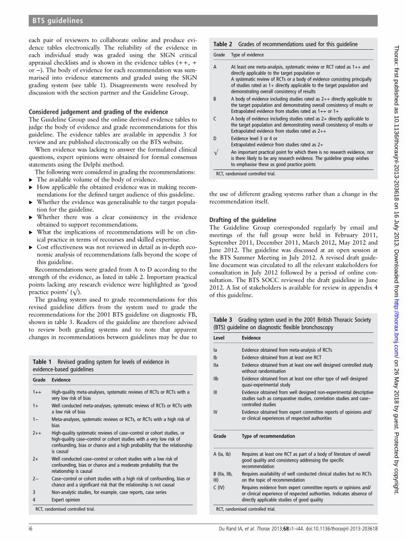
Table 1 Revised grading system for levels of evidence inevidence-based guidelines
Grade Evidence
1++ High-quality meta-analyses, systematic reviews of RCTs or RCTs with avery low risk of bias
1+ Well conducted meta-analyses, systematic reviews of RCTs or RCTs witha low risk of bias
1− Meta-analyses, systematic reviews or RCTs, or RCTs with a high risk ofbias
2++ High-quality systematic reviews of case–control or cohort studies, orhigh-quality case–control or cohort studies with a very low risk ofconfounding, bias or chance and a high probability that the relationshipis causal
2+ Well conducted case–control or cohort studies with a low risk ofconfounding, bias or chance and a moderate probability that therelationship is causal
2− Case–control or cohort studies with a high risk of confounding, bias orchance and a significant risk that the relationship is not causal
3 Non-analytic studies, for example, case reports, case series4 Expert opinion
RCT, randomised controlled trial.
Table 2 Grades of recommendations used for this guideline
Grade Type of evidence
A At least one meta-analysis, systematic review or RCT rated as 1++ anddirectly applicable to the target population orA systematic review of RCTs or a body of evidence consisting principallyof studies rated as 1+ directly applicable to the target population anddemonstrating overall consistency of results
B A body of evidence including studies rated as 2++ directly applicable tothe target population and demonstrating overall consistency of results orExtrapolated evidence from studies rated as 1++ or 1+
C A body of evidence including studies rated as 2+ directly applicable tothe target population and demonstrating overall consistency of results orExtrapolated evidence from studies rated as 2++
D Evidence level 3 or 4 orExtrapolated evidence from studies rated as 2+
√ An important practical point for which there is no research evidence, noris there likely to be any research evidence. The guideline group wishesto emphasise these as good practice points
RCT, randomised controlled trial.
Table 3 Grading system used in the 2001 British Thoracic Society(BTS) guideline on diagnostic flexible bronchoscopy
Level Evidence
Ia Evidence obtained from meta-analysis of RCTsIb Evidence obtained from at least one RCTIIa Evidence obtained from at least one well designed controlled study
without randomisationIIb Evidence obtained from at least one other type of well designed
quasi-experimental studyIII Evidence obtained from well designed non-experimental descriptive
studies such as comparative studies, correlation studies and case–controlled studies
IV Evidence obtained from expert committee reports of opinions and/or clinical experiences of respected authorities
Grade Type of recommendation
A (Ia, Ib) Requires at least one RCT as part of a body of literature of overallgood quality and consistency addressing the specificrecommendation
B (IIa, IIb,III)
Requires availability of well conducted clinical studies but no RCTson the topic of recommendation
C (IV) Requires evidence from expert committee reports or opinions and/or clinical experience of respected authorities. Indicates absence ofdirectly applicable studies of good quality
RCT, randomised controlled trial.
i6 Du Rand IA, et al. Thorax 2013;68:i1–i44. doi:10.1136/thoraxjnl-2013-203618
BTS guidelines
on 26 May 2018 by guest. P
rotected by copyright.http://thorax.bm
j.com/
Thorax: first published as 10.1136/thoraxjnl-2013-203618 on 16 July 2013. D
ownloaded from

The Guideline Group members adhered to the BTS policy forthe Declaration of Interests, and if appropriate, specific interestsare declared in appendix 1.
The guideline will be reviewed within 5 years from the dateof publication (2018).
AUDIT AND RESEARCH RECOMMENDATIONSAudit:▸ All those undertaking FB are advised to maintain personal
records of each procedure, including indication, outcomeand complications for audit purposes.
▸ Periodic audit of bronchoscopy practice, including patientsatisfaction surveys.
Research:▸ Utility of all bronchoscopic samples, including brush and
BAL for phenotyping and genotyping in patients withadvanced non-small cell lung cancer (NSCLC).
▸ Randomised assessment of the utility of bronchoscopy in therelief of lobar collapse/atelectasis in ventilated patients.
▸ Further assessment of the use of jet ventilation during bron-choscopy in mechanically ventilated patients.
▸ Further assessment of optimal analgesia and sedation for safebronchoscopy in mechanically ventilated patients.
TrainingTraining in FB does not fall in the scope of this guideline.
Audit standardsThe following standards provide criteria which may form thebasis of future audits:▸ A serious adverse event rate of <1% (box 1).▸ A ‘safe sedation policy’ and appropriate training in sedation
for all bronchoscopy unit staff, including trainees and inter-val audit of sedation practice.
▸ Utilisation of the bronchoscopy safety checklist (see appendix5).
▸ An 85% diagnostic rate for FB with visible endobronchialtumour.
▸ Periodic patient feedback to inform improvement and revi-sion of the endoscopy service.
MONITORING, PRECAUTIONS AND COMPLICATIONSFB is an increasingly important diagnostic, well tolerated pro-cedure that can be performed safely on an outpatient basis. Inthe largest retrospective series (n=20 986), serious complica-tions occurred in 1.1% with a mortality of 0.02%.4 The com-monest adverse events reported, though not universally in all
studies, included tachycardia/bradycardia, major and minorbleeding, bronchospasm/laryngospasm, cough, dyspnoea, sorethroat, apnoea, seizure, desaturation, pneumothorax and pul-monary oedema. Other smaller studies report complication ratesof 5–32%, and mortality rates of 0–0.8%, but these studies arelimited by their retrospective nature, the variable definition ofadverse events and limited follow up.
Smaller prospective studies suggest that the rate of adverseevents may be higher than previously reported. Hehn et al5
demonstrated respiratory complications in 4.3%, non-respiratory complications in 2.8% and mortality in 0.1%. Inaddition, Bechara et al6 reported adverse events in 35% of 300bronchoscopies performed, 60% of which were classified asmild and 8% as severe. Approximately 6% of patients were hos-pitalised and procedure-related deaths occurred in 1.4%.6
There is an increased risk of adverse events with increasingage but the absolute frequency is low. Chronological age shouldnot be a contraindication for bronchoscopy.5 7 Patient positionduring the procedure does not influence complication rates,with the exception that desaturation >4% is more common inthe sitting position.8
Many factors will influence the risk of complications, includ-ing patient characteristics and factors related to the broncho-scopic unit (including sedation practice and the samplingprocedures employed). Utilisation of a WHO safetychecklist aids in identifying specific possible complications (seeappendix 5) Complications, particularly serious adverse events(box 1), patient satisfaction and efficacy should be routinelymonitored by every bronchoscopy unit.Recommendation▸ All patients undergoing bronchoscopy should have heart
rate, blood pressure and oxygen saturation recorded repeat-edly, including before, during and after the procedure.(Grade D)
Good practice points▸ All bronchoscopy units should undertake periodic audit of
bronchoscopic performance, including efficacy, complicationsand patient satisfaction surveys. (√)
▸ All Trusts should have a ‘safe sedation policy’, and ensure allbronchoscopy unit staff, including trainees, receive appropri-ate training. (√)
HypoxaemiaMonitoring patients with pulse oximetry during bronchoscopy is anaccurate non-invasive method for assessing hypoxaemia.9–11
Significant decreases in oxygen saturation are commonly seenduring bronchoscopy, commencing with administration of sedationand worsening on passage through the vocal cords.8 9 12–16 Patientpositioning8 10 16 and intra-procedural sampling may also influenceoxygen saturations as may airway suctioning.17 18 Van Zwam et al8
described a twofold greater incidence of desaturation >4% andSpO2<90% in the sitting position compared with supine. The useof preprocedure oxygen and the specific sampling procedure (BALvs wash vs brush vs biopsy) was predictive of a higher rate of desat-uration episodes (<90%), but baseline saturations were not (desat-uration to <90% was seen in: BAL 89%, wash 44%, brush 15%,biopsy 10%).19 Milman et al11 (using benzodiazepine premedica-tion without oxygen supplementation) demonstrated that 38% ofpatients desaturate (SpO2<90%) before bronchoscopy, increasingto 80% of patients during the procedure. The proportion remainshigh after the procedure, with 60% of patients desaturating.Similarly, more severe desaturation (SpO2<85%) was seen in 10%,35% and 15%, respectively. No differences in oxygen saturation
Box 1 Suggested serious adverse events
▸ Severe bleeding (see table 5)▸ Cardiac arrhythmia requiring treatment▸ Seizures▸ Myocardial infarction/pulmonary oedema▸ Pneumothorax requiring aspiration/intercostal drain▸ Oversedation requiring ventilatory support or reversal▸ Hospitalisation▸ Admission to intensive care unit▸ Death
Du Rand IA, et al. Thorax 2013;68:i1–i44. doi:10.1136/thoraxjnl-2013-203618 i7
BTS guidelines
on 26 May 2018 by guest. P
rotected by copyright.http://thorax.bm
j.com/
Thorax: first published as 10.1136/thoraxjnl-2013-203618 on 16 July 2013. D
ownloaded from
were described in a comparison of trans-nasal or trans-oral broncho-scopic approaches.20
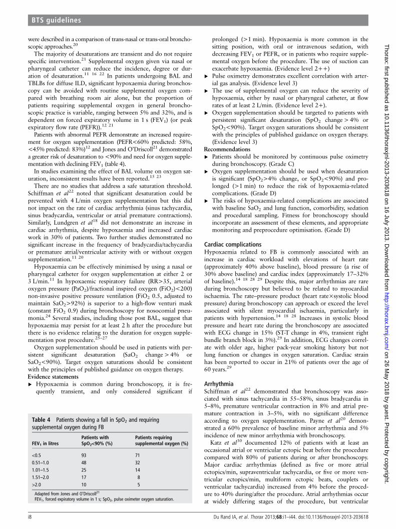
The majority of desaturations are transient and do not requirespecific intervention.21 Supplemental oxygen given via nasal orpharyngeal catheter can reduce the incidence, degree or dur-ation of desaturation.11 16 22 In patients undergoing BAL andTBLBs for diffuse ILD, significant hypoxaemia during bronchos-copy can be avoided with routine supplemental oxygen com-pared with breathing room air alone, but the proportion ofpatients requiring supplemental oxygen in general broncho-scopic practice is variable, ranging between 5% and 32%, and isdependent on forced expiratory volume in 1 s (FEV1) (or peakexpiratory flow rate (PEFR)).12 21
Patients with abnormal PEFR demonstrate an increased require-ment for oxygen supplementation (PEFR<60% predicted: 58%,<45% predicted: 83%)12 and Jones and O’Driscoll21 demonstrateda greater risk of desaturation to <90% and need for oxygen supple-mentation with declining FEV1 (table 4).
In studies examining the effect of BAL volume on oxygen sat-uration, inconsistent results have been reported.15 23
There are no studies that address a safe saturation threshold.Schiffman et al22 noted that significant desaturation could beprevented with 4 L/min oxygen supplementation but this didnot impact on the rate of cardiac arrhythmia (sinus tachycardia,sinus bradycardia, ventricular or atrial premature contractions).Similarly, Lundgren et al18 did not demonstrate an increase incardiac arrhythmia, despite hypoxaemia and increased cardiacwork in 30% of patients. Two further studies demonstrated nosignificant increase in the frequency of bradycardia/tachycardiaor premature atrial/ventricular activity with or without oxygensupplementation.11 20
Hypoxaemia can be effectively minimised by using a nasal orpharyngeal catheter for oxygen supplementation at either 2 or3 L/min.11 In hypoxaemic respiratory failure (RR>35, arterialoxygen pressure (PaO2)/fractional inspired oxygen (FiO2)<200)non-invasive positive pressure ventilation (FiO2 0.5, adjusted tomaintain SaO2>92%) is superior to a high-flow venturi mask(constant FiO2 0.9) during bronchoscopy for nosocomial pneu-monia.24 Several studies, including those post BAL, suggest thathypoxaemia may persist for at least 2 h after the procedure butthere is no evidence relating to the duration for oxygen supple-mentation post procedure.25–27
Oxygen supplementation should be used in patients with per-sistent significant desaturation (SaO2 change > 4% orSaO2<90%). Target oxygen saturations should be consistentwith the principles of published guidance on oxygen therapy.Evidence statements▸ Hypoxaemia is common during bronchoscopy, it is fre-
quently transient, and only considered significant if
prolonged (>1 min). Hypoxaemia is more common in thesitting position, with oral or intravenous sedation, withdecreasing FEV1 or PEFR, or in patients who require supple-mental oxygen before the procedure. The use of suction canexacerbate hypoxaemia. (Evidence level 2++)
▸ Pulse oximetry demonstrates excellent correlation with arter-ial gas analysis. (Evidence level 3)
▸ The use of supplemental oxygen can reduce the severity ofhypoxaemia, either by nasal or pharyngeal catheter, at flowrates of at least 2 L/min. (Evidence level 2+).
▸ Oxygen supplementation should be targeted to patients withpersistent significant desaturation (SpO2 change > 4% orSpO2<90%). Target oxygen saturations should be consistentwith the principles of published guidance on oxygen therapy.(Evidence level 3)
Recommendations▸ Patients should be monitored by continuous pulse oximetry
during bronchoscopy. (Grade C)▸ Oxygen supplementation should be used when desaturation
is significant (SpO2>4% change, or SpO2<90%) and pro-longed (>1 min) to reduce the risk of hypoxaemia-relatedcomplications. (Grade D)
▸ The risks of hypoxaemia-related complications are associatedwith baseline SaO2 and lung function, comorbidity, sedationand procedural sampling. Fitness for bronchoscopy shouldincorporate an assessment of these elements, and appropriatemonitoring and preprocedure optimisation. (Grade D)
Cardiac complicationsHypoxaemia related to FB is commonly associated with anincrease in cardiac workload with elevations of heart rate(approximately 40% above baseline), blood pressure (a rise of30% above baseline) and cardiac index (approximately 17–32%of baseline).14 18 28 29 Despite this, major arrhythmias are rareduring bronchoscopy but believed to be related to myocardialischaemia. The rate–pressure product (heart rate×systolic bloodpressure) during bronchoscopy can approach or exceed the levelassociated with silent myocardial ischaemia, particularly inpatients with hypertension.14 18 28 Increases in systolic bloodpressure and heart rate during the bronchoscopy are associatedwith ECG change in 15% (ST-T change in 4%, transient rightbundle branch block in 3%).29 In addition, ECG changes correl-ate with older age, higher pack-year smoking history but notlung function or changes in oxygen saturation. Cardiac strainhas been reported to occur in 21% of patients over the age of60 years.29
ArrhythmiaSchiffman et al22 demonstrated that bronchoscopy was asso-ciated with sinus tachycardia in 55–58%, sinus bradycardia in5–8%, premature ventricular contraction in 8% and atrial pre-mature contraction in 3–5%, with no significant differenceaccording to oxygen supplementation. Payne et al20 demon-strated a 60% prevalence of baseline minor arrhythmia and 5%incidence of new minor arrhythmia with bronchoscopy.
Katz et al30 documented 12% of patients with at least anoccasional atrial or ventricular ectopic beat before the procedurecompared with 80% of patients during or after bronchoscopy.Major cardiac arrhythmias (defined as five or more atrialectopics/min, supraventricular tachycardia, or five or more ven-tricular ectopics/min, multiform ectopic beats, couplets orventricular tachycardia) increased from 4% before the proced-ure to 40% during/after the procedure. Atrial arrhythmias occurat widely differing stages of the procedure, but ventricular
Table 4 Patients showing a fall in SpO2 and requiringsupplemental oxygen during FB
FEV1 in litresPatients withSpO2<90% (%)
Patients requiringsupplemental oxygen (%)
<0.5 93 710.51–1.0 48 321.01–1.5 25 141.51–2.0 17 8>2.0 10 5
Adapted from Jones and O’Driscoll21
FEV1, forced expiratory volume in 1 s; SpO2, pulse oximeter oxygen saturation.
i8 Du Rand IA, et al. Thorax 2013;68:i1–i44. doi:10.1136/thoraxjnl-2013-203618
BTS guidelines
on 26 May 2018 by guest. P
rotected by copyright.http://thorax.bm
j.com/
Thorax: first published as 10.1136/thoraxjnl-2013-203618 on 16 July 2013. D
ownloaded from
arrhythmias occur mainly on passage through the vocal cords.Maximum ventricular arrhythmia is correlated with minimumoxygen saturations. Asymptomatic ST-T wave changes occur in6% of patients, typically correlating with maximum heart rates.Oxygen saturations can remain lower than preprocedure levels3 h after bronchoscopy in 30% of patients.
Myocardial infarctionAcute MI is considered a contraindication to bronchoscopywithin 4–6 weeks. Dweik et al31 retrospectively analysed thesafety of bronchoscopies within 30 days of an acute MI andnoted that mortality (5%) was limited to patients with activeischaemia at the time of bronchoscopy.Evidence statements▸ Bronchoscopy increases cardiac rate, blood pressure and
cardiac index. The rate pressure product is often sufficient tocause myocardial ischaemia. (Evidence level 2+)
▸ Sinus tachycardia and atrial or ventricular premature contrac-tions are the commonest arrhythmia noted before, duringand after bronchoscopy. (Evidence level 3)
▸ Ventricular arrhythmia (mostly premature contraction, bi andtrigeminy) occurs most commonly on passage through thecords and is associated with low oxygen saturations. Oxygensaturations may remain below preprocedure levels for over3 h in a third of patients. (Evidence level 3)
▸ Myocardial ischaemia during bronchoscopy is related toheart rate and blood pressure, rather than oxygen saturationper se. It also correlates with increasing age and smokinghistory. (Evidence level 3)
▸ Bronchoscopy within 30-days of acute MI is associated witha 5% mortality (related to active ischaemia). In the absenceof active ischaemia, when good clinical justification is made,bronchoscopy can be performed. (Evidence level 3)
▸ Incidence of cardiac arrhythmia is not affected by oxygensupplementation. (Evidence level 3)
Recommendations▸ Continuous ECG monitoring should be used when there is a
high clinical risk of arrhythmia. (Grade D)▸ When there is a high risk of arrhythmia, oxygen saturations,
pulse rate and blood pressure should be optimised.Appropriate aftercare monitoring and instructions should begiven. (Grade D)
Good practice points▸ Resuscitation equipment should be readily available. (√)▸ Intravenous access should be established before sedation is
given and maintained until discharge. (√)
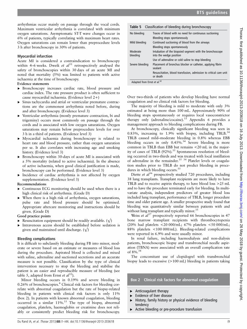
Bleeding complicationsIt is difficult to subclassify bleeding during FB into minor, mod-erate or severe based on an estimate or measures of blood lossduring the procedure. Aspirated blood is collected and mixedwith saline, adrenaline and suctioned secretions and an accuratemeasure is not possible. Classification by the type of clinicalintervention necessary to stop the bleeding and stabilise thepatient is an easier and reproducible measure of bleeding (seetable 5, adapted from Ernst et al32).
Minor bleeding occurs in 0.19% and severe bleeding in0.26% of bronchoscopies.4 Clinical risk factors for bleeding cor-relate with abnormal coagulation but the rate of biopsy-relatedbleeding in patients with clinical risk factors is only 11%(box 2). In patients with known abnormal coagulation, bleedingoccurred in a similar 11%.33 The type of biopsy, abnormalcoagulation, platelets, haemoglobin or creatinine does not reli-ably or consistently predict bleeding risk for bronchoscopy.
Over two-thirds of patients who develop bleeding have normalcoagulation and no clinical risk factors for bleeding.
The majority of bleeding is mild to moderate with only 3%estimated at being more than 100 mL. Approximately 90% ofbleeding stops spontaneously or requires local vasoconstrictortherapy only (adrenaline/cocaine).33 Appendix 6 provides amanagement approach to bleeding complications during FB.
At bronchoscopy, clinically significant bleeding was seen in0.83%, increasing to 1.9% with biopsy, including TBLB.34
TBLB caused mild–moderate bleeding in 0.8% whereas EBBbleeding occurs in only 0.45%.34 Severe bleeding is morecommon in TBLB than EBB but remains <20 mL in the major-ity of cases of TBLB (92%).35 Spontaneous resolution of bleed-ing occurred in two-thirds and was treated with local instillationof adrenaline in the remainder.35 34 Platelet levels or coagula-tion studies prior to TBLB do not predict bleeding in proce-dures in which bleeding occurs.35
Diette et al36 prospectively studied 720 procedures, including38 lung transplants. Transplant recipients are more likely to haveTBLB and to receive aspirin therapy, to have blood loss >25 mLand to have the procedure terminated early for bleeding. In multi-variate analysis, independent predictors of greater blood lossincluded lung transplant, performance of TBLB, longer proceduretime and older patient age. A smaller prospective study found thatbleeding was quantitatively similar between patients with andwithout lung transplant and typically minor.35
Weiss et al37 prospectively reported 66 bronchoscopies in 47bone marrow transplant recipients with thrombocytopenia(20% had platelets <20 000/mL; 67% platelets <50 000/mL;88% platelets <100 000/mL). Bleeding-related complicationswere reported in 6.9% and were usually minor.
In renal failure, including haemodialysis and non-dialysispatients, bronchoscopic biopsy and transbronchial needle aspir-ation (TBNA) were associated with an overall complication rateof 8%.38
The concomitant use of clopidogrel with transbronchialbiopsy leads to excessive (>100 mL) bleeding in patients taking
Table 5 Classification of bleeding during bronchoscopy
No bleeding Traces of blood with no need for continuous suctioningBleeding stops spontaneously
Mild bleeding Continued suctioning of blood from the airwaysBleeding stops spontaneously
Moderatebleeding
Intubation of the biopsied segment with the bronchoscopeinto the wedge positionUse of adrenaline or cold saline to stop bleeding
Severe bleeding Placement of bronchus blocker or catheter, applying fibrinsealant
Resuscitation, blood transfusion, admission to critical care unitor death
Adapted from Ernst et al.32
Box 2 Clinical risk factors for abnormal coagulation
▸ Anticoagulant therapy▸ Evidence of liver disease▸ History, family history or physical evidence of bleeding
tendency▸ Active bleeding or pre-procedure transfusion
Du Rand IA, et al. Thorax 2013;68:i1–i44. doi:10.1136/thoraxjnl-2013-203618 i9
BTS guidelines
on 26 May 2018 by guest. P
rotected by copyright.http://thorax.bm
j.com/
Thorax: first published as 10.1136/thoraxjnl-2013-203618 on 16 July 2013. D
ownloaded from
clopidogrel alone (89% vs 3.4%) and clopidogrel with aspirin(100% vs 3.4%).32 Bleeding rates are significantly higher in allcategories of bleeding (minor/moderate/severe) but can be con-trolled in most instances by bronchoscopic means. Need fortransfusion or death following haemorrhage secondary to clopi-dogrel is rare.32 See appendix 7 for an algorithm for the man-agement of patients on warfarin or clopidogrel undergoing FB.Evidence statements▸ Minor bleeding occurs in 0.19% and severe bleeding in
0.26% of bronchoscopies. (Evidence level 3)▸ The routine performance of coagulation studies, platelet or
haemoglobin counts are of no value in predicting the risk orseverity of bleeding. (Evidence level 3)
▸ Coagulation studies, platelet count and haemoglobin valuesshould be estimated when clinical risk factors indicate a like-lihood of abnormal coagulation. However, over two-thirdsof patients with significant bleeding possess normal coagula-tion and no clinical risk factors for bleeding. (Evidencelevel 3)
▸ Bleeding complications in patients with thrombocytopeniaundergoing bronchoscopy and lavage are approximately7%. No data are available regarding the safety of TBLB orEBB in thrombocytopenia but the majority of bleedingcomplications relate to epistaxis. (Evidence level 3)
▸ Clopidogrel causes bleeding, ranging from mild to severe,when performing TBLB. (Evidence level 2+)
▸ TBLB causes a twofold increase in the risk of mild–moderatebleeding and a threefold increase in the risk of severe bleed-ing compared with EBB. However, the overall risk remainssmall and TBLB rarely causes significant blood loss (92% ofpatients experience blood loss <20 mL) and typicallyresolves spontaneously or with endoscopic instillation ofcocaine/adrenaline. (Evidence level 3)
▸ Bronchoscopic biopsy and TBNA in patients receiving haemo-dialysis, or in patients with renal failure without dialysis, resultin a higher rate of bleeding complications (∼8%; 4% major,4% minor) than the general population. (Evidence level 3)
▸ Lung transplantation may predispose patients to greaterblood loss at bronchoscopic biopsy, including TBLB.(Evidence level 2−)
Recommendations▸ Perform coagulation studies, platelet count and haemoglobin
concentration when there are clinical risk factors for abnor-mal coagulation. (Grade D)
▸ Bronchoscopy with lavage can be performed with plateletcounts >20 000 per μL. Liaise with the local haematologyteam regarding the need for platelet transfusion before bron-choscopy if EBB or TBLB is planned. (Grade D)
▸ Discontinue clopidogrel 7 days prior to consideration of EBBand TBLB. Low-dose aspirin alone can be continued.(Grade C)
Good practice points▸ Anticoagulants should be managed according to published
guidelines as set out in appendix 7 of this guideline. (√)▸ The risk of biopsy needs to be weighed against the potential
for benefit and appropriate informed consent obtained. (√)
PneumothoraxPneumothorax following bronchoscopy for any indicationoccurs at a rate of 1 in 1000 (0.1–0.16%)4 39 but was as high as0.4–0.8% in some smaller series.40 41 In contrast the rate ofpneumothorax in TBLB has been reported to be significantlyhigher between 1% and 6%4 39 42 43 or higher still in TBLB of
diffuse abnormality (9%).43–46 TBLB remains a safe outpatientprocedure with a low incidence of delayed complications.47
In relation to all adverse events, pneumothorax representsapproximately 10% of all complications but rarely complicatesbronchoscopy without TBLB or therapeutic bronchoscopy.4
Pneumothorax is rarely total and often delayed (∼40% of pneu-mothoraces)4 and when present may require intercostal tubedrainage (40–70% of cases).39 42 43 47 The frequency ofpneumothorax is related to age and the number of TBLBs.5 44
Routine performance of a chest x-ray after TBLB rarely providesuseful clinical information in the absence of symptoms43 44 48
and may not be required. In the absence of a routine chest x-ray,monitoring for the development of symptoms associated withpneumothorax should continue for 2 h. The rate of pneumo-thorax following TBLB does not appear significantly differentwith or without fluoroscopy,45 but fluoroscopy may be of valueto improve the diagnostic yield in focal rather than diffuse lungdisease (focal 4.3%, diffuse 9%).43
Evidence statements▸ Risk of pneumothorax from all bronchoscopic procedures is
1 in 1000 (0.1%) but increases to between 1 in 100 to 1 in16 (1–6%) following TBLB. (Evidence level 3)
▸ Pneumothorax may be delayed (up to 2 h in 40% of cases)and may require intercostal tube drainage (Evidence level 3)
▸ Routine performance of a chest x-ray after TBLB rarely pro-vides useful clinical information in the absence of symptoms.(Evidence level 3)
▸ Fluoroscopy may reduce the rate of pneumothorax in focallung disease. (Evidence level 3)
Recommendations▸ A chest radiograph should be obtained if a patient is symp-
tomatic or there is a clinical suspicion of possible pneumo-thorax after TBLB. (Grade D)
▸ Fluoroscopic screening may improve diagnostic yield ofTBLB in focal but not diffuse lung disease. (Grade D)
▸ Patients should be advised of the potential for delayed compli-cations following TBLB and provided with written informationregarding likely symptoms and action required. (Grade D)
Fever and infectionPost bronchoscopy fever (PBF) is not reported in a large pro-spective study of complications in over 20 000 patients4 butappears relatively common in other smaller prospective studiesfocusing on PBF (5–10%).49 50 PBF is most typically seenapproximately 8 h (range 4–24 h) following BAL (13%) when itis associated with an acute inflammatory response characterisedby fever >38°C, neutrophilic leucocytosis, elevated C-reactiveprotein, fibrinogen and proinflammatory cytokines,50–52 and anabsence of bacteraemia.49 50 53 Fever is typically less than 40°C,lasts on average 14 h but is rarely accompanied by a chest x-rayinfiltrate.50 Antibiotic prophylaxis does not prevent PBF, pneu-monia or the proinflammatory cytokine response.53 54
A true bacteraemia post bronchoscopy occurs in 6–8% ofpatients,49 55 most commonly coagulase negative or positivestaphylococci, non-haemolytic or β-haemolytic streptococci,Citrobacter or Klebsiella species.49 54 55 National Institute forHealth and Clinical Excellence guidance in March 2008 also con-cluded that ‘Antibacterial prophylaxis is not recommended for theprevention of endocarditis in patients undergoing procedures ofthe upper and lower respiratory tract (including bronchoscopy)’.56
Evidence statements▸ PBF is common, particularly after BAL, and associated with a
non-infective acute inflammatory response, typically startingafter discharge from hospital. (Evidence level 2++)
i10 Du Rand IA, et al. Thorax 2013;68:i1–i44. doi:10.1136/thoraxjnl-2013-203618
BTS guidelines
on 26 May 2018 by guest. P
rotected by copyright.http://thorax.bm
j.com/
Thorax: first published as 10.1136/thoraxjnl-2013-203618 on 16 July 2013. D
ownloaded from

▸ Antibiotic prophylaxis does not prevent PBF or pneumonia.(Evidence level 1++)
▸ Bacteraemia occurs in 6–8% of patients undergoing bron-choscopy. (Evidence level 3)
Recommendations▸ Patients should receive written information regarding PBF
and appropriate management advice. (Grade C)▸ Antibiotic prophylaxis is not warranted before bronchoscopy
for the prevention of endocarditis, fever or pneumonia.(Grade B)
SAFETY OF FB IN SPECIFIC MEDICAL CONDITIONSPatients with asthmaThe safety of bronchoscopy in asthma has been studied inresearch and clinical settings. Bronchoscopy often causes a fallin FEV1. In healthy volunteers the mean fall in FEV1 has beenreported on average to be between 9% and 17%,15 57 howeverit can be over 20%.58 In patients with asthma the mean fall hasbeen reported to be 10–26%.15 57 59 The majority of studiescomparing the fall in FEV1 between patients with asthma andhealthy volunteers found no difference between thegroups.15 58 60 Patients with increased bronchial hyper-reactivitymay have a greater fall in FEV1
57 61 however this has not beenuniversally reported.58 In particular, BAL can cause a fall in theFEV1.
58 59
Complication rates post bronchoscopy range between 3.5%and 12% depending on how the complications are reported andwhat procedures were performed.15 60–63 In addition, patientswith severe asthma are more likely to require oral corticoster-oids post bronchoscopy.58 Many of the studies use bronchodila-tors prior to bronchoscopy to increase the FEV1.
57 58 60 62
Although this does not seem to reduce the percentage fall inFEV1 it may increase the absolute FEV1 at the end of the pro-cedure, as the patient starts from a higher baseline.Evidence statements▸ Up to 10% of patients with asthma may develop respiratory
symptoms post bronchoscopy. (Evidence level 2−)▸ BAL is associated with more symptoms in patients with asthma
compared with bronchoscopy alone. (Evidence level 2−)Recommendation▸ Patients’ asthma control should be optimised prior to bron-
choscopy, especially when BAL is likely to be performed.(Grade C)
Good practice point▸ Nebulised bronchodilators should be considered before bron-
choscopy in patients with asthma. (√)
Patients with COPDBronchoscopy in patients with COPD appears to carry a greaterrisk compared with those with normal lung function. Anincreased risk of complications has been reported in severeCOPD (defined as FEV1<50% predicted or FEV1<1 L andwith FEV1/forced vital capacity < 69%).64 Five percent ofpatients with COPD compared with 0.6% of controls experi-enced complication: pneumonia, respiratory failure and desatur-ation.64 In a separate study, bronchoscopy safety was studied inpatients with hypercapnia, 77% of whom had COPD.65
Desaturation occurred in 30% of the study population, 55% ofpatients developed wheezing and the procedure was terminatedearly in 20% of patients.65 A randomised control trial studiedthe effect of inhaled salbutamol (200 mg) on FEV1 after bron-choscopy in moderate to severe COPD.66 No difference wasobserved (change in FEV1) in the group receiving salbutamolcompared with placebo. Nine percent of patients experienced
desaturation during bronchoscopy, with no difference againbeing observed between treatment groups.
In research bronchoscopy comparable complication rates havebeen reported. Hattotuwa et al67 reported a complication rateof 9%, including haemoptysis (5%), pneumothorax (2%) andbronchospasm (2%). All patients were given 2.5 mg nebulisedsalbutamol prior to the procedure.Evidence statements▸ Patients with COPD have a higher risk of desaturation and
bronchoconstriction compared with controls, however this isnot a universal finding. (Evidence level 2−)
▸ Nebulised salbutamol administered prior to bronchoscopydoes not alter the post-bronchoscopy complication rate inpatients with COPD. (Evidence level 1−)
Recommendations▸ COPD treatment should be optimised prior to bronchoscopy
when possible. (Grade D)▸ Bronchoscopists should be cautious when sedating patients
with COPD. (Grade D)
Patients with ischaemic heart diseaseThe haemodynamic changes during FB might increase the riskof myocardial damage during the procedure. One study hasshown an increased risk of ischaemic ECG changes during theprocedure in patients over 60.29 A retrospective study investi-gated the safety of bronchoscopy in 20 patients after acute MI.The procedure was performed an average of 12 days after MI.31
One death occurred in this study—a patient with acute myocar-dial ischaemia before and during the procedure. No other com-plications were reported. A retrospective study of patientsundergoing bronchoscopy on a coronary care unit reported nodifference in complications in subjects after MI compared withthose without MI.68
The American perioperative guidelines recommend that elect-ive surgery is avoided for 4–6 weeks after an acute event result-ing in myocardial damage.69
Evidence statements▸ Active myocardial ischaemia is a contraindication to bron-
choscopy. (Evidence level 3)▸ FB can increase the risk of active ischaemia, haemodynamic
compromise, arrhythmia and further ischaemic events afterMI. (Evidence level 3)
Recommendations▸ Liaison with cardiologists should be considered in high-risk
patients with cardiac disease and if FB is indicated within 4–6 weeks after MI. (Grade D)
▸ FB should ideally be delayed for 4 weeks after MI.(Grade D)
Bronchoscopy for haemoptysisBronchoscopy can be used to investigate haemoptysis. CT scanshave changed the diagnostic pathway and should always be con-sidered prior to bronchoscopy. Two studies have investigated therole of bronchoscopy following a thoracic CT scan. One studyin Turkey found a bleeding site in 80% of 203 individuals evenif the CT and chest x-ray were normal.70 A different study(n=200) found an endobronchial diagnosis in 0.5% of indivi-duals when the CTwas normal.71
Recommendation▸ Consider bronchoscopy after a normal CT if the patient is
high risk for lung carcinoma or if the haemoptysis continues.(Grade D)
Du Rand IA, et al. Thorax 2013;68:i1–i44. doi:10.1136/thoraxjnl-2013-203618 i11
BTS guidelines
on 26 May 2018 by guest. P
rotected by copyright.http://thorax.bm
j.com/
Thorax: first published as 10.1136/thoraxjnl-2013-203618 on 16 July 2013. D
ownloaded from
Bronchoscopy in the older patientComorbid disease is more likely in the older patient with apotential increase in bronchoscopy risk. Several studies haveshown older patients tolerate the procedure well, with noincrease in complications.72–74 A prospective cohort study5 sug-gested that complication rates for pneumothorax and transienthypotension increased with age. When the safety of bronchos-copy was investigated in patients over 80 years old, higher com-plication rates and mortality rates were reported and thesepatients were also more likely to be mechanically ventilatedafter bronchoscopy.75
Recommendations▸ Age alone should not be a contraindication for bronchos-
copy. (Grade D)▸ The older patient may require reduced doses of benzodiaze-
pines/opioids for sedation. (Grade D)
Bronchoscopy in patients who are immunosuppressedFB can provide useful diagnostic information when treatingpatients with respiratory problems who are immunosuppressed(see ‘Diagnosis of infection’ in this guideline for details of diagnos-tic value). Bronchoscopy and associated diagnostic techniques arenot without risk in this patient population and this should betaken into account before performing a bronchoscopy. BAL has areported complication rate of 0–49%, depending on how the com-plications were defined, and deaths associated with BAL have beenreported.76–82 The main complications associated with BAL aredesaturation, a drop in FEV1 and haemorrhage.
Protected specimen brushes (PSBs) can sometimes be used forsimilar indications to BAL. One study involved patients whowere immunocompromised following bone marrow transplant78
and showed that the complication rates for PSBs were signifi-cantly higher than for BAL (36% vs 14%).
TBLB can carry a significant higher complication risk (30%)compared with BAL alone.79 81 Pneumothorax, haemorrhageand desaturation were reported to be complications associatedwith TBLB.79 81
Three studies comparing the overall mortality in patientsinvestigated with FB and patients investigated with non-invasivetechniques for obtaining diagnostic samples failed to demon-strate that obtaining samples by bronchoscopy significantlyreduces mortality.77 78 One study suggested that early FB toobtain diagnostic samples is associated with a lower mortalitycompared with delayed bronchoscopy for the same indication.82
In patients who have had lung transplantation, TBLB may bethe only means of diagnosing allograft rejection. It has previ-ously been reported that lung transplant patients have a higherrisk of bleeding (>25 mL blood) (44%) compared to otherpatients undergoing bronchoscopy, resulting in 5.4% of proce-dures being terminated early.36 Larger studies have suggestedthat significant bleeding occurs in 25%83 or 13% of patients84
depending on how bleeding is diagnosed. The largest study85
suggested that bronchoscopy in lung transplant patients carriedno increased risk compared with other patient groups andreported a complication risk of 0.7%. It should be noted thatonly 57% of procedures in this study included TBLB.85 Othergroups have reported higher complication rates of between4.8% and 22%.83 84 86 The main complications associated withTBLB are oversedation, pneumothorax and haemorrhage.Evidence statements▸ PSB has a low diagnostic yield, is rarely positive when BAL is
negative and has a higher complication rate than BAL.(Evidence level 2+)
▸ BAL in patients who are immunocompromised has a diag-nostic rate of 45–62% for infection with an associated com-plication rate of 0–49%. (Evidence level 2++)
▸ TBLB carries an increased complication rate compared withBAL of around 30%, which is mainly explained by theincreased risk of pneumothorax. (Evidence level 2++)
▸ Lung transplant patients may be at higher risk of bleeding thanother patients who are immunosuppressed. (Evidence level 2−)
▸ TBLB has a reported complication rate of between 0.7% and22%, with the main complications being oversedation,pneumothorax and haemorrhage. (Evidence level 2++)
Recommendations▸ When a diagnosis is not likely to be obtained through non-
invasive measures, bronchoscopy with BAL can be consid-ered to provide diagnostic information. (Grade C)
▸ TBLB is helpful in lung transplant recipients when rejectionis a possibility. (Grade C)
PREMEDICATION, SEDATION AND TOPICAL ANAESTHESIAFOR FBPremedicationVarious drugs have been considered to have a potential role aspremedication, including anticholinergics (atropine and glyco-pyrrolate), other drugs with cardiovascular activity (eg, cloni-dine and labetalol), fenoterol, benzodiazepines and opioids.
AnticholinergicsAnticholinergics, such as atropine and glycopyrrolate, have beenpostulated to reduce cough and improve bronchoscopic viewsby reducing airway secretions, and also to prevent vasovagalreactions and reduce reflex bronchoconstriction. Three rando-mised controlled trials (cumulatively studying more than 1300patients) examined anticholinergics and failed to demonstrateany consistent clinical benefit for patients undergoing bronchos-copy.87–89 Anticholinergics were associated with an increase inhaemodynamic fluctuations (tachycardia and hypertension).
Drugs with cardiovascular activityHypertension and tachycardia can commonly occur duringbronchoscopy. Several drugs that may blunt the cardiovascularresponse to bronchoscopy have been studied, postulating a rolein avoidance of myocardial ischaemia and arrhythmias.
Two small randomised controlled trials examined the role ofclonidine, a centrally acting antihypertensive, and demonstrateda blunting of cardiovascular responses (including blood pressure,heart rate and noradrenaline surges), although hypotensionrequiring treatment was seen at higher doses of clonidine andthere was no improvement in patient tolerance.90 91
A randomised study of intravenous labetalol failed to demon-strate any changes in haemodynamic parameters or procedural tol-erance associated with labetalol administration.92 Surprisingly,there was no tachycardia or hypertension seen in patients in thecontrol arm (who received relatively high doses of sedation), sug-gesting that the sedation regime is a significant determinant ofhaemodynamic responses.
Further studies are required to define potential patient benefitof cardiovascular-related premedication and should be poweredfor endpoints such as myocardial ischaemia and arrhythmias.
Premedication with other drugsOther small randomised controlled trials have indicated abenefit for several premedication drugs, but study limitations(size, design and lack of detailed participant characteristics)mean that further investigation is required: fenoterol may
i12 Du Rand IA, et al. Thorax 2013;68:i1–i44. doi:10.1136/thoraxjnl-2013-203618
BTS guidelines
on 26 May 2018 by guest. P
rotected by copyright.http://thorax.bm
j.com/
Thorax: first published as 10.1136/thoraxjnl-2013-203618 on 16 July 2013. D
ownloaded from
reduce cough rate and topical anaesthesia requirements93; dex-tromethorphan may lower sedation and topical anaesthesiarequirements while improving patient tolerance94; low-dose orallorazepam is associated with more favourable bronchoscopyrecall at 24 h, but not immediately after the procedure.95
The use of premedication with benzodiazepines (such as lor-azepam) and opioid-like drugs (such as dextromethorphan)should be discouraged if the same class of drug is to be adminis-tered intravenously during bronchoscopy.Evidence statements▸ Three randomised controlled trials have failed to demon-
strate significant clinical benefits associated with anticholiner-gics. Their use in bronchoscopy may be associated with anincreased rate of cardiovascular adverse effects. (Evidencelevel 1+)
▸ There is a lack of evidence suggesting benefit of routine pre-medication for bronchoscopy. Small randomised studiessuggest a potential role for several agents (clonidine, dextro-methorphan, fenoterol, lorazepam), but such findings requirevalidation in much larger studies of well characterisedpatients. (Evidence level 1−)
Recommendations▸ Anticholinergics (glycopyrrolate or atropine) should not rou-
tinely be used prior to bronchoscopy due to a lack of clinicalbenefit and a possible increased risk of haemodynamicchanges. (Grade A)
▸ Premedication for bronchoscopy is not routinely indicated.(Grade C)
SedationThe majority of bronchoscopists in the UK use sedation forbronchoscopy, with only 5–10% routinely performing bron-choscopy on unsedated patients.96 97 Five randomised con-trolled trials,95 98–101 one cohort study102 and one qualitativestudy103 examined patient and physician preference for sedationduring bronchoscopy. These studies provided a broad consensusthat patients and physicians usually preferred sedation for bron-choscopy, examining domains such as comfort, tolerance,bronchoscopic ease and willingness to undergo a repeat proced-ure. Some studies96 100 104 suggested that a subset of patientstolerate unsedated bronchoscopy well, suggesting that sedationshould remain an option dependent on patient preference andcomorbidities (which may limit suitability for sedation).Evidence statements▸ Patients and physicians usually prefer the use of sedation for
bronchoscopy. (Evidence level 1+)Recommendations▸ Intravenous sedation should be offered to patients undergo-
ing bronchoscopy, provided there are no contraindications.(Grade B)
▸ Some patients will tolerate unsedated bronchoscopy well,and patient preference should be sought. (Grade B)
Administration of sedationSedation for bronchoscopy is usually the responsibility ofbronchoscopists, although some centres use anaesthetist-delivered sedation. Bronchoscopist-delivered sedation should becarefully titrated using small incremental doses to avoid overse-dation, particularly given significant variability of patientresponse to sedatives. The practice of non-titrated single largebolus dose sedation regimes is strongly discouraged.105
The desired depth of sedation is usually ‘conscious’ sedation,in which the patient maintains airway patency and cardiorespira-tory function and verbal contact with the patient is possible at
all times,106 although interventional bronchoscopy may occa-sionally require deeper sedation provided by formal anaestheticsupport. Patients who are more deeply sedated should have thesame level of care and monitoring as those undergoing a formalgeneral anaesthetic. An Intercollegiate Working Party of the UKAcademy of Medical Royal Colleges, chaired by the RoyalCollege of Anaesthetists, has produced a summary of safe sed-ation practice (table 6).
Assessment and quantification of sedation depth may be aidedby various tools, including the Ramsay Scale107 and theModified Observer’s Assessment of Alertness/Sedation score108
(see appendix 9). These tools have particular utility in documen-tation of sedation level as part of the bronchoscopy record.Evidence statement▸ There is a significant and unpredictable inter-patient variabil-
ity in required doses of sedative drugs. (Evidence level 1+)Recommendations▸ Sedative drugs should be titrated to provide the desired
depth of sedation, given significant inter-patient variability inrequired doses. (Grade B)
▸ The desired depth of sedation is one in which verbal contactis possible at all times. (Grade D)
Good practice point▸ Bronchoscopists are encouraged to document an assessment
of sedation depth as part of the procedural report. (√)
Drugs used as sedatives for bronchoscopyPlease refer to appendix 8, tables 1 and 2.
A 2003 UK survey found that 78% of bronchoscopists rou-tinely use midazolam sedation, with midazolam and fentanyl/alfentanil being the most frequent combination sedation regimeused.96 Eleven randomised controlled trials101 108–118 haveinvestigated various drugs and drug combinations for bronchos-copy sedation, including benzodiazepines (eg, midazolam), pro-pofol (and its novel pro-drug fospropofol), ketamine andopioids (eg, fentanyl, alfentanil).
Benzodiazepines and propofolBenzodiazepines (eg, midazolam) cause sedation, anxiolysis andanterograde amnesia by binding to and increasing the activity of
Table 6 Intercollegiate Working Party of the UK Academy ofMedical Royal Colleges—summary of safe sedation practice106
Domain Safe practice
Patientassessment
‘Checklist’ identification of sedation risk factors prior toprocedure. Instructions on activities subsequent to procedureprovided
Level of sedation Sedation provided only to the level of ‘conscious’ sedation,in which verbal contact is possible
Intravenoussedation
Secure venous access mandatorySpecific antagonist drugs should be to hand (see appendix8)When combination sedation used, opioids should be givenfirst, and caution should be taken to avoid oversedation
Monitoring Defined professionals should have responsibility formonitoring patient safety and making a written record. Pulseoximeter monitoring should be continued until dischargefrom unit. Consider monitoring blood pressure and ECG inhigher risk patients
Oxygen therapy Nasal cannula and facial mask oxygen delivery should beavailable
Facilities Patient trolley should be capable of being tipped headdownResuscitation equipment should be immediately available
Du Rand IA, et al. Thorax 2013;68:i1–i44. doi:10.1136/thoraxjnl-2013-203618 i13
BTS guidelines
on 26 May 2018 by guest. P
rotected by copyright.http://thorax.bm
j.com/
Thorax: first published as 10.1136/thoraxjnl-2013-203618 on 16 July 2013. D
ownloaded from
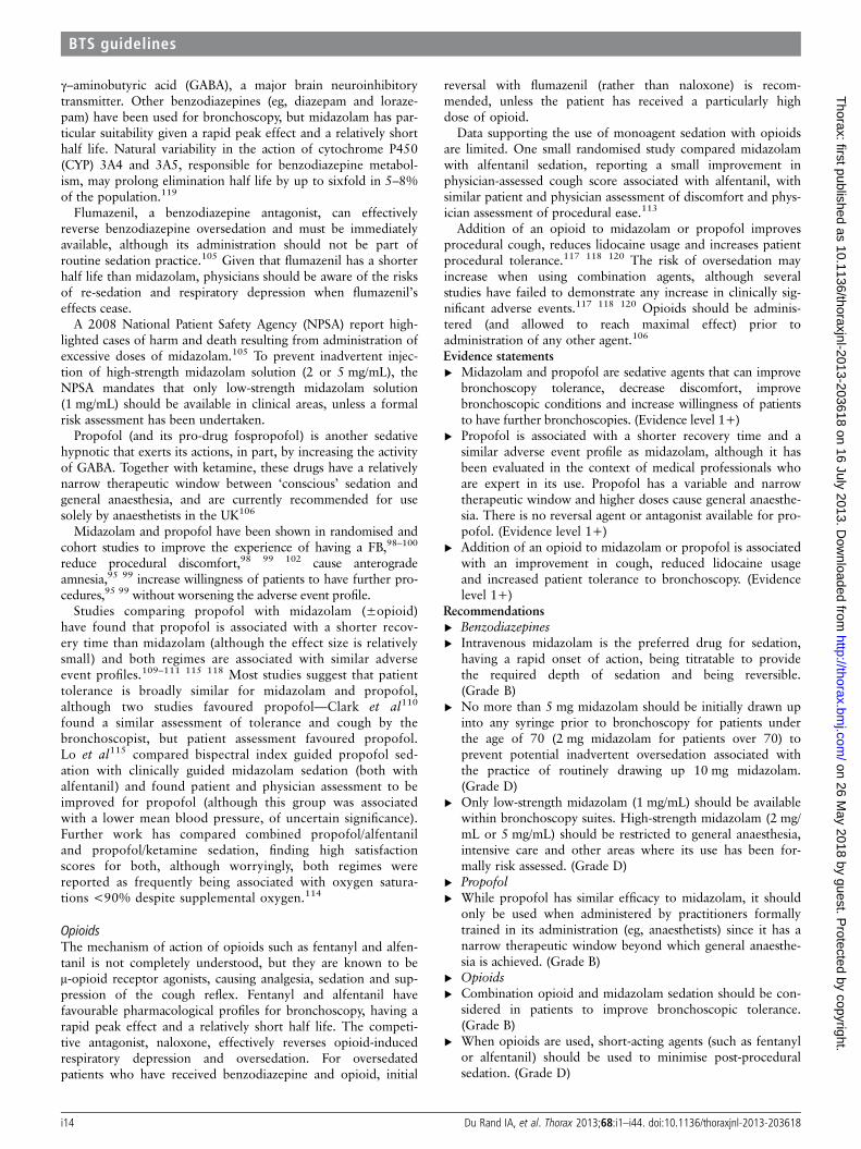
γ–aminobutyric acid (GABA), a major brain neuroinhibitorytransmitter. Other benzodiazepines (eg, diazepam and loraze-pam) have been used for bronchoscopy, but midazolam has par-ticular suitability given a rapid peak effect and a relatively shorthalf life. Natural variability in the action of cytochrome P450(CYP) 3A4 and 3A5, responsible for benzodiazepine metabol-ism, may prolong elimination half life by up to sixfold in 5–8%of the population.119
Flumazenil, a benzodiazepine antagonist, can effectivelyreverse benzodiazepine oversedation and must be immediatelyavailable, although its administration should not be part ofroutine sedation practice.105 Given that flumazenil has a shorterhalf life than midazolam, physicians should be aware of the risksof re-sedation and respiratory depression when flumazenil’seffects cease.
A 2008 National Patient Safety Agency (NPSA) report high-lighted cases of harm and death resulting from administration ofexcessive doses of midazolam.105 To prevent inadvertent injec-tion of high-strength midazolam solution (2 or 5 mg/mL), theNPSA mandates that only low-strength midazolam solution(1 mg/mL) should be available in clinical areas, unless a formalrisk assessment has been undertaken.
Propofol (and its pro-drug fospropofol) is another sedativehypnotic that exerts its actions, in part, by increasing the activityof GABA. Together with ketamine, these drugs have a relativelynarrow therapeutic window between ‘conscious’ sedation andgeneral anaesthesia, and are currently recommended for usesolely by anaesthetists in the UK106
Midazolam and propofol have been shown in randomised andcohort studies to improve the experience of having a FB,98–100
reduce procedural discomfort,98 99 102 cause anterogradeamnesia,95 99 increase willingness of patients to have further pro-cedures,95 99 without worsening the adverse event profile.
Studies comparing propofol with midazolam (±opioid)have found that propofol is associated with a shorter recov-ery time than midazolam (although the effect size is relativelysmall) and both regimes are associated with similar adverseevent profiles.109–111 115 118 Most studies suggest that patienttolerance is broadly similar for midazolam and propofol,although two studies favoured propofol—Clark et al110
found a similar assessment of tolerance and cough by thebronchoscopist, but patient assessment favoured propofol.Lo et al115 compared bispectral index guided propofol sed-ation with clinically guided midazolam sedation (both withalfentanil) and found patient and physician assessment to beimproved for propofol (although this group was associatedwith a lower mean blood pressure, of uncertain significance).Further work has compared combined propofol/alfentaniland propofol/ketamine sedation, finding high satisfactionscores for both, although worryingly, both regimes werereported as frequently being associated with oxygen satura-tions <90% despite supplemental oxygen.114
OpioidsThe mechanism of action of opioids such as fentanyl and alfen-tanil is not completely understood, but they are known to beμ-opioid receptor agonists, causing analgesia, sedation and sup-pression of the cough reflex. Fentanyl and alfentanil havefavourable pharmacological profiles for bronchoscopy, having arapid peak effect and a relatively short half life. The competi-tive antagonist, naloxone, effectively reverses opioid-inducedrespiratory depression and oversedation. For oversedatedpatients who have received benzodiazepine and opioid, initial
reversal with flumazenil (rather than naloxone) is recom-mended, unless the patient has received a particularly highdose of opioid.
Data supporting the use of monoagent sedation with opioidsare limited. One small randomised study compared midazolamwith alfentanil sedation, reporting a small improvement inphysician-assessed cough score associated with alfentanil, withsimilar patient and physician assessment of discomfort and phys-ician assessment of procedural ease.113
Addition of an opioid to midazolam or propofol improvesprocedural cough, reduces lidocaine usage and increases patientprocedural tolerance.117 118 120 The risk of oversedation mayincrease when using combination agents, although severalstudies have failed to demonstrate any increase in clinically sig-nificant adverse events.117 118 120 Opioids should be adminis-tered (and allowed to reach maximal effect) prior toadministration of any other agent.106
Evidence statements▸ Midazolam and propofol are sedative agents that can improve
bronchoscopy tolerance, decrease discomfort, improvebronchoscopic conditions and increase willingness of patientsto have further bronchoscopies. (Evidence level 1+)
▸ Propofol is associated with a shorter recovery time and asimilar adverse event profile as midazolam, although it hasbeen evaluated in the context of medical professionals whoare expert in its use. Propofol has a variable and narrowtherapeutic window and higher doses cause general anaesthe-sia. There is no reversal agent or antagonist available for pro-pofol. (Evidence level 1+)
▸ Addition of an opioid to midazolam or propofol is associatedwith an improvement in cough, reduced lidocaine usageand increased patient tolerance to bronchoscopy. (Evidencelevel 1+)
Recommendations▸ Benzodiazepines▸ Intravenous midazolam is the preferred drug for sedation,
having a rapid onset of action, being titratable to providethe required depth of sedation and being reversible.(Grade B)
▸ No more than 5 mg midazolam should be initially drawn upinto any syringe prior to bronchoscopy for patients underthe age of 70 (2 mg midazolam for patients over 70) toprevent potential inadvertent oversedation associated withthe practice of routinely drawing up 10 mg midazolam.(Grade D)
▸ Only low-strength midazolam (1 mg/mL) should be availablewithin bronchoscopy suites. High-strength midazolam (2 mg/mL or 5 mg/mL) should be restricted to general anaesthesia,intensive care and other areas where its use has been for-mally risk assessed. (Grade D)
▸ Propofol▸ While propofol has similar efficacy to midazolam, it should
only be used when administered by practitioners formallytrained in its administration (eg, anaesthetists) since it has anarrow therapeutic window beyond which general anaesthe-sia is achieved. (Grade B)
▸ Opioids▸ Combination opioid and midazolam sedation should be con-
sidered in patients to improve bronchoscopic tolerance.(Grade B)
▸ When opioids are used, short-acting agents (such as fentanylor alfentanil) should be used to minimise post-proceduralsedation. (Grade D)
i14 Du Rand IA, et al. Thorax 2013;68:i1–i44. doi:10.1136/thoraxjnl-2013-203618
BTS guidelines
on 26 May 2018 by guest. P
rotected by copyright.http://thorax.bm
j.com/
Thorax: first published as 10.1136/thoraxjnl-2013-203618 on 16 July 2013. D
ownloaded from
▸ When combination sedatives are used, opioids should beadministered first and allowed time to become maximallyeffective before administration of any other agent. (Grade D)
Sedation in specific patient groupsPlease refer to appendix 8, tables 1 and 2.
Respiratory failureRisk of oversedation and respiratory depression may beincreased when using combined sedation for patients in respira-tory failure, although one cohort study failed to demonstratethis. Dreher et al120 compared midazolam with combined mida-zolam/alfentanil sedation in 30 patients with type I or II respira-tory failure. A small, but similar increase in partial pressureof CO2 was seen in both groups, and combined therapy wasassociated with improved patient and physician satisfaction.Nevertheless, caution is recommended when administering sed-ation to patients in respiratory failure.
Older patientsOlder patients are likely to require lower doses of sedatives forbronchoscopy and may have prolonged after effects. A study offlunitrazepam (a benzodiazepine) found that, for the same doseof sedative, patients over 60 years old had a prolonged periodof amnesia, with a extended duration of impaired coordination(which should be considered when patients are being dis-charged)121 After reports of harm secondary to oversedation inolder patients undergoing therapeutic gastrointestinal endos-copy, a 2004 National Confidential Enquiry into PatientOutcome and Death recommended that the practice of routinelydrawing up 10 mg midazolam should stop and no more than2 mg should be initially drawn up for patients over 70 years old(5 mg for patients under 70 years old).122 123
ComorbiditiesOther conditions that are likely to require dose modificationinclude hepatic impairment, heart failure and renal impairment.
Concomitant medicationsBenzodiazepines, fentanyl and alfentanil are metabolised byCYP 3A4 and there is a risk of prolonged sedation in thosereceiving concomitant medications that inhibit these enzymes,such as antifungals, antiretrovirals, calcium channel blockers andmacrolide antibiotics.
Substance misusersA cohort study found that substance misusers are likely to needhigher doses of sedatives during bronchoscopy.124
Topical anaesthesiaMethods for providing airway topical anaesthesiaLidocaine is the most commonly used drug for topical anaesthe-sia of the upper airways and tracheobronchial tree, and a varietyof preparations and modes of application are used. Lidocainestabilises the neuronal membrane by inhibiting ionic fluxes,thereby preventing the initiation and conduction of actionpotentials. Topical anaesthesia has been achieved using 2% gel(nasal), 10% spray (nasal and oropharyngeal) and 1–4% solu-tion (‘spray-as-you-go’ via the bronchoscope working channel;injection into the trachea through the cricothryoid membrane;nebulised; using a dropper in the oropharynx; and applied tothe nasal passages using soaked pledgets).
Lidocaine administered to the larynx and tracheobronchialtree significantly reduces incidence of cough and stridor, and
reduces the requirement for sedative drugs.125 Other drugs,such as benzocaine and tetracaine, have been used, but lidocaineis the best characterised with a low risk of adverse effects(including methaemoglobinaemia)126 Cocaine has previouslybeen used for nasal anaesthesia, but is no longer routinely avail-able in the UK and can be associated with myocardial ischaemiaand infarction.127 128
Nasal topical anaesthesiaNasal lidocaine is most effectively provided using lidocaine gel.In the UK, 2% lidocaine gel preparations are available involumes of 6 mL, although smaller volumes may be sufficient.Several studies have demonstrated that patients prefer 2% lido-caine gel to lidocaine spray, lidocaine-soaked swabs or EMLAcream.129–131
Oropharyngeal topical anaesthesiaOropharyngeal topical anaesthesia is conventionally providedusing 10% lidocaine spray (10 mg per actuation, usually two tofive actuations), although nebulised lidocaine has also beenused. One small randomised study132 found similar efficacy incough suppression of 10% lidocaine spray and nebulised 4%lidocaine, although the groups were not well matched (particu-larly smoking status) and subsequent studies have suggested alack of efficacy of nebulised lidocaine (see below).
Laryngeal and tracheobronchial topical anaesthesiaSeveral techniques may be used to administer lidocaine to thelarynx and lower airways, including:▸ ‘Spray-as-you-go’ delivery, in which lidocaine is applied via
the bronchoscope working channel. Repeated applicationallows lidocaine delivery to the entire airway.
▸ Direct injection into the upper trachea using a needle passedthrough the cricothyroid membrane, allowing lidocainedelivery to the larynx and trachea prior to bronchoscopeinsertion. Further lidocaine doses to the bronchial tree maybe administered as required via the bronchoscope.Spray-as-you-go and cricothyroid lidocaine have had limited
comparative evaluation. One small randomised study found adecreased cough rate, but similar patient experience, associatedwith cricothyroid administration (vs spray-as-you-go).133 Afurther small randomised study found bronchoscopic condi-tions, but not patient assessment, were improved for cricothyr-oid administration compared with nebulised lidocaine.134
The evidence for use of nebulised lidocaine is conflicting. Thelargest randomised controlled trial 135 (150 patients) of nebu-lised 4% lidocaine versus saline placebo found no benefit tonebulised lidocaine (comparing additional doses of lidocainerequired, and cough and comfort scores), while doubling thetotal dose of lidocaine administered in those who received nebu-lised treatment. All patients also received lidocaine spray to thenasopharynx and oropharynx, a small volume of lidocaine solu-tion to the vocal cords and main bronchi, and sedation.
The optimal concentration of lidocaine solution has beeninvestigated in three studies, which demonstrated that volume,rather than concentration, of lidocaine determined efficacy ofairway topical anaesthesia. A randomised spray-as-you-go study,comparing the same volume of 1% and 2% lidocaine (deliveredto the larynx and tracheobronchial tree), found no difference insubjective and objective measures of cough suppression.136
Another randomised study compared similar volumes of 1%,1.5% and 2% lidocaine trickled onto the back of the patient’stongue, which was allowed to be aspirated into the larynx.137
Although a relatively small study, 1% was found to be equally
Du Rand IA, et al. Thorax 2013;68:i1–i44. doi:10.1136/thoraxjnl-2013-203618 i15
BTS guidelines
on 26 May 2018 by guest. P
rotected by copyright.http://thorax.bm
j.com/
Thorax: first published as 10.1136/thoraxjnl-2013-203618 on 16 July 2013. D
ownloaded from
efficacious at cough suppression and had similar requirementsfor additional lidocaine during bronchoscopy. In a randomisedawake fibre-optic intubation study,138 lidocaine was deliveredusing spray-as-you-go to the hypopharynx and larynx untiladequate topical anaesthesia for intubation was produced. Thesame volume of 2% lidocaine solution was found to be as effect-ive as 4% (1% lidocaine solution was not evaluated).Evidence statements▸ Topical anaesthesia with lidocaine reduces cough and
decreases sedation requirements. (Evidence level 1−)▸ While other topical anaesthetics such as benzocaine, tetra-
caine and cocaine have been studied, lidocaine is the bestcharacterised and has a lower risk of methaemoglobinaemia.(Evidence level 3)
▸ Good quality randomised studies suggest lower concentrationlidocaine is as effective as higher concentrations in achievingcough amelioration during bronchoscopy. (Evidence level 1+)
Recommendations▸ Lidocaine should be used for topical anaesthesia during
bronchoscopy, unless contraindicated. (Grade A)▸ Nasal topical anaesthesia is most effectively provided using
2% lidocaine gel. (Grade A)▸ Both cricothyroid and spray-as-you-go techniques are effect-
ive in delivering lidocaine to the vocal cords and trachea.(Grade B)
▸ Nebulisation is not recommended as a technique for deliver-ing lidocaine to the airways. (Grade B)
▸ 1% lidocaine solution should be used for spray-as-you-goadministration. (Grade A)
The maximal safe dose of lidocaineLidocaine has a favourable safety profile when used for bron-choscopy, although high topical doses may cause highserum concentrations, associated with subjective and life-threatening objective signs of toxicity (particularly affectingthe central nervous and cardiovascular systems—see appendix 8,table 1).
Peak serum concentrations usually occur from 20 to 45 minafter topical application,137–139 although concentrations ofactive (but less potent) metabolites (eg, monoethylglycinexyli-dide) continue to rise for at least 2 h.140 Several studies haveexamined the maximal safe dose of lidocaine, with conflictingresults. Lidocaine toxicity is conventionally accepted to be likelyabove serum concentrations of 5 mg/mL, although studiessuggest that subjective side effects may occur at much lowerserum concentrations;
Frey et al141 studied 154 patients who received a mean doseof 15.4 mg/kg lidocaine (including nebulised lidocaine and nasalgel lidocaine). A total of 62% of patients had euphoria or dizzi-ness (subjective signs of toxicity) despite only one patient havingserum concentration >5 mg/mL.
Martin et al142 studied 39 volunteer healthcare workershaving unsedated bronchoscopy who received a median dose of9.6 mg/kg lidocaine (including nebulised and nasal gel lido-caine). A total of 92% had subjective side effects.
Other studies failed to demonstrate side effects with averagelidocaine doses of 7.1–9.3 mg/kg.137 139 140 142 However, thesedata should be interpreted with caution given significant inter-patient variability between dose administered and serum con-centration (partly due to unpredictable absorption at differentparts of the respiratory tract), with serum concentrations>5 mg/mL occasionally seen even for lower doses of adminis-tered lidocaine.137 140 142 143 Given randomised studies suggest-ing that a low cough score is achievable with a total lidocaine
dose of less than 160 mg,118 135 bronchoscopists should striveto use the lowest dose of lidocaine possible, while still ensuringgood bronchoscopic conditions and patient comfort.
While lidocaine absorption varies throughout the respiratorytract, bronchoscopists should assess and record the total lidocainedose administered during bronchoscopy (including nasal, oropha-ryngeal, laryngeal and tracheobronchial lidocaine). Appendix 8,table 3 illustrates the lidocaine dose associated with various formula-tions and concentrations.
Factor influencing the risk of lidocaine toxicity duringbronchoscopyPlease refer to appendix 8.
Risk of toxicity increases in those with hepatic and cardiacdysfunction, and significant renal impairment. Care should betaken with older patients, although one small study found nodifference in adverse events associated with bronchoscopic lido-caine administration (mean dose 10.6 mg/kg) when comparingpatients aged 30–50 years with those aged 60–75 years.144
Evidence statements▸ For the same dose of administered lidocaine, there is signifi-
cant inter-patient variability in the corresponding peak serumlidocaine concentration. (Evidence level 1+)
▸ Doses up to 15.4 mg/kg may be used without serious adverseevents, but subjective symptoms of lidocaine toxicity (eg, diz-ziness, euphoria) were reported in studies using ≥9.6 mg/kglidocaine. (Evidence level 2+)
▸ Low total doses of lidocaine <160 mg may be associatedwith effective cough suppression. (Evidence level 1+)
Recommendations▸ To reduce the risk of lidocaine toxicity, bronchoscopists
should use the lowest dose of lidocaine sufficient to preventexcessive coughing and provide patient comfort. (Grade D)
▸ Bronchoscopists should remain vigilant for objective and sub-jective symptoms of lidocaine toxicity, particularly given sig-nificant inter-patient variability in lidocaine absorption andmetabolism. (Grade B)
Good practice point▸ Bronchoscopists should monitor and document the total
lidocaine dose delivered at all sites during bronchoscopy. (√)
SAMPLING AND DIAGNOSTIC ACCURACYBronchoscopy is an important tool in the diagnostic pathwayfor patients with potential lung cancer, with interstitial lungdisease and with pulmonary infection. A variety of samplingmethods can be used during FB. A how to guide on samplingtechniques can be found in appendix 10.
Lung cancerIn patients with lung cancer, a chest CT scan should beperformed prior to a bronchoscopy and patients with centrallesions on the CT scan should be offered bronchoscopy if thenodal status does not influence the management decision. Thediagnostic yield from bronchoscopy depends on whetherthe tumour is visible within the bronchial tree. Many studieshave demonstrated the sensitivity of bronchial biopsy withforceps, which ranges from 48% to 93% (American College ofChest Physicians guidelines 2007, table 2).145
Studies have shown that the addition of bronchial brush speci-mens and washings increases the diagnostic yield of bronchos-copy in patients with lung cancer. The systematic review andmeta-analysis carried out by the American College of ChestPhysicians145 identified 35 studies of patients with centraldisease undergoing bronchoscopy. The pooled data in 4507
i16 Du Rand IA, et al. Thorax 2013;68:i1–i44. doi:10.1136/thoraxjnl-2013-203618
BTS guidelines
on 26 May 2018 by guest. P
rotected by copyright.http://thorax.bm
j.com/
Thorax: first published as 10.1136/thoraxjnl-2013-203618 on 16 July 2013. D
ownloaded from

patients demonstrated an overall combined sensitivity of 88%for bronchoscopic techniques. Biopsy of visible lesions alonehad a yield of 74% while washings had a sensitivity of 48% andyield from brushings was 59%. McLean et al146 analysed 2238bronchoscopies carried out between January 1991 andDecember 1992 in five centres in Scotland. When endobron-chial tumour was seen, sensitivity for brushings and washingscombined was 47%. The yield from biopsy alone was 82% andwhen combined with brushings and washings the yield increasedto 87%. When the tumour was not visible the sensitivity ofbrushings and washings combined was 9%.146
The optimal number of bronchial biopsy specimens in casesof visible endobronchial tumour has also been studied.,Gellert147 demonstrated that at least five samples are required toachieve a 90% probability of a positive malignant biopsy for avisible tumour.147 Popovich et al148 demonstrated in peripherallung cancer lesions biopsied using TBLB that only a 45% diag-nostic yield was achieved with one biopsy and in some cases thediagnosis was not achieved before the fifth or sixth biopsy. Theysuggested that up to 10 biopsies may be required.
Based on recent evidence, it is reasonable to expect a diagnos-tic yield of 85% when the tumour is visible at bronchoscopyusing bronchial biopsy.149 150 Slade et al149 examined the diag-nostic yield from FB at a single centre between 2001 and 2007.In patients with tumour visible at bronchoscopy, the yieldincreased from 75% in 2001 to 94.5% in 2007.
The management of patients with advanced NSCLC nowdepends on the phenotyping and genotyping of lung cancer.151
However, the currently available evidence does not clarifywhether this is routinely possible with bronchoscopic samplesand in particular whether bronchial brushings and washings aresuitable for subclassification of NSCLC. Studies of thesequence of biopsies, brushings and washings suggest that theorder of sample acquisition is not important.152 However, tomaximise the volume of tissue to allow the phenotyping andgenotyping of advanced NSCLC, bronchial brushings andwashings may be taken after sufficient biopsy specimens havebeen taken.
In addition to providing pathological information, bronchos-copy enables the location and extent of an endobronchial lesionto be determined. Careful assessment of the vocal cords ismandatory, since the presence of vocal cord palsy may provideevidence of mediastinal invasion (and thus inoperability).Bronchoscopy also allows for accurate assessment of the loca-tion and therefore T staging of an endobronchial tumour whichmay have implications for surgical treatment.
Recommendations for FB in performing conventional TBNAand endobronchial ultrasound (EBUS) TBNA as a diagnostictechnique for lung cancer are discussed in the BTS guideline foradvanced diagnostic and therapeutic FB in adults, which alsoincludes a guide on how to perform TBNA.3
Evidence statements▸ In patients with visible endobronchial tumour a diagnostic
yield of at least 85% is achievable. (Evidence level 2+)▸ A minimum of five biopsies should be taken when endobron-
chial tumour is visible. (Evidence level 3)▸ The diagnostic yield from bronchoscopy can be increased by
undertaking brush and lavage specimens in addition to bron-chial biopsy. (Evidence level 3)
Recommendations▸ A diagnostic level of 85% should be attainable when definite
endobronchial tumour is visible. (Grade B)▸ At least five biopsy samples should be taken when endobron-
chial tumour is visible to maximise diagnostic yield and the
volume of biopsy tissue and to allow for tumour phenotyp-ing and genotyping. (Grade D)
▸ When endobronchial tumour is visible, brushings and wash-ings can increase the diagnostic yield of the procedure.(Grade D)
▸ A chest CT scan should be performed prior to a diagnosticbronchoscopy in patients with suspected lung cancer.(Grade D)
Interstitial lung diseaseBronchoscopy is a useful diagnostic tool in some ILDs, particu-larly sarcoidosis, hypersensitivity pneumonitis and organisingpneumonia.153 BAL and TBLB are frequently performed in sus-pected cases of ILD.
TBLB is a technique which uses biopsy forceps to obtainalveolar tissue; it is particularly useful in obtaining histologicalspecimens for suspected cases of ILD, although crush artefactduring the procedure may render the samples inadequate forhistological diagnosis, particularly in idiopathic pulmonaryfibrosis. The size of forceps used does not appear to affect thediagnostic yield (see appendix 10 for a step-by-step guide onhow to perform TBLB).153 A retrospective review of 452 bron-choscopies in localised and diffuse lesions examined by TBLBdemonstrated a diagnostic yield in 55.2% of patients with alocalised lesion and 67% in those with diffuse lesions. Thestudy also demonstrated an increase in the diagnostic yield withan increasing number of specimens, so that taking more thanfour biopsies increased the yield from 52% to more than70%.43 The complication rate in this series was 6%; 5.8%developed a pneumothorax (3.8% required intercostaldrainage) and significant bleeding was noted in 0.2%, suggestingthat TBLB is a useful diagnostic procedure with a low complica-tion rate43
The use of BAL may be controversial, since its use in diagno-sis and monitoring disease activity is limited in ILD, and itscurrent strength is in research in understanding the underlyingpathophysiological mechanisms in ILD.154 There is a suggestionthat combining techniques increases diagnostic yield. In a studyinvestigating patients with sarcoidosis, TBLB alone was diagnos-tic in 58%, TBNA alone was successful in diagnosing 17% ofcases as was BAL alone; combining all three procedures led to a100% diagnostic rate.155
No evidence currently exists for the coexistent use of BALwith TBLB, however it is reasonable to perform both these pro-cedures in cases of suspected ILD. BAL and TBLB should beperformed in areas shown to be affected on high-resolution CT(HRCT)156 and both procedures should be performed on thesame lung. In those with typical HRCT features of idiopathicpulmonary fibrosis, BAL and TBLB are not required fordiagnosis.
Diagnostic bronchoscopic procedures have moderate(67–74%) diagnostic sensitivity in patients with diffuseILD,157 158 but have high diagnostic sensitivity in cases of sar-coidosis. In a study of 530 consecutive TBLBs in patients withdiffuse infiltrates, the overall diagnostic yield was 50% and 75%for stage II–III sarcoidosis. The diagnostic yield of hypersensitiv-ity pneumonitis in this series was 92%. In a case series of 183patients with bilateral diffuse shadowing, Mitchell et al159
demonstrated that TBLB had a diagnostic yield of 77% forpatients with sarcoidosis.
The diagnostic sensitivity of TBLB and BAL in sarcoidosis isestimated to be between 56% and 77%, increasing with theseverity of the disease.157 159–164 One study demonstrated thesensitivity of TBLB to be 55% in stage I disease, 80% in stage II
Du Rand IA, et al. Thorax 2013;68:i1–i44. doi:10.1136/thoraxjnl-2013-203618 i17
BTS guidelines
on 26 May 2018 by guest. P
rotected by copyright.http://thorax.bm
j.com/
Thorax: first published as 10.1136/thoraxjnl-2013-203618 on 16 July 2013. D
ownloaded from
disease and 82% in stage III disease.162 In sarcoidosis, EBB mayalso prove useful, even without visible endobronchial abnormal-ity, particularly when parenchymal disease exists.163 Combiningtechniques will improve diagnostic yield.160 161 Mediastinallymphadenopathy can be sampled by conventional or ultra-sound guided TBNA—please refer to the BTS guideline foradvanced diagnostic and therapeutic FB in adults for specificrecommendations and evidence.3
A minimum of five to six TBLB samples should be takenand increasing the number of samples will increase the diag-nostic yield, particularly in those with a localised lesion.164
A direct correlation has been shown between the number ofTBLBs taken and diagnostic yield, with a 35% diagnosticyield for one to three biopsies, increasing to 69% with 6–10biopsies.164 Furthermore, Popovich et al148 demonstrated inperipheral lung cancer lesions biopsied using TBLB that onlya 45% diagnostic yield was achieved with one biopsy and insome cases the diagnosis was not achieved before the fifthor sixth biopsy. They suggested that up to 10 biopsies maybe required.
There is conflicting evidence regarding the use of fluoroscopyduring TBLB. A retrospective review by Anders et al of 112TBLBs performed with fluoroscopy and 135 without fluoros-copy found no significant difference in the yield (72.3% vs67.4%); however the yield for localised lesions was lowerwithout fluoroscopy.165 Fluoroscopy may be useful when per-forming TBLB on localised peripheral lesions. The diagnosticrate of TBLB without fluoroscopy varies between 60% and65%,159 166 and with fluoroscopy 55.2–73%43 165 for localisedlesions. It has therefore been suggested that fluoroscopy may beuseful when performing TBLB on localised peripheral lesions.Two studies have demonstrated that knowledge of the locationof the lesion from chest radiograph aids diagnostic yield. Toensure that peripheral localised lesions are being accessed cor-rectly, fluoroscopy may therefore be required.43 167 The inci-dence of pneumothorax following TBLB without fluoroscopy isbetween 3% and 5%,166 168 although one study demonstratedno increase in the incidence of pneumothoraces (0.015%).157 Achest x-ray may be performed immediately if pneumothorax issuspected or the patient is symptomatic, or be done at least 2 hafter TBLB to exclude pneumothorax.47
Evidence statements▸ EBBs should be performed in patients with suspected sar-
coidosis. Yield increases in patients with radiographic evi-dence of parenchymal disease. (Evidence level 2+)
▸ TBLBs should be performed in patients with radiographicstage II—IV sarcoidosis. (Evidence level 2+)
▸ At least five to six TBLBs should be performed in patientswith ILD. (Evidence level 2−)
▸ There is no convincing evidence that the use of fluoroscopyreduces pneumothorax rate or diagnostic yield in non-focallung disease. (Evidence level 3)
Recommendations▸ In suspected sarcoidosis, EBBs should be considered to
increase the diagnostic yield. (Grade C)▸ TBLB is recommended for the diagnosis of stage II–IV sar-
coidosis. (Grade C)▸ In patients with diffuse ILD, five to six TBLBs should be
taken from the same lung. (Grade D)▸ Fluoroscopy should be considered for TBLB in patients with
localised or focal parenchymal lung disease. (Grade D)Good practice point▸ Bronchoscopists should keep a record of their personal diag-
nostic accuracy for FB. (√)
Diagnosis of infectionBronchoscopy is a useful procedure to diagnose infection whenless invasive methods to diagnose the underlying aetiology arenot suitable.
This section includes basic sampling methods (bronchialwash, BAL, TBLB, EBB and bronchial brush) but does notinclude advanced techniques such as EBUS TBNA, which arecovered in the advanced bronchoscopy guidelines.3
The spectrum of infections encountered in immunocomprom-ised hosts differs from that seen in immunocompetent indivi-duals. The role of FB in obtaining diagnostic samples in thesetwo patient groups also differs
Bronchoscopy in the diagnosis of infection in theimmunocompromised hostThe underlying aetiology for pneumonia in patients who areimmunocompromised varies with the prevalence of disease inthe community and with geographical area. Studies performedin Africa where there is a high prevalence of TB have shown themost common organism causing pneumonia in patients withHIV-related immunosuppression is mycobacterium TB.169 170 InWestern countries with a lower prevalence of TB, commonlyencountered organisms are Pneumocystis jiroveci, fungal infec-tions, bacterial infections and viral infections such as cytomega-lovirs and Cryptococcus.171–173 A variety of bronchoscopictechniques can be used depending on the local population,disease prevalence and host defences (figure 1).
Bronchoalveolar lavageBAL has been shown to have high sensitivity in P jiroveci pneu-monia. In the absence of prior antibiotic use for treatment orprophylaxis, BAL is reported to have a sensitivity between 90%and 98% for P jiroveci.174–179 Prior use of empiric antibiotictreatment reduces the sensitivity to around 64%.178 BilateralBAL sampling has a higher diagnostic yield compared with uni-lateral BAL179 and BAL from upper lobes has higher sensitivitycompared with lower or middle lobe sampling techniques.180 181
The comparable sensitivity of BAL to transbronchial biopsy,176
but with fewer inherent risks, means that BAL is regarded as thegold standard investigation for suspected P jiroveci infection.
BAL in patients who are immunocompromised with smear-negative pulmonary TB has been reported to have a sensitivityof 10–30% based on microscopy182–184 and a sensitivity of52–95% on culture.182–184 When PCR techniques are used onBAL material, a rapid diagnosis of TB with reported sensitivityof 85.7% and specificity of 90.9%185 have been reported.Serological tests on BAL in patients with HIV (unlike thosewithout HIV) with suspected pulmonary TB have shown alower sensitivity, but the specificity remains high.185
In invasive aspergillosis, BAL is reported to show characteris-tic hyphae in 34–64%186–188 and to be positive on cultures forAspergillus in 23–85% of cases.186–188 If available, galactoman-nan antigen detection on BAL is useful. A meta-analysis lookingat the performance of BAL galactomannan testing in detectionof proven invasive aspergillosis suggested that the test has a sen-sitivity of 94% and a specificity of 79%.189 Unlike the serumgalactomannan test, BAL galactomannan is equally sensitive inpatients with haematological or other causes of immunosuppres-sion.190 There have been reports of false-positive results whenusing the serum galactomannan test, especially in patients whoare receiving certain β-lactam antibiotics.191–194 The reportedsensitivity for PCR on BAL in cases of invasive aspergillosis is67–100% and the specificity 96–100%.195 196 The PCR
i18 Du Rand IA, et al. Thorax 2013;68:i1–i44. doi:10.1136/thoraxjnl-2013-203618
BTS guidelines
on 26 May 2018 by guest. P
rotected by copyright.http://thorax.bm
j.com/
Thorax: first published as 10.1136/thoraxjnl-2013-203618 on 16 July 2013. D
ownloaded from
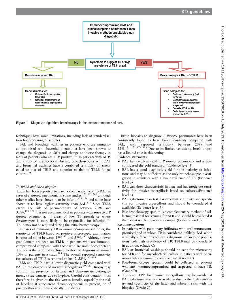
techniques have some limitations, including lack of standardisa-tion for processing of samples.
BAL and bronchial washings in patients who are immuno-compromised with bacterial pneumonia have been shown tochange the diagnosis in 50% and change antibiotic therapy in62% of patients who are HIV positive.197 In patients with AIDSand suspected cryptococcal disease, bronchoscopies with BALand bronchial washings have a combined sensitivity on smearequal to that of TBLB and superior to that of TBLB fungalculture.198
TBLB/EBB and brush biopsiesTBLB has been reported to have a comparable yield to BAL incases of P jiroveci pneumonia in some studies,171 199 200 althoughother studies have shown it to be inferior175 176 and some haveshown it to have higher sensitivity than BAL.177 Since TBLBcarries the risk of pneumothorax of between 2.5% and3.7%,176 177 it is not recommended in patients with suspected Pjiroveci pneumonia. In areas of low TB prevalence wherePneumocystis is more likely to be responsible for infection,173
TBLB may not be required during the initial bronchoscopy.In cases of pulmonary TB in immunocompromised hosts, the
sensitivity of TBLB based on positive microscopic examinationis reported to be between 19%183 and 39%.182 Although fewergranulomata are seen on TBLB in patients who are immuno-compromised compared with those who are immunocompetent,TBLB was the reported exclusive method of diagnosis in around13% of patients in a study.183 The overall reported sensitivityfor cultures of TBLB is reported to be 42–52%.182–184
EBB and TBLB have a lower diagnostic yield compared withBAL in the diagnosis of invasive aspergillosis.197 201 Biopsy mayconfirm the presence of hyphae and demonstrate pathogno-monic tissue damage due to hyphae. Careful consideration musttherefore be given to the risk versus benefit, especially the riskof bleeding if concurrent thrombocytopenia is present, or ofpneumothorax in these critically ill patients.
Brush biopsies to diagnose P jiroveci pneumonia have beenconsistently found to have lower sensitivity compared withBAL, with reported sensitivity between 29% and52%.171 175 176 199 Due to its limited sensitivity, brush biopsyhas a limited role in this setting.Evidence statements▸ BAL has excellent yield in P jiroveci pneumonia and is now
considered the gold standard. (Evidence level 3)▸ BAL has a good diagnostic yield for the majority of infec-
tions and may be sufficient as the only bronchoscopic investi-gation in countries with a low prevalence of TB. (Evidencelevel 3)
▸ BAL can show characteristic hyphae and has moderate sensi-tivity for invasive aspergillosis based on cultures.(Evidencelevel 3)
▸ BAL galactomannan test has excellent sensitivity and specifi-city for invasive aspergillosis and should be considered ifavailable. (Evidence level 3)
▸ Post-bronchoscopy sputum is a complementary method of col-lecting material for staining for AFB and should be collected ifthe patient is able to provide a sample. (Evidence level 3)
Recommendations▸ In patients with pulmonary infiltrates who are immunocom-
promised and in whom TB is considered unlikely, BAL aloneis usually sufficient to achieve a diagnosis. In areas or popula-tions with high prevalence of TB, TBLB may be consideredin addition. (Grade C)
▸ BAL or bronchial washings should be sent for microscopyfor AFB and for mycobacterial culture in patients with pneu-monia who are immunocompromised. (Grade C)
▸ Post-bronchoscopy sputum could be collected in patientswho are immunocompromised and suspected to have TB.(Grade D)
▸ TBLB and EBB for invasive aspergillosis may be avoided ifBAL galactomannan test is available due to the high sensitiv-ity and specificity of the latter and inherent risks with thebiopsies. (Grade C)
Figure 1 Diagnostic algorithm: bronchoscopy in the immunocompromised host.
Du Rand IA, et al. Thorax 2013;68:i1–i44. doi:10.1136/thoraxjnl-2013-203618 i19
BTS guidelines
on 26 May 2018 by guest. P
rotected by copyright.http://thorax.bm
j.com/
Thorax: first published as 10.1136/thoraxjnl-2013-203618 on 16 July 2013. D
ownloaded from
▸ In patients suspected to have invasive aspergillosis, BALshould be sent for microscopy for hyphae and fungalculture; a BAL galactomannan test should be considered tofurther improve diagnostic yield. (Grade C)
Bronchoscopy in the diagnosis of infection in theimmunocompetent hostThere are no randomised trials to compare the outcome of inva-sive tests such as bronchoscopy in the early phase of infectionwith non-invasive tests and/or empirical treatment in cases ofcommunity-acquired pneumonia. The current guidelines onmanagement of community-acquired pneumonia recommendthe use of non-invasive investigations. In patients with moderateto severe disease, bronchoscopy should be reserved for patientswhose condition fails to respond.202 203 The role of bronchos-copy in the early phase of management of a patient withcommunity-acquired pneumonia may be quite limited.
Bronchoscopy should be considered in patients whose condi-tion fails to respond to initial treatment for community-acquiredpneumonia. In this setting, FB can help to exclude underlyinglung cancer which can be present in a significant proportion ofpatients with non-resolving pneumonia204 FB is also useful inthis patient group to diagnose and remove aspirated organic orinorganic material. In adults, the presence of a non-resolvingpneumonia, lung abscess, atelectasis or collapse can indicatepossible aspiration.205 FB is the first-line investigation of choicein adults to remove aspirated material with reported successrates of up to 100% in experienced hands.205 A bronchoscopyin patients whose condition has failed to respond to treatmentprovides an opportunity to collect additional samples for micro-bial cultures.
In patients who are suspected to have TB but are unable toprovide an adequate sputum sample, bronchoscopy can be used asan alternative to induced sputum to obtain material for staining.The overall combined yield of bronchoscopy in patients with nega-tive sputum smears, with procedures such as BAL, bronchial biop-sies, bronchial wash and post-bronchoscopy sputum, is reported tobe 28.8–48.3%206 207 based on microscopy and smears for AFBand 69.2–94%206 208 209 based on cultures.
BAL and bronchial aspirateBronchial aspirates obtained for the purpose of culture and sen-sitivity have a significant false-positive rate due to contaminationwith upper respiratory tract secretions.210 Protected cathetershave a lower risk of contamination.211 Protected catheter andBAL samples have a reported sensitivity based on quantitativecultures of 64.7–70% and 73.8–77% respectively in patientswith community-acquired pneumonia who had a bronchoscopywithin 12 h of hospital admission.212 213 The yield is lowerwhen bronchoscopy is performed after treatment failure (lack ofresponse after 72 h).214
BAL also provides suitable material for other tests such asPCR and antigen detection. Performing the pneumococcalantigen test on BAL is reported to have a sensitivity of 54–95%and a specificity of 86.8–100% for pneumococcal pneumo-nia.215 216 When available, use of PCR on BAL has been shownto be useful in the detection of legionella,217 218 with reportedsensitivity and specificity approaching 100%.218 It has been sug-gested that samples should routinely be sent for legionella PCRif bronchoscopy is performed.202
In patients with suspected TB and negative sputum smear,BAL and bronchial aspirates range in sensitivity from 23% to28% on smears207 219 and 32–88% on cultures.207 208 219
Studies have shown a high sensitivity of PCR when performed
on BAL fluid with a range of 78–80.9%.220 221 The ability tomake an early diagnosis by using PCR can be further enhancedby performing BAL smears at the same time.221 BAL PCRappears to have a false-positive rate of around 4.4%.220 Tests onBAL, such as enzyme-linked immunospot have high sensitivityand specificity and can rapidly distinguish between smear-negative pulmonary TB and latent TB infection.222 223
EBB and TBLBEBB or TBLB are useful in the diagnosis of slowly or non-resolving pneumonia if there are any visible endobronchialabnormalities or if there is a possibility of TB. There is no evi-dence to suggest they are useful as a routine test in patients withpneumonia.
In patients with proven pulmonary TB, bronchial and brushbiopsies show typical granulomata in 3.84–53% ofcases.206 219 224 Post-bronchoscopy sputum is culture positive in14–46% of patients and sputum should be cultured if the patientis able to provide an adequate sample.207 208
Evidence statements▸ A significant proportion of patients (11%) with non-resolving
pneumonia, who are over 50 and are ex-smokers or currentsmokers, may have underlying carcinoma. Bronchoscopy canplay an important role in diagnosing endobronchial lesions,especially if there is an additional history of weight loss of>3 kg in the preceding 3 months. (Evidence level 2−)
▸ PCR for legionella on BAL specimens has a sensitivityapproaching 100%. (Evidence level 2+)
▸ In TB, BAL, bronchial aspirates and post-bronchoscopysputum all contribute to the overall diagnostic yield.(Evidence level 2+)
▸ In high-prevalence areas, routine samples for culture andmicroscopy for TB confirm TB in 6–8% of patients, even ifbronchoscopy is performed for another indication. (Evidencelevel 2+)
Recommendations▸ Bronchoscopy may be considered in patients with non-
resolving or slowly resolving pneumonia, especially if theyare current or ex smokers and older than 50 years. (Grade C)
▸ If bronchoscopy is performed for community-acquired pneu-monia, BAL specimens should be sent for legionella PCRand atypical pathogens. (Grade C)
▸ Bronchoscopy may be considered if the patient is suspectedto have TB when sputum smear is negative. (Grade C)
▸ In cases of suspected TB, BAL, bronchial aspirates, and post-bronchoscopy sputum appear to be complementary andshould all be analysed. (Grade C)
▸ In areas with high or intermediate prevalence of TB, patientsundergoing bronchoscopy for another indication should havesamples sent routinely for cultures for TB. (Grade C)
BRONCHOSCOPY IN THE ICUFB facilitates inspection of the upper airways and bronchial treein critically ill patients in the ICU. In non-intubated patients therisks of bronchoscopy are those described in the section on‘Monitoring, precautions and complications’. However, bron-choscopy in the ICU commonly involves intubated patients sup-ported by mechanical ventilation, with significant other illnessand comorbidities, and therefore bronchoscopy requires carefulconsideration in this patient population.
Bronchoscopes used in the ICUThe specifications of a bronchoscope suitable for use in the ICUrequire a degree of compromise. The internal diameter of the
i20 Du Rand IA, et al. Thorax 2013;68:i1–i44. doi:10.1136/thoraxjnl-2013-203618
BTS guidelines
on 26 May 2018 by guest. P
rotected by copyright.http://thorax.bm
j.com/
Thorax: first published as 10.1136/thoraxjnl-2013-203618 on 16 July 2013. D
ownloaded from
ET may restrict the size of the bronchoscope, while efficient suc-tioning requires a larger bronchoscope with a wide suctionchannel. The internal diameter of the tracheal tube relative tothe external diameter of the bronchoscope is an important con-sideration. Bronchoscopes in the non-intubated patient occupyonly 10–15% of the cross-sectional area of the trachea. In con-trast, a 5.7 mm bronchoscope occupies 40% of a 9 mm ET and66% of a 7 mm tracheal tube. Failure to recognise this may leadto inadequate ventilation of the patient and impaction of ordamage to the bronchoscope.
Lubrication is essential to facilitate passage of the broncho-scope. Tracheostomy tubes are prone to damage the broncho-scope, particularly during withdrawal when the rigid edge ofthe end of the tracheostomy tube can abrade the covering of thebronchoscope.
A single model (non-human) study addresses the issue ofairway support device and size of bronchoscope.225 In thisstudy, the laryngeal mask airway (LMA), reinforced LMA(RLMA) and ETwere assessed in a simulated model of ventila-tion, measuring flow resistance during simulated inspiration andthe maximum size of flexible bronchoscope (using oesophagealdilators to mimic six different sizes of bronchoscope(2.7–8.9 mm external diameter)). This study demonstrated thatthe LMA can accommodate a larger bronchoscope than the cor-responding sizes of RLMA and ET. Mean flow resistance was2.3 (1.7–3.5) times higher with the RLMA and 2.1 (1.2–4.2)times higher with the ET compared with the LMA, and 1.2(0.7–1.8) times lower with the ET compared with the RLMA.Removal of the LMA aperture bars resulted in a mean decreasein flow resistance of 3.6%. The study provides useful informa-tion on the maximum external diameter of the bronchoscope tobe used with different airway devices. This information is pro-vided in appendix 11 of this guideline.Recommendation▸ The external diameter of a bronchoscope used in the ICU
setting should be carefully selected according to the externaldiameter of the bronchoscope, the size of the airway supportdevice (ET or laryngeal mask) and the type of airway deviceused. (Grade D)
Indications for FB in the ICUFB may be performed in the ICU for a wide range of diagnosticor therapeutic indications. These are considered separately inthe sections below.
Treatment of lobar collapseBronchoscopy has been used to treat lobar collapse in intubatedpatients whose condition has failed to respond to treatmentssuch as physiotherapy. Retained bronchial secretions mayobstruct major airways, and local directed suction using a widechannel bronchoscope combined with saline instillation hasbeen used to treat this condition.
A total of four case series226–229 (totalling 47 patients) andone case report230 have reported the use of bronchoscopy inventilated patients to reverse lobar atelectasis, with good imme-diate outcome reported in all studies. All such studies are non-comparative and therefore subject to bias. However, bronchos-copy may be considered in patients with lobar atelectasis whichis not responding to conventional treatment.
One randomised trial has been conducted assessing the utilityof bronchoscopy in the prevention of atelectasis in 29 patients,ventilated after thoracic surgery (lobectomy).231 In this study,there was no difference in any outcome measured (includingblood gases, spirometry, the need for further bronchoscopy,
radiology, ICU and hospital stay) between those treated usingbronchoscopy and those treated with suction and physiotherapy(standard care).Evidence statement▸ A well conducted randomised trial demonstrates no benefit
in the use of bronchoscopy compared with physiotherapyand suction in the prevention of post-lobectomy atelectasis inventilated patients. (Evidence level 1+)
Recommendations▸ Prophylactic bronchoscopy and lavage should not be used to
prevent post-lobectomy atelectasis in ventilated patients.(Grade A)
▸ Bronchoscopy may be considered in specific circumstancesfor the relief of atelectasis in intubated and ventilatedpatients. (Grade D)
Diagnosis and management of haemoptysis/haemorrhageA minor degree of endotracheal bleeding is common duringroutine tracheal suctioning and may result from tracheal epithe-lial abrasions. If an intubated patient has persistent or excessivebleeding through the ET, bronchoscopy may identify the sourceand extent of the haemorrhage.
A total of two case series report the utility of bronchoscopyin the management or diagnosis of haemoptysis/haemorrhage inventilated patients.232 233 A case series of seven patients reportscontrol of haemorrhage in six of seven patients using broncho-scopic intervention (saline/adrenaline instillation) after anaverage of 5.9 treatments per patient, with one patient reportedas dying of haemorrhage.232
A larger case series has retrospectively compared the utility ofbronchoscopy and CT in the diagnosis and management ofhaemoptysis in 80 cases of haemoptysis in ventilated patients.233
A bleeding site was identified with bronchoscopy in 71 (89%)vs 64 (80%) with HRCT (p>0.3). The two techniques togetherdiagnosed the site of bleeding in 96%. The final cause of haem-optysis was identified by bronchoscopy in only two patients,compared with 49 patients using HRCT.
CT is therefore likely to be the first-line diagnostic test inhaemoptysis/haemorrhage in ventilated patients. Bronchoscopymay be diagnostically useful in specific circumstances whenhaemoptysis occurs in mechanically ventilated patients (eg, forthe diagnosis of diffuse alveolar haemorrhage on broncho-scopic samples), but there is no evidence that directly addressesthis issue. FB may be used in the management of haemoptysisin mechanically ventilated patients, but the evidence for its effi-cacy is scanty, and consideration should be given to localresources and other feasible techniques (eg, rigid bronchoscopy,surgery or interventional radiology) depending on the clinicalsituation.Recommendation▸ Bronchoscopy may be considered in ventilated patients with
haemoptysis if CT imaging has been performed and isunhelpful, or is not possible. (Grade D)
Diagnosis of infection in ICU/ventilated patientsBronchoscopically directed lavage or brushing techniques and‘non-invasive’ techniques (blind catheter brushes) can be used toobtain microbiological samples in ventilated patients with sus-pected ventilator-associated pneumonia. Either qualitative orquantitative microbiological techniques can be used to analysethe samples obtained.
There are a large number of case series assessing the utility ofbronchoscopy in the diagnosis of infection in ventilatedpatients234–257 which show great variability in the gold standard
Du Rand IA, et al. Thorax 2013;68:i1–i44. doi:10.1136/thoraxjnl-2013-203618 i21
BTS guidelines
on 26 May 2018 by guest. P
rotected by copyright.http://thorax.bm
j.com/
Thorax: first published as 10.1136/thoraxjnl-2013-203618 on 16 July 2013. D
ownloaded from
used for the diagnosis of ventilator-associated pneumonia(autopsy, clinical course, bronchoscopy results), the microbio-logical cut-off threshold for the diagnosis of confirmed infection(number of colony-forming units, number of intracellular organ-isms) and the exact bronchoscopic and non-bronchoscopic tech-niques used (BAL, protected telescope brush, telescopingcatheters). This results in a wide range of reported sensitivitiesof bronchoscopic techniques (51–100%), and variable compari-son to non-invasive techniques. Overall, bronchoscopy andlavage or brushing appears to be safe for microbiological diag-nosis in ventilated patients.
A well conducted systematic review conducted in 2008258 andthen repeated in 2012259 included five randomised trials (total-ling 1367 patients) which assessed the utility of invasive(bronchoscopic) versus non-invasive (blind catheter brush andother blind techniques) in reducing mortality and ICU stay inventilated patients with clinically diagnosed ventilator-associatedpneumonia. Comparing quantitative and qualitative cultures(1240 patients), there were no statistically significant differencesin mortality rates (RR=0.91, 95% CI 0.75 to 1.11). In addition,there was no evidence of mortality reduction in the invasivegroup versus the non-invasive group (RR=0.93, 95% CI 0.78to 1.11) and no significant differences between the interventionsin the number of days on mechanical ventilation, length of ICUstay or antibiotic change.
Thus, evidence from a well conducted systematic review259 sug-gests that bronchoscopic (invasive) techniques are not superior toless invasive techniques as a diagnostic strategy in ventilatedpatients with suspected pneumonia, with no improvement in clin-ically meaningful outcomes (mortality, ICU stay, days ventilated)using more invasive diagnostic strategies. As bronchoscopic techni-ques are more invasive with no demonstrated benefit over lessinvasive techniques, this evidence suggests bronchoscopy shouldnot be used in preference to non-invasive diagnostic strategies.However, when non-invasive diagnostic strategies are not avail-able, bronchoscopy to confirm diagnosis may be a reasonableoption, although there is no direct evidence to address this issue.
The evidence above does not address the issue of diagnosis ofnon-ventilator-associated pneumonia in ventilated patients (eg,patients with community-acquired pneumonia who subsequentlyrequire ventilation).Evidence statement▸ In the diagnosis of ventilator-associated pneumonia, invasive
diagnostic strategies using bronchoscopy are not associatedwith improvement in clinically important outcomes in venti-lated patients (mortality, length of ventilation and length ofICU stay) compared with ‘non-invasive’ techniques.(Evidence level 1−)
Recommendations▸ Directed non-invasive diagnostic strategies (eg, blind catheter
aspiration) should be used first line in preference to bron-choscopy in ventilated patients with suspected ventilator-associated pneumonia. (Grade A)
▸ When such non-invasive diagnostic techniques fail to identifya responsible organism, bronchoscopy should be consideredfor the diagnosis of ventilator-associated pneumonia.(Grade D)
Transbronchial biopsies and needle aspiration in the ICU/ventilated patientsTransbronchial biopsy may be considered in selected patients forhistological diagnosis. A single case series reports the utility oftransbronchial biopsy under fluoroscopy in 13 ventilated
patients with progressive infiltrates.260 Transbronchial biopsyestablished the diagnosis in six (46%), and was consideredhelpful in treatment of all these patients. However, significantcomplications occurred in 31% (two pneumothoraces requiringdrainage, one haemorrhage and one arrhythmia), although nodeaths were attributed to the procedure. On the basis of thislimited evidence, transbronchial biopsy should be cautiouslyused in intubated/ventilated patients, with poor diagnostic yields(<50%) and significant potential harm.
A small (eight patient) retrospective case series described theuse of TBNA in ventilated patients with abnormal lymph nodesidentified on CT scan.261 In this series, there were no complica-tions, and discounting a single case in which a definitive diagno-sis could not be obtained, TBNA had a sensitivity of 83% and aspecificity of 100% (giving positive predictive value of 100%,negative predictive value of 50%). In five of eight (63%) cases,TBNA led to a change in management.
This small case series suggests that TBNA may be a usefulprocedure in highly selected mechanically ventilated patients.
Diagnosis of acute respiratory distress syndromeA randomised crossover study has been conducted comparingthe diagnostic utility of bronchoscopy and catheter lavage in dif-ferentiating acute respiratory distress syndrome (ARDS) (n=21)from those at risk of ARDS (n=20) using cell profiles.262
Although this study was well conducted and demonstrated thatbronchoscopy and BAL can distinguish between ARDS andacute lung injury when catheter lavage cannot, the diagnosticmethods used (total protein, protein permeability, epitheliallining fluid volume and myeloperoxidase) are research tools andnot in current standard practice, and the clinical implicationsare unknown.
Precautions and contraindications to bronchoscopy on the ICUCritically ill patients represent a high risk group for most inva-sive procedures. They will often present with hypoxia, electro-lyte disturbances, clotting abnormalities and arrhythmias. It istherefore important that the potential benefits of bronchoscopyoutweigh the risks. Elevated prothrombin time, increased acti-vated partial thromboplastin time, reduced fibrinogen titre orthrombocytopenia indicate clotting dysfunction which makesbiopsy procedures hazardous. Critically ill patients may be moresusceptible to the toxic effects of local anaesthetics. However, inthe ventilated patient intravenous sedation or anaesthesia isprobably the most appropriate alternative.
A case series of 37 ventilated patients with severe thrombocyto-penia (platelets<50) undergoing bronchoscopy (33 BALs, 5TBLBs)263 demonstrated only two deaths associated with the pro-cedure (5.4%), one of which was secondary to a major airwaybleed. Minor tracheobronchial bleeds requiring no treatment wereseen in 32%. In this study, it is not clear which patients requiredplatelet transfusions. However, overall, it would appear that bron-choscopy in patients with low platelets is feasible (if corrected) butassociated with significant potential harm.
In a case series of 110 mechanically ventilated, highly selectedpatients with ARDS on standard criteria (excluded if partialpressure of O2 <80 mm Hg on FiO2 of 1.0, hypotension,ischaemic heart disease, raised ICP or ET <7 mm diameter),bronchoscopy and BAL appeared to be relatively safe in theshort term (1 h) with no clinically significant changes in oxygen-ation.264 One episode of pneumothorax (without transbronchialbiopsy) occurred. Thus bronchoscopy and BAL appear to be
i22 Du Rand IA, et al. Thorax 2013;68:i1–i44. doi:10.1136/thoraxjnl-2013-203618
BTS guidelines
on 26 May 2018 by guest. P
rotected by copyright.http://thorax.bm
j.com/
Thorax: first published as 10.1136/thoraxjnl-2013-203618 on 16 July 2013. D
ownloaded from
safe in the short term in highly selected patients with ARDS interms of oxygenation.Recommendations▸ Patients in the ICU should be considered at high risk from
complications when undergoing bronchoscopy. (Grade D)▸ All potential risk factors (ventilator parameters, clotting dys-
function) should be corrected as far as possible before under-taking bronchoscopy. (Grade D)
Good practice point▸ The risks and benefits of bronchoscopy should be carefully
considered in mechanically ventilated patients. (√)
MonitoringA full range of physiological monitoring is available in the ICU,including ECG (for heart rate and rhythm), continuousintra-arterial blood pressure or intermittent cuff blood pressuremeasurement, pulse oximetry (SpO2) and end-tidal CO2 moni-toring. Setting appropriate alarm limits for heart rate, bloodpressure and SpO2 and requesting other attendant staff tomonitor physiological variables during the bronchoscopy allowsclose monitoring for deterioration during the procedure.Adverse events require immediate withdrawal of the broncho-scope and resuscitation of the patient. The clinician must thenweigh the benefits against the risks of proceeding further.
A total of 13 studies have addressed potential physiologicalparameters which may be altered by bronchoscopy in ventilated/ICU patients.239 243 246 263–271 A randomised trial of differentBAL volumes using bronchoscopy in mechanically ventilatedpatients demonstrated the procedure to be associated with aworsening of PaO2/FiO2 ratio.265 A number of case seriessuggest bronchoscopy and BAL are associated with variable andsometimes non-significant increases in mean arterial pressure,mean pulmonary artery pressure and CO2 in ventilatedpatients.264 266–271 Bronchoscopy and BAL are associated withworsened hypoxia and tachycardia in several studies in mechan-ically ventilated patients,239 243 263–269 271 although thesechanges were clinically insignificant in most patients. In a spe-cific study with a specific subgroup of patients with ARDS, amore than 30% drop in PaO2 was observed in 35% ofpatients.271 A more detailed physiological study in 18 patientsundergoing bronchoscopy and BAL has demonstrated adversechange in lung compliance and resistance.267 Post-bronchoscopypneumothorax has been reported in ventilated patients in whomtransbronchial biopsy was not undertaken.246 264
Evidence statements▸ Bronchoscopy with BAL in mechanically ventilated patients
has been shown to be associated with a worsened oxygen-ation, with an 80–86% reduction in PaO2/FiO2 ratio frombaseline regardless of the volume of BAL used. (Evidencelevel 1+)
▸ BAL has been associated with a change in lung compliance,an increase in mean arterial pressure, mean pulmonary arterypressure and rise in CO2 in ventilated patients. Hypoxia andtachycardia have been shown to be a result of BAL in severalstudies in mechanically ventilated patients, although thesewere clinically insignificant in most patients. (Evidencelevel 3)
▸ Post-bronchoscopy pneumothorax can occur in mechanicallyventilated patients in the absence of TBLB. (Evidence level 3)
Recommendations▸ Continuous multimodal physiological monitoring should
be undertaken during and after bronchoscopy in the ICUsetting. (Grade C)
▸ Patients should be monitored post procedure for complica-tions, including pneumothorax even when a biopsy has notbeen taken. (Grade D)
Ventilator settingsPreoxygenation should be achieved by increasing the inspiredoxygen concentration to 100%. One hundred percent oxygenshould be given during bronchoscopy and in the immediaterecovery period. The ventilator should be adjusted to a manda-tory setting. Triggered modes such as pressure support or assistcontrol will not reliably maintain ventilation during bronchos-copy. The ventilator pressure limit should be increased to ensurethat adequate tidal volumes are delivered during each respira-tory cycle and the ventilator rate increased if necessary. Mostmodern microprocessor-controlled ventilators will monitor tidalvolume and minute ventilation.
A special swivel connector (Portex, Hythe, UK) with a perfo-rated diaphragm, through which the bronchoscope can beinserted, allows continued ventilation and maintenance of (posi-tive end-expiratory pressure PEEP)/CPAP. This is particularlyimportant when performing a bronchoscopy in patients withhypoxia (eg, ARDS) when maintenance of PEEP is important.
There is a small case series in normal human volunteers272
and a single study conducted in a dog model and six healthyhuman volunteers273 in support of the use of jet ventilation viathe bronchoscope in ventilated patients. An 11-patient caseseries in healthy volunteers is in support of the use of high-frequency positive pressure ventilation during bronchoscopy.274
These techniques may be useful during bronchoscopy in theICU, but more robust comparative studies are needed to quan-tify any potential benefit.
A total of three case series (totalling 46 patients)24 275 276
suggest non-invasive ventilation via mask or helmet may behelpful in preventing progression to mechanical ventilation innon-ventilated patients with hypoxia undergoing bronchoscopy,but further studies are needed in this area. A small (n=30) butwell conducted randomised trial suggests CPAP plus oxygen issuperior to oxygen alone during bronchoscopy of patients withhypoxia, preventing reduction in saturations and the need forventilator support post procedure.268 A single case series of 40patients assessed those requiring non-invasive ventilation forrespiratory failure prior to a decision to conduct bronchos-copy.277 In these patients, 4/40 (10%) required mechanical ven-tilation within 8 h. Within 48 h, 45% were intubated, and intotal 55% required intubation (median time from bronchoscopyto intubation=27 h). This implies that patients requiring non-invasive ventilator support prior to bronchoscopy should beconsidered high risk for requiring intubation post procedure,and that the procedure should therefore be conducted by anexperienced operator in a setting where intubation and ventila-tion can occur.
Monitoring intracranial pressure (ICP) in patients with a headinjury is essential if sudden rises in ICP are to be avoided due toCO2 retention or other causes. Monitoring endotracheal CO2 insuch patients may also help to detect falls in minute ventilationcaused by the presence of the bronchoscope within the ET.Profound anaesthesia, including effective neuromuscular blockade,is often used in patients with head injury while undergoing bron-choscopy. Four studies (one randomised controlled trial270 andthree others278–280) addressing this issue have consistently demon-strated that bronchoscopy raises ICP. A small but well conductedrandomised study assessing hyperventilation via the ventilatorduring bronchoscopy demonstrated no difference in peak ICP,270
but suggested hyperventilation may accelerate return to baseline
Du Rand IA, et al. Thorax 2013;68:i1–i44. doi:10.1136/thoraxjnl-2013-203618 i23
BTS guidelines
on 26 May 2018 by guest. P
rotected by copyright.http://thorax.bm
j.com/
Thorax: first published as 10.1136/thoraxjnl-2013-203618 on 16 July 2013. D
ownloaded from
ICP after bronchoscopy in patients with closed head injury. Thesignificance of this treatment effect on clinically important recov-ery is unknown. Three small non-randomised studies suggest thatwhile ICP is significantly raised during bronchoscopy, cerebral per-fusion pressure is probably maintained.278–280
Evidence statements▸ A small but well conducted randomised study suggests hyper-
ventilation via the ventilator is useful in reducing theexpected rise in ICP seen during bronchoscopy in ventilatedpatients with closed head injury. (Evidence level 1−)
▸ A single but well conducted randomised trial suggests CPAPplus oxygen is superior to oxygen alone during bronchos-copy of patients with hypoxia, preventing reduction insaturations and the need for ventilator support post proced-ure. (Evidence level 1+)
▸ A total of three case series suggest non-invasive ventilationmay be helpful in preventing mechanical ventilation inpatients with hypoxia undergoing bronchoscopy, but furtherstudies are needed in this area. (Evidence level 3)
Recommendations▸ CPAP plus oxygen support may be considered in patients
with hypoxia undergoing bronchoscopy to prevent desatur-ation and post-procedure requirement for mechanical ventila-tion. (Grade B)
▸ When patients require non-invasive ventilation prior to bron-choscopy, the procedure should be conducted in an environ-ment where intubation and ventilatory support are readilyaccessible. (Grade D)
▸ Bronchoscopy should be undertaken cautiously in patientswith documented or suspected raised ICP. (Grade D)
Good practice point▸ Care must be exercised to ensure adequate ventilation and
oxygenation is maintained during bronchoscopy in intubatedpatients. (√)
Sedation and analgesiaThe clinical status of the patient will often determine the typeand level of sedation required for bronchoscopy. Synthetic nar-cotics such as alfentanil or fentanyl will suppress cough andprovide profound analgesia. Sedation can be induced usingincremental doses of a benzodiazepine or propofol. Somepatients may only require light sedation for comfort duringmechanical ventilation. In such cases bronchoscopy can beundertaken with supplemental topical anaesthesia with ligno-caine injected through the bronchoscope. However, care mustbe exercised in patients with combinations of renal failure, liverdysfunction and congestive heart failure when accumulation oflignocaine may be associated with side effects (please seesection on ‘Tpoical anaesthesia’).
One case series of 104 patients undergoing bronchoscopyduring mechanical ventilation indirectly addressed the issue ofsedation level during bronchoscopy.271 The primary purpose ofthe study was to assess gas exchange differences before and afterthe procedure, however the study demonstrated significant fallsin oxygenation (PaO2< 60 mm Hg with FiO2= 0.8) in 14patients, and this fall was independently associated in multivari-ate analysis with the presence of ARDS and a clinician’s observa-tion of the patient ‘fighting the ventilator’. This study thereforeprovides weak indirect evidence that adequate sedation, to thepoint of the patient not fighting the ventilator, may be helpfulin preventing hypoxia during bronchoscopy in ventilatedpatients.
Evidence statement▸ Adequate sedation, to the point of the patient not fighting
the ventilator, may be helpful in preventing hypoxia duringFB in ventilated patients. (Evidence level 3)
Recommendations▸ Adequate sedation and analgesia should be provided for
patients undergoing bronchoscopy in an intensive caresetting. The risks of these procedures should be carefullybalanced with their potential benefit in ventilated patients.(Grade D)
▸ Clinicians administering sedation/anaesthesia/analgesiashould be acquainted with the use of these agents, and theanaesthetist/intensivist is usually best placed to fulfil this role.(Grade D)
DECONTAMINATIONFlexible bronchoscopes are delicate, complex devices, withchannels and attachments that make them difficult to clean.Although bronchoscopy carries a low risk of infection it isessential that all stages of the decontamination process are con-ducted correctly.
Since the last bronchoscopy guidelines were published in20012 the body of new evidence relating to decontamination ofbronchoscopes is very limited (eight articles). Many of therecommendations are based on expert opinion.
Most bronchoscopy procedures are carried out in endoscopyunits where British Society of Gastroenterology (BSG) guidelineson decontamination are followed.281 These guidelines werereviewed in 2008282; the recommendations made by BSGequally apply to bronchoscopes.
The NHS National Endoscopy Programme document‘Decontamination standards for flexible endoscopes 2008’,283
which was produced following the introduction of the JointAdvisory Group on Gastrointestinal Endoscopy accreditation,looks at safety and quality issues. This document may be consid-ered as a basis for departmental policy. Guidelines from theMedical Devices Agency (MDA) are also available.284 A cleaningand disinfecting procedure can be found in appendix 12 of thisguideline.
Many of the problems identified in the literature suggest thatlack of training and poor adherence to decontamination proce-dures are the main cause of infection/pseudo infection. AnMDA alert published in 2004 highlighted concerns with inad-equate decontamination, including that of auxiliary channels.285
The Health Act 2006 stipulates the roles of decontaminationleads, the need for training programmes and monitoringsystems.286 Newer bronchoscopes, such as endobronchial ultra-sound scopes, have extra channels, and therefore it is essentialthat all staff are aware of the configuration of all scopes anddecontamination processes.Recommendation▸ All personnel involved in cleaning and decontaminating
bronchoscopes must receive specific training in infectioncontrol practices and decontamination processes. (Grade D)Traditionally bronchoscopes are decontaminated at the begin-
ning of a list. When a bronchoscope has been stored in a dryingcabinet or storage chamber the decontamination process priorto its use may be omitted provided it is used within the manu-facturer’s recommended time.Recommendation▸ Decontamination and disinfection should be carried out at
the beginning and end of each list and after each patient use.
i24 Du Rand IA, et al. Thorax 2013;68:i1–i44. doi:10.1136/thoraxjnl-2013-203618
BTS guidelines
on 26 May 2018 by guest. P
rotected by copyright.http://thorax.bm
j.com/
Thorax: first published as 10.1136/thoraxjnl-2013-203618 on 16 July 2013. D
ownloaded from
If drying cabinets or storage chambers are unavailablebronchoscopes should be decontaminated no more than 3 hbefore the procedure to eliminate colonisation of pathogens.(Grade D)Designated cleaning areas should ensure safe decontamination
practice that allow adequate ventilation of hazardous substancesand do not harm staff and patients. A one-way flow for equip-ment, with separate dirty and clean areas, should prevent crosscontamination.Recommendation▸ Bronchoscopes should be cleaned in designated cleaning
areas. Used scopes must be separated from clean scopes toprevent cross contamination. (Grade D)All stages of the decontamination process should be carried out
every time. Pre-cleaning prevents drying of particulate matter ifthere is unavoidable delay in the decontamination process.Standardised manual cleaning, immediately after use, removesorganic material from bronchoscopes to enable thorough disinfec-tion. All channels must always be decontaminated.Recommendation▸ Thorough cleaning, brushing and flushing of all accessible
channels with enzymatic or low foaming detergent remainsthe most important initial stage of the cleaning process.(Grade D)Poor cleaning and dismantling of suction valves can harbour
debris and can result in continuing contamination of mycobac-teria and viruses.Recommendations▸ Single-use suction valves should replace reusable valves wher-
ever possible. Single-use valves must be discarded after eachprocedure. (Grade D)
▸ Reusable valves should be used only with one bronchoscopeand stored alongside the scope for traceability. (Grade D)The use of single-use accessories helps minimise the risk of
transmission of prions.Recommendations▸ Single-use accessories should be selected over reusable acces-
sories wherever possible. (Grade D)▸ When it is necessary to use reusable accessories they must be
cleaned according to manufacturer’s recommendations.(Grade D)
▸ Tracking of patient use of equipment and cleaning processesmust be completed after each use. (Grade D)Chemical disinfectants are often toxic to the skin, mucous
membranes and/or by vapour inhalation. Exposure of staff isregulated by COSHH guidance. Gluteraldehyde has been identi-fied as the fifth highest cause of occupational asthma, thereforereduction of its use is recommended in favour of newer, lesshazardous substances.287 There are a number of disinfectantsavailable. The Health and Safety Executive 288 offers advice onsuitable alternatives.Recommendations▸ On the grounds of staff safety, manual disinfection is no
longer recommended. (Grade D)▸ Bronchoscopes should be processed in AERs. (Grade D)▸ Aldehyde-based disinfectants are no longer recommended.
(Grade C)▸ Alternative, recommended disinfectants should be used in
accordance with the manufacturer’s instructions. (Grade D)The choice of disinfectant will depend on compatibility with
bronchoscopes and AER brand and advice should be soughtfrom the manufacturer. Disinfectants should be used accordingto the manufacturer’s instructions as they differ in recom-mended exposure time, temperature, dilution etc. The material
safety data sheet must be obtained to ensure precautions are fol-lowed if applicable.Recommendations▸ Disinfectant times should be those recommended by disin-
fectant manufacturers. (Grade D)▸ Universal decontamination procedures should be performed
before and after all procedures to avoid transmission of HIV.(Grade D)
▸ The use of 70% alcohol after final rinse is no longer recom-mended as it is considered to act as a fixative. (Grade D)
Good practice points▸ Compatibility of bronchoscopes with disinfectant and AER
manufacturers’ instructions should be checked. (√)▸ A record of which bronchoscope and other reusable equip-
ment are used on an individual patient should be kept andalso of the decontamination procedure. (√)
▸ There is currently no known decontamination method thatprevents transmission of vCJD. Record keeping and identifi-cation of high-risk cases are advised. (√)Purpose built drying cabinets and storage chambers are now
available. These devices deliver particulate filtered air throughthe scope channels at individual instrument appropriate tem-peratures and flow rates. Manufacturers claim they have beenshown to prevent colonisation for up to 72 h or longer; theiruse would negate the need for bronchoscopes to undergo thedecontamination cycle at the beginning of a list.Recommendations▸ Drying cabinets/storage chambers are recommended for
storing clean bronchoscopes. Compatibility of bronchoscopesmust be confirmed with individual instrument manufacturers.(Grade D)
▸ Bronchoscopes stored in drying cabinets or storage chambersshould be reprocessed in accordance with the manufacturer’srecommendations. (Grade D)Bronchoscopes require storing in a manner that keeps them
free from contamination from other instruments and environ-mental organisms. Moisture can accumulate around suctionvalves and ports if valves are left in situ during storage.Recommendations▸ When drying cabinets or storage chambers are not available,
bronchoscopes must be stored in a hanging position, withsufficient space between instruments to avoid cross contamin-ation. (Grade D)
▸ Valves must not be attached to bronchoscopes duringstorage. (Grade D)
▸ Bronchoscopes must be cleaned and disinfected before andafter placing in carrying cases as these cases cannot be disin-fected. Bronchoscopes should not be stored in carrying cases.(Grade D)Mycobacteria (and subspecies), fungi, bacteria, including
Pseudomonas aeruginosa and Legionella pneumophilia, have allbeen isolated from bronchoscope washings. Poor cleaning,storing of bronchoscopes and suction valves and contaminatedtap water and AERs have all been identified as sources ofcontamination.Recommendation▸ A record must be kept of each bronchoscope and reusable
accessory used on each individual patient. Tracking each stepof the decontamination cycle and personnel involved shouldalso be recorded. This will facilitate tracing if an increase incontamination by organisms is identified among bronchos-copy patients. (Grade D)AER’s are the recommended method of disinfecting broncho-
scopes, but have been shown to be a potential source of
Du Rand IA, et al. Thorax 2013;68:i1–i44. doi:10.1136/thoraxjnl-2013-203618 i25
BTS guidelines
on 26 May 2018 by guest. P
rotected by copyright.http://thorax.bm
j.com/
Thorax: first published as 10.1136/thoraxjnl-2013-203618 on 16 July 2013. D
ownloaded from
infection. Validation and quality control are essential for ensur-ing bronchoscopes are not being contaminated with organismsduring the disinfection cycle. Guidance is provided inHTM-2030 (http://www.environmentalwatersystems.co.uk/HTM2030.htm) and from individual hospital authorisedpersons. The use of mains water of inappropriate quality canalso be a contributory factor to the contamination of themachine and the endoscope.Recommendations▸ AERs should be self-disinfected at the beginning of each day.
(Grade D)▸ AERs must be validated on instillation and following intro-
duction of new disinfectants according to HTM-01.(Grade D)
▸ Sterile water or filtered water should be used for the finalrinse. Tap water is not recommended. (Grade D)
▸ Regular testing of AERs and final rinse water for mycobac-teria must be carried out according to HTM-01. (Grade D)
STAFF SAFETYDisinfectants can be hazardous to staff, they can produce irritat-ing and sensitising vapours. Toxic effects include severe derma-titis, conjunctivitis, sinusitis and asthma.
To comply with the Health and Safety Act (1974) a detailedCOSHH risk assessment must be completed and should includestaff health surveillance, adequate personal protective equip-ment and ventilation control. A policy and equipment fordealing with spillages or disposal of disinfectants must be avail-able. The ‘Decontamination standards for flexible endoscopes2008’ document contains detailed information regarding safetyissues, including staff safety.283
When handling disinfectants full personal protective equip-ment should be worn, including full face visors and long nitrilegloves to protect against splashes. Charcoal impregnated face-masks will protect against disinfectant vapours. Wounds andabrasions should be covered. Sealed or exhaust ventilation facil-ities are required to protect staff from exposure to hazardoussubstances such as disinfectants.Recommendations▸ Open troughs of disinfectant are not recommended.
(Grade D)▸ Staff handling disinfectants should always wear full personal
protective equipment in line with COSHH risk assessment.(Grade D)
▸ Medical histories of staff should be recorded including pre-existing asthma, skin and mucosal sensitivities. (Grade D)
▸ Pre-employment baseline lung function, such as spirometry,should be measured and recorded. (Grade D)
▸ Annual lung function measurements, such as spirometry,should be performed on all personnel directly exposed todisinfectants. (Grade D)Bronchoscopy personnel are potentially at risk of transmis-
sion of infections such as mycobacteria. Cough-inducing pro-cedures such as bronchoscopy pose a risk to staff fromairborne mycobacteria TB. Full personal protective equipmentprotects staff from airborne contaminants and contaminatedsecretions.
There is also a potential risk to bronchoscopy personnel ofblood-borne infections such as hepatitis B and HIV. Needle stickinjuries can occur during re-sheathing of needles or removal oftissue from biopsy forceps. Hypodermic needles should not beused for removing tissue from forceps.
Recommendations▸ Immunisation against hepatitis B and TB should be con-
firmed in all bronchoscopy personnel before employment.Vaccinations should be offered if necessary. (Grade D)
▸ Hypodermic needles or other sharp instruments should notbe used to remove tissue samples from biopsy forceps.Blunt-ended needles or sterile plastic toothpicks are prefer-able. (Grade D)
▸ Reusable spiked forceps are not recommended. (Grade D)Good practice point▸ In patients with suspected TB, bronchoscopy should be per-
formed in an appropriately engineered and ventilated area,and the bronchoscopy team should use adequate protection,including masks. (√)
Nursing skill mix and staffing levelsThere is limited evidence available regarding skill mix andnurse staffing levels required during bronchoscopy procedures(www.nrls.npsa.nhs.uk/resources/?entryid45=59896NPSA/2008/RRR011). National Patient Safety Alert RRR011 (2008), con-cerning administration of sedatives during procedures such asbronchoscopy, must be considered when assessing nurse skill mixand staffing levels. The BSG guidelines (2001)289 have maderecommendations for two service models for endoscopy proce-dures with the recommendation that levels increase when morethan one procedure room is available.
Patients require very careful monitoring of sedation levelsduring the procedure and throughout the recovery phase. Thisrequires knowledge and experience of the effects and adverseeffects of the sedatives and the ability to administer drugs whenrequired.Recommendations▸ A minimum of two qualified nurses are required during
bronchoscopy procedures: one assistant nurse and anotherdedicated to monitoring patients response to the medicationand procedure. (Grade D)
▸ A qualified nurse is required to recover a patient after bron-choscopy. (Grade D)
▸ Advanced procedures may require additional staff. (Grade D)Numbers of ancillary staff, for support and cleaning of equip-
ment, may vary according to location and cleaning methods ofbronchoscopes.
PATIENT SATISFACTIONFB is necessary for the diagnosis and endobronchial treatmentof respiratory disease. Many patients are fearful of the proced-ure and experience anxiety prior to and discomfort during thebronchoscopy, irrespective of sedation and analgesia.290 291
Increasingly emphasis is placed on patient satisfaction as anoutcome measure for bronchoscopy, along with the diagnosticand therapeutic value of the procedure.292
Patient satisfaction is complex and influenced by multiplefactors, measured in varied ways, and needs to be interpreted inthe context of the specific procedure evaluated. Multiple factorsappear to influence patient satisfaction when assessed for FB.Patient characteristics, previous healthcare experience, expecta-tions, the procedure and post-procedure care all play a role indetermining how satisfied a patient is following a FB. Patient’swillingness to return for another bronchoscopy,292 visual ana-logue scoring (VAS) systems, adherence to post-procedureinstructions and recorded numbers of complaints and medical
i26 Du Rand IA, et al. Thorax 2013;68:i1–i44. doi:10.1136/thoraxjnl-2013-203618
BTS guidelines
on 26 May 2018 by guest. P
rotected by copyright.http://thorax.bm
j.com/
Thorax: first published as 10.1136/thoraxjnl-2013-203618 on 16 July 2013. D
ownloaded from
negligence claims are mentioned as indicators of satisfaction inbronchoscopy.
Determining factors for patient satisfaction with bronchos-copy can be divided into patient characteristics, process orprocedure factors and factors associated with the setting.Lechtzin et al292 found that patients more willing to returnfor a repeat bronchoscopy had a better general health status,experienced less pain with bronchoscope insertion, rated theinformation given to them better and rated the quality of thebronchoscopist higher. Hirose et al293 found that malegender, shorter examination time, more experienced bronch-oscopist and less discomfort from coughing, pharyngeal painand swallowing were associated with higher patient satisfac-tion. VAS for cough perception during a FB appear to correl-ate best with patient satisfaction in one study.294 This studyfurther demonstrates a positive correlation between VAS forsatisfaction as reported by the bronchoscopist and thepatient and the total number of coughs recorded duringthe procedure. Bernasconi et al reported the length ofthe procedure as an important determining factor for satisfac-tion, irrespective of procedures performed during thebronchoscopy.295
Many patients undergoing a bronchoscopy are fearful of amalignant diagnosis and are anxious prior to the procedure.The discomfort experienced by patients during bronchoscopystrongly correlates to anxiety before the procedure.295 296
Adequate sedation reduces patient anxiety and improves toler-ance and satisfaction with the procedure.98 Detailed pre-procedure information explaining ‘how’ the procedure will bedone and ‘why’ it is indicated helps to reduce pre-procedurefear in patients290 (appendix 13 provides an example of apatient information leaflet that is also available electronically onthe BTS website).
Distraction methods have been used to reduce patients’ levelsof fear and anxiety prior to bronchoscopy. Distraction therapywith nature sounds and sights improved self-reported anxiety ina randomised trial done by Diette et al.297 Two randomisedtrials using music to reduce stress in patients failed to demon-strate a difference in physiological responses during the proced-ure298 or procedure-related anxiety state scores.299
There is insufficient evidence to support a specific time forpatients to starve prior to bronchoscopy. A Cochrane review andrecent systematic review on fasting times for surgery found noevidence that participants given fluids 2–3 h preoperatively wereat an increased risk of aspiration/regurgitation.300 301 The 2001guideline recommended fasting time for solids of 4 h and clearfluids up to 2 h.2 No new evidence was found of increasedadverse events with this regime or evidence to support longerfasting times for FB.
Patient satisfaction is complex and influenced by multiplefactors; it is increasingly recognised that measuring the patientexperience in a specific setting is important to improve andtransform services. The Department of Health in the UK pub-lished a document on best practice in 2009 entitled‘Understanding what matters: a guide to using patient feedbackto transform care’.302 In this document the importance ofpatient involvement in developing and changing healthcare ser-vices is emphasised. The process of collecting and reviewingpatient experience feedback is important in how healthcare ser-vices are delivered.302 The benefits of including patient feedbackin review of FB services include:▸ improvement in communication between patients and staff;▸ building trust and confidence in the healthcare provider and
service;
▸ informs planning and service improvement;▸ patient-centred, accessible and responsive services;▸ patients help to shape the services that they use.Evidence statements▸ Patient satisfaction is determined by multiple factors and
depends on patient specific factors, the procedure, thesetting, information given prior to the procedure,sedation used and post-procedure management. (Evidencelevel 3)
▸ Verbal and written information given prior to FB, explainingthe indications and what the patient can expect during theprocedure, improve patient tolerance and satisfaction.(Evidence level 3)
▸ Patients offered sedation during FB tolerate the procedurebetter and are less likely to have a failed procedure.(Evidence level 2)
Recommendations▸ Verbal and written patient information explaining indications
and what to expect during the procedure, and potential com-plications should be provided to improve patient tolerance.(Grade C)
▸ Patients should be offered sedation during FB to improvepatient tolerance. (Grade B)
▸ It is sufficient for patients to have no food by mouth for 4 hand to allow clear fluids by mouth up to 2 h before bron-choscopy. (Grade D)
Good practice point▸ Patients who had sedation should be advised not to drive,
sign legally binding documents or operate machinery for24 h after the procedure (√)
CONSENTPatients have a right to information about their condition andthe treatment options open to them and practitioners should besatisfied that they have consent from a patient, or other validauthority, before undertaking FB. Giving and obtaining consentis usually a process, not a one-off event. Patients need time toread and understand the information provided before FB andshould be given the opportunity to ask questions. Patients canchange their minds and withdraw consent at any time. Toproceed with bronchoscopy in the absence of appropriateconsent may lead to legal proceedings for assault and battery.Failure to provide adequate information to a patient, includinginformation about the potential risks and adverse outcomes of aprocedure, may lead to a negligence action against the clinician.In the UK, the principles and guidance for obtaining informedconsent is outlined by the General Medical Council in a docu-ment entitled ‘Consent guidance: patients and doctors makingdecisions together’.303
Good practice point▸ Practitioners undertaking FB should be familiar with, and
adhere to the national and local guidance for obtaininginformed consent. (√)The practitioner undertaking the bronchoscopy or responsible
for the trainee doing the bronchoscopy is responsible for ensur-ing valid consent was obtained prior to the procedure. If it isnot practical for this clinician to discuss the procedure with thepatient, the responsibility can be delegated to someone else,provided the person the task is delegated to:▸ has been suitably trained and is qualified;▸ has sufficient knowledge of the proposed procedure and/or
treatment to understand and discuss the risks involved withthe patient;
Du Rand IA, et al. Thorax 2013;68:i1–i44. doi:10.1136/thoraxjnl-2013-203618 i27
BTS guidelines
on 26 May 2018 by guest. P
rotected by copyright.http://thorax.bm
j.com/
Thorax: first published as 10.1136/thoraxjnl-2013-203618 on 16 July 2013. D
ownloaded from
▸ has a good understanding, and agrees to act in accordancewith national and local guidance on obtaining informedconsent.The patient should be provided with timely and sufficient
information about the proposed FB before the proceduredate. The exchange of information between the clinician andthe patient is central to the process of informed consent andgood decision-making. The amount of information sharedwith patients will vary, depending on their individual circum-stances. The approach to discussions with patients should betailored to:▸ the patient’s individual needs, wishes and priorities;▸ the patient’s unique level of understanding and knowledge of
their condition, prognosis and the treatment options;▸ the nature of the condition that the bronchoscopy is indi-
cated for;▸ the nature and level of risk associated with the proposed
procedure.Patients should be given the opportunity to ask any ques-
tions and practitioners must answer all questions honesty andtruthfully. Care should be taken not to make any assumptionsabout the amount of information the patient may need orwant or the patient’s knowledge or understanding of theproposed procedure. A suggested patient information leafletaccompanies this guideline in appendix 13.
CONCLUSIONFB is an essential, established and ever expanding tool inrespiratory medicine. Its practice, however, needs to be safe,effective and for the right indications.
This guideline seeks to provide a detailed, evidence-based andpractical overview of best practice in this field. Extensive litera-ture has been published in the various aspects of bronchoscopyover the last three decades, and this document has been devel-oped by carefully appraising all the available publications, apply-ing a rigorous, standardised methodology, and summarising thewealth of data that are available.
Particular emphasis has been placed on the safety aspectsof bronchoscopy, such as patient monitoring, precautions inspecific conditions, prevention and management of complica-tions, adequate staffing, optimal sedation and disinfection. Atthe same time, the importance of continually auditing thepractice of this indispensable technique, with the aim ofachieving and maintaining the high diagnostic utility that itoffers is outlined.
The primary objective of this guideline is to provide practi-tioners in the UK and beyond with a comprehensive guide tothe technique of basic FB, using up-to-date information. Forinformation on more advanced bronchoscopic procedures, thisdocument is best read in conjunction with the BTS guideline foradvanced diagnostic and therapeutic bronchoscopy, published inNovember 2011.3
REFERENCES1 Ikeda S, Yanai N, Ishikawa S. Flexible bronchofiberscope. Keio J Med
1968;17:1–16.2 British Thoracic Society Bronchoscopy Guidelines Committee. British Thoracic
Society guidelines on diagnostic flexible bronchoscopy. Thorax 2001;56(Suppl 1):i1–21.
3 Du Rand IA, Barber PV, Goldring J, et al. British Thoracic Society guideline foradvanced diagnostic and therapeutic flexible bronchoscopy in adults. Thorax2011;66(Suppl 3):iii1–iii21.
4 Facciolongo N, Patelli M, Gasparini S, et al. Incidence of complications inbronchoscopy. Multicentre prospective study of 20,986 bronchoscopies. MonaldiArch Chest Dis 2009;71:8–14.
5 Hehn BT, Haponik E, Rubin HR, et al. The relationship between age andprocess of care and patient tolerance of bronchoscopy. J Am Geriatr Soc2003;51:917–22.
6 Bechara R, Beamis J, Simoff M, et al. Practice and complications of flexiblebronchoscopy with biopsy procedures. J Bronchol 2005;12:139–42.
7 Hehn B, Haponik EF. Flexible bronchoscopy in the elderly. Clin Chest Med2001;22:301–9.
8 Van Zwam JP, Kapteijns EFG, Lahey S, et al. Flexible bronchoscopy in supine orsitting position: a randomized prospective analysis of safety and patient comfort.J Bronchol 2010;17:29–32.
9 Yildiz P, Ozgul A, Yimaz V. Changes in oxygen saturation in patients undergoingfiberoptic bronchoscopy. Chest 2002;121:1007–8.
10 Maranetra N, Pushpakom R, Bovornkitti S. Oxygen desaturation during fibreopticbronchoscopy. J Med Assoc Thai 1990;73:258–63.
11 Milman N, Faurschou P, Grode G, et al. Pulse oximetry during fibreopticbronchoscopy in local anaesthesia: frequency of hypoxaemia and effect of oxygensupplementation. Respiration, 1994;61:342–7.
12 Attaran D, Towhidi M, Amini M, et al. The relationship between peak expiratoryflow rate before bronchoscopy and arterial oxygen desaturation duringbronchoscopy. Acta Medica Iranica 2008;46:95–8.
13 Lin CC, Wu JL, Huang WC. Pulmonary function in normal subjects afterbronchoalveolar lavage. Chest 1988;93:1049–53.
14 Markou NK, Kanakaki MC, Boutzouka E, et al. Fluctuations in gas exchange andcardiovascular parameters during flexible bronchoscopy. J Bronchol 1999;6:241–6.
15 Van Vyve T, Chanez P, Bousquet J, et al. Safety of bronchoalveolar lavage andbronchial biopsies in patients with asthma of variable severity. Am Rev Respir Dis1992;146:116–21.
16 Meghjee SP, Marshall M, Redfern EJ, et al. Influence of patient posture on oxygensaturation during fibre-optic bronchoscopy. Respir Med 2001;95:5–8.
17 Arai T, Hatano Y, Komatsu K, et al. Real-time analysis of the change in arterialoxygen tension during endotracheal suction with a fiberoptic bronchoscope. CritCare Med 1985;13:855–8.
18 Lundgren R, Haggmark S, Reiz S. Hemodynamic effects of flexible fiberopticbronchoscopy performed under topical anesthesia. Chest 1982;82:295–9.
19 Fang W-F, Chen Y-C, Chung Y-H, et al. Predictors of oxygen desaturation inpatients undergoing diagnostic bronchoscopy. Chang Gung Med J2006;29:306–12.
20 Payne CB Jr, Goyal PC, Gupta SC. Effects of transoral and transnasal fiberopticbronchoscopy on oxygenation and cardiac rhythm. Endoscopy 1986;18:1–3.
21 Jones AM, O’Driscoll R. Do all patients require supplemental oxygen duringflexible bronchoscopy? Chest 2001;119:1906–9.
22 Schiffman PL, Westlake RE, Fourre JA, et al. Arterial oxygen saturation andcardiac rhythm during transoral fiberoptic bronchoscopy. J Med Soc N J1982;79:723–6.
23 Pirozynski M, Sliwinski P, Zielinski J. Effect of different volumes of BAL fluid onarterial oxygen saturation. Eur Respir J 1988;1:943–7.
24 Antonelli M, Pennisi MA, Conti G, et al. Fiberoptic bronchoscopy duringnoninvasive positive pressure ventilation delivered by helmet. Intensive Care Med2003;29:126–9.
25 Cole P, Turton C, Lanyon H, et al. Bronchoalveolar lavage for the preparation offree lung cells: technique and complications. Br J Dis Chest 1980;74:273–8.
26 Sharma GD, Bansal SK, Kashyap S, et al. Effect of fiberoptic bronchoscopy onarterial blood gases and cardiac rhythm at a moderate altitude of 2250 meters.J Assoc Physicians India 1999;47:1056–9.
27 Hendy MS, Bateman JR, Stableforth DE. The influence of transbronchial lungbiopsy and bronchoalveolar lavage on arterial blood gas changes occurring inpatients with diffuse interstitial lung disease. Br J Dis Chest 1984;78:363–8.
28 Ouellette DR, Diaz J. Elevation of the double product during flexible bronchoscopy:effects of uncontrolled hypertension and the use of beta-blockade. J Bronchol2008;15:73–7.
29 Davies L, Mister R, Spence DP, et al. Cardiovascular consequences of fibreopticbronchoscopy. Eur Respir J 1997;10:695–8.
30 Katz AS, Michelson EL, Stawicki J, et al. Cardiac arrhythmias. Frequency duringfiberoptic bronchoscopy and correlation with hypoxemia. Arch Intern Med1981;141:603–6.
31 Dweik RA, Mehta AC, Meeker DP, et al. Analysis of the safety of bronchoscopyafter recent acute myocardial infarction. Chest 1996;110:825–8.
32 Ernst A, Eberhardt R, Wahidi M, et al. Effect of routine clopidogrel use onbleeding complications after transbronchial biopsy in humans. Chest2006;129:734–7.
33 Kozak EA, Brath LK. Do ‘screening’ coagulation tests predict bleeding in patientsundergoing fiberoptic bronchoscopy with biopsy? Chest 1994;106:703–5.
34 Cordasco EM Jr, Mehta AC, Ahmad M. Bronchoscopically induced bleeding.A summary of nine years’ Cleveland clinic experience and review of the literature.Chest 1991;100:1141–7.
35 Bjortuff O, Brosstad F, Boe J. Bronchoscopy with transbronchial biopsies:measurement of bleeding volume and evaluation of the predictive value ofcoagulation tests. Eur Respir J 1998;12:1025–7.
i28 Du Rand IA, et al. Thorax 2013;68:i1–i44. doi:10.1136/thoraxjnl-2013-203618
BTS guidelines
on 26 May 2018 by guest. P
rotected by copyright.http://thorax.bm
j.com/
Thorax: first published as 10.1136/thoraxjnl-2013-203618 on 16 July 2013. D
ownloaded from
36 Diette GB, Wiener CM, White P Jr. The higher risk of bleeding in lung transplantrecipients from bronchoscopy is independent of traditional bleeding risks: results ofa prospective cohort study. Chest 1999;115:397–402.
37 Weiss SM, Hert RC, Gianola FJ, et al. Complications of fiberoptic bronchoscopy inthrombocytopenic patients. Chest 1993;104:1025–8.
38 Mehta NL, Harkin TJ, Rom WN, et al. Should renal insufficiency be a relativecontraindication to bronchoscopic biopsy? J Bronchol 2005;12:81–3.
39 Pue CA, Pacht ER. Complications of fiberoptic bronchoscopy at a universityhospital. Chest 1995;107:430–2.
40 Prickett C, LeGrand P. Complications of fiberoptic bronchoscopy in a communityhospital. Ala Med 1984;53:25–7.
41 D’Ippolito R, Foresi A, Castagnetti C, et al. Indications for flexible fiberopticbronchoscopy and its safety in the very elderly. Monaldi Arch Chest Dis2007;67:23–9.
42 Colt HG, Matsuo T. Hospital charges attributable to bronchoscopy-relatedcomplications in outpatients. Respiration 2001;68:67–72.
43 Milman N, Faurschou P, Munch EP, et al. Transbronchial lung biopsy through thefibre optic bronchoscope. Results and complications in 452 examinations. RespirMed 1994;88:749–53.
44 Izbicki G, Shitrit D, Yarmolovsky A, et al. Is routine chest radiography aftertransbronchial biopsy necessary? A prospective study of 350 cases. Chest2006;129:1561–4.
45 Ibrahim AS, Allangawi MH, Sattar HA, et al. Indications, diagnostic yields andcomplications of transbronchial biopsy over 5 years in the State of Qatar. SaudiMed J 2005;26:641–5.
46 Hue SH. Complications in transbronchial lung biopsy. Korean J Intern Med1987;2:209–13.
47 Ahmad M, Livingston DR, Golish JA, et al. The safety of outpatient transbronchialbiopsy. Chest 1986;90:403–5.
48 Milam MG, Evins AE, Sahn SA. Immediate chest roentgenography followingfiberoptic bronchoscopy. Chest 1989;96:477–9.
49 Sharif-Kashani B, Shahabi P, Behzadnia N, et al. Incidence of fever andbacteriemia following flexible fiberoptic bronchoscopy: a prospective study. ActaMedica Iranica 2010;48:385–8.
50 Um S-W, Choi C-M, Lee C-T, et al. Prospective analysis of clinical characteristicsand risk factors of postbronchoscopy fever. Chest 2004;125:945–52.
51 Huang JCT, Bassett MA, Levin D, et al. Acute phase reaction in healthy volunteersafter bronchoscopy with lavage. Chest 2006;129:1565–9.
52 Georgiades G, Myrianthefs P, Venetsanou K, et al. Temperature and serumproinflammatory cytokine changes in patients with NSCLC after BAL. Lung2003;181:35–47.
53 Park JS, Lee CH, Yim JJ, et al. Impact of antibiotic prophylaxis onpostbronchoscopy fever: a randomised controlled study. Int J Tuberc Lung Dis2011;15:528–35.
54 Haynes J, Greenstone MA. Fibreoptic bronchoscopy and the use of antibioticprophylaxis. Br Med J Clin Res ed 1987;294:1199.
55 Yigla M, Oren I, Bentur L, et al. Incidence of bacteraemia following fibreopticbronchoscopy. Eur Respir J 1999;14:789–91.
56 Centre for Clinical Practice at NICE. Prophylaxis against infective endocarditis(CG64). London: National Institute for Health and Clinical Excellence (NICE),2008.
57 Djukanovic R, Wilson JW, Lai CK, et al. The safety aspects of fiberopticbronchoscopy, bronchoalveolar lavage, and endobronchial biopsy in asthma. AmRev Respir Dis 1991;143(4 Pt 1):772–7.
58 Moore WC, Evans MD, Bleecker ER, et al. Safety of investigative bronchoscopy inthe Severe Asthma Research Program. J Allergy Clin Immunol 2011;128:328–36.e3.
59 Kariyawasam HH, Aizen M, Kay AB, et al. Safety and tolerability of threeconsecutive bronchoscopies after allergen challenge in volunteers with mildasthma. Thorax 2007;62:557–8.
60 Rankin JA, Snyder PE, Schachter EN, et al. Bronchoalveolar lavage. Its safety insubjects with mild asthma. Chest 1984;85:723–8.
61 Humbert M, Robinson DS, Assoufi B, et al. Safety of fibreoptic bronchoscopy inasthmatic and control subjects and effect on asthma control over two weeks.Thorax 1996;51:664–9.
62 Elston WJ, Whittaker AJ, Khan LN, et al. Safety of research bronchoscopy, biopsyand bronchoalveolar lavage in asthma. Eur Respir J 2004;24:375–7.
63 Tapanainen L, Lindqvist A, Halme M, et al. Investigative bronchoscopy andendobronchial biopsy is well tolerated in hyperreactive asthma patients. RespirMed 2002;96:466–8.
64 Peacock AJ, Benson-Mitchell R, Godfrey R. Effect of fibreoptic bronchoscopy onpulmonary function. Thorax 1990;45:38–41.
65 Chechani V. Flexible bronchoscopy in patients with hypercapnia. J Bronchol2000;7:226–32.
66 Stolz D, Pollak V, Chhajed PN, et al. A randomized, placebo-controlled trial ofbronchodilators for bronchoscopy in patients with COPD. Chest 2007;131:765–72.
67 Hattotuwa KL, Gizycki MJ, Ansari TW, et al. The effects of inhaled fluticasone onairway inflammation in chronic obstructive pulmonary disease: a double-blind,placebo-controlled biopsy study. Am J Respir Crit Care Med 2002;165:1592–6.
68 Dunagan DP, Burke HL, Aquino SL, et al. Fiberoptic bronchoscopy in coronary careunit patients: indications, safety, and clinical implications. Chest1998;114:1660–7.
69 Fleisher LA, Beckman JA, Brown KA, et al. ACC/AHA 2007 guidelines onperioperative cardiovascular evaluation and care for noncardiac surgery. Circulation2007;116:e418–500.
70 Ozgul MA, Turna A, Yildiz P, et al. Risk factors and recurrence patterns in 203patients with hemoptysis. Tuberk Toraks 2006;54:243–8.
71 Thirumaran M, Sundar R, Sutcliffe IM, et al. Is investigation of patients withhaemoptysis and normal chest radiograph justified? Thorax 2009;64:854–6.
72 O’Hickey S, Hilton AM. Fibreoptic bronchoscopy in the elderly. Age Ageing1987;16:229–33.
73 Macfarlane JT, Storr A, Wart MJ, et al. Safety, usefulness and acceptability offibreoptic bronchoscopy in the elderly. Age Ageing 1981;10:127–31.
74 Knox AJ, Mascie-Taylor BH, Page RL. Fibreoptic bronchoscopy in the elderly: 4years’ experience. Br J Dis Chest 1988;82:290–3.
75 Rokach A, Fridlender ZG, Arish N, et al. Bronchoscopy in octogenarians. AgeAgeing 2008;37:710–13.
76 Glazer M, Breuer R, Berkman N, et al. Use of fiberoptic bronchoscopy in bonemarrow transplant recipients. Acta Haematol 1998;99:22–6.
77 Azoulay E, Mokart D, Rabbat A, et al. Diagnostic bronchoscopy in hematology andoncology patients with acute respiratory failure: prospective multicenter data. CritCare Med 2008;36:100–7.
78 Dunagan DP, Baker AM, Hurd DD, et al. Bronchoscopic evaluation of pulmonaryinfiltrates following bone marrow transplantation. Chest 1997;111:135–41.
79 Hofmeister CC, Czerlanis C, Forsythe S, et al. Retrospective utility of bronchoscopyafter hematopoietic stem cell transplant. Bone Marrow Transplant 2006;38:693–8.
80 Feinstein MB, Mokhtari M, Ferreiro R, et al. Fiberoptic bronchoscopy in allogeneicbone marrow transplantation: findings in the era of serum cytomegalovirus antigensurveillance. Chest 2001;120:1094–100.
81 Jain P, Sandur S, Meli Y, et al. Role of flexible bronchoscopy inimmunocompromised patients with lung infiltrates. Chest 2004;125:712–22.
82 Shannon VR, Andersson BS, Lei X, et al. Utility of early versus late fiberopticbronchoscopy in the evaluation of new pulmonary infiltrates followinghematopoietic stem cell transplantation. Bone Marrow Transplant2010;45:647–55.
83 Chhajed PN, Glanville AR. Management of hypoxemia during flexiblebronchoscopy. Clin Chest Med 2003;24:511–16.
84 McWilliams TJ, Williams TJ, Whitford HM, et al. Surveillance bronchoscopy in lungtransplant recipients: risk versus benefit. J Heart Lung Transplant 2008;27:1203–9.
85 Smith L, Singer JP, Hayes M, et al. An analysis of potential risk factors for earlycomplications from fiberoptic bronchoscopy in lung transplant recipients. TransplInt 2012;25:172–8.
86 Hopkins PM, Aboyoun CL, Chhajed PN, et al. Prospective analysis of 1,235transbronchial lung biopsies in lung transplant recipients. J Heart Lung Transplant2002;21:1062–7.
87 Malik JA, Gupta D, Agarwal AN, et al. Anticholinergic premedication for flexiblebronchoscopy: a randomized, double-blind, placebo-controlled study of atropineand glycopyrrolate. Chest 2009;136:347–54.
88 Williams T, Brooks T, Ward C. The role of atropine premedication in fiberopticbronchoscopy using intravenous midazolam sedation. Chest 1998;113:1394–8.
89 Cowl CT, Prakash UB, Kruger BR. The role of anticholinergics in bronchoscopy.A randomized clinical trial. Chest 2000;118:188–92.
90 Matot I, Kramer MR. Sedation in outpatient bronchoscopy. Respir Med2000;94:1145–53.
91 de Padua AI, de Castro M, Schmidt A, et al. Clonidine as a pre-anesthetic agentfor flexible bronchoscopy. Respir Med 2004;98:746–51.
92 Fox BD, Krylov Y, Leon P, et al. Benzodiazepine and opioid sedation attenuate thesympathetic response to fiberoptic bronchoscopy. Prophylactic labetalol gave noadditional benefit. Results of a randomized double-blind placebo-controlled study.Respir Med 2008;102:978–83.
93 Vesco D, Kleisbauer JP, Orehek J. Attenuation of bronchofiberscopy-induced coughby an inhaled beta 2-adrenergic agonist, fenoterol. Am Rev Respir Dis1988;138:805–6.
94 Schwarz Y, Greif J, Lurie O, et al. Dextromethorphan premedication reducesmidazolam requirement: objective and subjective parameters in peribronchoscopy.Respiration 2007;74:314–19.
95 Maltais F, Laberge F, Laviolette M. A randomized, double-blind, placebo-controlledstudy of lorazepam as premedication for bronchoscopy. Chest 1996;109:1195–8.
96 Pickles J, Jeffrey M, Datta A, et al. Is preparation for bronchoscopy optimal? EurRespir J 2003;22:203–6.
97 Smyth CM, Stead RJ. Survey of flexible fibreoptic bronchoscopy in the UnitedKingdom. Eur Respir J 2002;19:458–63.
98 Putinati S, Ballerin L, Corbetta L, et al. Patient satisfaction with conscious sedationfor bronchoscopy. Chest 1999;115:1437–40.
99 Gonzalez R, De-La-Rosa-Ramirez I, Maldonado-Hernandez A, et al. Shouldpatients undergoing a bronchoscopy be sedated? Acta Anaesthesiol Scand2003;47:411–15.
Du Rand IA, et al. Thorax 2013;68:i1–i44. doi:10.1136/thoraxjnl-2013-203618 i29
BTS guidelines
on 26 May 2018 by guest. P
rotected by copyright.http://thorax.bm
j.com/
Thorax: first published as 10.1136/thoraxjnl-2013-203618 on 16 July 2013. D
ownloaded from
100 Hatton MQ, Allen MB, Vathenen AS, et al. Does sedation help in fibreopticbronchoscopy? BMJ 1994;309:1206–7.
101 Silvestri GA, Vincent BD, Wahidi MM, et al. A phase 3, randomized, double-blindstudy to assess the efficacy and safety of fospropofol disodium injection formoderate sedation in patients undergoing flexible bronchoscopy. Chest2009;135:41–7.
102 Maguire GP, Rubinfeld AR, Trembath PW, et al. Patients prefer sedation forfibreoptic bronchoscopy. Respirology 1998;3:81–5.
103 De S. Assessment of patient satisfaction and lidocaine requirement during flexiblebronchoscopy without sedation. J Bronchol 2009;16:176–9.
104 Morris LG, Zeitler DM, Amin MR. Unsedated flexible fiberoptic bronchoscopy inthe resident clinic: technique and patient satisfaction. Laryngoscope2007;117:1159–62.
105 Reducing risk of overdose with midazolam injection in adults. NPSA/2008/RRR011. 2008. http://www.nrls.npsa.nhs.uk/resources/?entryid45=59896(accessed Jun 2013).
106 The Royal College of Anaesthestists. Implementing and ensuring safe sedationpractice for healthcare procedures in adults. UK Academy of Medical RoyalColleges and their Faculties. 2001. http://www.rcoa.ac.uk/node/2270 (accessed Jun2013).
107 Ramsay MA, Savege TM, Simpson BR, et al. Controlled sedation withalphaxalone-alphadolone. Br Med J 1974;2:656–9.
108 Chernik DA, Gillings D, Laine H, et al. Validity and reliability of the Observer’sAssessment of Alertness/Sedation Scale: study with intravenous midazolam. J ClinPsychopharmacol 1990;10:244–51.
109 Clarkson K, Power CK, O’Connell F, et al. A comparative evaluation of propofoland midazolam as sedative agents in fiberoptic bronchoscopy. Chest1993;104:1029–31.
110 Clark G, Licker M, Younossian AB, et al. Titrated sedation with propofol ormidazolam for flexible bronchoscopy: a randomised trial. Eur Respir J2009;34:1277–83.
111 Crawford M, Pollock J, Anderson K, et al. Comparison of midazolam with propofolfor sedation in outpatient bronchoscopy. Br J Anaesth 1993;70:419–22.
112 Greig JH, Cooper SM, Kasimbazi HJ, et al. Sedation for fibre optic bronchoscopy.Respir Med 1995;89:53–6.
113 Houghton CM, Raghuram A, Sullivan PJ, et al. Pre-medication for bronchoscopy:a randomised double blind trial comparing alfentanil with midazolam. Respir Med2004;98:1102–7.
114 Hwang J, Jeon Y, Park HP, et al. Comparison of alfetanil and ketamine incombination with propofol for patient-controlled sedation during fiberopticbronchoscopy. Acta Anaesthesiol Scand 2005;49:1334–8.
115 Lo YL, Lin TY, Fang YF, et al. Feasibility of bispectral index-guided propofolinfusion for flexible bronchoscopy sedation: a randomized controlled trial. PLoSONE 2011;6:e27769.
116 Schlatter L, Pflimlin E, Fehrke B, et al. Propofol versus propofol plus hydrocodonefor flexible bronchoscopy: a randomised study. Eur Respir J 2011;38:529–37.
117 Stolz D, Chhajed PN, Leuppi JD, et al. Cough suppression during flexiblebronchoscopy using combined sedation with midazolam and hydrocodone: arandomised, double blind, placebo controlled trial. Thorax 2004;59:773–6.
118 Stolz D, Kurer G, Meyer A, et al. Propofol versus combined sedation in flexiblebronchoscopy: a randomised non-inferiority trial. Eur Respir J 2009;34:1024–30.
119 Dundee JW, Collier PS, Carlisle RJ, et al. Prolonged midazolam eliminationhalf-life. Br J Clin Pharmacol 1986;21:425–9.
120 Dreher M, Ekkernkamp E, Storre JH, et al. Sedation during flexible bronchoscopy inpatients with pre-existing respiratory failure: midazolam versus midazolam plusalfentanil. Respiration 2010;79:307–14.
121 Korttila K, Saarnivaara L, Tarkkanen J, et al. Effect of age on amnesia andsedation induced by flunitrazepam during local anaesthesia for bronchoscopy. Br JAnaesth 1978;50:1211–18.
122 Gray AJG, Bell GD. Elderly patients vulnerable because of excessive doses ofsedatives. National Confidential Enquiry into Patient Outcome and Death(NCEPOD). http://www.ncepod.org.uk/pdf/current/NPSA%20sedation%20article.pdf(accessed Jun 2013).
123 Cullinane M, Gray AJG, Hargraves CMK, et al. Scoping our practice. The 2004Report of the National Confidential Enquiry into Patient Outcome and Death.2004.
124 Diaz-Fuentes G, Dalvi A, Blum S, et al. Requirement of sedation during flexiblebronchoscopy among substance and nonsubstance users. J Bronchol2006;13:58–60.
125 Antoniades N, Worsnop C. Topical lidocaine through the bronchoscope reducescough rate during bronchoscopy. Respirology 2009;14:873–6.
126 Guay J. Methemoglobinemia related to local anesthetics: a summary of 242episodes. Anesth Analg 2009;108:837–45.
127 Osula S, Stockton P, Abdelaziz MM, et al. Intratracheal cocaine inducedmyocardial infarction: an unusual complication of fibreoptic bronchoscopy. Thorax2003;58:733–4.
128 Benzaquen BS, Cohen V, Eisenberg MJ. Effects of cocaine on the coronary arteries.Am Heart J 2001;142:402–10.
129 Randell T, Yli-Hankala A, Valli H, et al. Topical anaesthesia of the nasal mucosafor fibreoptic airway endoscopy. Br J Anaesth 1992;68:164–7.
130 Webb AR, Woodhead MA, Dalton HR, et al. Topical nasal anaesthesia forfibreoptic bronchoscopy: patients’ preference for lignocaine gel. Thorax1989;44:674–5.
131 Zainudin BM, Rafia MH, Sufarlan AW. Topical nasal anaesthesia for fibreopticbronchoscopy: lignocaine spray or gel? Singapore Med J 1993;34:148–9.
132 Keane D, McNicholas WT. Comparison of nebulized and sprayed topicalanaesthesia for fibreoptic bronchoscopy. Eur Respir J 1992;5:1123–5.
133 Webb J. Local anaesthesia for fibreoptic bronchoscopy—where are we now?Respir Med 1990;84:349–50.
134 Isaac PA, Barry JE, Vaughan RS, et al. A jet nebuliser for delivery of topicalanesthesia to the respiratory tract. A comparison with cricothyroid puncture anddirect spraying for fibreoptic bronchoscopy. Anaesthesia 1990;45:46–8.
135 Stolz D, Chhajed PN, Leuppi J, et al. Nebulized lidocaine for flexible bronchoscopy:a randomized, double-blind, placebo-controlled trial. Chest 2005;128:1756–60.
136 Hasmoni MH, Rani MFA, Harun R, et al. Randomized-controlled trial to study theequivalence of 1% versus 2% lignocaine in cough suppression and satisfactionduring bronchoscopy. J Bronchol Interv Pulmonol 2008;15:78–82.
137 Mainland PA, Kong AS, Chung DC, et al. Absorption of lidocaine during aspirationanesthesia of the airway. J Clin Anesth 2001;13:440–6.
138 Xue FS, Liu HP, He N, et al. Spray-as-you-go airway topical anesthesia in patientswith a difficult airway: a randomized, double-blind comparison of 2% and 4%lidocaine. Anesth Analg 2009;108:536–43.
139 Efthimiou J, Higenbottam T, Holt D, et al. Plasma concentrations of lignocaineduring fibreoptic bronchoscopy. Thorax 1982;37:68–71.
140 Milman N, Laub M, Munch EP, et al. Serum concentrations of lignocaine and itsmetabolite monoethylglycinexylidide during fibre-optic bronchoscopy in localanaesthesia. Respir Med 1998;92:40–3.
141 Frey WC, Emmons EE, Morris MJ. Safety of high dose lidocaine in flexiblebronchoscopy. J Bronchol 2008;15:33–7.
142 Martin KM, Larsen PD, Segal R, et al. Effective nonanatomical endoscopytraining produces clinical airway endoscopy proficiency. Anesth Analg2004;99:938–44.
143 Langmack EL, Martin RJ, Pak J, et al. Serum lidocaine concentrations in asthmaticsundergoing research bronchoscopy. Chest 2000;117:1055–60.
144 Ameer B, Burlingame MB, Harman EM. Systemic absorption of topical lidocaine inelderly and young adults undergoing bronchoscopy. Pharmacotherapy 1989;9:74–81.
145 Rivera MP, Mehta AC. Initial diagnosis of lung cancer. Chest 2007;132(3Suppl):131S–48S.
146 McLean AN, Semple PA, Franklin DH, et al. The Scottish multi-centre prospectivestudy of bronchoscopy for bronchial carcinoma and suggested audit standards.Respir Med 1998;92:1110–15.
147 Gellert AR, Rudd RM, Sinha G, et al. Fibreoptic bronchoscopy: effect of multiplebronchial biopsies on diagnostic yield in bronchial carcinoma. Thorax1982;37:684–7.
148 Popovich J Jr, Kvale PA, Eichenhorn MS, et al. Diagnostic accuracy of multiplebiopsies from flexible fiberoptic bronchoscopy. A comparison of central versusperipheral carcinoma. Am Rev Respir Dis 1982;125:521–3.
149 Slade MG, Rahman NM, Stanton AE, et al. Improving standards in flexiblebronchoscopy for lung cancer. Eur Respir J 2011;37:895–901.
150 Hetzel J, Eberhardt R, Herth FJF, et al. Cryobiopsy increases the diagnostic yield ofendobronchial biopsy: a multicentre trial. Eur Respir J 2012;39:685–90.
151 Navani N, Brown JM, Nankivell M, et al. Suitability of EBUS-TBNA specimens forsubtyping and genotyping of NSCLC: a multi-centre study of 774 patients. Am JRespir Crit Care Med 2012;185:1316–22.
152 van der Drift MA, van der Wilt G-J, Thunnissen FBJM, et al. A prospective study ofthe timing and cost-effectiveness of bronchial washing during bronchoscopy forpulmonary malignant tumors. Chest 2005;128:394–400.
153 Bradley B, Branley HM, Egan JJ, et al. Interstitial lung disease guideline: theBritish Thoracic Society in collaboration with the Thoracic Society of Australiaand New Zealand and the Irish Thoracic Society. Thorax 2008;63(Suppl 5):v1–58.
154 Reynolds HY. Present status of bronchoalveolar lavage in interstitial lung disease.Curr Opin Pulm Med 2009;15:479–85.
155 Leonard C, Tormey VJ, O’Keane C, et al. Bronchoscopic diagnosis of sarcoidosis.Eur Respir J 1997;10:2722–4.
156 Agusti C, Xaubet A, Arriols R, et al. Multinuclear giant cells in bronchoalveolarlavage in interstitial lung diseases. Respiration 1987;51:307–11.
157 Puar HS, Young RC Jr, Armstrong EM. Bronchial and transbronchial lung biopsywithout fluoroscopy in sarcoidosis. Chest 1985;87:303–6.
158 Milman N, Graudal N, Jacobsen GK. Bronchoalveolar lavage in radiologicallydetected diffuse lung disease. Diagnostic value of total and differential cell countin a series of 130 patients. APMIS 1995;103:764–8.
159 Mitchell DM, Emerson CJ, Collins JV, et al. Transbronchial lung biopsy with thefibreoptic bronchoscope: analysis of results in 433 patients. Br J Dis Chest1981;75:258–62.
160 Shorr AF, Torrington KG, Hnatiuk OW. Endobronchial biopsy for sarcoidosis:a prospective study. Chest 2001;120:109–14.
i30 Du Rand IA, et al. Thorax 2013;68:i1–i44. doi:10.1136/thoraxjnl-2013-203618
BTS guidelines
on 26 May 2018 by guest. P
rotected by copyright.http://thorax.bm
j.com/
Thorax: first published as 10.1136/thoraxjnl-2013-203618 on 16 July 2013. D
ownloaded from
161 Velardocchio JM, Boutin C, Irisson M. Broncho alveolar lavage in diffuse lungdiseases, comparison with transbronchial and thoracoscopic lung biopsy. Eur JRespir Dis 1983;62(Suppl 126):457–8.
162 Rohatgi PK, Kuzmowych TV, Delaney MD. Indications for transbronchial lungbiopsy in the diagnosis of intrathoracic sarcoidosis. Respiration 1981;42:155–60.
163 Navani N, Booth HL, Kocjan G, et al. Combination of endobronchialultrasound-guided transbronchial needle aspiration with standard bronchoscopictechniques for the diagnosis of stage I and stage II pulmonary sarcoidosis.Respirology 2011;16:467–72.
164 Descombes E, Gardiol D, Leuenberger P. Transbronchial lung biopsy: an analysis of530 cases with reference to the number of samples. Monaldi Arch Chest Dis1997;52:324–9.
165 Anders GT, Johnson JE, Bush BA, et al. Transbronchial biopsy without fluoroscopy.A seven-year perspective. Chest 1988;94:557–60.
166 Rhee CK, Kang HH, Kang JY, et al. Diagnostic yield of flexible bronchoscopywithout fluoroscopic guidance in evaluating peripheral lung lesions. J Bronchol2010;17:317–22.
167 Chechani V. Bronchoscopic diagnosis of solitary pulmonary nodules and lungmasses in the absence of endobronchial abnormality. Chest 1996;109:620–5.
168 de Fenoyl O, Capron F, Lebeau B, et al. Transbronchial biopsy without fluoroscopy:a five year experience in outpatients. Thorax 1989;44:956–9.
169 McLeod DT, Neill P, Gwanzura L, et al. Pneumocystis carinii pneumonia in patientswith AIDS in Central Africa. Respir Med 1990;84:225–8.
170 Malin AS, Gwanzura LK, Klein S, et al. Pneumocystis carinii pneumonia inZimbabwe. Lancet 1995;346:1258–61.
171 Abramson MJ, Stone CA, Holmes PW, et al. The role of bronchoalveolar lavage in thediagnosis of suspected opportunistic pneumonia. Aust N Z J Med 1987;17:407–12.
172 Rano A, Agusti C, Jimenez P, et al. Pulmonary infiltrates in non-HIVimmunocompromised patients: a diagnostic approach using non-invasive andbronchoscopic procedures. Thorax 2001;56:379–87.
173 Jensen BN, Gerstoft J, Hojlyng N, et al. Pulmonary pathogens in HIV-infectedpatients. Scand J Infect Dis 1990;22:413–20.
174 Golden JA, Hollander H, Stulbarg MS, et al. Bronchoalveolar lavage as theexclusive diagnostic modality for Pneumocystis carinii pneumonia. A prospectivestudy among patients with acquired immunodeficiency syndrome. Chest1986;90:18–22.
175 Heurlin N, Elvin K, Lidman C, et al. Fiberoptic bronchoscopy and sputumexamination for diagnosis of pulmonary disease in AIDS patients in Stockholm.Scand J Infect Dis 1990;22:659–64.
176 Orenstein M, Webber CA, Cash M, et al. Value of bronchoalveolar lavage in thediagnosis of pulmonary infection in acquired immune deficiency syndrome. Thorax1986;41:345–9.
177 Chuang MT, Rosen MJ, Teirstein AS, et al. Flexible bronchoscopy in the diagnosisof Pneumocystis carinii pneumonia in patients with acquired immune deficiencysyndrome. South Med J 1986;79:1363–5.
178 Gracia JD, Miravitlles M, Mayordomo C, et al. Empiric treatments impair thediagnostic yield of BAL in HIV-positive patients. Chest 1997;111:1180–6.
179 Meduri GU, Stover DE, Greeno RA, et al. Bilateral bronchoalveolar lavage in thediagnosis of opportunistic pulmonary infections. Chest 1991;100:1272–6.
180 Baughman RP, Dohn MN, Shipley R, et al. Increased Pneumocystis carinii recoveryfrom the upper lobes in Pneumocystis pneumonia. The effect of aerosolpentamidine prophylaxis. Chest 1993;103:426–32.
181 Read CA, Cerrone F, Busseniers AE, et al. Differential lobe lavage for diagnosis ofacute Pneumocystis carinii pneumonia in patients receiving prophylactic aerosolizedpentamidine therapy. Chest 1993;103:1520–3.
182 Salzman SH, Schindel ML, Aranda CP, et al. The role of bronchoscopy in thediagnosis of pulmonary tuberculosis in patients at risk for HIV infection. Chest1992;102:143–6.
183 Kennedy DJ, Lewis WP, Barnes PF. Yield of bronchoscopy for the diagnosis oftuberculosis in patients with human immunodeficiency virus infection. Chest1992;102:1040–4.
184 Miro AM, Gibilara E, Powell S, et al. The role of fiberoptic bronchoscopy fordiagnosis of pulmonary tuberculosis in patients at risk for AIDS. Chest1992;101:1211–14.
185 Kibiki GS, Mulder B, van der Ven AJ, et al. Laboratory diagnosis of pulmonarytuberculosis in TB and HIV endemic settings and the contribution of real time PCRfor M. tuberculosis in bronchoalveolar lavage fluid. Trop Med Int Health2007;12:1210–17.
186 Baughman RP, Dohn MN, Loudon RG, et al. Bronchoscopy with bronchoalveolarlavage in tuberculosis and fungal infections. Chest 1991;99:92–7.
187 Levy H, Horak DA, Tegtmeier BR, et al. The value of bronchoalveolar lavage andbronchial washings in the diagnosis of invasive pulmonary aspergillosis. RespirMed 1992;86:243–8.
188 Kahn FW, Jones JM, England DM. The role of bronchoalveolar lavage in thediagnosis of invasive pulmonary aspergillosis. Am J Clin Pathol 1986;86:518–23.
189 Guo YL, Chen YQ, Wang K, et al. Accuracy of BAL galactomannan in diagnosinginvasive aspergillosis: a bivariate metaanalysis and systematic review. Chest2010;138:817–24.
190 Meersseman W, Lagrou K, Maertens J, et al. Galactomannan in bronchoalveolarlavage fluid: a tool for diagnosing aspergillosis in intensive care unit patients. AmJ Respir Crit Care Med 2008;177:27–34.
191 Mattei D, Rapezzi D, Mordini N, et al. False-positive Aspergillus galactomannanenzyme-linked immunosorbent assay results in vivo during amoxicillin-clavulanicacid treatment. J Clin Microbiol 2004;42:5362–3.
192 Adam O, Auperin A, Wilquin F, et al. Treatment with piperacillin-tazobactam andfalse-positive Aspergillus galactomannan antigen test results for patients withhematological malignancies. Clin Infect Dis 2004;38:917–20.
193 Viscoli C, Machetti M, Cappellano P, et al. False-positive galactomannan plateliaAspergillus test results for patients receiving piperacillin-tazobactam. Clin Infect Dis2004;38:913–6.
194 Aubry A, Porcher R, Bottero J, et al. Occurrence and kinetics of false-positiveAspergillus galactomannan test results following treatment with beta-lactamantibiotics in patients with hematological disorders. J Clin Microbiol2006;44:389–94.
195 Luong ML, Clancy CJ, Vadnerkar A, et al. Comparison of an Aspergillus real-timepolymerase chain reaction assay with galactomannan testing of bronchoalvelolarlavage fluid for the diagnosis of invasive pulmonary aspergillosis in lung transplantrecipients. Clin Infect Dis 52:1218–26.
196 Musher B, Fredricks D, Leisenring W, et al. Aspergillus galactomannan enzymeimmunoassay and quantitative PCR for diagnosis of invasive aspergillosis withbronchoalveolar lavage fluid. J Clin Microbiol 2004;42:5517–22.
197 Lewin SR, Hoy J, Crowe SM, et al. The role of bronchoscopy in the diagnosis andtreatment of pulmonary disease in HIV-infected patients. Aust N Z J Med1995;25:133–9.
198 Malabonga VM, Basti J, Kamholz SL. Utility of bronchoscopic sampling techniquesfor cryptococcal disease in AIDS. Chest 1991;99:370–2.
199 Dugan JM, Avitabile AM, Rossman MD, et al. Diagnosis of Pneumocystis cariniipneumonia by cytologic evaluation of Papanicolaou-stained bronchial specimens.Diagn Cytopathol 1988;4:106–12.
200 Hartman B, Koss M, Hui A, et al. Pneumocystis carinii pneumonia in the acquiredimmunodeficiency syndrome (AIDS). Diagnosis with bronchial brushings, biopsy,and bronchoalveolar lavage. Chest 1985;87:603–7.
201 Albelda SM, Talbot GH, Gerson SL, et al. Role of fiberoptic bronchoscopy in thediagnosis of invasive pulmonary aspergillosis in patients with acute leukemia. Am JMed 1984;76:1027–34.
202 Lim WS, Baudouin SV, George RC, et al. BTS guidelines for the management ofcommunity acquired pneumonia in adults: update 2009. Thorax 2009;64(Suppl 3):iii1–55.
203 Mandell LA, Wunderink RG, Anzueto A, et al. Infectious Diseases Society ofAmerica/American Thoracic Society consensus guidelines on the management ofcommunity-acquired pneumonia in adults. Clin Infect Dis 2007;44(Suppl 2):S27–72.
204 Gibson SP, Weir DC, Burge PS. A prospective audit of the value of fibre opticbronchoscopy in adults admitted with community acquired pneumonia. Respir Med1993;87:105–9.
205 Dikensoy O, Usalan C, Filiz A. Foreign body aspiration: clinical utility of flexiblebronchoscopy. Postgrad Med J 2002;78:399–403.
206 Dasgupta KS, Mundada PS, Soni N. Diagnostic role of fibreoptic bronchoscopy inpulmonary tuberculosis. Indian J Otolaryngol Head Neck Surg 2000;52:347–9.
207 Jaiswal AK, Kulpati DD, Jain NK, et al. Role of bronchoscopy in the early diagnosisof suspected smear negative cases of pulmonary tuberculosis. Ind J Tub1989;36:233.
208 de Gracia J, Curull V, Vidal R, et al. Diagnostic value of bronchoalveolar lavage insuspected pulmonary tuberculosis. Chest 1988;93:329–32.
209 Altaf Bachh A, Gupta R, Haq I, et al. Diagnosing sputum/smear-negativepulmonary tuberculosis: does fibre-optic bronchoscopy play a significant role? LungIndia 2010;27:58–62.
210 Bartlett JG, Alexander J, Mayhew J, et al. Should fiberoptic bronchoscopy aspiratesbe cultured? Am Rev Respir Dis 1976;114:73–8.
211 Wimberley N, Faling LJ, Bartlett JG. A fiberoptic bronchoscopy technique to obtainuncontaminated lower airway secretions for bacterial culture. Am Rev Respir Dis1979;119:337–43.
212 Manali E, Papadopoulos A, Tsiodras S, et al. The impact on community acquiredpneumonia empirical therapy of diagnostic bronchoscopic techniques. Scand JInfect Dis 2008;40:286–92.
213 Jimenez P, Saldias F, Meneses M, et al. Diagnostic fiberoptic bronchoscopy inpatients with community-acquired pneumonia. Comparison betweenbronchoalveolar lavage and telescoping plugged catheter cultures. Chest1993;103:1023–7.
214 Ortqvist A, Kalin M, Lejdeborn L, et al. Diagnostic fiberoptic bronchoscopy andprotected brush culture in patients with community-acquired pneumonia. Chest1990;97:576–82.
215 Jimenez P, Meneses M, Saldias F, et al. Pneumococcal antigen detection inbronchoalveolar lavage fluid from patients with pneumonia. Thorax 1994;49:872–4.
216 Jacobs JA, Stobberingh EE, Cornelissen EI, et al. Detection of Streptococcuspneumoniae antigen in bronchoalveolar lavage fluid samples by a rapidimmunochromatographic membrane assay. J Clin Microbiol 2005;43:4037–40.
Du Rand IA, et al. Thorax 2013;68:i1–i44. doi:10.1136/thoraxjnl-2013-203618 i31
BTS guidelines
on 26 May 2018 by guest. P
rotected by copyright.http://thorax.bm
j.com/
Thorax: first published as 10.1136/thoraxjnl-2013-203618 on 16 July 2013. D
ownloaded from
217 Jaulhac B, Nowicki M, Bornstein N, et al. Detection of Legionella spp. inbronchoalveolar lavage fluids by DNA amplification. J Clin Microbiol 1992;30:920–4.
218 Reischl U, Linde HJ, Lehn N, et al. Direct detection and differentiation ofLegionella spp. and Legionella pneumophila in clinical specimens by dual-colorreal-time PCR and melting curve analysis. J Clin Microbiol 2002;40:3814–17.
219 Charoenratanakul S, Dejsomritrutai W, Chaiprasert A. Diagnostic role of fiberopticbronchoscopy in suspected smear negative pulmonary tuberculosis. Respir Med1995;89:621–3.
220 Liam CK, Chen YC, Yap SF, et al. Detection of Mycobacterium tuberculosis inbronchoalveolar lavage from patients with sputum smear-negative pulmonarytuberculosis using a polymerase chain reaction assay. Respirology 1998;3:125–9.
221 Tueller C, Chhajed PN, Buitrago-Tellez C, et al. Value of smear and PCR inbronchoalveolar lavage fluid in culture positive pulmonary tuberculosis. Eur Respir J2005;26:767–72.
222 Jafari C, Thijsen S, Sotgiu G, et al. Bronchoalveolar lavage enzyme-linkedimmunospot for a rapid diagnosis of tuberculosis: a Tuberculosis NetworkEuropean Trials group study. Am J Respir Crit Care Med 2009;180:666–73.
223 Jafari C, Ernst M, Kalsdorf B, et al. Rapid diagnosis of smear-negative tuberculosisby bronchoalveolar lavage enzyme-linked immunospot. Am J Respir Crit Care Med2006;174:1048–54.
224 Fujii H, Ishihara J, Fukaura A, et al. Early diagnosis of tuberculosis by fibreopticbronchoscopy. Tuber Lung Dis 1992;73:167–9.
225 Brimacombe J, Dunbar-Reid K. The effect of introducing fibreoptic bronchoscopeson gas flow in laryngeal masks and tracheal tubes. Anaesthesia 1996;51:923–8.
226 Haenel JB, Moore FA, Moore EE, et al. Efficacy of selective intrabronchial airinsufflation in acute lobar collapse. Am J Surg 1992;164:501–5.
227 Harada K, Mutsuda T, Saoyama N, et al. Re-expansion of refractory atelectasisusing a bronchofiberscope with a balloon cuff. Chest 1983;84:725–8.
228 Lee TS, Wright BD. Selective insufflation of collapsed lung with fiberopticbronchoscope and Swan-Ganz catheter. Intensive Care Med 1981;7:241–3.
229 Tsao TC, Tsai YH, Lan RS, et al. Treatment for collapsed lung in critically illpatients. Selective intrabronchial air insufflation using the fiberoptic bronchoscope.Chest 1990;97:435–8.
230 Tabboush ZS, Ayash RH, Badran HM. When fiberoptic bronchoscopy is indicated inthe management of postoperative atelectasis. Acta Anaesthesiol Scand1998;42:384.
231 Jaworski A, Goldberg SK, Walkenstein MD, et al. Utility of immediate postlobectomyfiberoptic bronchoscopy in preventing atelectasis. Chest 1988;94:38–43.
232 Dupree HJ, Lewejohann JC, Gleiss J, et al. Fiberoptic bronchoscopy of intubatedpatients with life-threatening hemoptysis. World J Surg 2001;25:104–7.
233 Khalil A, Soussan M, Mangiapan G, et al. Utility of high-resolution chest CT scanin the emergency management of haemoptysis in the intensive care unit: severity,localization and aetiology. Br J Radiol 2007;80:21–5.
234 Baigelman W, Bellin S, Cupples LA, et al. Bacteriologic assessment of the lowerrespiratory tract in intubated patients. Crit Care Med 1986;14:864–8.
235 Barreiro B, Dorca J, Manresa F, et al. Protected bronchoalveolar lavage in thediagnosis of ventilator-associated pneumonia. Eur Respir J 1996;9:1500–7.
236 Bello S, Tajada A, Chacon E, et al. ‘Blind’ protected specimen brushing versusbronchoscopic techniques in the aetiolological diagnosis of ventilator-associatedpneumonia. Eur Respir J 1996;9:1494–9.
237 Casetta M, Blot F, Antoun S, et al. Diagnosis of nosocomial pneumonia in cancerpatients undergoing mechanical ventilation: a prospective comparison of theplugged telescoping catheter with the protected specimen brush. Chest1999;115:1641–5.
238 Humphreys H, Winter R, Baker M, et al. Comparison of bronchoalveolar lavageand catheter lavage to confirm ventilator-associated lower respiratory tractinfection. J Med Microbiol 1996;45:226–31.
239 Jorda R, Parras F, Ibanez J, et al. Diagnosis of nosocomial pneumonia inmechanically ventilated patients by the blind protected telescoping catheter.Intensive Care Med 1993;19:377–82.
240 Jourdain B, Novara A, Joly-Guillou ML, et al. Role of quantitative cultures ofendotracheal aspirates in the diagnosis of nosocomial pneumonia. Am J Respir CritCare Med 1995;152:241–6.
241 Kirtland SH, Corley DE, Winterbauer RH, et al. The diagnosis ofventilator-associated pneumonia: a comparison of histologic, microbiologic, andclinical criteria. Chest 1997;112:445–57.
242 Lambert RS, Vereen LE, George RB. Comparison of tracheal aspirates andprotected brush catheter specimens for identifying pathogenic bacteria inmechanically ventilated patients. Am J Med Sci 1989;297:377–82.
243 Leal-Noval SR, Alfaro-Rodriguez E, Murillo-Cabeza F, et al. Diagnostic value of theblind brush in mechanically ventilated patients with nosocomial pneumonia.Intensive Care Med 1992;18:410–14.
244 Marquette CH, Herengt F, Mathieu D, et al. Diagnosis of pneumonia inmechanically ventilated patients. Repeatability of the protected specimen brush.Am Rev Respir Dis 1993;147:211–14.
245 Marquette CH, Herengt F, Saulnier F, et al. Protected specimen brush in theassessment of ventilator-associated pneumonia. Selection of a certain lungsegment for bronchoscopic sampling is unnecessary. Chest 1993;103:243–7.
246 Pham LH, Brun-Buisson C, Legrand P, et al. Diagnosis of nosocomial pneumonia inmechanically ventilated patients. Comparison of a plugged telescoping catheter withthe protected specimen brush. Am Rev Respir Dis 1991;143(5 Pt 1):1055–61.
247 Potgieter PD, Hammond JM. Etiology and diagnosis of pneumonia requiring ICUadmission. Chest 1992;101:199–203.
248 Pugin J, Auckenthaler R, Mili N, et al. Diagnosis of ventilator-associatedpneumonia by bacteriologic analysis of bronchoscopic and nonbronchoscopic‘blind’ bronchoalveolar lavage fluid. Am Rev Respir Dis 1991;143(5 Pt 1):1121–9.
249 Rodriguez de Castro F, Sole-Violan J, Aranda Leon A, et al. Do quantitativecultures of protected brush specimens modify the initial empirical therapy inventilated patients with suspected pneumonia? Eur Respir J 1996;9:37–41.
250 Rouby JJ, Rossignon MD, Nicolas MH, et al. A prospective study of protectedbronchoalveolar lavage in the diagnosis of nosocomial pneumonia. Anesthesiology1989;71:679–85.
251 Sanchez-Nieto JM, Torres A, Garcia-Cordoba F, et al. Impact of invasive andnoninvasive quantitative culture sampling on outcome of ventilator-associatedpneumonia: a pilot study. Am J Respir Crit Care Med 1998;157:371–6.
252 Souweine B, Veber B, Bedos JP, et al. Diagnostic accuracy of protected specimenbrush and bronchoalveolar lavage in nosocomial pneumonia: impact of previousantimicrobial treatments. Crit Care Med 1998;26:236–44.
253 Swanson JM, Wood GC, Croce MA, et al. Utility of preliminary bronchoalveolar lavageresults in suspected ventilator-associated pneumonia. J Trauma 2008;65:1271–7.
254 Timsit JF, Misset B, Francoual S, et al. Is protected specimen brush a reproduciblemethod to diagnose ICU-acquired pneumonia? Chest 1993;104:104–8.
255 Timsit JF, Misset B, Renaud B, et al. Effect of previous antimicrobial therapy on theaccuracy of the main procedures used to diagnose nosocomial pneumonia inpatients who are using ventilation. Chest 1995;108:1036–40.
256 Torres A, el-Ebiary M, Padro L, et al. Validation of different techniques for the diagnosisof ventilator-associated pneumonia. Comparison with immediate postmortempulmonary biopsy. Am J Respir Crit Care Med 1994;149(2 Pt 1):324–31.
257 Torres A, Puig de la Bellacasa J, Rodriguez-Roisin R, et al. Diagnostic value oftelescoping plugged catheters in mechanically ventilated patients with bacterialpneumonia using the Metras catheter. Am Rev Respir Dis 1988;138:117–20.
258 Berton DC, Kalil AC, Cavalcanti M, et al. Quantitative versus qualitative cultures ofrespiratory secretions for clinical outcomes in patients with ventilator-associatedpneumonia. Cochrane Database Syst Rev 2008;(4):CD006482.
259 Berton DC, Kalil AC, Teixeira PJ. Quantitative versus qualitative cultures ofrespiratory secretions for clinical outcomes in patients with ventilator-associatedpneumonia. Cochrane Database Syst Rev 2012;(1):CD006482.
260 Pincus PS, Kallenbach JM, Hurwitz MD, et al. Transbronchial biopsy duringmechanical ventilation. Crit Care Med 1987;15:1136–9.
261 Ghamande S, Rafanan A, Dweik R, et al. Role of transbronchial needle aspirationin patients receiving mechanical ventilation. Chest 2002;122:985–9.
262 Perkins GD, Chatterjie S, McAuley DF, et al. Role of nonbronchoscopic lavage forinvestigating alveolar inflammation and permeability in acute respiratory distresssyndrome. Crit Care Med 2006;34:57–64.
263 Kim YH, Suh GY, Kim MH, et al. Safety and usefulness of bronchoscopy inventilator-dependent patients with severe thrombocytopenia. Anaesth IntensiveCare 2008;36:411–17.
264 Steinberg KP, Mitchell DR, Maunder RJ, et al. Safety of bronchoalveolar lavage inpatients with adult respiratory distress syndrome. Am Rev Respir Dis1993;148:556–61.
265 Bauer TT, Torres A, Ewig S, et al. Effects of bronchoalveolar lavage volume onarterial oxygenation in mechanically ventilated patients with pneumonia. IntensiveCare Med 2001;27:384–93.
266 Hilbert G, Gruson D, Vargas F, et al. Bronchoscopy with bronchoalveolar lavagevia the laryngeal mask airway in high-risk hypoxemic immunosuppressed patients.Crit Care Med 2001;29:249–55.
267 Klein U, Karzai W, Zimmermann P, et al. Changes in pulmonary mechanics afterfiberoptic bronchoalveolar lavage in mechanically ventilated patients. IntensiveCare Med 1998;24:1289–93.
268 Maitre B, Jaber S, Maggiore SM, et al. Continuous positive airway pressure duringfiberoptic bronchoscopy in hypoxemic patients. A randomized double-blind studyusing a new device. Am J Respir Crit Care Med 2000;162(3 Pt 1):1063–7.
269 Papazian L, Colt HG, Scemama F, et al. Effects of consecutive protected specimenbrushing and bronchoalveolar lavage on gas exchange and hemodynamics inventilated patients. Chest 1993;104:1548–52.
270 Previgliano IJ, Ripoll PI, Chiappero G, et al. Optimizing cerebral perfusion pressureduring fiberoptic bronchoscopy in severe head injury: effect of hyperventilation.Acta Neurochir Suppl 2002;81:103–5.
271 Trouillet JL, Guiguet M, Gibert C, et al. Fiberoptic bronchoscopy in ventilatedpatients. Evaluation of cardiopulmonary risk under midazolam sedation. Chest1990;97:927–33.
272 MacIntyre NR, Ramage JE, Follett JV. Jet ventilation in support of fiberopticbronchoscopy. Crit Care Med 1987;15:303–7.
273 Ramanathan S, Sinha K, Arismendy J, et al. Humidification and airway pressuresduring high-frequency jet ventilation delivered through the suction-biopsy channelof a flexible bronchofiberscope. Crit Care Med 1984;12:820–3.
i32 Du Rand IA, et al. Thorax 2013;68:i1–i44. doi:10.1136/thoraxjnl-2013-203618
BTS guidelines
on 26 May 2018 by guest. P
rotected by copyright.http://thorax.bm
j.com/
Thorax: first published as 10.1136/thoraxjnl-2013-203618 on 16 July 2013. D
ownloaded from
274 Flatau E, Lewinsohn G, Konichezky S, et al. Mechanical ventilation infiberoptic-bronchoscopy: comparison between high frequency positive pressureventilation and normal frequency positive pressure ventilation. Crit Care Med1982;10:733–5.
275 Antonelli M, Conti G, Riccioni L, et al. Noninvasive positive-pressure ventilation viaface mask during bronchoscopy with BAL in high-risk hypoxemic patients. Chest1996;110:724–8.
276 Chiner E, Sancho-Chust JN, Llombart M, et al. Fiberoptic bronchoscopy duringnasal non-invasive ventilation in acute respiratory failure. Respiration2010;80:321–6.
277 Baumann HJ, Klose H, Simon M, et al. Fiber optic bronchoscopy in patients withacute hypoxemic respiratory failure requiring noninvasive ventilation—a feasibilitystudy. Crit Care 2011;15:R179.
278 Bajwa MK, Henein S, Kamholz SL. Fiberoptic bronchoscopy in the presence ofspace-occupying intracranial lesions. Chest 1993;104:101–3.
279 Kerwin AJ, Croce MA, Timmons SD, et al. Effects of fiberoptic bronchoscopy onintracranial pressure in patients with brain injury: a prospective clinical study. JTrauma 2000;48:878–82; discussion 882–3.
280 Peerless JR, Snow N, Likavec MJ, et al. The effect of fiberoptic bronchoscopy oncerebral hemodynamics in patients with severe head injury. Chest 1995;108:962–5.
281 Cleaning and disinfection of equipment for gastrointestinal endoscopy. Report of aWorking Party of the British Society of Gastroenterology Endoscopy Committee.Gut 1998;42:585–93.
282 Beilenhoff U, Neumann CS, Rey JF, et al. The ESGE Guidelines Committee. ESGE–ESGENA guideline: cleaning and disinfection in gastrointestinal endoscopy. Update2008. Endoscopy 2008;40:939–57.
283 National Endoscopy Programme. Decontamination standards for flexibleendoscopes 2008. 2009. http://www.thejag.org.uk/downloads%5CUnit%20Resources%5CDecontamination%20Standards%20for%20Flexible%20Endoscopy.pdf (accessed Jun 2013).
284 MHRA. Medical Devices Agency (MDA) Device Bulletin DB2002 (05) ondecontamination of endoscopes. 2005. http://www.mhra.gov.uk/Publications/Safetyguidance/DeviceBulletins/CON007329
285 MHRA. MDA/2004/028—Flexible and rigid endoscopes. June 2004. http://www.mhra.gov.uk/Publications/Safetywarnings/MedicalDeviceAlerts/CON008543(accessed Jun 2013).
286 The Health Act 2006: Code of practice for the prevention and control of healthcareassociated infections. 2006. http://www.hpa.org.uk/Topics/InfectiousDiseases/InfectionsAZ/HCAI/GuidelinesForProfessionalsHCAI/ (accessed Jun 2013).
287 Niven K. An evaluation of chemical disinfecting agents used in endoscopy suites in theNHS. 2007. http://www.hse.gov.uk/research/rrpdf/rr445.pdf (accessed Jun 2013).
288 Health and Safety Executive (HSE) UK. An evaluation of disinfecting agents used inendoscopy suites. 2007. http://www.hse.gov.uk/research/rrhtm/rr445.htm (accessedJun 2013).
289 British Society of Gastroenterology Provision of Endoscopy Related Services inDistrict General Hospitals. 2001. http://www.bsg.org.uk/images/stories/docs/clinical/guidelines/endoscopy/endo_related_services.pdf (accessed Jun 2013).
290 Poi PJ, Chuah SY, Srinivas P, et al. Common fears of patients undergoingbronchoscopy. Eur Respir J 1998;11:1147–9.
291 Diette GB, White P Jr, Terry P, et al. Quality assessment through patient self-reportof symptoms prefiberoptic and postfiberoptic bronchoscopy. Chest1998;114:1446–53.
292 Lechtzin N, Rubin HR, White P Jr, et al. Patient satisfaction with bronchoscopy.Am J Respir Crit Care Med 2002;166:1326–31.
293 Hirose T, Okuda K, Ishida H, et al. Patient satisfaction with sedation for flexiblebronchoscopy. Respirology 2008;13:722–7.
294 Hadzri H, Azarisman S, Fauzi A, et al. Can a bronchoscopist reliably assess apatient’s experience of bronchoscopy? JRSM Short Rep 2010;1:35.
295 Bernasconi M, Chhajed PN, Muller P, et al. Patients’ satisfaction withflexible bronchoscopy in a hospital-based community practice. Respiration2009;78:440–5.
296 Mitsumune T, Senoh E, Adachi M. Prediction of patient discomfort duringfibreoptic bronchoscopy. Respirology 2005;10:92–6.
297 Diette GB, Lechtzin N, Haponik E, et al. Distraction therapy with nature sights andsounds reduces pain during flexible bronchoscopy: a complementary approach toroutine analgesia. Chest 2003;123:941–8.
298 Dubois JM, Bartter T, Pratter MR. Music improves patient comfort level duringoutpatient bronchoscopy. Chest 1995;108:129–30.
299 Colt HG, Powers A, Shanks TG. Effect of music on state anxiety scores in patientsundergoing fiberoptic bronchoscopy. Chest 1999;116:819–24.
300 Brady M, Kinn S, Stuart P. Preoperative fasting for adults to prevent perioperativecomplications. Cochrane Database Syst Rev 2003;(4):CD004423.
301 Molina JA, Lobo CA, Goh HK, et al. Review of studies and guidelines on fastingand procedural sedation at the emergency department. Int J Evid Based Healthc2010;8:75–8.
302 Department of Health UK. Understanding what matters: a guide to using patientfeedback to transform care. May 2009. http://webarchive.nationalarchives.gov.uk/+/www.dh.gov.uk/en/Publicationsandstatistics/Publications/PublicationsPolicyAndGuidance/DH_099780
303 General Medical Council UK. Consent guidance: patients and doctors makingdecisions together. 2008. http://www.gmc-uk.org/guidance/ethical_guidance/consent_guidance_index.asp
Du Rand IA, et al. Thorax 2013;68:i1–i44. doi:10.1136/thoraxjnl-2013-203618 i33
BTS guidelines
on 26 May 2018 by guest. P
rotected by copyright.http://thorax.bm
j.com/
Thorax: first published as 10.1136/thoraxjnl-2013-203618 on 16 July 2013. D
ownloaded from
APPENDIX 1 CONTRIBUTIONS AND DECLARATIONS OF INTEREST
Name Affiliation Declaration of interest
Dr IA Du RandConsultant Physician in Respiratory Medicine with a specialinterest in lung cancer and evidence based medicineWorcestershire Acute Hospitals NHS TrustMSc University of Oxford, Evidence Based Health Care
Worcestershire Royal Hospital, WorcestershireAcute Hospitals NHS Trust, Worcester, UK
None
Dr J BlaikleyAcademic Clinical Lecturer and specialist registrar inRespiratory MedicineThe University of Manchester
University of Manchester, Manchester, UK None
R BootonConsultant Physician in Respiratory Medicine and ClinicalSenior Lecturer in Respiratory Medicine Respiratory ResearchGroup, School of Translational Medicine, The University ofManchester
The University of Manchester, ManchesterAcademic Health Science Centre, UniversityHospital South Manchester NHS Foundation Trust,Manchester, UK
None
N ChaudhuriConsultant Physician in Respiratory MedicineUnited Hospital of South manchester, Wythenshawehospital, Manchester.
University Hospital of South Manchester,Manchester, UK
None
V GuptaAcademic Clinical Lecturer and specialist registrar inRespiratory MedicineThe University of Manchester
University of Manchester, Manchester, UK None
S KhalidConsultant Physician in Respiratory MedicineRoyal Blackburn Hospital, Lancashire, UK
Royal Blackburn Hospital, Lancashire, UK None
S MandalSpecialist Registrar in Respiratory MedicineLane Fox Unit, St Thomas’ Hospital, London, UK
Lane Fox Unit, St Thomas’ Hospital, London, UK None
J MartinConsultant nurse bronchoscopist.South Manchester University Hospital NHS Trust
University Hospital of South Manchester,Manchester, UK
None
J MillsEndoscopy Nurse & Respiratory Research NurseLancashire Teaching HospitalsPreston
Lancashire Teaching Hospitals NHS Trust, Preston,UK
None
N NavaniConsultant Physician in Respiratory MedicineUniversity College London Hospital & MRC Clinical TrialsUnit, London
University College London Hospital, NationalInstitute for Health Research University CollegeLondon Hospitals Biomedical Research Centre,London, UK
Dr Navani is chair of the EBUS Live courses, sponsoredby Olympus. He has no financial interest in Olympus andthere is no conflict of interest in relation to theseguidelines.
NM RahmanConsultant Physician in Respiratory Medicine and SeniorLecturerNIHR Oxford Biomedical Research Centre, University ofOxford and Oxford Centre for Respiratory Medicine, ChurchillHospital, Oxford
Oxford Centre for Respiratory Medicine, NIHROxford Biomedical Research Centre, OxfordRespiratory Trials Unit, University of Oxford,Oxford, UK
Involved in evaluation of interventional bronchoscopy/thoracoscopy related equipment for Olympus andSpiration. Involved in Interventional Bronchoscopy/Thoracoscopy and Basic Bronchoscopy courses, which aresponsored by numerous companies including Olympus,ERBE, Wolf, Sonosite, Rocket, UK Medical, Nucleotron,Pentax, Fujinon, Conmed, Cook, Spiration and others. Nofinancial interest in any of these companies and there isno conflict of interest in relation to the guidelines.
JM WrightsonSpecialist Registrar in Respiratory Medicine and ClinicalResearch FellowNIHR Oxford Biomedical Research Centre, University ofOxford and Oxford Centre for Respiratory Medicine, ChurchillHospital, Oxford
Oxford Centre for Respiratory Medicine, NIHROxford Biomedical Research Centre, OxfordRespiratory Trials Unit, University of Oxford,Oxford, UK
None
M MunavvarConsultant Respiratory Physician with a special interest ininterventional bronchoscopy and lung cancerLancashire Teaching Hospitals NHS Trust Preston
Lancashire Teaching Hospitals NHS Trust, Preston,UK
Dr M Munavvar has been involved in prototypeevaluation of interventional bronchoscopy/ thoracoscopyrelated equipment for Olympus, Boston Scientific, Cookand Spiration. He organises Interventional Bronchoscopy/Thoracoscopy and Basic Bronchoscopy courses, which aresponsored by numerous companies including Olympus,ERBE, Wolf, Sonosite, Rocket, UK Medical, Nucleotron,Pentax, Fujinon, Conmed, Cook, Spiration and others. DrMunavvar has no financial interest in any of thesecompanies and there is no conflict of interest in relationto the guidelines.
Mrs A McCloy Patient representative for this guideline None
i34 Du Rand IA, et al. Thorax 2013;68:i1–i44. doi:10.1136/thoraxjnl-2013-203618
BTS guidelines
on 26 May 2018 by guest. P
rotected by copyright.http://thorax.bm
j.com/
Thorax: first published as 10.1136/thoraxjnl-2013-203618 on 16 July 2013. D
ownloaded from
The assistance of the following colleagues is gratefullyacknowledged:▸ Nicholas Smith BA(Hons) MsC, Information Support
Librarian.Rowlands Library, Worcestershire Royal Hospital
▸ Emma Gibbs, Senior Library Assistant.Rowlands Library, Worcestershire Royal Hospital.
▸ Philip Adams, Illustrator and artistMaster of Arts (Art Education)
▸ Prof Richard Lewis (Chairman) and the GuidelineCommittee of the BTS guideline for advanced diagnostic andtherapeutic flexible bronchoscopy in adults.
▸ Sally Welham, Deputy Chief Executive of the BTS
APPENDIX 2 - SEARCH STRATEGY: BRITISH THORACICSOCIETY GUIDELINE FOR DIAGNOSTIC FLEXIBLEBRONCHOSCOPY IN ADULTSThe search strategy is available on-line on the BTS website.Please also see Data supplement 1 (http://dx.doi.org/10.1136/thoraxjnl-2013-203618)
APPENDIX 3 THE EVIDENCE TABLESThe evidence tables are available on-line on the BTS websitefor review. Please also see Data supplement 2 (http://dx.doi.org/10.1136/thoraxjnl-2013-203618)
APPENDIX 4 LIST OF STAKEHOLDERS
List of Stakeholders
Association of Respiratory Nurse SpecialistsAssociation of Chartered Physiotherapists in Respiratory CareRoyal College of PhysiciansRoyal College of Physicians, EdinburghRoyal College of Surgeons of EnglandRoyal College of Surgeons of EdinburghRoyal College of Physicians and Surgeons of GlasgowRoyal College of AnaesthetistsThe Intensive Care SocietyBritish Geriatrics SocietyBritish Cardiovascular SocietySociety for Acute MedicineTSANZEndoscopy UKRoyal College of NursingJoint Royal Colleges Ambulance Liaison CommitteeRoyal College of General PractitionersBritish Pharmacological SocietyBritish Society for RheumatologyBritish Infection SocietyAssociation for Palliative Medicine of Great Britain and IrelandThe British Heart & Lung Transplant AssociationRoyal College of PathologistsSociety for Cardiothoracic Surgery in GB and Ireland
College of Emergency MedicineBritish Society for ImmunologyThe Association for Clinical BiochemistryBritish Society of Clinical CytologyRoyal College of RadiologistsKeyMed Olympus, Pentax
Du Rand IA, et al. Thorax 2013;68:i1–i44. doi:10.1136/thoraxjnl-2013-203618 i35
BTS guidelines
on 26 May 2018 by guest. P
rotected by copyright.http://thorax.bm
j.com/
Thorax: first published as 10.1136/thoraxjnl-2013-203618 on 16 July 2013. D
ownloaded from
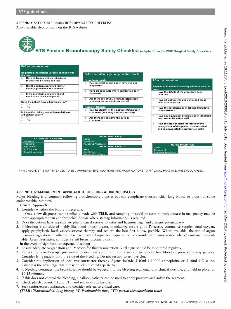
APPENDIX 5: FLEXIBLE BRONCHOSCOPY SAFETY CHECKLISTAlso available electronically on the BTS website
APPENDIX 6: MANAGEMENT APPROACH TO BLEEDING AT BRONCHOSCOPYMajor bleeding is uncommon following bronchoscopic biopsies but can complicate transbronchial lung biopsy or biopsy of someendobronchial tumours.
General Approach:1. Consider whether the biopsy is necessary.
Only a few diagnoses can be reliably made with TBLB, and sampling of nodal or extra thoracic disease in malignancy may bemore appropriate than endobronchial disease where staging information is required.
2. Does the patient have appropriate physiological reserve to withstand haemorrhage, and a secure patent airway3. If bleeding is considered highly likely and biopsy urgent/ mandatory, ensure good IV access, commence supplemental oxygen,
apply prophylactic local vasoconstrictor therapy and achieve the best first biopsy possible. Where available, the use of argonplasma coagulation or other similar haemostatic biopsy technique could be considered. Ensure senior advice/ assistance is avail-able. As an alternative, consider a rigid bronchoscopic biopsy.In the event of significant unexpected bleeding:
1. Ensure adequate oxygenation and IV access for fluid resuscitation. Vital signs should be monitored regularly.2. Retract the bronchoscope proximally to maintain vision, and apply suction to remove free blood to preserve airway patency.
Consider lying patient onto the side of the bleeding. Do not suction to remove clot.3. Consider the application of local vasoconstrictor therapy. Agents include 5-10ml 1:10000 epinephrine or 5-10ml 4°C saline.
Saline has the advantage that it may be administered repeatedly.4. If bleeding continues, the bronchoscope should be wedged into the bleeding segmental bronchus, if possible, and held in place for
10-15 minutes.5. If this does not control the bleeding, a balloon catheter can be used to apply pressure and isolate the segment.6. Check platelet count, PTand PTT, and recheck drug history.7. Seek senior/expert assistance, and consider referral to critical care.
(TBLB : Transbronchial lung biopsy, PT: Prothrombin time, PTT: partial thromboplastin time)
i36 Du Rand IA, et al. Thorax 2013;68:i1–i44. doi:10.1136/thoraxjnl-2013-203618
BTS guidelines
on 26 May 2018 by guest. P
rotected by copyright.http://thorax.bm
j.com/
Thorax: first published as 10.1136/thoraxjnl-2013-203618 on 16 July 2013. D
ownloaded from
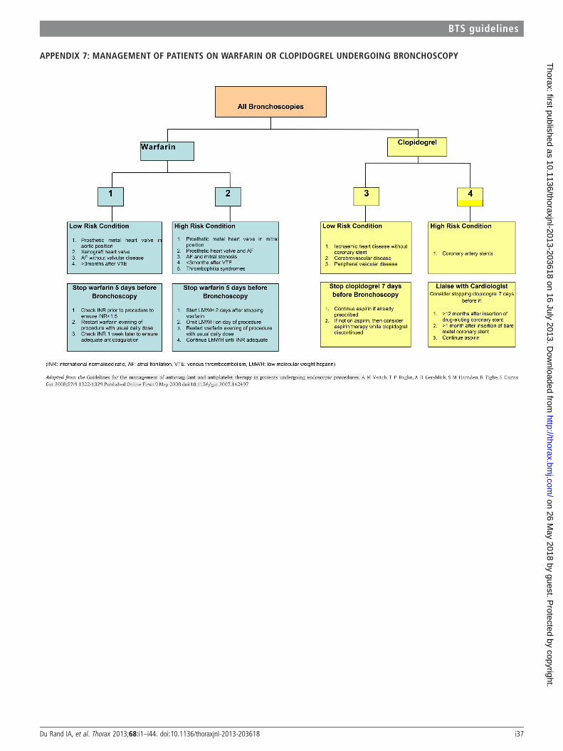
APPENDIX 7: MANAGEMENT OF PATIENTS ON WARFARIN OR CLOPIDOGREL UNDERGOING BRONCHOSCOPY
Du Rand IA, et al. Thorax 2013;68:i1–i44. doi:10.1136/thoraxjnl-2013-203618 i37
BTS guidelines
on 26 May 2018 by guest. P
rotected by copyright.http://thorax.bm
j.com/
Thorax: first published as 10.1136/thoraxjnl-2013-203618 on 16 July 2013. D
ownloaded from
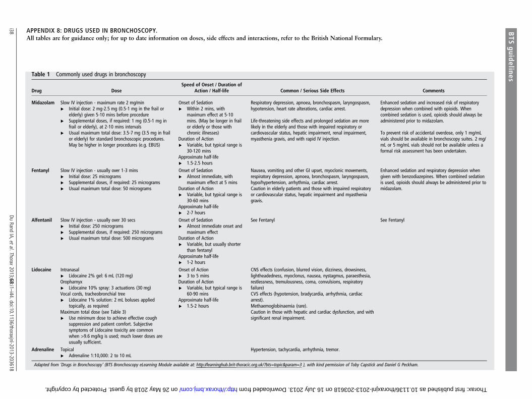
APPENDIX 8: DRUGS USED IN BRONCHOSCOPY.All tables are for guidance only; for up to date information on doses, side effects and interactions, refer to the British National Formulary.
Table 1 Commonly used drugs in bronchoscopy
Drug DoseSpeed of Onset / Duration of
Action / Half-life Common / Serious Side Effects Comments
Midazolam Slow IV injection - maximum rate 2 mg/min▸ Initial dose: 2 mg-2.5 mg (0.5-1 mg in the frail or
elderly) given 5-10 mins before procedure▸ Supplemental doses, if required: 1 mg (0.5-1 mg in
frail or elderly), at 2-10 mins intervals▸ Usual maximum total dose: 3.5-7 mg (3.5 mg in frail
or elderly) for standard bronchoscopic procedures.May be higher in longer procedures (e.g. EBUS)
Onset of Sedation▸ Within 2 mins, with
maximum effect at 5-10mins. (May be longer in frailor elderly or those withchronic illnesses)
Duration of Action▸ Variable, but typical range is
30-120 minsApproximate half-life▸ 1.5-2.5 hours
Respiratory depression, apnoea, bronchospasm, laryngospasm,hypotension, heart rate alterations, cardiac arrest.
Life-threatening side effects and prolonged sedation are morelikely in the elderly and those with impaired respiratory orcardiovascular status, hepatic impairment, renal impairment,myasthenia gravis, and with rapid IV injection.
Enhanced sedation and increased risk of respiratorydepression when combined with opioids. Whencombined sedation is used, opioids should always beadministered prior to midazolam.
To prevent risk of accidental overdose, only 1 mg/mLvials should be available in bronchoscopy suites. 2 mg/mL or 5 mg/mL vials should not be available unless aformal risk assessment has been undertaken.
Fentanyl Slow IV injection - usually over 1-3 mins▸ Initial dose: 25 micrograms▸ Supplemental doses, if required: 25 micrograms▸ Usual maximum total dose: 50 micrograms
Onset of Sedation▸ Almost immediate, with
maximum effect at 5 minsDuration of Action▸ Variable, but typical range is
30-60 minsApproximate half-life▸ 2-7 hours
Nausea, vomiting and other GI upset, myoclonic movements,respiratory depression, apnoea, bronchospasm, laryngospasm,hypo/hypertension, arrhythmia, cardiac arrest.Caution in elderly patients and those with impaired respiratoryor cardiovascular status, hepatic impairment and myastheniagravis.
Enhanced sedation and respiratory depression whengiven with benzodiazepines. When combined sedationis used, opioids should always be administered prior tomidazolam.
Alfentanil Slow IV injection - usually over 30 secs▸ Initial dose: 250 micrograms▸ Supplemental doses, if required: 250 micrograms▸ Usual maximum total dose: 500 micrograms
Onset of Sedation▸ Almost immediate onset and
maximum effectDuration of Action▸ Variable, but usually shorter
than fentanylApproximate half-life▸ 1-2 hours
See Fentanyl See Fentanyl
Lidocaine Intranasal▸ Lidocaine 2% gel: 6 mL (120 mg)Oropharnyx▸ Lidocaine 10% spray: 3 actuations (30 mg)Vocal cords, tracheobronchial tree▸ Lidocaine 1% solution: 2 mL boluses applied
topically, as requiredMaximum total dose (see Table 3)▸ Use minimum dose to achieve effective cough
suppression and patient comfort. Subjectivesymptoms of Lidocaine toxicity are commonwhen >9.6 mg/kg is used; much lower doses areusually sufficient.
Onset of Action▸ 3 to 5 minsDuration of Action▸ Variable, but typical range is
60-90 minsApproximate half-life▸ 1.5-2 hours
CNS effects (confusion, blurred vision, dizziness, drowsiness,lightheadedness, myoclonus, nausea, nystagmus, paraesthesia,restlessness, tremulousness, coma, convulsions, respiratoryfailure)CVS effects (hypotension, bradycardia, arrhythmia, cardiacarrest).Methaemoglobinaemia (rare).Caution in those with hepatic and cardiac dysfunction, and withsignificant renal impairment.
Adrenaline Topical▸ Adrenaline 1:10,000: 2 to 10 mL
Hypertension, tachycardia, arrhythmia, tremor.
Adapted from ’Drugs in Bronchoscopy’ (BTS Bronchoscopy eLearning Module available at: http://learninghub.brit-thoracic.org.uk/?bts=topic¶m=3 ), with kind permission of Toby Capstick and Daniel G Peckham.
i38Du
RandIA,etal.Thorax
2013;68:i1–i44.doi:10.1136/thoraxjnl-2013-203618
BTS
guidelines
on 26 May 2018 by guest. Protected by copyright. http://thorax.bmj.com/ Thorax: first published as 10.1136/thoraxjnl-2013-203618 on 16 July 2013. Downloaded from
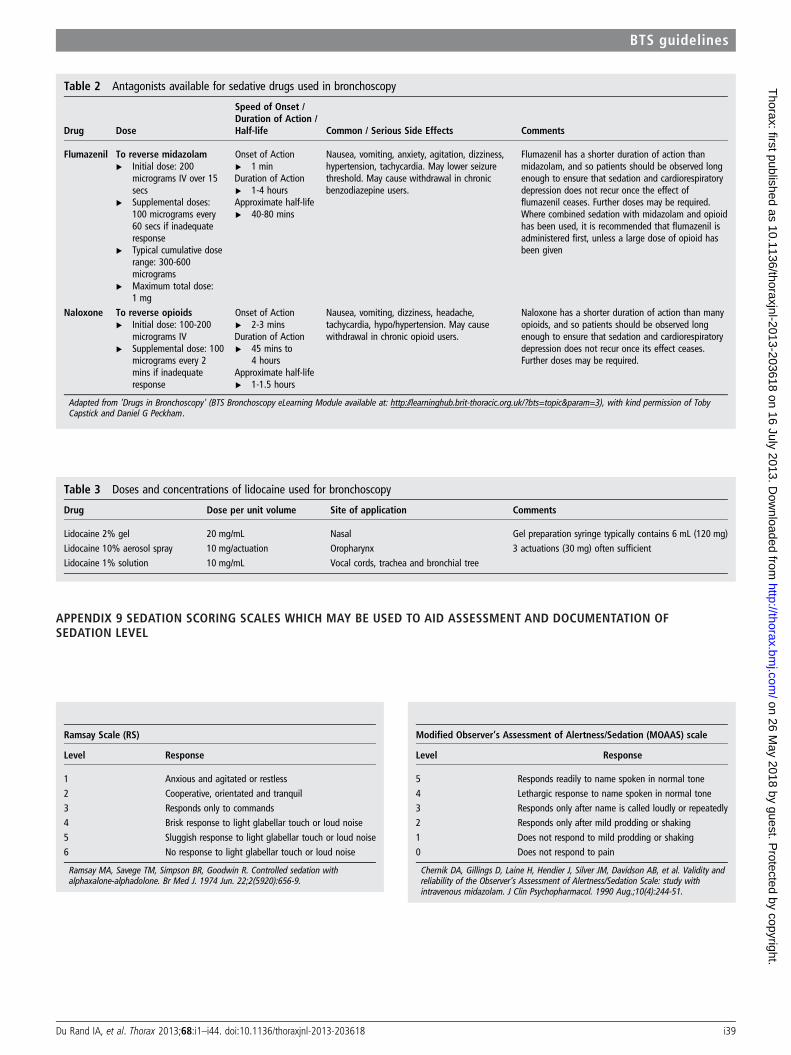
APPENDIX 9 SEDATION SCORING SCALES WHICH MAY BE USED TO AID ASSESSMENT AND DOCUMENTATION OFSEDATION LEVEL
Ramsay Scale (RS)
Level Response
1 Anxious and agitated or restless2 Cooperative, orientated and tranquil3 Responds only to commands4 Brisk response to light glabellar touch or loud noise
5 Sluggish response to light glabellar touch or loud noise6 No response to light glabellar touch or loud noise
Ramsay MA, Savege TM, Simpson BR, Goodwin R. Controlled sedation withalphaxalone-alphadolone. Br Med J. 1974 Jun. 22;2(5920):656-9.
Modified Observer’s Assessment of Alertness/Sedation (MOAAS) scale
Level Response
5 Responds readily to name spoken in normal tone4 Lethargic response to name spoken in normal tone3 Responds only after name is called loudly or repeatedly2 Responds only after mild prodding or shaking
1 Does not respond to mild prodding or shaking0 Does not respond to pain
Chernik DA, Gillings D, Laine H, Hendíer J, Silver JM, Davidson AB, et al. Validity andreliability of the Observer’s Assessment of Alertness/Sedation Scale: study withintravenous midazolam. J Clin Psychopharmacol. 1990 Aug.;10(4):244-51.
Table 2 Antagonists available for sedative drugs used in bronchoscopy
Drug Dose
Speed of Onset /Duration of Action /Half-life Common / Serious Side Effects Comments
Flumazenil To reverse midazolam▸ Initial dose: 200
micrograms IV over 15secs
▸ Supplemental doses:100 micrograms every60 secs if inadequateresponse
▸ Typical cumulative doserange: 300-600micrograms
▸ Maximum total dose:1 mg
Onset of Action▸ 1 minDuration of Action▸ 1-4 hoursApproximate half-life▸ 40-80 mins
Nausea, vomiting, anxiety, agitation, dizziness,hypertension, tachycardia. May lower seizurethreshold. May cause withdrawal in chronicbenzodiazepine users.
Flumazenil has a shorter duration of action thanmidazolam, and so patients should be observed longenough to ensure that sedation and cardiorespiratorydepression does not recur once the effect offlumazenil ceases. Further doses may be required.Where combined sedation with midazolam and opioidhas been used, it is recommended that flumazenil isadministered first, unless a large dose of opioid hasbeen given
Naloxone To reverse opioids▸ Initial dose: 100-200
micrograms IV▸ Supplemental dose: 100
micrograms every 2mins if inadequateresponse
Onset of Action▸ 2-3 minsDuration of Action▸ 45 mins to
4 hoursApproximate half-life▸ 1-1.5 hours
Nausea, vomiting, dizziness, headache,tachycardia, hypo/hypertension. May causewithdrawal in chronic opioid users.
Naloxone has a shorter duration of action than manyopioids, and so patients should be observed longenough to ensure that sedation and cardiorespiratorydepression does not recur once its effect ceases.Further doses may be required.
Adapted from ’Drugs in Bronchoscopy’ (BTS Bronchoscopy eLearning Module available at: http://learninghub.brit-thoracic.org.uk/?bts=topic¶m=3), with kind permission of TobyCapstick and Daniel G Peckham.
Table 3 Doses and concentrations of lidocaine used for bronchoscopy
Drug Dose per unit volume Site of application Comments
Lidocaine 2% gel 20 mg/mL Nasal Gel preparation syringe typically contains 6 mL (120 mg)Lidocaine 10% aerosol spray 10 mg/actuation Oropharynx 3 actuations (30 mg) often sufficientLidocaine 1% solution 10 mg/mL Vocal cords, trachea and bronchial tree
Du Rand IA, et al. Thorax 2013;68:i1–i44. doi:10.1136/thoraxjnl-2013-203618 i39
BTS guidelines
on 26 May 2018 by guest. P
rotected by copyright.http://thorax.bm
j.com/
Thorax: first published as 10.1136/thoraxjnl-2013-203618 on 16 July 2013. D
ownloaded from
APPENDIX 10 BTS FLEXIBLE BRONCHOSCOPY - SUGGESTED GUIDES ON HOW TO PERFORM STANDARD PROCEDURES
BRONCHOALVEOLAR LAVAGE
This procedure is performed during flexible bronchoscopy and is useful in the diagnosis of pulmonary infections and may be useful in the diagnosis of parenchymal lungdisease.The site of BAL should be guided by available radiology.i. Once the site for BAL has been chosen, the bronchoscope should be advanced until the desired site is reached.ii. The bronchoscope should then be wedged in to a position where the lumen of the bronchus is occluded by the bronchoscopeiii. Whilst the bronchoscope is in this position, 60-180mls of normal saline should be instilled in to the segmentiv. Low-pressure suction should then be applied to retrieve the sample. The aim should be to apply enough suction to retrieve the sample without causing airway collapse.v. To ensure an adequate sample is retrieved it is useful to have tubing with several containers in series attached to the suction pump.vi. Some centres employ a syringe to retrieve the sample from the same port that the saline is instilled through.ENDOBRONCHIAL BIOPSYThis procedure is performed for visible airway abnormalities, most commonly in the diagnosis of suspected lung cancer. Once again, radiology should be available to planwhere sampling should occur. There are several types of biopsy forceps, most commonly alligator or open cup forceps are used. Studies have not demonstrated anyadvantage of one type of forceps over the other in diagnostic yield.i. The bronchoscope should be advanced until the endobronchial lesion is visualised.ii. Secure the bronchoscope in this position so that the abnormality can be fully visualised, then advance the forceps in the closed position through the working channel
of the bronchoscope.iii. Once the forceps are visualised at the distal end of the bronchoscope the forceps can be opened.iv. Whilst keeping the bronchoscope still the forceps should be advanced in the open position towards the abnormality.v. Once the target is reached the forceps should be closed trapping as much tissue as possible.vi. The forceps should be retracted and removed from the working channel of the bronchoscope.vii. A "tugging" sensation may be felt whilst retracting the forceps.viii. Once the sample has been removed from the forceps, it should be reinserted into the working channel and the procedure repeated 5-6 times.ENDOBRONCHIAL BRUSHThis procedure is performed to gain cytology samples in areas of abnormal mucosa or endobronchial lesions. Although no evidence exists for which procedure is performedfirst we suggest that endobronchial biopsies are performed prior to brushings.i. Once the area of abnormality is detected the bronchial brush is inserted through the working channel of the bronchoscope in its retracted position in its protective
sheathii. Once the bronchial brush is seen at the distal end of the bronchoscope the brush can be pushed out (opened)iii. The brush is then advanced towards the lesion and moved back and forth over the lesion several timesiv. The brush is then retracted into its sheath (closed) and removed from the working channelv. The procedure is repeatedTRANSBRONCHIAL LUNG BIOPSYThis procedure is performed via flexible bronchoscopy, to aid the diagnosis of parenchymal lung disease. Unlike endobronchial biopsy this procedure can be performed"blind", without direct visualisation of the lesion. Some centres chose to perform this procedure with radiological guidance (fluoroscopy), diagnostic yield does not differbetween methods. Accurate radiological imaging is essential to guide the bronchoscopist to the lung parenchyma with the greatest potential diagnostic yield in non-diffuseinterstitial disease.i. The bronchoscope is advanced as far as possible into the area of the lung to be biopsiedii. The forceps are then inserted into the working channel and advanced as far as possibleiii. At this point the forceps are retracted 1 cm to avoid a biopsy of the pleura Instruct the patient to take a slow deep breath in. The forceps are then opened during
inspirationiv. Instruct the patient to breathe out slowly, whilst the patient is breathing out the forceps are advanced and closedv. The forceps are then removed and the sample retrievedWarning: Do not take the biopsy if the patient experience pain when forceps is pulled back or removed, pleura
could have been caught in the forceps. Open forceps and remove without biopsy.vi. The procedure is repeated a further 5-6 timesClinical Tip: Whilst performing a TBLB co-operation of the patient is necessary. Patients should be able to follow
commands and light sedation is therefore advised.
i40 Du Rand IA, et al. Thorax 2013;68:i1–i44. doi:10.1136/thoraxjnl-2013-203618
BTS guidelines
on 26 May 2018 by guest. P
rotected by copyright.http://thorax.bm
j.com/
Thorax: first published as 10.1136/thoraxjnl-2013-203618 on 16 July 2013. D
ownloaded from
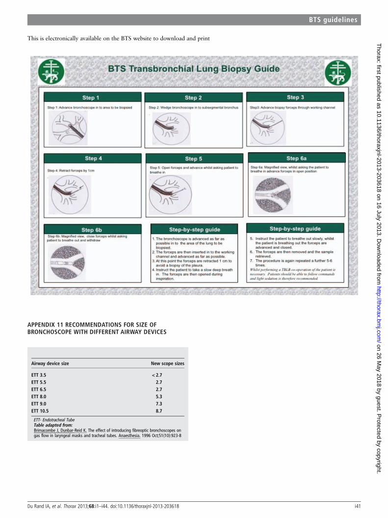
Airway device size New scope sizes
ETT 3.5 < 2.7ETT 5.5 2.7ETT 6.5 2.7ETT 8.0 5.3ETT 9.0 7.3ETT 10.5 8.7
ETT- Endotracheal TubeTable adapted from:Brimacombe J, Dunbar-Reid K. The effect of introducing fibreoptic bronchoscopes ongas flow in laryngeal masks and tracheal tubes. Anaesthesia. 1996 Oct;51(10):923-8
This is electronically available on the BTS website to download and print
APPENDIX 11 RECOMMENDATIONS FOR SIZE OFBRONCHOSCOPE WITH DIFFERENT AIRWAY DEVICES
Du Rand IA, et al. Thorax 2013;68:i1–i44. doi:10.1136/thoraxjnl-2013-203618 i41
BTS guidelines
on 26 May 2018 by guest. P
rotected by copyright.http://thorax.bm
j.com/
Thorax: first published as 10.1136/thoraxjnl-2013-203618 on 16 July 2013. D
ownloaded from
APPENDIX 12: CLEANING AND DISINFECTING PROCEDURE
Wear appropriate, protective clothing(Gloves, aprons, visors or goggles, face mask and forearm covers)
1 Before removing bronchoscope from light source or video processor suction with enzymatic detergent, followed by 10 seconds of air. This will remove blood, mucus anddebris from internal channels. Wipe the external surface of the scope with a soft lint free cloth soaked in detergent. Check scope for damage. Remove, flush and wipebiopsy and suction ports. Discard single use valves.
2 Transport bronchoscope to the decontamination area in an appropriately sized and covered receptacle.3 Leak test bronchoscope according to manufacturer’s recommendations.4 Manual cleaning of bronchoscope to be performed in a dedicated sink filled with water and detergent according to manufacturer’s recommendations. Water and
detergent to be discarded after each use.5 Brush suction and biopsy channels and ports with a single-use brush, according to manufacturer’s guidelines. Ensure the brush is visibly clean at the end of the process.6 Clean the external surface of the scope, around the angulation control and distal tip of the bronchoscope.7 Brush the biopsy and suction channel ports with a single-use short brush.8 Irrigate the channels with an enzymatic detergent followed by water, then air.9 Transfer the bronchoscope to a separate sink for rinsing, to remove residual detergent.10 Transfer bronchoscope to AER in an appropriately sized receptacle.11 Disinfect in Automated Endoscope Reprocessor alongside reusable valves. Ensure all channels are exposed to disinfectant process.12 Store hanging in cupboard or drying cabinet following manufacturers recommendations.13 Maintain record of decontamination process for bronchoscope and accessories.
i42 Du Rand IA, et al. Thorax 2013;68:i1–i44. doi:10.1136/thoraxjnl-2013-203618
BTS guidelines
on 26 May 2018 by guest. P
rotected by copyright.http://thorax.bm
j.com/
Thorax: first published as 10.1136/thoraxjnl-2013-203618 on 16 July 2013. D
ownloaded from
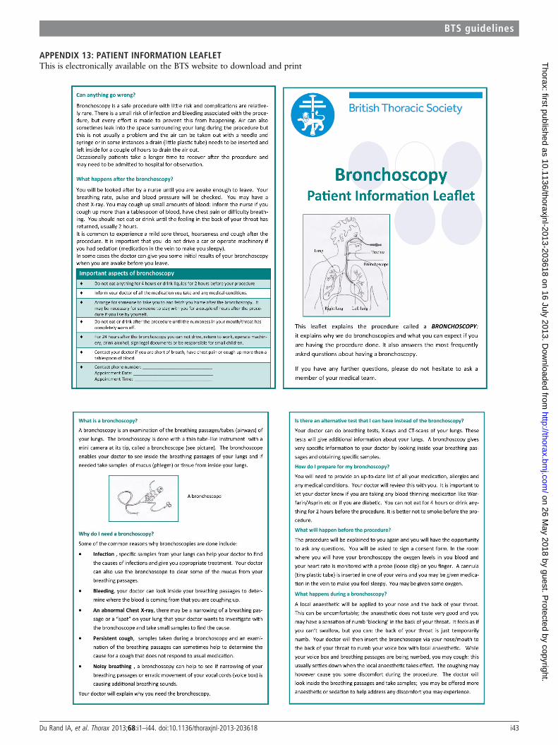
APPENDIX 13: PATIENT INFORMATION LEAFLETThis is electronically available on the BTS website to download and print
Du Rand IA, et al. Thorax 2013;68:i1–i44. doi:10.1136/thoraxjnl-2013-203618 i43
BTS guidelines
on 26 May 2018 by guest. P
rotected by copyright.http://thorax.bm
j.com/
Thorax: first published as 10.1136/thoraxjnl-2013-203618 on 16 July 2013. D
ownloaded from

on 26 May 2018 by guest. P
rotected by copyright.http://thorax.bm
j.com/
Thorax: first published as 10.1136/thoraxjnl-2013-203618 on 16 July 2013. D
ownloaded from