brucella in tajikistan - zoonotic risks of urbanized...
TRANSCRIPT

Brucella in Tajikistan - Zoonotic Risks of Urbanized Livestock in a
Low-Income Country
Elisabeth Lindahl RajalaFaculty of Veterinary Medicine and Animal Science
Department of Clinical SciencesUppsala
Doctoral ThesisSwedish University of Agricultural Sciences
Uppsala 2016

Acta Universitatis agriculturae Sueciae2016:111
ISSN 1652-6880ISBN (print version) 978-91-576-8725-8ISBN (electronic version) 978-91-576-8726-5© 2016 Elisabeth Lindahl Rajala, UppsalaPrint: SLU Service/Repro, Uppsala 2016
Cover: Tajik small-scale farmer(photo: E. Lindahl Rajala)

Brucella in Tajikistan – Zoonotic risks of urbanized livestock in a low-income country
AbstractOne of the most powerful megatrends of our time is urbanization. Urban and peri-urban(UPU) farming is a common practice in many low-income countries because itincreases the incomes of families that are often restrained by limited economicresources. However, there is a concern that the growing number of people and livestock living close together in UPU areas will increase the transmission of different zoonotic pathogens such as Brucella. Brucellosis is one of the most common and economically important zoonoses globally and Central Asia represents an area with high incidence among humans and livestock. This thesis aims to assess the occurrence of Brucella among livestock in an UPU area and to elucidate how farmers understand and respondto this zoonosis. The results of this thesis might contribute to raising awareness of how livestock in urban areas can constitute a public health risk if they are infected with Brucella or other zoonoses. The four studies included in this thesis were conducted among small-scale livestock farmers in the UPU region of Dushanbe, the capital of Tajikistan. Blood samples were collected from 904 dairy cows and 667 sheep and goatsand analysed with enzyme-linked immunosorbent assay (ELISA). The Brucellaseroprevalence was 4.1% among the dairy cows at the herd level and ranged between1.0 and 15.6% among sheep and goats at the individual level in the four included districts. Furthermore, 564 cow milk samples were analysed for Brucella DNA by IS711-based real-time PCR and 13.7% were found to be positive. All seropositive cows were positive by PCR, but 11.8% of the seronegative cows were also positive by PCR.Further characterization of the Brucella DNA suggests that there is a reservoir of B. abortus in the cattle population and a spillover of B. melitensis from small ruminants to cattle. A knowledge, attitudes and practice study targeting 441 households revealedpoor knowledge of brucellosis and several high-risk behaviours, such as consumption of unpasteurized dairy products and not wearing protective clothing when handling potentially infectious materials like aborted foetuses and discharges.
Brucella is widespread among the livestock in the UPU area of Dushanbe and this might constitute a serious risk to public health and cause significant economic losses. The discrepancy between serology and PCR results suggests that implementing complementary diagnostic strategies to detect false serological negative individualsmight be warranted in Brucella control programmes. Poor knowledge, several high-risk behaviours and a willingness to learn more provide the rationale for developing campaigns to raise awareness of brucellosis and its associated risks among farmers.
Keywords: Brucella, ELISA, IS711, KAP, PCR, rpoB, Tajikistan, urban/peri-urban
Author’s address: Elisabeth Lindahl Rajala, SLU, Department of Clinical Sciences, P.O. Box 7054, 750 07 Uppsala, Sweden E-mail: [email protected]

DedicationTo my family
“The disease rarely kills anybody, but it often makes a patient wish he were dead”
TIME magazine 1943 reporting on brucellosis

ContentsList of Publications 7
Abbreviations 8
1 Introduction 91.1 Brucella – a neglected disease of the poor 101.2 Brucellosis in Tajikistan 101.3 Urbanization and UPU livestock production in low-income countries 111.4 Brucella spp. 13
1.4.1 Brucella: species and biovars 131.4.2 Diagnostic methods 14
1.5 Control of brucellosis 171.5.1 Brucellosis vaccine 18
1.6 Brucella in mammals 191.6.1 Brucella in humans 191.6.2 Brucella in livestock 201.6.3 Brucella in other animal species 21
2 Aims of this thesis 23
3 Methodological considerations 253.1 Study area (papers I–IV) 253.2 Study population (papers I and II) 263.3 Epidemiological unit (papers I and II) 263.4 Data collected anonymously 273.5 Serology (papers I and II) 273.6 Detection and analysis of Brucella DNA (paper III) 283.7 Interviews (paper IV) 29
4 Main results and discussion 314.1 Small-scale UPU livestock farming 314.2 Brucella seropositivity (papers I and II) 354.3 Factors associated with Brucella seropositivity (papers I and II) 364.4 Detection of Brucella DNA by PCR and comparison with serology
(paper III) 394.5 Sequence analysis of Brucella DNA (paper III) 414.6 Knowledge of brucellosis (paper IV) 41

4.7 Attitudes towards brucellosis (paper IV) 444.8 Self-reported practices (paper IV) 44
5 Conclusions 49
6 Future perspectives 51
7 Populärvetenskaplig sammanfattning 53
References 55
Acknowledgements 63

7
List of PublicationsThis thesis is based on the work contained in the following papers, referred to by Roman numerals in the text:
I Lindahl E, Sattorov N, Boqvist S, Sattori I, Magnusson U (2014). Seropositivity and risk factors for Brucella in dairy cows in urban and peri-urban small-scale farming in Tajikistan. Tropical Animal Health and Production 46(3), 563-569.
II Lindahl Rajala E, Grahn C, Ljung I, Sattorov N, Boqvist S, Magnusson U (2016). Prevalence and risk factors for Brucella seropositivity among sheep and goats in a peri-urban region of Tajikistan. Tropical Animal Health and Production 48(3), 553-558.
III Lindahl Rajala E, Hoffman T, Fretin D, Godfroid J, Sattorov N, Boqvist S, Lundkvist Å, Magnusson U. Detection and characterization of Brucella spp. in bovine milk in small-scale urban and peri-urban farming in Tajikistan. Submitted manuscript.
IV Lindahl E, Sattorov N, Boqvist S, Magnusson U (2015). A study of knowledge, attitudes and practices relating to brucellosis among small-scale dairy farmers in an urban and peri-urban area of Tajikistan. PLoS One10(2), doi: 10.1371/journal.pone.0117318.
Papers I, II and IV are reproduced with the permission of the publishers.

8
Abbreviations
C-ELISACtDNA
Competitive ELISACycle thresholdDeoxyribonucleic acid
ELISA Enzyme-linked immunosorbent assayFAOI-ELISAILRIKAP
OR
Food and Agriculture Organization of the United NationsIndirect ELISAInternational Livestock Research InstituteKnowledge, Attitudes and Practices
Odds ratioOIE World Organisation for Animal Health
PCR Polymerase chain reaction qPCRSNP
quantitative or real-time PCRSingle nucleotide polymorphism
UPU Urban/peri-urbanWHO World Health Organization

9
1 IntroductionBrucellosis is one of the most common and economically important zoonoses globally (McDermott et al., 2013). Central Asia and the Middle East are among the regions with the highest incidence of brucellosis in humans and livestock worldwide and the incidence is rising (Pappas, 2010; Seleem et al., 2010; Pappas et al., 2006). Humans can become chronically infected if not treated adequately and osteoarticular manifestation is a common complication (Deanet al., 2012). Inadequate treatment might result in loss of work and income atthe individual level (Seleem et al., 2010). Furthermore, loss of human productivity and increased costs within the public health sector cause economic losses at the national level. Brucellosis in livestock mainly affects the reproductive organs and causes abortion, reduced fertility and a reduced milk production (Corbel, 2006). A decrease in productivity, loss of export and costs caused by movement restrictions and vaccinations of livestock also causesignificant economic losses within the livestock sector (McDermott et al.,2013). Brucellosis is a zoonosis, meaning that the disease is transmissible between animals and humans. The rapid urbanization currently prevailing in many countries is drastically changing how societies are organised and is laying the foundation for great economic advancements (United Nations, 2014; WHO, 2010). However, urbanization of humans also implies urbanization of their livestock. This development raises concerns that the growing number of people and livestock living close together in urban/peri-urban (UPU) areas will increase the transmission of different zoonotic pathogens such as Brucella(Steinfeld, 2004). Like in many other low-income countries, the people of Tajikistan are dependent on small-scale farming (Jackson et al., 2007) and at the same time are quickly urbanizing (United Nations, 2014). Brucella is endemic among the livestock in the region, there is no control programme in place and resources are scarce. The need for dealing with zoonotic infections in

10
the face of accelerating urbanization and economic constraints is shared by many low-income countries.
1.1 Brucella – a neglected disease of the poor
The World Health Organization (WHO) has classified brucellosis as one of seven neglected zoonotic diseases (WHO, 2005). These diseases are rarely in the spotlight for research and mainly affect poor, marginalized people.Furthermore, the International Livestock Research Institute (ILRI) has listed brucellosis as one of the 13 most important zoonotic infections in terms of impact on human and livestock health, amenability to agricultural-based control and emergence or severity of disease in people (Grace et al., 2012).According to the WHO, there are three underlying mechanisms for why the consequences of this group of neglected diseases are so serious, especially for poor people (WHO, 2005). First, poor people are more likely to acquire a zoonotic infection like brucellosis because they often live in close proximity with animals. Poor people are also more likely to consume low-quality food products from informal markets such as unpasteurized dairy products or meat from sick animals and thus are at greater risk of becoming infected. Second,poor people are less likely to get a proper diagnosis and to receive adequatetreatment. For Brucella, this implies that they are more likely to become chronically infected and be at greater risk of permanent disability. Third, poor households have lower economic margins and often only keep a small number of livestock. Loss of income due to a family member´s inability to work, additional costs for medical treatment and production losses among the few livestock they own can be devastating for a poor household´s economy.
1.2 Brucellosis in Tajikistan
Tajikistan is located in Central Asia and borders Afghanistan to the south,China to the east, Kyrgyzstan to the north and Uzbekistan to the west and is populated by approximately 8 million people (CIA, 2016; Worldbank, 2016)(Figure 1). The pace of poverty reduction has been among the top 10% in the world over the past 15 years (Worldbank, 2016) and the poverty rate, measured as the percentage of the population that lives at or below USD 1.90 a day, has decreased from over 80% in 1999 to around 30% today. According to the 2015 Global Hunger Index report, 33% of the Tajik population suffer from malnutrition and Tajikistan is ranked as the country with the highest malnutrition rate among the former countries of the Soviet Union (von Grebmer et al., 2015). Due to economic slowdown in Russia, tighter migration

11
restrictions in Russia resulting in a decline in remittances and weak global demand for its key export commodities, Tajikistan´s GDP growth hasdecreased from 6.7% in 2014 to 4.2% in 2015 – although this is still a considerable growth rate.
During the Soviet era, brucellosis among livestock was fairly well controlled in Tajikistan through a strategy of vaccination and test-and-slaughter(Ward et al., 2012). After the collapse of the Soviet Union in 1991, the animal health sector was seriously affected and control for zoonotic infections like brucellosis became markedly impaired in the region. Over the last two decades, Tajikistan - as well as other Central Asian countries - have seen increased numbers of small farm units (Beauvais et al., 2015; Ward et al., 2012; Pappas, 2010), uncontrolled movement of livestock, poor infrastructure, deregulations of trade and decreased border controls (Ward et al., 2012; Pappas, 2010). This could be one set of explanations for why Central Asia is currently a hotspot for Brucella infection among humans and livestock.
The Food and Agricultural Organization of the United Nations (FAO)started a Brucella control programme in 2004 in eight districts in Tajikistan with high Brucella burden in sheep and goats (Jackson et al., 2007). Thatprogramme did not include the region around Dushanbe, which is the focus of this thesis. The program comprised mass-vaccination of sheep and goats in the first two years followed by biannual vaccination of young animals and non-vaccinated adults. The vaccine used was Brucella melitensis Rev 1 applied as eye drops. After six years, the seroprevalence had dropped from 8.9% to 1.8%in the eight best-vaccinated districts, making it one of the most successful programmes in the region (FAO, 2014; Ward et al., 2012) .
1.3 Urbanization and UPU livestock production in low-income countries
One of the most powerful megatrends of our time is urbanization (United Nations, 2014). Today, 54% of the world’s human population lives in urban areas and by 2050 the number is anticipated to rise to 66%. An often forgotten consequence of human urbanization is the urbanization of their livestock. Today, Asia and Africa are among the most rural regions of the world, but also the regions with the highest urbanization rates. As the farmers of these highly populated regions move themselves and their livestock closer to cities at an unprecedented scale, there are increased concerns for the spread of zoonotic diseases (Steinfeld, 2004). Due to population growth, increasing urbanization and higher disposable income, the demand for animal food products is expected to double by 2030 in low-income countries. This will likely result in

12
increasing large-scale livestock production close to urban centres. In Tajikistan, 27% of the population lives in urban areas (United Nations, 2014)and by 2050 this number is expected to rise to 41%. Living in a city often implies many advantages compared to living in rural areas, including better access to health services and higher levels of literacy and education. UPU livestock production offers an opportunity for people to improve their livelihood but it also poses a public health threat if the risk of zoonotic infections is not addressed wisely (Flynn, 1999).
Figure 1. World map and close up of Central Asia and Tajikistan (ArcGIS® software by Esri, www.esri.com).

13
1.4 Brucella spp.
Brucella is a genus of gram negative bacteria named after David Bruce (1855-1931) who was the first person to isolate B. melitensis. This strain was isolated from the spleen of a British soldier, suffering from a febrile illness called Malta fever in 1887 (Nicoletti, 2002).
1.4.1 Brucella: species and biovars
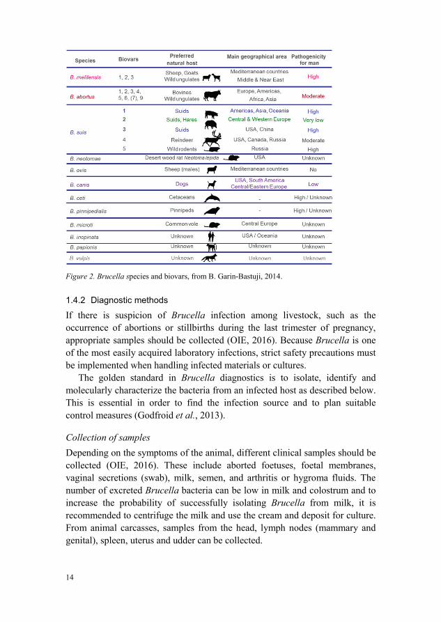
To date, twelve different Brucella species have been described (Scholz et al.,2016; Whatmore et al., 2014) (Figure 2). The six classical species are B. melitensis and B. abortus (Meyer & Shaw, 1920), B. suis (Huddleson, 1929),B. ovis (Buddle, 1956), B. neotomae (Stoenner & Lackman, 1957) and B. canis(Carmichael & Bruner, 1968). Brucella melitensis, B. abortus and B. suis are further classified into biovars. After B. canis was isolated in the late 1960s, no novel Brucella species were identified for many decades (Pappas, 2010). In the 1990s, two new Brucella species were found in marine mammals (Ewalt et al.,1994; Ross et al., 1994) and these were subsequently categorized as B. ceti and B. pinnipedialis (Foster et al., 2007), both with zoonotic potential (Whatmoreet al., 2008). Another newly described species, B. microti was isolated from common voles and red foxes (Scholz et al., 2008b). Two additional novel strains have recently been isolated from humans and the first one was isolated from an infected human breast implant (Scholz et al., 2010). This strain was named B. inopinata and the second strain showed similarity to B. inopinata and was isolated from a patient with chronic lung disease (Tiller et al., 2010). The two most recently described species are B. papionis, which was isolated from two baboons with retained placenta (Whatmore et al., 2014) and B. vulpiswhich was isolated in Austria from the mandibular lymph nodes of two red foxes (Scholz et al., 2016) (Figure 2).
14
Figure 2. Brucella species and biovars, from B. Garin-Bastuji, 2014.
1.4.2 Diagnostic methods
If there is suspicion of Brucella infection among livestock, such as the occurrence of abortions or stillbirths during the last trimester of pregnancy, appropriate samples should be collected (OIE, 2016). Because Brucella is one of the most easily acquired laboratory infections, strict safety precautions must be implemented when handling infected materials or cultures.
The golden standard in Brucella diagnostics is to isolate, identify and molecularly characterize the bacteria from an infected host as described below.This is essential in order to find the infection source and to plan suitable control measures (Godfroid et al., 2013).
Collection of samples
Depending on the symptoms of the animal, different clinical samples should be collected (OIE, 2016). These include aborted foetuses, foetal membranes, vaginal secretions (swab), milk, semen, and arthritis or hygroma fluids. The number of excreted Brucella bacteria can be low in milk and colostrum and toincrease the probability of successfully isolating Brucella from milk, it is recommended to centrifuge the milk and use the cream and deposit for culture. From animal carcasses, samples from the head, lymph nodes (mammary and genital), spleen, uterus and udder can be collected.

15
Staining methods
Brucella bacteria can be demonstrated in smears of organs or biological fluids by different staining methods like Gram or Stamp staining (OIE, 2016; Godfroid et al., 2010). Brucella are coccobacilli or short rods approximately 0.5-1.5 m long and 0.5-0.7 m wide. They are usually arranged singly or occasionally in pairs. Brucella are non-motile and do not form spores.
Culture
Due to the very low infectious dose, Brucella is an easily acquired laboratory infection and bio-safety level 3 laboratories are recommended for culturing of all zoonotic Brucella spp. that infect livestock (Schwarz et al., 2015).Phenotypic analysis of Brucella includes growth on different media, colonymorphology and organism morphology after different staining methods like Gram or Stamp staining (OIE, 2016; Godfroid et al., 2010). Further identification of Brucella spp. includes the requirement of CO2 enrichedatmosphere for growth, metabolic profile (oxidase production and ureaseactivity), growth in the presence of dyes, production of H2S, lysis by Brucella-specific bacteriophage and agglutination with Brucella antiserum.
Culture of Brucella can be performed on both basal and selective media(OIE, 2016). Direct isolation is often performed on basal solid media whereas liquid media can be used for enrichment purpose.
Selective media can be prepared from basal media by adding antibiotics to suppress the growth of other microorganisms (OIE, 2016). One of the mostcommonly used selective media is Farrell´s medium, which contains six different antibiotics (OIE, 2016; Farrell, 1974).
The Brucella colonies can be visible on solid media after two to three days of incubation. After four days they can be seen as round colonies 1-2 mm in diameter with smooth margins, except for B. ovis and B. canis, which presentwith rough margins (OIE, 2016; Godfroid et al., 2010).
Molecular DNA technology Species within the genus Brucella show a high level of nucleotide similarity (> 90%) and most genetic differences between the species consist of single nucleotide polymorphisms (SNPs) (Foster et al., 2009; Whatmore, 2009; Verger et al., 1985). Culture of Brucella and further characterization into species and biovars is time consuming, requires experienced personnel and involves exposure to living Brucella organisms (Whatmore, 2009; Marianelliet al., 2006). Therefore, methods of genetic characterization using molecular DNA technology have been developed. Today, there are several molecular methods described for the detection, identification and to some extent

16
differentiation of Brucella spp. and biovars. Specific sequences of Brucellaspp. such as the 16S rRNA (Romero et al., 1995; Herman & De Ridder, 1992),the bcsp31 gene (Bricker, 2002; Baily et al., 1992) and the IS711 insertion sequence (Halling et al., 1993) have been used for the detection of BrucellaDNA with conventional polymerase chain reaction (PCR) assays. There are also real-time or quantitative PCR (qPCR) assays that have been developed for rapid and safe detection of Brucella, including assays targeting the bcsp31gene or the IS711 insertion sequence (Bounaadja et al., 2009). To enable epidemiological tracking, further typing is necessary. A number of different PCR-based assays have been used to find DNA markers to enable further molecular typing of Brucella, including assays based on the rpoB gene (Marianelli et al., 2006). Furthermore, different multiplex PCR assays such asthe Bruce ladder (García-Yoldi et al., 2006) and the AMOS-PCR (Bricker & Halling, 1994), are widely used. Recently developed techniques, such as multiple variable number of tandem repeat (VNTR) loci and multi-locus VNTR analysis (MLVA), have made it possible to classify Brucella into biovars as well as to discriminate Brucella isolates within a given biovar (Allenet al., 2015; Godfroid et al., 2010; Le Flèche et al., 2006).
Serology
Serological methods are valuable tools for screening purposes in Brucellasurveillance, control and eradication programmes but all serological tests have limitations, especially when it comes to testing of individual animals. When performing serology, it is important to use standardized serological methods and reference sera according to the World Organisation for Animal Health (OIE) standards (OIE, 2016). There are different serological tests for detecting Brucella-specific antibodies and the OIE recommends that positive samples should be confirmed as positive with a suitable confirmation test. The buffered plate agglutination test (BPAT), the rose bengal test (RBT), the complement fixation test (CFT), the fluorescence polarisation assay (FPA) and the indirect enzyme-linked immunosorbent assay (I-ELISA) are suitable screening tests among cattle, buffaloes, sheep, goats and camelids. The competitive ELISA (C-ELISA) may be used in some situations but is considered costly (OIE, 2016).
An efficient way to screen a dairy herd for Brucella infection is to analysebulk milk with I-ELISA (OIE, 2016). A shortcoming with bulk milk is that pregnant individuals in the last trimester are not producing milk and hence will not be discovered as seropositive if only analysing bulk milk. Therefore, the testing of these individuals should be repeated after parturition. Bulk milk sampling is more convenient than performing individual blood sampling and

17
also more cost effective. If a positive bulk milk result is obtained, it is recommended to investigate the herd further by collecting individual blood samples and analysing them with appropriate methods. Another test commonly used for analysing bulk milk is the milk ring test (OIE, 2016; Godfroid et al.,2010). None of the serological tests have the ability to differentiate with 100% certainty antibodies induced by recent vaccination for brucellosis and antibodies induced by natural infection (OIE, 2016; Godfroid et al., 2010).Therefore, it is important to have knowledge of the animals’ vaccination status in order to draw the right conclusions from the serology results. Another shortcoming with serology is the difficulty in discriminating between serological reactions due to Brucella infection and those due to cross-reacting bacteria like Yersinia enterocolitica O:9 (OIE, 2016; Godfroid et al., 2010).According to the OIE Terrestrial Manual, the C-ELISA can eliminate some but not all false positive reactions due to cross-reacting bacteria (OIE, 2016; Munoz et al., 2005).
1.5 Control of brucellosis
In order to reduce the incidence of many zoonotic infections among humans, the pathogens must be controlled in the animal population (WHO, 2005).Unfortunately, few low-income countries have the capacity and resources necessary to monitor and control emerging zoonotic infections. The Brucellaspecies mainly concerning livestock and their principal farm animal hosts are B. abortus (cattle), B. melitensis (sheep and goats) and B. suis (swine) and all have zoonotic potential (Godfroid et al., 2011; Seleem et al., 2010). The most common cause of human brucellosis worldwide is B. melitensis (Blasco & Molina-Flores, 2011).
The economic cost of bovine brucellosis can be high. For example in Argentina, with a prevalence around 5%, has been estimated to be about USD 60 million per year or USD 1.20 per bovine (McDermott et al., 2013). The corresponding figure in Nigeria, with a prevalence of 7% to 12%, has been estimated to be USD 3.16 annually per bovine. A study from India reports that the economic loss is USD 6.8 per bovine, USD 0.7 per sheep and USD 0.5 per goat and emphasize that the economic costs and social consequences of human infection are not included in these figures (Singh et al., 2015). As long assufficient funding is provided, the knowledge and tools on how to control and eventually eradicate Brucella and many other neglected diseases often already exist (WHO, 2005). Brucella in livestock has been eradicated from many high-income countries. This has been achieved by an initial compulsory whole-flock vaccination strategy until the prevalence drops to 1–2% followed by a test-and-

18
slaughter strategy. A prerequisite for success has been financial compensationto farmers for loss of livestock as well as financial incentives to farmers to gain and preserve the status of a Brucella-free herd (Godfroid et al., 2013; WHO, 2005). Sweden was one of the first countries to eradicate the disease among livestock in the 1950s (SVA, 2015; Cerenius, 2010).
Countries suffering from high rates of Brucella infection among livestock are often poor countries with limited financial resources, hence endemic Brucella infection among livestock is often ignored (WHO, 2005). In these countries, test-and-slaughter programs are not feasible for many reasons, especially lack of funding and weak institutions (Blasco & Molina-Flores, 2011). In such cases, a mass vaccination program targeting livestock can be a way forward to reduce the incidence among humans and livestock (FAO, 2014; Smits, 2012).
1.5.1 Brucellosis vaccine
There are effective brucellosis vaccines both for cattle and for sheep and goats(OIE, 2016). Among sheep and goats, the best and most commonly used vaccine is the live B. melitensis strain Rev. 1 vaccine (OIE, 2016; Blasco & Molina-Flores, 2011; Blasco, 1997). Rev. 1 is often given to young animals (aged 3–6 months) as a single subcutaneous or conjunctival inoculation. However, subcutaneous vaccination can induce long-term persistence of vaccinal antibodies that interfere with serological tests for Brucella infectionand the use of conjunctival vaccines has been reported to minimize this problem (OIE, 2016). Despite this, in an eradication programme where massvaccination has been conducted, it is recommended to avoid serological testing within two years after vaccination to avoid culling of healthy but seropositive adult vaccinated animals (Blasco, 2010). Also, vaccinated animals kept in an infected environment can be exposed to Brucella field strains and produce antibodies that can be detected with serological tests, which makes the interpretation of the results difficult. Conjunctival vaccines are quick and easy to administer and these are the most preferred vaccines in control programmes(OIE, 2016). Reported side effects for Rev. 1 vaccine include induced abortions in pregnant animals. As Rev. 1 is a live vaccine, bacteria can be excreted in the milk of the vaccinated animal and might therefore constitute a public health risk. In control programmes, the recommendation is to use conjunctival vaccine only on non-pregnant sheep and goats or during the last month of pregnancy. Another reported side effect is accidental human infection when handling the vaccine. Therefore, the vaccine should be handled with protective glasses and gloves (Blasco & Molina-Flores, 2011).
19
In cattle, the most widely used vaccine is the live B. abortus S 19 vaccine(OIE, 2016). It is often given to calves (aged 3–6 months) as a single subcutaneous dose. The vaccine can also be given conjunctival as eye drops.For adult cattle, conjunctival inoculation is often preferred to minimize the risk of persistent antibody response which can interfere with serological tests. Furthermore, S 19 vaccine given via the conjunctival route decrease the risk for abortions and milk shedding in adult cattle to less than 1% (Godfroid et al.,2011). Another commonly used vaccine for cattle is the B. abortus strain RB51 vaccine. The side effects reported for S19 and RB51 are similar to those for Rev. 1 in sheep and goats. Vaccination of pregnant cattle should therefore be avoided and care should be taken to avoid accidental human infection when handling the vaccines (OIE, 2016).
1.6 Brucella in mammals
Despite the knowledge and tools for controlling Brucella among cattle, sheep and goats, the disease is re-emerging among humans and livestock in many regions of the world (Pappas, 2010). The regions with the highest incidence rates among humans and livestock are Central Asia and the Middle East but increasing numbers of human and animal brucellosis cases have recently beenreported from the Balkan Peninsula and sub-Saharan Africa. The genusBrucella can infect a wide range of hosts, including humans, livestock and wild animals. Over the last two decades, six novel species have been discovered and the complexity of the Brucella genus has become evident (Scholz et al., 2016; Whatmore et al., 2014; Pappas, 2010). Although each Brucella species has apreferred host, cross-infection between animal species can occur (Corbel, 2006).
Brucella bacteria can survive and replicate within a variety of host cells and reproductive failure in the host is due to the replication of Brucella within placental trophoblasts (Roop II et al., 2009). Brucella can persist withinmacrophages for prolonged periods and can therefore produce chronic and sometimes lifelong infections.
1.6.1 Brucella in humans
The incidence of human brucellosis is reported to be 500.000 new cases every year and it is considered to be one of the world’s most widespread zoonotic infections (Pappas et al., 2006). The true number of human cases is believed to be much higher because many cases are never diagnosed or reported (Pappas et al., 2006; WHO, 2005). Recently published data suggests that the incidence ofhuman brucellosis exceeds 800.000 cases per year with a 95% uncertainty level

20
of 0.34 – 19.6 million cases (Kirk et al., 2015). Close to 50% of these cases are estimated to be caused by contaminated food (Havelaar et al., 2015).Furthermore, 40% of the Brucella cases are estimated to result in chronic infection and 10% of cases to result in orchitis in men (Kirk et al., 2015).
Brucella melitensis is the most frequently reported Brucella spp. causing human infection (Blasco & Molina-Flores, 2011) and other Brucella species with high zoonotic potential are B. abortus and B. suis (biovars 1, 3, 4 and 5)(Whatmore, 2009). Despite the low zoonotic potential of B. canis, small outbreaks have been reported in humans (Lucero et al., 2010). The recently described species B. ceti and B. pinnipedialis (Foster et al., 2007), both isolated from marine mammals, are also considered to be zoonotic (Whatmore et al.,2008) (Figure 2, p. 14).
The most prevalent routes of human infection are through consumption of unpasteurized milk products and close contact with infected animals (Corbel, 2006). Furthermore, laboratory staff working with Brucella-infected material and cultures are exposed to the risk of contracting infection (OIE, 2016; Corbel, 2006). Indirect transmission through a contaminated environment, such as water sources contaminated by aborted animals, might also play a significant role in transmission to humans (Corbel, 2006). Infected humans often present with weakness, undulant fever, anorexia, headache and joint and muscle pain (Dean et al., 2012; Solera et al., 1999; Young, 1995). If left untreated, the infection can become chronic with osteoarticular manifestation which in turn can have a disabling outcome.
1.6.2 Brucella in livestock
The most common cause of cattle brucellosis B. abortus (Godfroid et al., 2010; Whatmore, 2009; Corbel, 2006), but B. suis and B. melitensis also have the potential to infect cattle (Corbel, 2006). The predominant cause of brucellosis in sheep and goats is B. melitensis (Godfroid et al., 2010; Whatmore, 2009; Corbel, 2006) and although B. ovis also infects sheep, it is reported to lackzoonotic potential. Brucellosis in pigs is mainly caused by B. suis (Figure 2, p. 14).
The most prevalent route of transmission to livestock is through direct contact between an infected animal and a susceptible animal (Whatmore, 2009; Corbel, 2006). Large numbers of bacteria are shed with aborted foetuses and discharges and contaminated pastures or animal barns can constitute important transmission sites (Corbel, 2006). Venereal transmission can also occur and is mainly a problem in dogs, sheep and pigs (Whatmore, 2009). Therefore, male individuals used for natural breeding should be proven Brucella-free before being introduced into a herd (Corbel, 2006). Furthermore, semen collected for

21
artificial insemination must come from animals tested free from Brucella infection.
Brucella has a predilection for the reproductive organs of both males and females (Whatmore, 2009; Corbel, 2006). Sexually mature female animals are most susceptible to infection and symptoms of Brucella infection includeabortions in susceptible replacement animals, stillbirth, weak offspring, retained placenta, metritis, reduced fertility and decreased milk production(Corbel, 2006). Infected males can present with orchitis, epididymitis and decreased fertility. Young animals are often resistant to infection but if infected in utero or in the early post-natal period the infection can become latent. These individuals can become important disease transmitters after their first abortionor parturition. Approximately 3.5% of infected cows are estimated to deliver latent-infected offspring (Saegerman et al., 2010).
1.6.3 Brucella in other animal species
In addition to causing infection in humans and livestock, Brucella spp. havebeen isolated from a wide range of animal species, including B. neotomaeisolated from rodents (Stoenner & Lackman, 1957), B. microti isolated from common voles and red foxes (Scholz et al., 2008b), B. vulpis isolated from red foxes (Scholz et al., 2016) and B. papionis isolated from baboons (Whatmoreet al., 2014) (Figure 2, p. 14). Furthermore, B. abortus has been isolated from buffalo, elk, yak and camels and wild boar, reindeer, caribou and rodents have all been shown to be infected with B. suis (Pappas, 2010). Two novel species were described in 2007, B. ceti isolated from porpoises, dolphins and whales and B. pinnipedialis isolated from seals (Foster et al., 2007). Furthermore, B. melitensis have been isolated from fish (El-Tras et al., 2010) and B. microti hasbeen recognized as a soil contaminant (Scholz et al., 2008a).
There are still many unanswered questions regarding the epidemiology of Brucella in wildlife, but the findings of wildlife infected with Brucella have raised concerns that wildlife might act as an important reservoir for disease transmission (Godfroid et al., 2010). This is the case with transmission of Brucella between wild boars and domestic pigs in some parts of the world(Pappas, 2010). Furthermore, in the USA, there have been concerns that elks might act as Brucella disease transmitters to cattle during the grazing season in the Yellowstone National Park area (Beja-Pereira et al., 2009). This has led to elk being trapped, tested for Brucella, and, if positive, being killed (Pappas, 2010). When implementing control strategies, it is important to demonstrate whether Brucella infection in wildlife is a spillover from domestic animals or if it is a persistent infection in the wild-life population (Godfroid et al., 2010).

22
The major cause of brucellosis among dogs is B. canis (Whatmore, 2009).However, dogs can also become infected with B. abortus, B. melitensis and B. suis, most commonly due to consumption of placental or foetal material(Corbel, 2006). Thus dogs can constitute a zoonotic risk as well as serve as a disease transmitter to livestock. Disease transmission between dogs is similar to that described for livestock, and the primary route is through direct contact between a susceptible animal and an infected female either aborting or giving birth. Venereal transmission can also be an important factor because infected male dogs can excrete large numbers of bacteria in their semen (Corbel, 2006).

23
2 Aims of this thesisThe overall aim of this thesis was to assess the occurrence of Brucella among livestock in Tajikistan and to elucidate how the farmers understand and respond to the threat posed by this neglected zoonotic disease. The results presented in this thesis might contribute to raising awareness of how livestock in UPU areas can constitute a public health risk if infected with Brucella or other zoonoses.
The more specific objectives were to:
Describe small-scale UPU livestock farming in Dushanbe, Tajikistan.
Assess the Brucella seroprevalence in dairy cows, sheep and goats in small-scale UPU farming in Dushanbe, Tajikistan.
Identify factors associated with Brucella seropositivity among cattle, sheep and goats.
Investigate the presence of Brucella DNA in bovine milk, compare the results to serology and perform sequence analysis of Brucella DNAextracted from bovine milk.
Identify and evaluate knowledge, attitudes and risk practices with regards to brucellosis among dairy farmers involved in small-scale UPU farming in Dushanbe, Tajikistan.


25
3 Methodological considerationsThis section presents an overview of the methodological considerations that led to the choice of methods in papers I – IV. The overview focuses on why the methods were chosen. How the materials and methods were applied is described in detail in each paper.
3.1 Study area (papers I–IV)
In papers I, III and IV the study area was set to a radius of 20 km from the central part of Dushanbe. The goal was to collect data as close as possible to centres of dense human and livestock populations in Dushanbe. Figure 6(p. 33) shows the location of herds included in paper I and Figure 7 (p. 34)shows the locations of the study subjects included in paper III. Paper IItargeted the peri-urban area of Dushanbe (areas more distant from the city centre than in papers I, III and IV) and the radius was therefore extended to 30 km (Figure 8, p. 34).
As the distance from an urban centre increases, the characteristics of the environment gradually changes from urban to peri-urban. A clear distinction between urban and peri-urban farming is difficult to make, but the following guidelines from a report published by the Special Programme for Food Security within the FAO (FAO, 2001) were used when categorizing different parts of the study area.
Urban areas have higher population densities than peri-urban areas.Urban farming most commonly consists of small-scale subsistence, whereas peri-urban areas most commonly have access to more land with a market-oriented production.Urban farming is often a part-time job, whereas peri-urban farming is more commonly a full time job.

26
Urban areas have more infrastructure/construction compared to peri-urban areas.Urban areas have more services (banks, schools, medical centres etc.) compared to peri-urban areas.There is a lower availability of natural resources in urban areas compared to peri-urban areas.Urban areas have easy access to markets, whereas peri-urban areas have more difficult access to markets.Labour and land costs are higher in urban areas compared to peri-urban areas.
3.2 Study population (papers I and II)
In papers I and II sexually mature female animals were targeted because they play an important role in transmission Brucella (Corbel, 2006). In none of the studies did we include male individuals. In paper I, this was mainly due to difficulties in performing safe sample collection of bulls because it was not possible to immobilize the animals. There were a few farms raising bulls in each village, and these bulls comingled with the cows during grazing season in villages that practiced communal grazing. In the villages where the cows were kept within limited pastures or were tethered, the dairy farmers could pay the farmers raising bulls a mating-fee each season. Because each village used the same few bulls for mating, it would have been interesting to also include the bulls in paper I because Brucella infection can be transmitted during natural breeding (Corbel, 2006).
Mating among the small ruminants included in paper II took place during collective grazing in August. The remaining part of the year the rams and bucks were kept separated from the ewes and dams. Sexually mature females are considered to play the major role in disease transmission because Brucella invades the reproductive organs and causes placentitis followed by abortionwith large numbers of organisms shed in the birth fluids of pregnant females. Sexual transmission is probably more important among sheep and goats compared to cattle (Corbel, 2006). In retrospect, extending the selection in paper II to also include male individuals could potentially have contributed to a deeper understanding of a less studied disease transmitter.
3.3 Epidemiological unit (papers I and II)
The main route of transmitting Brucella infection is through direct contact between an infected and susceptible animal (Corbel, 2006). Hence, when

27
defining the epidemiological units, animals raised together were assumed tohave similar probabilities of being exposed to the pathogen.
In paper II, we considered the village to be the epidemiological unit becauseall sheep and goats within every village grazed collectively during the summer and winter. In paper I, the herd was considered to be the epidemiological unit because a majority of the cows were kept in limited pastures or tethered in a pasture for most of the year. Furthermore, most farmers in the area kept their cows within the farms instead of on the open pastures/natural rangelands for the month prior to and during calving (Sattorov, personal communication 2016). This practice might decrease the risk of contaminating the pastures and natural rangelands with Brucella spp. excreted with aborted foetuses and the discharges of infected animals and might reduce the importance of disease exposure in such pastures.
3.4 Data collected anonymously
In none of the four studies did we collect any data regarding the identity of the animals or farmers. The owners of animals that tested positive by serology or PCR could therefore not be informed that their animals might be infectious to themselves or other animals. Additionally, we would not have had the capacity to take action such as compensating farmers financially for test-positive animals that were subsequently culled. A previous study of brucellosis performed in Tajikistan had a similar set-up as the current studies with no collection of personal data (Jackson et al., 2007). The authors of that study concluded that informing the owners of test-positive animals would not have had any real impact on the overall distribution of Brucella-infected animals or on the risk that Brucella-positive animals might be sold to other farmers rather than being slaughtered.
3.5 Serology (papers I and II)
To assess the Brucella seroprevalence among cattle, sheep and goats in the study area, blood samples were collected and analysed with ELISA. Animals were regarded as seropositive for Brucella spp. if they tested positive in both I-ELISA and C-ELISA. Because none of the animals in the two studies had been vaccinated against brucellosis, seropositivity was considered to be caused by natural exposure to Brucella. Also, because papers I and II were based on serology, it was not possible to conclude which Brucella spp. had induced antibodies in the hosts.

28
There are different serology tests that can be used to assess serological responses to Brucella antigens. In papers I and II, all samples were analysed with I-ELISA (SVANOVA Biotech AB, Uppsala, Sweden) because this test is according to the OIE suitable for testing of individual animals (OIE, 2016).Positive samples were confirmed with C-ELISA (SVANOVA Biotech AB, Uppsala, Sweden) in an attempt to avoid false positive reactors. Because the two ELISAs detect different classes of antibodies, an option would have been to analyse all samples with I-ELISA and C-ELISA and present the results separately. This set up would have enabled us to detect true infected individuals that were positive only in I-ELISA or C-ELISA. Due to economic and time constraints, this was not possible.
3.6 Detection and analysis of Brucella DNA (paper III)
Because cultivation of Brucella requires strict safety measures, it was not a feasible option to culture Brucella in the milk samples taken in paper IIIbecause there was no bio-safety level 3 laboratory available in Dushanbe. A possibility would have been to perform bacteriological cultivations in Sweden, but because milk samples have to be heat inactivated in order to be transportedto Sweden and cultivation of milk should preferably be performed directly from fresh samples, it was decided to exclude cultivation of the milk samples.According to the OIE, milk samples collected for cultivation should contain milk from all quarters of the udder and a minimum of 10–20 ml should be collected from each teat (OIE, 2016). Collecting more milk from each cowthan was done in paper III would probably have increased the amount of DNA extracted from each sample and thus increased the number of samples that were successfully characterized at the species level. Unfortunately, it was not possible to transport such large amounts of milk to Sweden.
To investigate the presence of Brucella DNA in the milk samples, we chose to analyse all samples with primers targeting the IS711 insertion sequencebecause IS711 is a specific and highly sensitive method for the safe detection of the genus Brucella (Bounaadja et al., 2009). The first choice for further typing of Brucella would have been to perform a MLVA analysis. Unfortunately, the amount of DNA extracted from the milk samples was too low to enable such analysis. Therefore, further typing of Brucella was performed by using primers targeting the rpoB gene, which allows for rapid differentiation of all Brucella species (Marianelli et al., 2006).
29
3.7 Interviews (paper IV)
Paper IV is based on face-to-face interviews performed during householdvisits. A questionnaire with approximately 50 questions was developed by the authors and pre-tested with three students at the Tajik Agrarian University to allow for improvements. The same questionnaire was used for all interviews and we chose to target the family member responsible for the daily management of the cows, such as milking the cows and handling the milk. In 78% (342/441) of the households this person was a female. The option to engage all adults within the family so as to obtain a comprehensive understanding of the knowledge, attitudes and risk practices relating to brucellosis was considered. The reason for targeting the main personresponsible for the daily management of the cows was to avoid all members of the family participating during the interviews because this could have causedinformation bias.


31
4 Main results and discussion
4.1 Small-scale UPU livestock farming
Papers I, III and IV were focused in the UPU area of Dushanbe, while paper II specifically targeted the peri-urban area. Figure 3 shows a local breed of dairy cows in an urban area of Dushanbe and Figure 4 shows tethered cattle in a peri-urban village. The reason for excluding the urban area in paper II was becauseit was more common to keep small ruminants in peri-urban areas compared to urban areas, while cattle could be found in both areas. A reason for not rearing small ruminants in urban areas could be due to the lack of land and peri-urban areas often have easier access to natural rangelands (Figure 5). A previous study from Tajikistan reported that urban households have higher proportionsof cattle in relation to sheep and goats compared to rural areas (Jackson et al.,2007)
Figure 3. Urban farming in Dushanbe (author’s photo).

32
Figure 4. Tethered cattle in the peri-urban region of Dushanbe (author’s photo).
Figure 5. Natural rangeland (author’s photo).
In paper I, 904 serum samples were collected from dairy cows of breeding age belonging to 443 herds in 32 villages (Figure 6). The median herd size was four cattle (range 1–24 cattle), which is in line with previously published data on small-scale farming in Tajikistan (Jackson et al., 2007). Twenty per cent of the herds included small ruminants and 10% of the farmers reported having bought new cattle during the previous year. Rearing small ruminants togetherwith cattle increases the risk of transmitting B. melitensis infection from sheep and goats to cattle and trading of livestock is a common cause of transmitting Brucella infection between herds (Godfroid et al., 2013; Blasco, 2010). Four per cent of the herds were reported as having cows that aborted during the previous year which corresponds to the seroprevalence at herd level. In some areas abortion is reported to be a relatively uncommon sign of brucellosis and most infected animals only abort once (Corbel, 2006).

33
Figure 6. Map of the study area and Brucella serology results at herd level (n = 441). Brucellaseropositive herds (n = 18) are represented by red dots and seronegative herds (n = 423) are represented by blue dots. © OpenStreetMap contributors (www.openstreetmap.org)
In paper III, the study population was the same as in paper I with two exceptions – non-lactating cows were excluded and milk samples were only collected during October 2011. In total, 564 cow milk extracts from 326 herds in 21 villages were analysed for Brucella DNA (Figure 7).
In paper II, 667 individual blood samples were included from 260 sheep and 407 goats in 21 villages located in the peri-urban area of Dushanbe in four different districts (Figure 8). Only one goat was reported to have a history of abortion/stillbirth. Because abortion is a common consequence of Brucellainfection (Corbel, 2006), this result might not represent the true number of abortions in the study area considering seroprevalence in
districts. A reason behind this low reported number could be that the farmers fail to observe the abortions/stillbirths during the grazing season. Also, it has been reported that abortions/stillbirths may be uncommon in some areas whereBrucella infection is prevalent (Corbel, 2006). All villages in paper II used natural rangelands for communal grazing, which corresponds to the characteristics of peri-urban areas that commonly have access to more openland compared to urban areas (FAO, 2001).
34
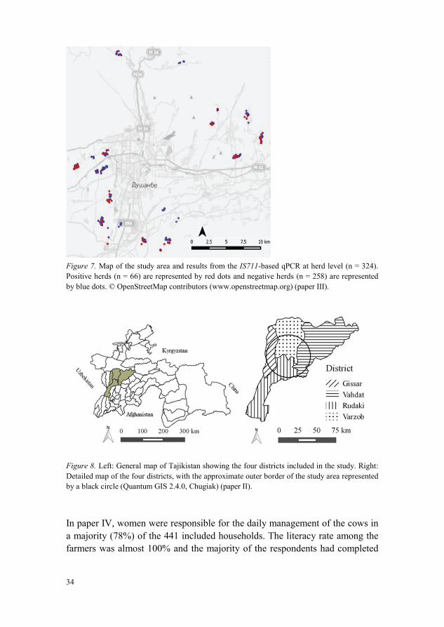
Figure 7. Map of the study area and results from the IS711-based qPCR at herd level (n = 324). Positive herds (n = 66) are represented by red dots and negative herds (n = 258) are represented by blue dots. © OpenStreetMap contributors (www.openstreetmap.org)
Figure 8. Left: General map of Tajikistan showing the four districts included in the study. Right: Detailed map of the four districts, with the approximate outer border of the study area represented by a black circle (Quantum GIS 2.4.0, Chugiak)
In paper IV, women were responsible for the daily management of the cows in a majority (78%) of the 441 included households. The literacy rate among the farmers was almost 100% and the majority of the respondents had completed

35
secondary school which is in accordance with statistics from UNICEF (Unicef, 2013).
4.2 Brucella seropositivity (papers I and II)
In paper I, the herd seroprevalence among the dairy cows was 4.1% (95% CI, 2.6–6.3%) and the individual seroprevalence was 2.0% (95% CI, 1.3–3.1%). The distribution of Brucella-seropositive herds is shown in Figure 6 (p. 33).The seroprevalence at the individual level is in line with the findings in aprevious study from 2007 comprising both small-scale and large-scale herds in Tajikistan (Jackson et al., 2007). A nationwide study conducted in the neighbouring country of Kyrgyzstan showed similar results to those presented in paper I, with an individual seroprevalence in cattle of 2.8% (Bonfoh et al.,2012). Higher figures have been reported from Kazakhstan, another neighbouring country to Tajikistan, where it was estimated that 14% of the lactating cows were Brucella seropositive (Beauvais et al., 2016).
In countries where brucellosis has been eradicated, the typical eradication strategy has been compulsory whole-flock vaccination until the prevalence drops to 1–2%, followed by a test-and-slaughter strategy (Godfroid et al.,2013). This eradication strategy has often taken more than 10 years in high-income countries. In Tajikistan and other low-income countries with scarce resources, an eradication programme is not a realistic approach because itrequires elements such as financial compensation to farmers for production losses and to replace culled animals (Blasco & Molina-Flores, 2011), a legal framework for enforcing eradication measures, well-developed transportation systems, control of animal movements and an identification system forindividual animals (FAO, 2014; Smits, 2012; Blasco & Molina-Flores, 2011).Instead, a mass vaccination strategy to reduce the incidence of Brucella among dairy cows together with campaigns promoting pasteurization of milk and dairy products might be a way forward to reduce the risk of human infection(Godfroid et al., 2013).
In paper II, the true individual seroprevalence among sheep and goats ranged from 1.0% to 15.6% in the four districts. Table 1 shows the apparent individual seroprevalences and the total number of samples collected from sheep and goats. Fourteen villages had at least one seropositive sheep or goat, resulting in an apparent prevalence at the village level of 67%. In a serosurvey performed in 2009, the overall individual seroprevalence was 4.2% in rural districts near the Dushanbe area in the western part of Tajikistan (Ward et al.,2012). From a public health perspective, it is unfortunate that the higher seroprevalence in sheep and goats as shown here geographically coincides with

36
higher human population density in the peri-urban areas. A recent studyconducted in Mexico modelled whether a control programme targeting goats would be economically profitable or not (Montiel et al., 2015). The results showed that a control programme including mass vaccination with Rev 1 would be economically profitable for the farmers whereas a test-and-slaughter strategy would not. This implies that a test-and-slaughter strategy in Mexico would need to include financial compensation to the farmers for the culling of infected animals. To accomplish effective control of the high disease burden among sheep and goats in some districts in Tajikistan, long-term massvaccination with Rev 1 would probably be the best alternative.
Table 1. Descriptive results of Brucella seropositivity at the individual level (n = 667) (paper II).
Variable Category % (Number) Seropositive % (Number)
Species
District a
SheepGoatVarzobRudakiGissar Vahdat
39 (260)61 (407)26 (174)23 (156)23 (156)27 (181)
11 (28)5 (20)3 (5)2 (3)8 (12)15 (28)
a Including both sheep and goats
4.3 Factors associated with Brucella seropositivity (papers I and II)
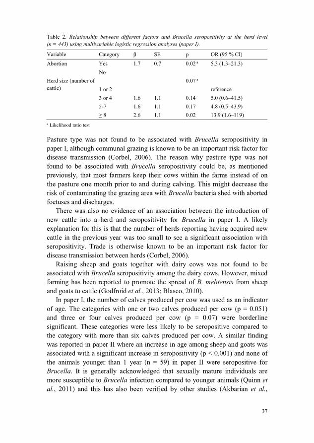
In paper I, the multivariable logistic regression analysis at herd level showedthat abortions were significantly associated with seropositivity (p = 0.02) (Table 2). This finding is in line with the pathobiology of Brucella (Corbel, 2006), and similar results have been described in other field studies (Matope et al., 2011; Al-Majali et al., 2009; Silva et al., 2000). It is generally acknowledged that abortions and decreasing milk yield can be of major economic importance in an infected herd (McDermott et al., 2013).
In paper I, herds with more than eight cattle were significantly associated with seropositivity (p = 0.02) compared with herds with only one or twocattle (Table 2). This is consistent with other studies from Uganda, Jordan, sub-Saharan Africa and Kenya (Mugizi et al., 2015; Al-Majali et al., 2009; McDermott & Arimi, 2002; Kadohira et al., 1997). Because Brucella is most commonly transmitted through direct contact between cattle following abortion, it is likely that the large herds provide more opportunities for transmission of Brucella infection between cattle.
37
Table 2. Relationship between different factors and Brucella seropositivity at the herd level (n = 443) using multivariable logistic regression analyses (paper I).
a Likelihood ratio test
Pasture type was not found to be associated with Brucella seropositivity in paper I, although communal grazing is known to be an important risk factor for disease transmission (Corbel, 2006). The reason why pasture type was not found to be associated with Brucella seropositivity could be, as mentioned previously, that most farmers keep their cows within the farms instead of on the pasture one month prior to and during calving his might decrease the risk of contaminating the grazing area with Brucella bacteria shed with aborted foetuses and discharges.
There was also no evidence of an association between the introduction of new cattle into a herd and seropositivity for Brucella in paper I. A likely explanation for this is that the number of herds reporting having acquired new cattle in the previous year was too small to see a significant association with seropositivity. Trade is otherwise known to be an important risk factor for disease transmission between herds (Corbel, 2006).
Raising sheep and goats together with dairy cows was not found to be associated with Brucella seropositivity among the dairy cows. However, mixed farming has been reported to promote the spread of B. melitensis from sheep and goats to cattle (Godfroid et al., 2013; Blasco, 2010).
In paper I, the number of calves produced per cow was used as an indicator of age. The categories with one or two calves produced per cow (p = 0.051) and three or four calves produced per cow (p = 0.07) were borderline significant. These categories were less likely to be seropositive compared to the category with more than six calves produced per cow. A similar finding was reported in paper II where an increase in age among sheep and goats was associated with a significant increase in seropositivity (p < 0.001) and none ofthe animals younger than 1 year (n = 59) in paper II were seropositive forBrucella. It is generally acknowledged that sexually mature individuals are more susceptible to Brucella infection compared to younger animals (Quinn et al., 2011) and this has also been verified by other studies (Akbarian et al.,
Variable Category SE p OR (95 % CI)
Abortion
Herd size (number of cattle)
YesNo
1 or 23 or 45-7
1.7
1.61.62.6
0.7
1.11.11.1
0.02 a
0.07 a
0.140.170.02
5.3 (1.3–21.3)
reference5.0 (0.6–41.5)4.8 (0.5–43.9)13.9 (1.6–119)

38
2015; Boukary et al., 2013; Megersa et al., 2011; Al-Majali et al., 2009; Silvaet al., 2000). However, in papers I and II where we only sampled sexually mature individuals, the older animals had lived a longer time at risk of being exposed to infection compared to the younger animals.
In paper II, there was a significant difference in seroprevalence between the four districts (Table 3). Sheep and goats in Rudaki (p = 0.003) and Varzob (p = 0.024) were less likely to be seropositive than sheep and goats in Vahdat.One reason for the high seroprevalence observed in Vahdat district could be that many villages from other districts use part of Vahdat district as the main route for the movement of sheep and goats between summer and winter pastures. This could increase the density of animals grazing together and hence the risk for transmission of Brucella through direct contact between animals from different villages. Furthermore, there are three large animal markets inVahdat district that could play an important role in transmitting Brucellainfection (Figure 9). To reduce the risk of transmission between villages and districts, trade in animals should be restricted (Blasco & Molina-Flores, 2011).
Table 3. Relationship between different factors and Brucella seropositivity at the individual level (n=667) using multivariable logistic regression analyses with village as the random effect, Tajikistan, 2012 (paper II).
Variable Category p OR (95% CI)
Species
District
Age (in years)
SheepGoat
VarzobGissarRudakiVahdatContinuous
1.0
1.30.52.3
0.3
0.009
0.0080.0240.1910.003
<0.001
2.7 (1.3-5.5)
0.3 (0.09 0.8)0.6 (0.3 1.3)0.1 (0.03 0.4)Reference1.4 (1.2 1.6)

39
Figure 9. Animal market (author’s photo).
In paper II, sheep were more likely to be seropositive than goats (p = 0.009) (Table 3). However, other literature suggests that goats are more susceptible to B. melitensis infection than sheep (Quinn et al., 2011). There was no difference in seroprevalence between non-vaccinated sheep and goats in a previous studyconducted in Tajikistan (Ward et al., 2012), but differences in susceptibility have been observed among sheep where the milking breeds seem to be mostsusceptible to B. melitensis (Corbel, 2006). More research is required to allow firm conclusions to be drawn on whether the fat-tailed Gissar breed of sheepthat are common in the region are more susceptible to Brucella infection than goats.
4.4 Detection of Brucella DNA by PCR and comparison with serology (paper III)
In the current study, 564 cow milk extracts from 326 herds in 21 villages were analy ed for Brucella DNA. In total, Brucella DNA was detected in 13.7% (n = 77) of the milk samples with IS711 qPCR. A sample was considered to be positive if the Cycle threshold (Ct) was (Al Dahouk et al., 2007) in two runs. The apparent individual seroprevalence measured previously with I-ELISA and C-ELISA was 2.1%. All seropositive cows (n = 12) were positiveby qPCR with Ct-values ranging between 26.9 and 31.9. Out of the 552seronegative cows, 11.8% (n = 65) were qPCR positive for Brucella DNA with Ct-values ranging between 26.5 and 39.8. At herd level, 20% (n = 66) had at least one positive cow by qPCR (Figure 7, p. 34). Similar discrepancy between the serology and qPCR results was demonstrated in a study comparing IS711-based qPCR, serology and culture among wild boars. In that study, BrucellaDNA was detected in tissue samples of 11.1% of the seronegative individuals

40
et al., 2009). The discrepancy between the serology and PCR resultsobserved in the current study might indicate that the true number of Brucella-infected cattle within the study area could be underestimated by serology screening. False serological negative results have been reported previously(Mailles et al., 2012; Al Dahouk et al., 2003; Godfroid et al., 2002) and one explanation could be that antibody titers reduce over time (Godfroid et al.,2010). Hence, seronegative animals in the current study, which tested positive by qPCR, could have been exposed to Brucella and turned seronegative after a certain time period. Alternatively, if sampling at an early stage of the infection, i.e. within the first 14 days, the humoral immune response has not yet induced detectable levels of antibodies in the host (Gardner et al., 2000). Furthermore, individuals infected in utero or in the early post-natal period can become latently infected and hence never become seropositive (Corbel, 2006).Approximately 3.5% of infected cows are estimated to deliver latent-infected offspring (Saegerman et al., 2010). Furthermore, it has previously been reported that B. suis infection in cattle generates a shorter duration of antibody in the host ���������� (Godfroid et al., 2002). Whether this is also
B. melitensis infection in cattle is not known and needs to be investigated further.If this is the case, it might partially explain the discrepancy between the serology and qPCR results observed in the current study. Hypothetically, the discrepancy between the serology and qPCR results could be previous vaccination against brucellosis as reported from a study in Egypt where cattle vaccinated with RB51 tested negative by serology tests but positive by qPCR(Gwida et al., 2016). However, in the current study, the information given from the local official veterinarians that none of the cattle had been vaccinated against brucellosis is considered reliable because there is no national control programme for brucellosis among livestock in Tajikistan. The potentially significant number of serological false negative individuals observed in paper III highlights the importance of determining if there is a need for implementingcomplementary diagnostic strategies to detect false serological negative individuals in Brucella surveillance, control, and eradication programmes.
To draw firm conclusions regarding the zoonotic risk of consuming the milk from the qPCR-positive individuals is difficult because the qPCR can detect DNA from live, damaged or dead bacteria. However, becauseconsumption of and trading with unpasteurized dairy products is commonly occurring among small-scale farmers in the study area, the significant numbers of cows with detectable levels of Brucella DNA in their milk could constitute a serious health .

41
4.5 Sequence analysis of Brucella DNA (paper III)
In total, only two samples had sufficient amounts of DNA to perform sequence analysis. The first sample was collected from a seropositive cow and the SNP allelic profiles corresponded to the SNP profiles described for B. melitensis and B. suis at codon positions 716 (CCG) and 737 (GTT) (Marianelli et al., 2006).Because there is almost no pig production in Tajikistan, it is highly likely that this cow was infected with B. melitensis. This individual was not being kept together with small ruminants and the infection source in this particular case remains unknown. The prevailing epidemiological situation in the study area, with endemic Brucella infection among sheep and goats and where cattle are often kept in close proximity with small ruminants, could lead to a spillover of B. melitensis from small ruminants to cattle (Godfroid et al., 2013; Blasco, 2010). A similar finding has been reported in a study from the neighbouring country of Kyrgyzstan where B. melitensis has been isolated from cattle(Kasymbekov et al., 2013).
The other sample with a sufficient amount of DNA to perform molecular characterization came from a seronegative cow, and the SNP allelic profiles corresponded to B. abortus at codon positions 716 (CCA), 969 (CGT) and 985 (GCC). At one position – the codon at 737 (GTT) – the SNP was not described for B. abortus, although it has previously been reported to be GTC for B. abortus. Whether this SNP is a new marker for B. abortus in the region remains unclear, and more research is required to draw firm conclusions from this observation. A report by the FAO stresses that B. melitensis infection is much more common than B. abortus in Central Asia and the Middle East (FAO, 2010) but the findings of the current study suggests that B. abortusinfection might also constitute a problem in the region hus vaccination ofcattle with S19 in addition to vaccination of small ruminants with Rev 1 mightbe needed in order to control Brucella infections in the livestock population.To obtain a comprehensive understanding of the Brucella spp. circulation within the livestock population in this region, further research, including isolation of Brucella from cattle, sheep and goats, is required.
4.6 Knowledge of brucellosis (paper IV)
In paper IV, a majority (85%) of the 441 respondents had never heard of brucellosis. Of those who had heard of the disease (n = 65), about half (55%)had received information from relatives or friends and the majority (82%)knew that cattle, sheep or goats could become infected (Table 4). Allinterviewees who had heard of brucellosis (n = 65) knew that humans could become infected and 52 of these persons knew that arthritis was a common

42
symptom in humans. A majority (78%) did not know that cattle could be vaccinated against the disease and 91% of those who had heard of brucellosis knew at least one correct route of transmission from animals to humans, most commonly through the consumption of unpasteurized milk from infected cows.Fewer (22%) knew one or more correct route of transmission between animals. In five of the households that had heard of brucellosis, a family member had been diagnosed with the disease by a physician and in two of the households a veterinarian had diagnosed brucellosis among cattle, sheep or goats. A high awareness of brucellosis among farmers has been shown in Egypt where the majority of the farmers were aware of brucellosis, which the authors explained by endemic brucellosis in the study area (Holt et al., 2011). The difference in awareness among farmers in Egypt and the current study might be explained by a lower herd seroprevalence among the dairy cows in UPU areas of Dushanbe compared to Egypt. However, it is noteworthy that the awareness of brucellosis was poor among the farmers in paper IV despite a control programme among small ruminants initiated in 2004 by the FAO. The programme did not include the region of Dushanbe, but included several areas in Tajikistan with high seroprevalences of Brucella spp. in sheep and goats (FAO, 2014; Ward et al., 2012).
A study from Kyrgyzstan showed that a good knowledge of the transmission routes for brucellosis had a protective effect against human infection (Kozukeev et al., 2006) and a study from Iran showed that knowledge of the mode of brucellosis transmission through fresh cheese was protective against disease transmission in humans (Sofian et al., 2008). The human incidence of brucellosis is increasing in Tajikistan (Pappas, 2010) and the majority of the farmers in the current study with a low awareness of brucellosis could be exposed to a higher risk of contracting Brucella infection than the farmers with a high awareness of the disease.
The multivariable logistic regression model showed that participants with alower level of education were less likely to have knowledge of brucellosis compared to those who had attended technical college or university (p < 0.001). The relationship between educational level and brucellosis has also been investigated in a study from Yemen, which showed that humans diagnosed with brucellosis were more likely to have a lower education level compared to controls (Al-Shamahy et al., 2000). If this also is true for the current study area, farmers with a lower level of education could be at higher risk of contracting brucellosis than their peers with a higher level of education. Respondents who discussed animal health issues with family members or friends were less likely to have heard of brucellosis compared to those who often talked to veterinarians (p = 0.03). Discussing animal health issues with

43
veterinarians was common among the farmers (Table 5, p. 46) and information campaigns regarding the epidemiology of Brucella targeting veterinarians might thus be an effective way to transfer knowledge to farmers.
Table 4. Knowledge about brucellosis among the respondents who had heard of the disease (n = 65) (paper IV).
a Stated at least one correct route of transmission b Stated no correct route of transmission
Category %
Information source
Which animal species can become infected? Can humans become infected? Symptoms in humans
Does any vaccination for animals exist?
Modes of transmission: Animal-to-animal Animal-to-human
Previous Brucella infection within the household: Among humans
Among cattle/sheep/goats
Relatives/friendsVeterinarianBook TelevisionDon´t knowCattle/Sheep/GoatsAll mammalsDon´t knowYesNoArthritisFever and arthritisFatigueSkin lesionsDon´t knowYesNo
Correct a
Incorrect b
Correct a
Incorrect b
Yes NoYesNo
5522111.511824.6141000803.11.53.1122278
2278919.2
7.7923.197

44
4.7 Attitudes towards brucellosis (paper IV)
Sixty-three per cent (n = 279) of the households wanted more information about brucellosis, while the remaining 37% claimed that they did not need more information. Of the 279 respondents who wanted more information, the majority (58%) preferred to receive the information through an educational booklet while 23% preferred a course or information meeting in the village. The high literacy rate and educational standard, together with a positive attitude towards learning more, provides a good foundation for integratinginformation campaigns for brucellosis into future control programmes in Tajikistan.
Of the respondents who had heard of brucellosis (n = 65), the majority (n = 52) did not consider any family member to be at risk of contracting Brucella infection and of all respondents (n = 441), only 2.5% perceived themselves as being at risk of contracting brucellosis. Among those who had heard of brucellosis (n = 65), 17% perceived a risk of contracting the diseaseand that the person in the household working most with the cows was exposed to the highest risk. Thus increasing the share of farmers who are knowledgeable of the existence of brucellosis might increase the number of farmers perceiving themselves as being at risk of contracting brucellosis. This could be a first step in building a platform for discussions regarding risk behaviours.
4.8 Self-reported practices (paper IV)
Consumption of unpasteurized dairy products is a well-known risk factor for human brucellosis (Earhart et al., 2009; Sofian et al., 2008; Kozukeev et al.,2006) and close to 30% of the households reported consuming unpasteurized dairy products from the cows on a regular basis (Table 5). Seventeen per cent (n = 76) of the respondents sold unpasteurized milk or unpasteurized milk products directly to consumers on a regular basis. The majority (66%) of these 76 respondents sold their unpasteurized dairy products on an everyday basis.The results from paper I show that 4% of the households had at least one Brucella-seropositive cow. Furthermore, paper III shows that 13.7% of the dairy cows included in that study had detectable levels of Brucella DNA in their milk. Hence, the consumption of unpasteurized dairy products reported in paper IV could constitute a risk to public health. Changes in the political andeconomic situation in the region have led to increased privatization of collective farms in Tajikistan and other Central Asian countries (Jackson et al.,2007; Kozukeev et al., 2006) and Kozukeev et al. suggest that this development has led to more frequent trading with home-made animal source

45
foods in Kyrgyzstan and thus to decreased food safety (Kozukeev et al., 2006).Because the pattern of privatization of collective farms has been similar in Tajikistan, there are reasons to believe that trading in Tajik home-made animalsource foods has increased, putting food safety at risk.
Seventy-eight per cent reported hand washing as the only protective measure after having handled aborted foetuses or discharges, and only 21% used gloves (Table 5). If abortions in livestock occur due to brucellosis, the foetuses and aborted material will be heavily infected by Brucella (OIE, 2016),and using hand washing as the only protective measure to avoid transmission of infection to humans might not be sufficient. One explanation for why the majority of farmers were only using hand washing as a protective measure could be poor knowledge of the risk with this practice but also lack of access to protective clothing like gloves. Similar results have been reported in a study from Egypt (Holt et al., 2011) and this practice is a known risk factor for humans to contract brucellosis from livestock (Earhart et al., 2009; Kozukeevet al., 2006).
Females were more likely to assist during calving (56% of the households)compared to males (31% of the households) (Table 5). This finding, together with the previously reported finding that females were responsible for the daily management of the cows in a majority of the households, could imply that females are exposed to a high risk of contracting Brucella infection through direct contact with Brucella-infected dairy cows in the study area. This is supported by a study from Mongolia that showed that women were more likely to be seropositive for Brucella compared to men (Zolzaya et al., 2014). The authors of that study suggest that reasons for this could be that women more often take care of weak newborn animals and are more responsible for milking livestock.
It was almost as common to discuss animal health issues with veterinarians (48%) as it was with family members or friends (52%) and the majority of the farmers (81%) contacted a veterinarian if a cow showed symptoms of disease(Table 5). This is in line with findings from a study conducted in Egypt where most respondents would contact the local veterinarian if they suspected Brucella infection among their livestock (Holt et al., 2011). The well-established relationship between many veterinarians and farmers could be useful for implementing information campaigns as part of a future brucellosis control programme.

46
Table 5. Descriptive results of self-reported practices among dairy farmers (paper IV).
Category n %
Does the respondent sell unpasteurized milk or unpasteurized milk products directly to consumers? (n = 438)
Yes 76 17No 362 83
How frequently does the respondent sell unpasteurized milk or unpasteurized milk products? (n = 76)
Every day 50 66One to two times per week
14 18
Once a month/sometimes
12 16
Does the respondent consume unpasteurized milk or unpasteurized milk products? (n = 441)
Yes 123 28No 318 72
Who in the household assist during calving? (n = 441) Female 246 56Male 138 31Female & Male 56 13Always call veterinarian
1 0.2
Who does the respondent talk to about animal health issues?(n = 441)
Family member/friend
229 52
Veterinarian 212 48What does the respondent do with dead cattle foetuses?(n = 441)
Bury 413 94Call veterinarian 9 2Food for dogs 7 1.6Burn 2 0.5Don´t know 10 2.3
Does the respondent use protection when dealing with cows having an abortion or with aborted materials? (n = 441)
Use gloves 93 21Wash hands 344 78Always call veterinarian
1 0.2
No / Don´t know 3 0.7If the respondent buys a new cattle, does he/she take any action to assure it is healthy? (n = 441)
No 280 63Use more experienced people in the village
142 32
Use veterinary inspection
19 4.3
What does the respondent do if a cattle is sick or shows signs of disease? (n = 441)
Seek veterinary assistance
359 81
Treat 77 17Slaughter 4 0.9Don´t know 1 0.2

47
In paper IV, almost all households (94%) stated that they usually buried dead cattle foetuses (Table 5). This practice might reduce the risk of dogs consuming placental or foetal materials from aborting cows and thus reduce the risk of dogs acting as transmitters of Brucella to humans and livestock (Warethet al., 2016). In contrast to the finding in Tajikistan, a Mongolian study showed that almost half of the respondents usually fed their dogs with aborted foetuses and placentas (Zolzaya et al., 2014). Burying dead animals (that were not slaughtered) is reported to be common practice in Tajikistan for religious reasons (Sattorov, personal communication, 2016).
When purchasing new cattle, the majority (63%) stated that they did not take any specific action to make sure the animal was healthy, whereas 32% used more experienced people in the village for help (Table 5). The introduction of new livestock into a herd from a source not declared free from Brucella infection is an important risk factor for transmitting infection between herds (Blasco & Molina-Flores, 2011). The practice of uncontrolled trading of cattle around Dushanbe might therefore increase the risk for transmission of Brucella infection between herds.
Households with a history of reported Brucella infection among humans, cattle, sheep or goats were equally inclined to sell and consume unpasteurized dairy products as those who had not had the infection within the household or who had never heard of the disease. This might imply that there is a lack of information from physicians and veterinarians to affected farmers regarding the modes of transmission of brucellosis. It could also reflect the fact that awareness of risks might not translate into changed behaviors similar to what was shown in a study of avian influenza in Cambodia (Osbjer et al., 2015).


49
5 ConclusionsThis thesis reports on the occurrence of Brucella among livestock in an UPU area of Tajikistan, identifies the Brucella spp. extracted from bovine milk and elucidates farmers´ knowledge, attitudes and self-reported risk practices relatedto brucellosis. The following are the main conclusions of this thesis.
Brucella infection is widespread among livestock in the UPU area of Dushanbe, with high seroprevalence among sheep and goats in Vahdat and Gissar districts. This can constitute a public health risk and cause significant production losses.Previously known risk factors for Brucella seropositivity were also demonstrated here including herd size (among cattle) and age (among cattle, sheep and goats). Contrary to existing data, sheep are more likely to be seropositive compared to goats in the study area.Brucella DNA was commonly detected in milk from cows in the UPU area of Dushanbe, both among Brucella seropositive and seronegative individuals. The discrepancies between the serology and PCR resultshighlight the need to further investigate whether there is a need for implementing complementary diagnostic strategies to detect false serologically negative individuals in Brucella surveillance, control and eradication programmes.There is likely a reservoir of B. abortus in the cattle population and aspillover of B. melitensis from small ruminants to cattle. This suggests thatvaccination cattle with S19 in addition to vaccination of small ruminants with Rev 1 might be needed in order to control Brucella infections in the livestock population.
50
The knowledge of Brucella is poor among UPU dairy farmers and a high literacy rate and willingness to learn more strengthens the logic for including campaigns aiming to raise awareness of brucellosis and its associated risks in control programmes.Several known high-risk practices, such as consumption of unpasteurized dairy products and not wearing protective clothing when dealing with aborted foetuses or discharges, were reported among the UPU dairy farmers. Health education aiming to change such risk behaviours should therefore be promoted in future control programmes.
51
6 Future perspectivesTajikistan is a low-income country in a politically turbulent part of the world.With continued development and economic growth, gaining control of some important zoonotic infections as many high-income countries have done, is a likely progression. However, such a positive development cannot be guaranteed with ongoing conflicts in neighbouring countries, weak institutions and strong economic dependency o Russia. Many high-income countries have successfully gained control of Brucella through the implementation of control and eradication programmes in livestock and the question of whether Tajikistan can follow suit needs to be addressed.
A cost-benefit analysis conducted in Mongolia, another low-income , suggests that implementation of a brucellosis control programme
consisting of vaccinations of livestock can be cost effective (Roth et al., 2003).To determine whether a control programme among livestock in Tajikistan would also be cost effective, the cost of implementing a control programme as well as the national economic gain from such a programme needs to be investigated (Smits, 2012). Also, it must be acknowledged that Tajikistan is a country with limited economic resources that is forced to make difficultprioritizations. Although a control programme might be profitable, it mightvery well be the case that it should not be implemented if there are other national interests of even higher urgency. Furthermore, even if a control programme makes economic sense after having considered other national needs, the feasibility of conducting a successful and long-lasting control programme for brucellosis in Tajikistan must be critically analysed. Not only is Tajikistan an economically constrained country, it is also a young nation. The functioning of infrastructure, border control, enforcement of policies and regulations, long-term political commitment and funding of the veterinary sector cannot be taken for granted. Understanding if Tajikistan will have the

52
institutional functions in place to support a long-term control programme must therefore complement the economic analysis.
Long-term changes must be taken into consideration when evaluating the benefits of long-term interventions. One important driver of change in Tajikistan, and many other low-income countries, is urbanization. Today, every fourth Tajik citizen lives in an urban area and in 2050 almost every second Tajik is projected to live in an urban area (United Nations, 2014). Such a drastic development will surely affect the practice of livestock keeping and therefore needs to be better understood. One plausible development might bean increasing number of small-scale UPU farmers continuing to trade with home-made animal source foods and thus putting food safety at even greater risk. Another trajectory might be that urbanization leads to specialisation with large scale livestock production and well-controlled food value chains. These two scenarios would imply different set-ups of a proper Brucella control programme. Whether the described scenarios, or some other scenarios, are the most likely to materialize would be of great value to understand whenprojecting the future benefits of a control programme.
This thesis shows that the knowledge of brucellosis is poor among UPU dairy farmers in Dushanbe, Tajikistan. Furthermore, several known high-risk practices, such as consumption of unpasteurized dairy products and not wearing protective clothing when dealing with aborted foetuses or discharges, were commonly reported among the farmers. A future control programme should therefore promote health education aiming to increase public awareness of brucellosis and change such risk-behaviours among livestock farmers.
The fact that the majority of published brucellosis studies rely on serology is a matter that has recently received some critical attention because serology does not reveal which Brucella spp. is causing the infection in the host and therefore precludes the possibility of identifying the source of infection (Godfroid et al., 2013). Some of the factors in favour of serology are that serology is rapid, easy to perform and does not require a well-equipped laboratory, all of which are beneficial in low-income countries. Today, there are different rapid typing tools for molecular characterization of Brucella, but the challenge is to transfer the technology and knowledge to low-income countries. The introduction of such tools in low-income countries would probably increase the number of publications investigating which Brucella spp. is causing infection in humans and livestock and thereby contribute to the understanding of how to better control Brucella in the future.
53
7 Populärvetenskaplig sammanfattning
Brucellos är en av de vanligaste zoonotiska sjukdomarna i världen och drabbar framför allt människor och djur i fattiga länder. En zoonos är en sjukdom som kan smitta mellan djur och människor. Brucellos orsakar stora ekonomiska förluster i de länder där sjukdomen är vanlig. Centralasien Mellanöstern är hårt drabbade områden med många fall av brucellos hos människor och djur.
En av vår tids stora samhällsförändrande krafter är urbaniseringen, dvs. att människor flyttar från landsbygden till städer. När människor flyttar till städertar de ofta med sig sina livsmedelsproducerande djur såsom kor, får, getter,grisar och fjäderfä. Att bedriva småskalig djurhållning i städer kan ge fattiga människor en extra inkomst och även utgöra en stor del av familjens
men urbaniseringen bidrar också till att djur och tät tillsammans. I dessa miljöer är handel med mjölk, kött och ägg
essa livsmedel kan nå många människor i staden, ofta via informellahandelskanaler. Om livsmedelsproducerande djur, som hålls i en stadsmiljö, är infekterade med zoonotiska sjukdomar såsom brucellos, kan de därför utgöra en stor risk för folkhälsan.
Målen med denna avhandling är att inventera förekomsten av Brucella hos kor, får och getter och att hur människor i ett låginkomstland, med en snabb förväntad urbaniseringstakt, förstår och agerar utifrån de risker denna sjukdom för med sig.
De fyra studier som ingår i denna avhandling är utförda i hushåll med småskalig djurhållning belägna i och i utkanten av Dushanbe, som är huvudstaden i Tajikistan. Totalt togs blodprov från 904 kor och 667 får och getter samt mjölkprover från 564 kor. Vidare intervjuades 441 hushåll om kunskaper, attityder och riskbeteenden med anknytning till brucellos. Andelen av kor, får och getter som bar på antikroppar mot Brucella undersöktes. Ett

54
djur som bär på antikroppar Brucella, och som tidigare inte vaccinerats för brucellos, har någon gång stött på smittämnet och räknas därmed som infekterad med Brucella. Fyra procent av de hushåll som ägde kor hade minst en Brucella-positiv ko. Positiva samband påvisades mellan antikroppar Brucella och antal kor i hushållet, ålder på korna samt tidigare aborter hos djuren. Bland får och getter var det en skillnad i förekomst av antikroppar Brucella i de fyra distrikten som undersöktes. I det distrikt med lägst förekomst bar 1.0% av de testade djuren på antikroppar mot Brucella medan det i med högst förekomst var 16% av fåren och getterna som Brucella. Ett positivt samband mellan antikroppar Brucella och ålder på djuren påvisades och får visade högre sannolikhet att vara antikroppspositiva jämfört med getter.
Brucella DNA påvisades i 14% av mjölkproverna med polymeras-kedjereaktion (PCR). Alla kor som hade antikroppar brucellos hade även Brucella DNA i sin mjölk. Brucella DNA kunde också påvisas i mjölken hos 12% av de kor som inte hade antikroppar brucellos. Analyser av DNA:t från två mjölkprover tyder på att korna utgör en reservoar för B. abortus och att det även med hög sannolikhet skett en spridning av B. melitensis från får och getter till korna.
Kunskapen om brucellos bland de tillfrågade hushållen visade sig vara mycket bristfällig. Ett flertal riskbeteenden såsom konsumtion av opastöriserad mjölk och att inte bära skyddskläder vid hantering av aborterade foster och fostervätskor var vanligt förekommande.
Denna avhandling visar att Brucella finns bland livsmedelproducerande djur i och i utkanterna av Dushanbe. Dessutom pekar denna avhandling på hög förekomst bland får och getter i vissa av de undersökta distrikten samt stor förekomst av Brucella DNA i komjölk. Detta kan utgöra en risk för folkhälsan och orsaka stora ekonomiska förluster i området. Skillnaden i resultaten mellan antikroppstesterna och PCR visar på ett behov av att undersöka om PCR böranvändas som ett komplement till antikroppsanalyser i olika övervaknings-,och kontrollprogram för brucellos. För att bättre kunna förebygga brucellos i framtiden behövs fler studier som syftar till att skapa en helhetsbild över vilka Brucella arter som förekommer bland både djur och människor i Tajikistan.
Med tanke på den låga kunskapsnivån, den frekventa förekomsten av riskbeteenden och en vilja bland hushållen att lära sig mer, bör en informationskampanj riktad mot hushåll med livsmedelsproducerande djur vara en del av insatsen i ett framtida kontrollprogram mot brucellos i Tajikistan.

55
ReferencesAkbarian, Z., Ziay, G., Schauwers, W., Noormal, B., Saeed, I., Qanee, A.H., Shahab, Z.,
Dennison, T., Dohoo, I. & Jackson, R. (2015). Brucellosis and Coxiella burnetii infection in householders and their animals in secure villages in Herat province, Afghanistan: A cross-sectional study. PLoS Neglected Tropical Diseases, 9, doi: 10.1371/journal.pntd.0004112.
Al Dahouk, S., Tomaso, H., Nöckler, K., Neubauer, H. & Frangoulidis, D. (2003). Laboratory-based diagnosis of brucellosis--a review of the literature. Part II: serological tests for brucellosis. Clinical Laboratory, 49(11-12), pp. 577-589.
Al Dahouk, S., Nöckler, K., Scholz, H.C., Pfeffer, M., Neubauer, H. and Tomaso, H. (2007). Evaluation of genus-specific and species-specific real-time PCR assays for the identification of Brucella spp. Clinical Chemical Laboratory Medicine, 45(11), pp.1464-1470.
Al-Majali, A.M., Talafha, A.Q., Ababneh, M.M. & Ababneh, M.M. (2009). Seroprevalence and risk factors for bovine brucellosis in Jordan. Journal of Veterinary Science, 10(1), pp. 61-65.
Al-Shamahy, H.A., Whitty, C.J.M. & Wright, S.G. (2000). Risk factors for human brucellosis in Yemen: a case control study. Epidemiology and infection, 125, pp. 309-313.
Allen, A., Breadon, E., Byrne, A., Mallon, T., Skuce, R., Groussaud, P., Dainty, A., Graham, J., Jones, K., Pollock, L. & Whatmore, A. M. (2015). Molecular Epidemiology of Brucella abortus in Northern Ireland—1991 to 2012. PloS one, 10(9), doi: 10.1371/journal.pone.0136721.
Baily, G.G., Krahn, J.B., Drasar, B.S. & Stoker, N.G. (1992). Detection of Brucella melitensisand Brucella abortus tion. Journal of Tropical Medicine and Hygiene, 95, 271-275.
Beauvais, W., Coker, R., Nurtazina, G. & Guitian, J. (2015). Policies and Livestock Systems Driving Brucellosis Re-emergence in Kazakhstan. EcoHealth, doi: 10.1007/s10393-015-1030-7.
Beauvais, W., Orynbayev, M. & Guitian, J. (2016). Empirical Bayes estimation of farm prevalence adjusting for multistage sampling and uncertainty in test performance: a Brucella cross-sectional serostudy in southern Kazakhstan. Epidemiology & Infection, doi: 10.1017/S0950268816001825.
Beja-Pereira, A., Bricker, B., Chen, S., Almendra, C., White, P. & Luikart, G. (2009). DNA genotyping suggests that recent brucellosis outbreaks in the Greater Yellowstone Area originated from elk. Journal of Wildlife Diseases, 45(4), pp. 1174-1177.

56
Blasco, J.M. (1997). A review of the use of B. melitensis Rev 1 vaccine in adult sheep and goats. Preventive Veterinary Medicine, 31, pp. 275-283.
Blasco, J.M. (2010). Control and eradication strategies for Brucella melitensis infection in sheep and goats. Prilozi, 31(1), pp. 145-165.
Blasco, J. M. & Molina-Flores, B. (2011). Control and eradication of Brucella melitensisinfection in sheep and goats. Veterinary Clinics of North America: Food Animal Practice, 27, pp. 95-104.
Bonfoh, B., Kasymbekov, J., Dürr, S., Toktobaev, N., Doherr, M. G., Schueth, T., Zinsstag, J. & Schelling, E. (2012). Representative seroprevalences of brucellosis in humans and livestock in Kyrgyzstan. EcoHealth, 9, pp. 132-138.
Boukary, A. R., Saegerman, C., Abatih, E., Fretin, D., Bada, R. A., De Deken, R., Harouna, H. A., Ytnikoye, A. & Thys, E. (2013). Seroprevalence and potential risk factors for Brucella spp. infection in traditional cattle, sheep and goats reared in urban, periurban and rural areas of Niger. PloS one, 8, doi: 10.1371/journal.pone.0083175.
Bounaadja, L., Albert, D., Chénais, B., Hénault, S., Zygmunt, M.S., Poliak, S. & Garin-Bastuji, B. (2009). Real-time PCR for identification of Brucella spp.: A comparative study of IS711, bcsp31 and per target genes. Veterinary microbiology, 137, pp. 156-164.
Bricker, B. J. (2002). PCR as a diagnostic tool for brucellosis. Veterinary microbiology, 90, pp. 435-446.
Bricker, B. J. & Halling, S. M. (1994). Differentiation of Brucella abortus bv. 1, 2, and 4, Brucella melitensis, Brucella ovis, and Brucella suis bv. 1 by PCR. Journal of Clinical Microbiology, 32(11), pp. 2660-2666.
Buddle, M. B. (1956). Studies on Brucella ovis (n. sp.), a cause of genital disease of sheep in New Zealand and Australia. Journal of Hygiene, 54, pp. 351-364.
Carmichael, L. & Bruner, D. (1968). Characteristics of a newly-recognized species of Brucella responsible for infectious canine abortions. The Cornell Veterinarian, 48(4), 579-592.
Cerenius, F. (2010). Det svenska djursmittskyddets historia fram till 2000. In Folkhälsa-Djurhälsa: Ny ansvarsfördelning mellan stat och näring, Regeringen SOU 2010:106 (in Swedish, Swedish Government’s official reports).
CIA (2016). Tajikistan. Available: www.cia.gov/library/publications/resources/the-world-factbook/geos/ti.html [Accessed 13 October 2016].
Corbel, M.J. (2006). Brucellosis in humans and animals, World Health Organization in collaboration with the Food and Agriculture Organization of the United Nations and World Organisation for Animal Health. Available: www.who.int/csr/resources/publications/Brucellosis.pdf [Accessed 12 October 2016].
Dean, A. S., Crump, L., Greter, H., Hattendorf, J., Schelling, E. & Zinsstag, J. (2012). Clinical manifestations of human brucellosis: a systematic review and meta-analysis. PLoS Neglected Tropical Diseases, 6, doi: 10.1371/journal.pntd.0001929.
Earhart, K., Vafakolov, S., Yarmohamedova, N., Michael, A., Tjaden, J. & Soliman, A. (2009). Risk factors for brucellosis in Samarqand Oblast, Uzbekistan. International Journal of Infectious Diseases, 13, pp. 749-753.

57
El-Tras, W., Tayel, A. A., Eltholth, M. M. & Guitian, J. (2010). Brucella infection in fresh water fish: Evidence for natural infection of Nile catfish, Clarias gariepinus, with Brucella melitensis. Veterinary microbiology, 141, pp. 321-325.
Ewalt, D. R., Payeur, J. B., Martin, B. M., Cummins, D. R. & Miller, W. G. (1994). Characteristics of a Brucella species from a bottlenose dolphin (Tursiops truncatus). Journal of Veterinary Diagnostic Investigation, 6, pp. 448-452.
FAO (2001). Urban and peri-urban agriculture – A briefing guide for the successful implementation of urban and peri-urban agriculture in developing countries and countries of transition. The special programme for food security. Food and agriculture Organization of the United Nations, Rome, Italy.
FAO (2010). Brucella melitensis in Eurasia and the Middle East. FAO Animal Production and Health Proceedings. No. 10. Rome.
FAO (2014). FAO works to curb the burden of brucellosis in endemic countries: Case studies from Eurasia and the Near East. FOCUS ON, No. 8, July 2014. Rome.
Farrell, I. (1974). The development of a new selective medium for the isolation of Brucella abortus from contaminated sources. Research in veterinary science, 16, pp. 280-286.
Flynn, K. (1999). An overview of public health and urban agriculture: Water, soil, and crop contamination and emerging urban zoonoses. Cities Feeding People Series, Report, 30.
Foster, G., Osterman, B. S., Godfroid, J., Jacques, I. & Cloeckaert, A. (2007). Brucella ceti sp. nov. and Brucella pinnipedialis sp. nov. for Brucella strains with cetaceans and seals as their preferred hosts. International journal of systematic and evolutionary microbiology, 57, pp. 2688-2693.
Foster, J.T., Beckstrom-Sternberg, S.M., Pearson, T., Beckstrom-Sternberg JS, Chain, P.S., Roberto, F.F., Hnath, J., Brettin, T. & Keim, P. (2009). Whole-genome-based phylogeny and divergence of the genus Brucella. Journal of bacteriology, 191(8), pp. 2864-2870.
García-Yoldi, D., Marín, C.M., de Miguel, M.J., Muñoz, P.M., Vizmanos, J.L. & López-Goñi, I. (2006). Multiplex PCR assay for the identification and differentiation of all Brucella species and the vaccine strains Brucella abortus S19 and RB51 and Brucella melitensis Rev1. Clinical Chemistry, 52(4), pp. 779-781.
Gardner, I. A., Stryhn, H., Lind, P. & Collins, M. T. (2000). Conditional dependence between tests affects the diagnosis and surveillance of animal diseases. Preventive veterinary medicine, 45, pp. 107-122.
Godfroid, J., Saegerman, C., Wellemans, V., Walravens, K., Letesson, J.J., Tibor, A., Mc Millan, A., Spencer, S., Sanna, M., Bakker, D., Pouillot, R. & Garin-Bastuji B. (2002). How to substantiate eradication of bovine brucellosis when aspecific serological reactions occur in the course of brucellosis testing. Veterinary microbiology, 90, pp. 461-477.
Godfroid, J., Nielsen, K. & Saegerman, C. (2010). Diagnosis of brucellosis in livestock and wildlife. Croatian medical journal, 51, pp. 296-305.
Godfroid, J., Scholz, H., Barbier, T., Nicolas, C., Wattiau, P., Fretin, D., Whatmore, A., Cloeckaert, A., Blasco, J. & Moriyon, I. (2011). Brucellosis at the animal/ecosystem/human interface at the beginning of the 21st century. Preventive veterinary medicine, 102(2), pp. 118-131.

58
Godfroid, J., Al Dahouk, S., Pappas, G., Roth, F., Matope, G., Muma, J., Marcotty, T., Pfeiffer, D. & Skjerve, E. (2013). A “One Health” surveillance and control of brucellosis in developing countries: moving away from improvisation. Comparative immunology, microbiology and infectious diseases, 36, pp. 241-248.
Grace, D., Mutua, F., Ochungo, P., Kruska, R., Jones, K., Brierley, L., Lapar, L., Said, M., Herrero, M., Phuc, P., Thao, N.B., Akuku, I. & Ogutu, F. (2012). Mapping of poverty and likely zoonoses hotspots. Zoonoses Project 4. Report to the UK Department for International Development. Nairobi, Kenya: ILRI.
Gwida, M., El-Ashker, M., Melzer, F., El-Diasty, M., El-Beskawy, M. & Neubauer, H. (2016). Use of serology and real time PCR to control an outbreak of bovine brucellosis at a dairy cattle farm in the Nile Delta region, Egypt. Irish veterinary journal, 69(3), doi: 10.1186/s13620-016-0062-9.
Halling, S. M., Tatum, F. M. & Bricker, B. J. (1993). Sequence and characterization of an insertion sequence, IS711, from Brucella ovis. Gene, 133, pp. 123-127.
Havelaar, A.H., Kirk, M.D., Torgerson, P.R., Gibb, H.J., Hald, T., Lake, R.J., Praet, N., Bellinger, D.C., De Silva, N.R.,Gargouri, N., Speybroeck, N., Cawthorne, A., Mathers, C., Stein, C., Angulo, F.J. & Devleesschauwer, B. (2015). World Health Organization Global estimates and regional comparisons of the burden of foodborne disease in 2010. PLoS Medicine, 12(12), e1001923.
Herman, L. & De Ridder, H. (1992). Identification of Brucella spp. by using the polymerase chain reaction. Applied and Environmental Microbiology, 58(6), pp. 2099-2101.
Brodard, I., Thomann, A., Holub, M., Miserez, R. & Abril, C. (2009). IS711-based real-time PCR assay as a tool for detection of Brucella spp. in wild boars and comparison with bacterial isolation and serology. BMC Veterinary Research, 5, doi: 10.1186/1746-6148-5-22.
Holt, H. R., Eltholth, M. M., Hegazy, Y. M., El-Tras, W. F., Tayel, A. A. & Guitian, J. (2011). Brucella spp. infection in large ruminants in an endemic area of Egypt: cross-sectional study investigating seroprevalence, risk factors and livestock owner's knowledge, attitudes and practices (KAPs). BMC Public Health, 11, doi: 10.1186/1471-2458-11-341.
Huddleson, I. F. (1929). Differentiation of the Species of the Genus Brucella. Michigan State College Agricultural Experimental Station Technical Bulletin, 100, pp. 1–16.
Jackson, R., Ward, D., Kennard, R., Amirbekov, M., Stack, J., Amanfu, W., El-Idrissi, A. & Otto, H. (2007). Survey of the seroprevalence of brucellosis in ruminants in Tajikistan. Veterinary Record, 161, pp. 476-482.
Kadohira, M., McDermott, J., Shoukri, M. & Kyule, M. (1997). Variations in the prevalence of antibody to Brucella infection in cattle by farm, area and district in Kenya. Epidemiology and Infection, 118, pp. 35-41.
Kasymbekov, J., Imanseitov, J., Ballif, M., Schürch, N., Paniga, S., Pilo, P., Tonolla, M., Benagli, C., Akylbekova, K., Jumakanova, Z., Schelling, E. & Zinsstag, J. (2013). Molecular epidemiology and antibiotic susceptibility of livestock Brucella melitensis isolates from Naryn Oblast, Kyrgyzstan. PLoS neglected tropical Diseases, 7, doi: 10.1371/journal.pntd.0002047.

59
Kirk, M.D., Pires, S.M., Black, R.E., Caipo, M., Crump, J.A., Devleesschauwer, B., Döpfer, D., Fazil, A., Fischer-Walker, C.L., Hald, T., Hall, A.J., Keddy, K.H., Lake, R.J., Lanata, C.F., Torgerson, P.R., Havelaar, A.H. & Angulo, F. (2015). World Health Organization estimates of the global and regional disease burden of 22 foodborne bacterial, protozoal, and viral diseases, 2010: a data synthesis. PLoS Medicine, 12, e1001921.
Kozukeev, T. B., Ajeilat, S., Maes, E. & Favorov, M. (2006). Risk factors for brucellosis - Leylek and Kadamjay districts, Batken Oblast, Kyrgyzstan, January-November, 2003. Morbidity and Mortality Weekly Report, 55, p. 31-34.
Le Flèche, P., Jacques, I., Grayon, M., Al Dahouk, S., Bouchon, P., Denoeud, F., Nöckler, K., Neubauer, H., Guilloteau, L.A. & Vergnaud, G. (2006). Evaluation and selection of tandem repeat loci for a Brucella MLVA typing assay. BMC Microbiology, 6, doi: 10.1186/1471-2180-6-9.
Lucero, N.E., Corazza, R., Almuzara, M.N., Reynes, E., Escobar, G.I., Boeri, E. & Ayala, S.M.(2010). Human Brucella canis outbreak linked to infection in dogs. Epidemiology and infection, 138, pp. 280-285.
Mailles, A., Rautureau, S., Le Horgne, J., Poignet-Leroux, B., d’Arnoux, C., Dennetière, G., Faure, M., Lavigne, J.P., Bru, J.P. & Garin-Bastuji, B. (2012). Re-emergence of brucellosis in cattle in France and risk for human health. Eurosurveillance. 17(30):pii=20227.
Marianelli, C., Ciuchini, F., Tarantino, M., Pasquali, P. & Adone, R. (2006). Molecular characterization of the rpoB gene in Brucella species: new potential molecular markers for genotyping. Microbes and infection, 8, pp. 860-865.
Matope, G., Bhebhe, E., Muma, J. B., Oloya, J., Madekurozwa, R. L., Lund, A. & Skjerve, E. (2011). Seroprevalence of brucellosis and its associated risk factors in cattle from smallholder dairy farms in Zimbabwe. Tropical animal health and production, 43, p. 975-982.
McDermott, J. & Arimi, S. (2002). Brucellosis in sub-Saharan Africa: epidemiology, control and impact. Veterinary microbiology, 90, pp. 111-134.
McDermott, J., Grace, D. & Zinsstag, J. (2013). Economics of brucellosis impact and control in low-income countries. Revue scientifique et technique (International Office of Epizootics), 32(1), pp. 249-261.
Megersa, B., Biffa, D., Niguse, F., Rufael, T., Asmare, K. & Skjerve, E. (2011). Cattle brucellosis in traditional livestock husbandry practice in Southern and Eastern Ethiopia, and its zoonotic implication. Acta Veterinaria Scandinavica, 53(24), doi: 10.1186/1751-0147-53-24.
Meyer, K. & Shaw, E. (1920). A Comparison of the Morphologic, Cultural and Biochemical Characteristics of B. Abortus and B. Melitensis: Studies on the Genus Brucella Nov. Gen. I. The Journal of Infectious Diseases, 27(3), pp. 173-184.
Montiel, D. O., Bruce, M., Frankena, K., Udo, H., van der Zijpp, A. & Rushton, J. (2015). Financial analysis of brucellosis control for small-scale goat farming in the Bajío region, Mexico. Preventive veterinary medicine, 118, pp. 247-259.
Mugizi, D. R., Boqvist, S., Nasinyama, G. W., Waiswa, C., Ikwap, K., Rock, K., Lindahl, E., Magnusson, U. & Erume, J. (2015). Prevalence of and factors associated with Brucella sero-positivity in cattle in urban and peri-urban Gulu and Soroti towns of Uganda. The Journal of veterinary medical science, 77(5), pp. 557-564.

60
Muñoz, P.M., Marín, C.M., Monreal, D., González, D., Garin-Bastuji, B., Díaz, R., Mainar-Jaime, R.C., Moriyón, I. & Blasco, J.M. (2005). Efficacy of several serological tests and antigens for diagnosis of bovine brucellosis in the presence of false-positive serological results due to Yersinia enterocolitica O: 9. Clinical and Diagnostic Laboratory Immunology, 12(1), pp. 141-151.
Nicoletti, P. (2002). A short history of brucellosis. Veterinary microbiology, 90, pp. 5-9.OIE (2016). OIE Terrestial manual. Brucellosis (Brucella abortus, B. melitensis and B. suis)
(infection with B. abortus, B. melitensis and B. suis). Available from: http://www.oie.int/fileadmin/Home/eng/Health_standards/tahm/2.01.04_BRUCELLOSIS.pdf [Accessed 12 October 2016].
Osbjer, K., Boqvist, S., Sokerya, S., Kannarath, C., San, S., Davun, H. & Magnusson, U. (2015). Household practices related to disease transmission between animals and humans in rural Cambodia. BMC public health, 15, doi: 10.1186/s12889-015-1811-5.
Pappas, G. (2010). The changing Brucella ecology: novel reservoirs, new threats. International journal of antimicrobial agents, 36, pp. 8-11.
Pappas, G., Papadimitriou, P., Akritidis, N., Christou, L. & Tsianos, E. V. (2006). The new global map of human brucellosis. The Lancet infectious diseases, 6, pp. 91-99.
Quinn, P.J., Markey, B.K., Leonard, F.C., FitzPatrick, E. S., Fanning, S. & Hartigan, P.J. (2011). Veterinary microbiology and microbial disease. Blackwell Science, Oxford, pp. 334-340.
Romero, C., Gamazo, C., Pardo, M. & López-Goñi, I. (1995). Specific detection of Brucella DNAby PCR. Journal of clinical microbiology, 33, pp. 615-617.
Roop II, R. M., Gaines, J. M., Anderson, E. S., Caswell, C. C. & Martin, D. W. (2009). Survival of the fittest: how Brucella strains adapt to their intracellular niche in the host. Medical microbiology and immunology, 198, pp. 221-238.
Ross, H., Foster, G., Reid, R., Jahans, K. & MacMillan, A. (1994). Brucella species infection in sea-mammals. Veterinary Record, 134(14), doi:10.1136/vr.134.14.359-b.
Roth, F., Zinsstag, J., Orkhon, D., Chimed-Ochir, G., Hutton, G., Cosivi, O., Carrin, G. & Otte, J. (2003). Human health benefits from livestock vaccination for brucellosis: case study. Bulletin of the World health Organization, 81, pp. 867-876.
Saegerman, C., Berkvens, D., Godfroid, J. & Walravens, K. (2010). Bovine brucellosis. In: Lefévre PC, Blancou J, Chermette R, Uilenberg G, editors. Infectious and parasitic disease of livestock. Paris, France. Lavoisier and Commonwealth Agricultural Bureau – International,pp. 971-1001.
Scholz, H.C., Hubalek, Z., Nesvadbova, J., Tomaso, H., Vergnaud, G., Le Flèche, P., Whatmore, A.M., Al Dahouk, S., Krüger, M., Lodri, C. & Pfeffer, M. (2008a). Isolation of Brucella microti from soil. Emerging Infectious Diseases, 14(8), pp. 1316-1317.
Scholz H. C., Hubalek, Z., Sedlácek, I., Vergnaud, G., Tomaso, H., Al Dahouk, S., Melzer, F., Kämpfer, P., Neubauer, H., Cloeckaert, A., Maquart, M., Zygmunt, M.S., Whatmore, A.M., Falsen, E., Bahn, P., Göllner, C., Pfeffer, M., Huber, B., Busse, H.-J. & Nöckler, K. (2008b). Brucella microti sp. nov., isolated from the common vole Microtus arvalis. International Journal of Systematic and Evolutionary Microbiology, 58, pp. 375-382.

61
Scholz, H.C., Nöckler, K., Göllner, C., Bahn, P., Vergnaud, G., Tomaso, H., Al Dahouk, S., Kämpfer, P., Cloeckaert, A., Maquart, M., Zygmunt, M.S., Whatmore, A.M., Pfeffer, M., Huber, B., Busse, H.-J. & De, B.K. (2010). Brucella inopinata sp. nov., isolated from a breastimplant infection. International journal of systematic and evolutionary microbiology, 60, pp. 801-808.
Scholz, H.C., Revilla-Fernández, S., Al Dahouk, S., Hammerl, J.A., Zygmunt, M.S., Cloeckaert, A., Koylass, M., Whatmore, A.M., Blom, J., Vergnaud, G., Witte, A., Aistleitner, K. & Hofer, E. (2016). Brucella vulpis sp. nov., isolated from mandibular lymph nodes of red foxes (Vulpes vulpes). International journal of systematic and evolutionary microbiology, 66, pp. 2090-2098.
Schwarz, N.G., Loderstaedt, U., Hahn, A., Hinz, R., Zautner, A.E., Eibach, D., Fischer, M., Hagen, R.M. & Frickmann, H. (2015). Microbiological laboratory diagnostics of neglected zoonotic diseases (NZDs). Acta tropica, doi: 10.1016/j.actatropica.2015.09.003.
Seleem, M.N., Boyle, S. M. & Sriranganathan, N. (2010). Brucellosis: a re-emerging zoonosis. Veterinary microbiology, 140, pp. 392-398.
Silva, I., Dangolla, A. & Kulachelvy, K. (2000). Seroepidemiology of Brucella abortus infection in bovids in Sri Lanka. Preventive Veterinary Medicine, 46, pp. 51-59.
Singh, B.B., Dhand, N. K. & Gill, J., P., S. (2015). Economic losses occurring due to brucellosis in Indian livestock populations. Preventive veterinary medicine, 119, pp. 211-215.
Smits, H. L. (2012). Control and prevention of brucellosis in small ruminants: time for action. Veterinary Record, 170, pp. 97-98.
Sofian, M., Aghakhani, A., Velayati, A. A., Banifazl, M., Eslamifar, A. & Ramezani, A. (2008). Risk factors for human brucellosis in Iran: a case–control study. International Journal of Infectious Diseases, 12, pp. 157-161.
Solera, J., Lozano, E., Martínez-Alfaro, E., Espinosa, A., Castillejos, M. L. & Abad, L. (1999). Brucellar spondylitis: review of 35 cases and literature survey. Clinical infectious diseases,29, pp. 1440-1449.
Steinfeld, H. (2004). The livestock revolution—a global veterinary mission. Veterinary parasitology, 125, pp. 19-41.
Stoenner, H. & Lackman, D. (1957). A new species of Brucella isolated from the desert wood rat, Neotoma lepida Thomas. American journal of veterinary research, 18, pp. 947-951.
SVA (2015). Brucellos. Available: http://www.sva.se/djurhalsa/epizootier/brucellos1 [Accessed 13 October 2016].
Tiller, R. V., Gee, J. E., Lonsway, D. R., Gribble, S., Bell, S. C., Jennison, A. V., Bates, J., Coulter, C., Hoffmaster, A. R. & De, B. K. (2010). Identification of an unusual Brucella strain (BO2) from a lung biopsy in a 52 year-old patient with chronic destructive pneumonia. BMC microbiology, 10(23), doi: 10.1186/1471-2180-10-23.
UNICEF (2013). Tajikistan. Available: http://www.unicef.org/infobycountry/Tajikistan_statistics.html#117. [Accessed 13 October 2016].
United Nations, Department of Economic and Social Affairs, Population Division (2014). World Urbanization Prospects: The 2014 Revision, Highlights (ST/ESA/SER.A/352).

62
Verger, J.-M., Grimont, F., Grimont, P. A. & Grayon, M. (1985). Brucella, a monospecific genus as shown by deoxyribonucleic acid hybridization. International Journal of Systematic Bacteriology, 35(3), pp. 292-295.
von Grebmer, K., Bernstein, J., de Waal, A., Prasai, N., Yin, S. & Yohannes, Y. (2015). 2015 Global Hunger Index: Armed Conflict and the Challenge of Hunger. Bonn, Washington, DC, and Dublin: Welthungerhilfe, International Food Policy Research Institute, and Concern Worldwide.
Ward, D., Jackson, R., Karomatullo, H., Khakimov, T., Kurbonov, K., Amirbekov, M., Stack, J., El-Idrissi, A. & Heuer, C. (2012). Brucellosis control in Tajikistan using Rev 1 vaccine: change in seroprevalence in small ruminants from 2004 to 2009. Veterinary Record, 170, doi: 10.1136/vr.100012.
Wareth, G., Melzer, F., El Diasty, M., Schmoock, G., Elbauomy, E., Abdel Hamid, N., Sayour, A. & Neubauer, H. (2016). Isolation of Brucella abortus from a Dog and a Cat Confirms their Biological Role in Re emergence and Dissemination of Bovine Brucellosis on Dairy Farms. Transboundary and Emerging Diseases. doi: 10.1111/tbed.12535.
Whatmore, A. M. (2009). Current understanding of the genetic diversity of Brucella, an expanding genus of zoonotic pathogens. Infection, Genetics and Evolution, 9, pp. 1168-1184.
Whatmore, A. M., Dawson, C. E., Groussaud, P., Koylass, M. S., King, A. C., Shankster, S. J., Sohn, A. H., Probert, W. S. & McDonald, W. L. (2008). Marine mammal Brucella genotype associated with zoonotic infection. Emerging Infectious Diseases, 14(3), pp. 517-518.
Whatmore, A.M., Davison, N., Cloeckaert, A., Al Dahouk, S., Zygmunt, M.S., Brew, S.D., Perrett, L.L., Koylass, M.S., Vergnaud, G., Quance, C., Scholz, H.C., Dick, E.J. Jr, Hubbard, G. & Schlabritz-Loutsevitch, N.E. (2014). Brucella papionis sp. nov., isolated from baboons (Papio spp.). International journal of systematic and evolutionary microbiology, 64, pp. 4120-4128.
WHO (2005). The control of neglected zoonotic diseases - A route to poverty alleviation. Report of a joint WHO/DFID-AHP meeting, WHO Headquarters, Geneva. Available: www.who.int/zoonoses/Report_Sept06.pdf [Accessed 12 October 2016].
WHO (2010). Hidden Cities: unmasking and overcoming health inequities in urban settings, World Health Organization, WHODOX, xviii, Geneva, Switzerland.
Worldbank (2016). Tajikistan. Available: http://www.worldbank.org/en/country/tajikistan/overview [Accessed 13 October 2016].
Young, E. J. (1995). An overview of human brucellosis. Clinical infectious diseases, 21, pp. 283-289.
Zolzaya, B., Selenge, T., Narangarav, T., Gantsetseg, D., Erdenechimeg, D., Zinsstag, J. & Schelling, E. (2014). Representative seroprevalences of human and livestock brucellosis in two Mongolian provinces. EcoHealth, 11, pp. 356-371.

63
AcknowledgementsThe work presented in this thesis was performed at the Division of Reproduction, Department of Clinical Sciences at the Swedish University of Agricultural Sciences (SLU). Financial support was provided by The Swedish Ministry of Foreign Affairs’ special investment in food security.
The work in this thesis would not have been possible without the help and support from many people. I would like to express my utmost gratitude to:
My main-supervisor, Professor Ulf Magnusson, who gave me the opportunity to be enrolled in this project, for your positive, can-do attitude and supervision. Your flexibility and your trust in me to take on responsibility in the project have been of great value. To me, you have been an excellent supervisor and I have enjoyed our many discussions, both of the professional and sometimes not so professional sort.
My co-supervisor, Associate Professor Sofia Boqvist, for your positive attitude and for always finding the time to answer my questions and assisting me whenever needed. Without your epidemiological input when designing the studies, I am sure the reviewers would have had a lot more to comment on.
My dear friend and colleague Dr. Nosirjon Sattorov, without you none of this work would have been possible. Thank you for devoting so much of your time during the field work, for always responding quickly regardless of the hour and for welcoming me into your family for dinners and celebration of Eid. There is not enough room here to express my gratitude to you, so I just say Rahmati Kalon!

64
All the farmers in Dushanbe for your participation and overwhelming hospitality.
Dr. Izatullo Sattori, Agriculture Minister and former rector of the Tajik Agrarian University (TAU), for welcoming me to Tajikistan and for allowing me to perform this research.
Professor Jacques Godfroid at the Department of Arctic and Marine Biology, University of Tromsø - the Arctic University of Norway, for your immense enthusiasm, great experience and invaluable help in facilitating the diagnostic work.
Dr. David Fretin at the Veterinary and Agrochemical Research Centre, Brussels, Belgium, for sharing your expertise and kindly supporting us during the DNA sequencing work.
Dr. Bruno Garin-Bastuji at the French Agency for Food, Environmental and Occupational Health & Safety (ANSES) for kindly sharing your great experience in the Brucella field and for providing me with a nice figure for this thesis.
Professor Åke Lundqvist and Dr. Tanja Strand at the Department of Medical Biochemistry and Microbiology, Uppsala University for welcoming me into the Zoonosis family and facilitating the molecular diagnostic work.
My dear friend Tove Hoffman at the Department of Medical Biochemistry and Microbiology, Uppsala University for devoting so much time in the laboratory. Your expertise has been of great value to me and I really appreciate all our conversations at “Primaten” and elsewhere.
Dr. Krishna Gopaul at the Animal Health and Veterinary Laboratories Agency, Weybridge, UK, for your kind support and for sharing your expertise with Brucella diagnostics.
My colleagues Cecilia Grahn and Isabella Ljung for the great work you didduring your Minor Field Studies in Tajikistan.
Dr. Barotov Safarali at the TAU for your hard work during late evenings with sample preparation in the laboratory.
65
All the veterinary students at the TAU for chasing and immobilizing livestock and for devoting so much of your time to this project.
Dr. David Ward for guidance and for sharing your great experience from many years of working in Central Asia.
All colleagues at the Division of Reproduction, animal barn and KV laboratory including Eva Axnér, Ann-Sofi Bergqvist, Renée Båge, Anne-Marie Dalin,Yongzhi Guo, Hans Gustafsson, Bodil Ström Holst, Patrice Humblot,Ann-Louise Jansson, Carola Jansson, Anders Johannisson, Marta Kot,Dennis Larsson, Jane Morrell, Annika Rikberg, Ylva Sjunnesson, Anna Svensson, Lennart Söderkvist, Mari Wallbring, Margareta Wallgren,Karin Selin Wretling and Karin Östensson for nice chats during fika and lunch.
Former and present PhD students Jenna Anderson, Celina Abraham, Essraa Al-Essawe, Ziyad Al-Kass, Jean-Baptiste Ndahetuye, Krister Blodörn,Wiruntita Chankeaw, Metasu Chanrot, Anna Duse, Karolina Enlund,Lisa Gustafsson, Panisara Kunkitti, Branislav Lakic, Johanna Lindahl,Ida Lindgren, Åsa Lundberg, Anna Malmsten, Jonas Malmsten, Thanapol Nongbua, Kristina Nordeus, Theodoros Ntallaris, Sara Persson and Kim Rock for making every day enjoyable at work.
My room-mate and colleague Kristina Osbjer for nice chats, fruitful discussions and sharing the ups and downs during these years.
Denise Laskowski for friendship and nice lunch get togethers at “Yukikos” and “Skarholmen”, which I hope will continue in the future, Gunilla Ström forsharing much laughter and for being the best companion at conferences in Sweden and abroad, and Ola Thomson for your great sense of humour and every-day-singing at work.
My colleague Karin Sjöström for friendship and assistance during
Present and former Heads of the Department of Clinical Sciences, Björn Ekesten and Torkel Ekman.KV-administration including Anette Forsberg, Annika Nyström, Susanne Pettersson and Mikael Rosenius for all your help with various administrative issues over the years.

66
Ett stort tack till alla vänner för trevliga middagar och weekends som gett massa energi under dessa år.
Ett stort tack till dig mamma och dig pappa, för att ni alltid ställer upp med kort varsel oavsett om det gäller barnpassning, hundpassning, logistikövningar på avancerad nivå eller möbelförvaring.
Tack också till min syster Anna för stöttning och kloka ord.
Slutligen vill jag tacka dig Fredrik, för din eviga tro på mig och för allstöttning under dessa år. Sist, och även minst, våra älskade barn Liv och Valter, tack för att ni gör varje dag till ett nytt spännande äventyr.