brush-border membrane cation conducting channels from rat
TRANSCRIPT
Brush-border membrane cation conducting channels from rat kidney proximal tubules
SHIMON MAROM, DANIEL DAGAN, JOSEPH WINAVER, AND YORAM PALTI Department of Physiology and Biophysics, Faculty of Medicine, and the Rappaport Institute for Research in the Medical Sciences, Technion, Haifa 31096, Israel
MAROM, SHIMON, DANIEL DAGAN, JOSEPH WINAVER, AND YORAM PALTI. Brush-border membrane cation conducting channels from rat kidney proximal tubules. Am. J. Physiol. 257 (Renal Fluid Electrolyte Physiol. 26): F328-F335, 1989.-This is a description and kinetic characterization of cation channels from rat kidney brush-border membrane vesicles and from apical membranes of proximal tubule cells in culture. Channel activity was demonstrated and characterized in both artificial phospholipid bilayers and in tissue culture. Intermediate con- ductance, -50 pS, cation-selective channels were observed by both methods. Channels were characterized by a Na permea- bility (p&/K permeability (pk) of l-5:1. Open-channel cur- rent-voltage curves were linear in symmetric 300 mM NaCl. In tissue culture the gating kinetics are described by two open- time constants and two closed-time constants. Channel activity was neither voltage nor Ca”’ dependent and the probability of being in the open state ranged from 0.6 to 0.95. In tissue culture experiments the channel demonstrated nonstationary gating activity. A second, l5-pS cation channel, seen in planar bilay- ers, demonstrated a higher selectivity for Na+ with a (&/PK ratio of X0).
patch clamp; membrane vesicles; primary culture; model mem- brane; ion channels
THE APICAL MEMBRANE of epithelial cells from mam- malian renal proximal convoluted tubules (PCT), are inaccessible to direct measurements of ionic transport in situ. Several preparations, including brush-border mem- brane vesicles (BBMV) and epithelial cell cultures are being used to circumvent this problem. The use of these preparations has been limited primarily to studies at the level of macroscopic ion fluxes. In an attempt to gain further understanding of transport mechanisms in these membranes, we resorted to two single-channel monitor- ing techniques; reconstitution of single channels from BBMV into artificial lipid membranes and patch-clamp- ing of apical membranes of PCT epithelial cells in cul- ture. Two complementary variants of lipid bilayer sys- tems, one in a chamber partition and the other at the tip of a patch electrode, “tip dipping,” were employed for channel reconstitution.
A reconstituted channel in a bilayer is a well-defined system since the channel is infinitely diluted and isolated from all other cellular components. Cell cultures on the other hand provide a model system approximating more closely native conditions. Both of these approaches pro- vide access to the apical membrane channels and serve as a means for ionic current analysis at the molecular
level. Characteristics including conductance, selectivity, and gating kinetics of single channels from the inacces- sible apical membranes in situ can thus be obtained and compared under the two experimental conditions. We report here that a variety of cation-permeable channels derived from apical membranes of rat PCT epithelial cells can be reconstituted in both “tip dip” and conven- tional planar bilayers. Epithelial cells from the same source grown in culture, demonstrated cation channel activity in both cell-attached and inside-out patches of apical membranes.
METHODS
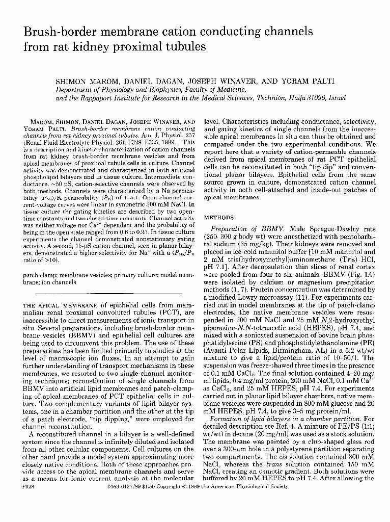
Preparation of BBMV. Male Sprague-Dawley rats (250-300 g body wt) were anesthetized with pentobarbi- tal sodium (35 mg/kg). Their kidneys were removed and placed in ice-cold mannitol buffer [lo mM mannitol and 2 mM tris( hydroxymethyl)aminomethane (Tris) . HCI, pH 7.11. After decapsulation thin slices of renal cortex were pooled from four to six animals. BBMV (Fig. 1A) were isolated by calcium or magnesium precipitation methods (1,7). Protein concentration was determined by a modified Lowry microassay (11). For experiments car- ried out in model membranes at the tip of patch-clamp electrodes, the native membrane vesicles were resus- pended in 200 mM NaCl and 25 mM N,Z-hydroxyethyl piperazine-N,N-tetraacetic acid (HEPES), pH 7.4, and mixed with a sonicated suspension of bovine brain phos- phatidylserine (PS) and phosphatidylethanolamine (PE) (Avanti Polar Lipids, Birmingham, AL) in a 52 wt/wt mixture to give a lipid/protein ratio of 10-50/l. The suspension was freeze-thawed three times in the presence of 0.1 mM CaC12. The final solution contained 4-20 mg/ ml lipids, 0.4 mg/ml protein, 200 mM NaC1,O.l mM Ca2+ as CaC12, and 25 mM HEPES, pH 7.4. For experiments carried out in planar lipid bilayer chambers, native mem- brane vesicles were suspended in 800 mM glucose and 20 mM HEPES, pH 7.4, to give 3-5 mg protein/ml.
Formation of lipid bilayers in a chamber partition. For detailed description see Ref. 4. A mixture of PE/PS (1:l; wt/wt) in decane (20 mg/ml) was used as a stock solution. The membrane was painted by a club-shaped glass rod over a 300-pm hole in a polystyrene partition separating two compartments. The cis solution contained 300 mM NaCl, whereas the trans solution contained 150 mM NaC1, creating an osmotic gradient. Both solutions were buffered by 20 mM HEPES to pH 7.4. After allowing the
F328 0363-6127/89 $1.50 Copyright 0 1989 the American Physiological Society
CATION CHANNELS IN KIDNEY PROXIMAL TUHIJLE F329
* :
FIG. 1. A: negative staining of brush-border membrane vesicles (scale bar, 0.5 PM). B: phase micrograph of proximal convoluted tubule fraction purified in Percoll gradient (scale bar, 200 Mm). C: epithelial monolayer 4 days in culture (scale bar, 40 am).
bilayer to thin, monitored by its capacitance to >300 pF, we tested the membrane for absence of channel-like activity. When this criterion was satisfied, 20 ~1 of native membrane preparation was added to the cis side while the chamber was thoroughly stirred. In most cases Ca” was added to a final concentration of 3 mM to enhance fusion. Fusion usually occurred within 30 min. When channel gating was observed, stirring was stopped and ethylene glycol-bis(@-aminoethyl ether)-N,N,N’,iV’-tet- raacetic acid (EGTA) was added to chelate the Ca’+. The experimental solutions were then added to the cis side.
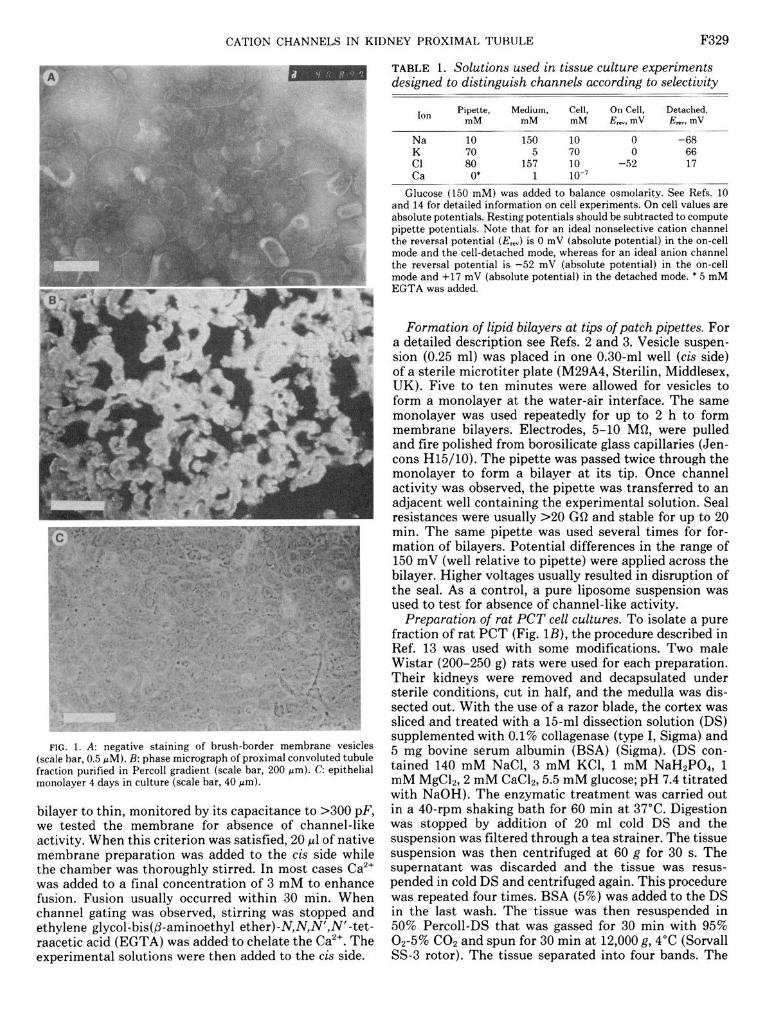
TABLE 1. Solutions used in tissue culture experiments designed to distinguish channels according to selectivity
-- - -_ -_-
Ion Pipette, Medium, Cell, On Cell, Detached, mM mM mM E,,, mV E,,, mV
Na 10 - 150 10 0 -68
El 70 5 70 0 66 80 157 10 -52 17
Ca 0’ 1 10-l --~ - Glucose-(150 mM) was added to balance osmolarity. See Refs. 10
and 14 for detailed information on cell experiments. On cell values are absolute potentials. Resting potentials should be subtracted to compute pipette potentials. Note that for an ideal nonselective cation channel the reversal potential (E,,) is 0 mV (absolute potential) in the on-cell mode and the cell-detached mode, whereas for an ideal anion channel the reversal potential is -52 mV (absolute potential) in the on-cell mode and +I7 mV (absolute potential) in the detached mode. * 5 mM EGTA was added.
Formation of lipid bilayers at tips of patch pipettes. For a detailed description see Refs. 2 and 3. Vesicle suspen- sion (0.25 ml) was placed in one 0.30-ml well (cis side) of a sterile microtiter plate (M29A4, Sterilin, Middlesex, UK). Five to ten minutes were allowed for vesicles to form a monolayer at the water-air interface. The same monolayer was used repeatedly for up to 2 h to form membrane bilayers. Electrodes, 5-10 MR, were pulled and fire polished from borosilicate glass capillaries (Jen- cons H15/10). The pipette was passed twice through the monolayer to form a bilayer at its tip. Once channel activity was observed, the pipette was transferred to an adjacent well containing the experimental solution. Seal resistances were usually ~20 GQ and stable for up to 20 min. The same pipette was used several times for for- mation of bilayers. Potential differences in the range of 150 mV (well relative to pipette) were applied across the bilayer. Higher voltages usually resulted in disruption of the seal. As a control, a pure liposome suspension was used to test for absence of channel-like activity.
Preparation of rat PCT cell cultures. To isolate a pure fraction of rat PCT (Fig. lB), the procedure described in Ref. 13 was used with some modifications. Two male Wistar (200-250 g) rats were used for each preparation. Their kidneys were removed and decapsulated under sterile conditions, cut in half, and the medulla was dis- sected out. With the use of a razor blade, the cortex was sliced and treated with a 15-ml dissection solution (DS) supplemented with 0.1% collagenase (type I, Sigma) and 5 mg bovine serum albumin (BSA) (Sigma). (DS con- tained 140 mM NaCl, 3 mM KCl, 1 mM NaH2P04, 1 mM MgClz, 2 mM CaC&, 5.5 mM glucose; pH 7.4 titrated with NaOH). The enzymatic treatment was carried out in a 40-rpm shaking bath for 60 min at 37°C. Digestion was stopped by addition of 20 ml cold DS and the suspension was filtered through a tea strainer. The tissue suspension was then centrifuged at 60 g for 30 s. The supernatant was discarded and the tissue was resus- pended in cold DS and centrifuged again. This procedure was repeated four times. BSA (5%) was added to the DS in the last wash. The tissue was then resuspended in 50% Percoll-DS that was gassed for 30 min with 95% 02-5% CO:! and spun for 30 min at 12,000 g, 4°C (Sorvall SS-3 rotor). The tissue separated into four bands. The

0
470 C
0
-50
-70
-90
C
0
C
0 -. . -- ---
F . u
0
-100 c
FIG. 2. Brush-border membrane ves- icle cation channel activity in planar bi- layers. A: 60-pS channel. Cis solution contained 300 mM NaCl, 1 mM CaC12, and 20 mM HEPES (pH 7.4). Trans solution contained 150 mM NaCl, 0.5 mM CaC12, and 10 mM HEPES (pH 7.4). Voltages on left are given as tram3 rela- tive to cis. Downward current stands for movement of cations from ck to trans. Scale, 4 pA, 100 ms. B: gating pattern of 60-pS channel demonstrating bursting behavior. A selected section is shown on an expanded time scale (bottom trace). Upward deflections denote channel openings. Recorded under symmetrical 140 mM KCl. V = 50 mV. C: 15 pS channel. Solutions and conventions as in A. Top trace, V = +40 mV; bottom trace, V = -40 mV. Scale, 0.5 pA, 200 ms.
50 ms F330

CATION CHANNELS IN KIDNEY PROXIMAL TUBULE F331
C
-40 FIG. 2. See facing page for legend.
bottom band contained the PCT fragments. This band plates were not coated with collagen or any other sub- was resuspended in 1-2 ml of Dulbecco’s modified Eagle’s strate, nor were any cells used as an underlayer. Several medium (DMEM) (Bet-Haemek, Israel). Twenty micro- experiments were conducted without the addition of the liters of the suspension were then added to each of 20 antibiotic antimycotic mixture to exclude amphotericin- tissue culture dishes (35 mm, no. 153066, Nunclon, Den- induced channels. Similar results were obtained under mark) containing 2 ml DMEM supplemented with 10% these conditions. All supplements were added to the FCS (Bet-Haemek) and 5% antibiotic antimycotic mix- medium immediately before use. The tissue culture was ture (10,000 units/ml penicillin, 10 mg/ml streptomycin, kept at 37°C in 5% COZ. Monolayers containing up to and 250 pg/ml amphotericin B, Bet-Haemek). Culture 100 cells were present after 48 h (Fig. 1C). The experi-
A
T OPEN
Taul = 24. 80 ms < 0. so>
Tau2 - 559.00 ms < 0. so>
. B
OPEN
Taul = 17.60 me < 0. 62)
Tau2 - 74.00 ms C 0. 38)
250 500 250 500
CLOSED -
Taul - 23. 70 ms < 0. 85) --
Tau2 = 422. 00 - ms <0.15> x w
/ . 25 I
I I i 250 500
Time. ms
CLOSED
Taul - 12.74 ms ( 0. 45>
Tau2 - 112.00 ms < 0.55)
Time. ms
FIG. 3. A: open- and closed-time distributions for 60-pS channel. Bin size = 5 ms, V = -50 mV. B: open- and closed-time distributions for 15pS channel. Bin size = 7 ms, V = -40 mV; filtered at 300 Hz. Taul and Tau2, time constants. Values in parentheses represent relative coefficient of each exponent in overall activity.

F332 CATION CHANNELS IN KIDNEY PROXIMAL TUBULE
PA
’ 5
4 a x I
250 mr,
I Leakage trace
tllV
FIG. 4. A: currents induced by clamping membrane with a voltage ramp from -80 to 80 mV, recorded in a bilayer on tip of a patch-clamp pipette. Top part shows raw data of 2 traces. In 1 trace, no channel opening events are seen. Other trace includes 1 opening and 1 closing event. Trace without gating activity is defined as “leakage trace.” This trace is subtracted from trace containing an opening event. Leak- corrected trace is put on I- V axes to compute conductance and reversal potential (bottom). All steps are computer controlled. In record shown, pipette and well solutions were 140 mM KCl, 5 mM NaCl, and 140 mM NaCl, 5 mM KCl, respectively. Reversal potential of - 7 mV yields a ~WSJ~ = 0.7 computed using Goldman-Hodgkin-Katz equation. In voltage range of -48 to 20 mV conductance is linear (50 pS). B: I- V relationship of channel under different ionic conditions. For right hand I-V curve (crosses) solutions were 80 mM KC1 well and 140 mM KC1 pipette. Under these conditions Cl potential = -14 mV and K potential = +l4 mV. Average reversal potential = +16 mV (n = 4, SD = 7), indicating cation over anion selectivity. After changing well solution to 140 mM NaCl, left I- V curve (diamonds), reversal potential shifts to negative values, indicating a slight preference for Na over K ions. Channel conductance varies with ion concentration (9). Reversal po- tential is obtained by linear extrapolation and thus calculated perme- ability ratios are estimates.
ments were carried out 2-7 days after plating. During 2- A this period cells formed monolayer islands of hundreds P of cells. In no case were recordings made, either from single cells or from completely confluent culture plates.
Recording and analysis. Membranes at tips of patch 1 --
pipettes allow a high temporal resolution of gating, how- ever, they are fragile and tend to break on moving the pipette from one well to another. Planar bilayers are EK E Cl
much more stable, enabling longer experiments but at a 1 I 1 I I I lower temporal resolution. The solutions used in tissue +O -40 -20
culture experiments (see Table 1) were designed to dis- tinguish between different channels according to their
FIG. 5. An I-V relationship of l5-pS Na channel in a planar bilayer with 150 NaC12 KC1 cis and 300 NaCl20 KC1 trans. Reversal potential is close to Na potential (EN*), demonstrating a selectivity for Na over K (n = 9) and over Cl ions (n = 3).
mV

CATION CHANNELS IN KIDNEY PROXIMAL TUBULE F333
-L 100 ms
B
100 ms
FIG. 6. Activity of a 50-pS cation-permeable channel in apical membrane of proximal convoluted tubule cells in tissue culture. A: “on cell” configuration voltage = 0 mV pipette relative to bath. B: cell-detached configuration voltage = 80 mV pipette relative to bath. Filtered at 1 kHz. For composition of solutions see Table 1.
selectivity while minimizing experimental manipulations of the patches, which are very fragile. A Yale (New Haven, CT) patch-clamp amplifier with a 10 GQ feedback resistor was used. Data were sampled at 22 kHz (16 bit) and taped using a digital VCR recording adaptor (Medi- cal Instrument PCM). Experiments were handled by a PDP 11/23+ computer using an INDEC laboratory dis- play and analysis system (INDEC, Sunnyvale, CA).
RESULTS
Cation channels from BBMV. Data analyses are based on 24 records in which only a single channel was active in the membrane. In most cases channels tended to appear in clusters, even when the vesicles were diluted
to a point in which fusion events were rare, thus making individual channel analysis impossible (3).
At least two types of cation-permeable channels, dif- fering in their kinetics and conductance were observed (Fig. 2). A 60-pS channel (n = 15, SD = 13) was observed in both tip dipping and planar bilayers. The channel current-voltage (I-V) curve is linear in a symmetric 300 mM NaCl solution. This channel is characterized by long open times in the range of hundreds of milliseconds. Analysis of the distribution of open- and closed-times is shown in Fig. 3A, demonstrating time constants one to two orders of magnitude apart (n = 5). Closed-time distributions can be fitted to two rate constants repre- senting the bursting behaviour of this channel. The

CATION CHANNELS IN KIDNEY PROXIMAL TUBULE
cu
OPEN
Taul = 4. 36 me
Tau2 - 156. 00 ms
< 0. 69>
( 0.31)
l T
2’00
CLOSED
Taul = 4.48 me < 0. 86>
Tau2 - 74.00 ms ( 0. 14)
Time. ms
FIG. 7. Open - and closed-t ime distributions for 50-pS channel from ltured cells in cell-detached mode. V = -80 m V, bin size 5 ms; filtered
at 1 kHz. Values in parentheses exponent in overall activity.
represent relative coefficient of each
probability of the channel to be in the open state ranged from 0.60 to 0.95 and was independent of voltage, in the tlO0 mV range. Calcium concentrations in the range of 1O-7 to lo-:’ M ( using EGTA as a chelator) did not affect the open-time probabilities. Measurements of permea- bility ratios of the channel using the Goldman-Hodgkin- Katz voltage equation (see legend of Fig. 4 for details) revealed selectivity for cations over anions [ratio of Na permeability to Cl permeability (&lpcJ > lo] and a ratio of Pk/& = 1.1 (n = 7, SD = 0.23).
A second, 15 pS (14.8 pS, n = 9, SD = 1.8) cation- selective channel, was detected in planar bilayers only. The channel I-V curve is linear in a symmetric 300 mM NaCl solution. This channel is selective to Na+ over K+ (&I& > 10) and is not permeable to Cl- (PN,/Pcl > 10) (Fig. 5). The channel activity is characterized by bursts and both the open- and close-time distributions can be fitted by two exponentials (Fig. 3B) with time constants one to two orders of magnitude apart (n = 3). The probability of the channel to be in the open state ranged from 0.4 to 0.6 independent of voltage. Calcium concentrations in the range of 10B7 to 10s3 M (using EGTA as a chelator) did not effect the open-time prob- ability.
Cation channels in the apical membrane of PCT cells in culture. Patch-clamping the apical membrane of PCT cells in culture uncovered an assortment of channel types
in almost every patch. Among these channels were 15 pS (15.4 pS, n = 7, SD = 2.2) K+-selective (pK/& = 16,
= 2) calcium-activated channels, and 4O-pS (n = 3) El- channels. These and other channels are beyond the scope of the present study and are not discussed further.
A 50-pS (n = 6, SD = 19) cation-selective channel was seen in patches on apical membranes (Fig. 6). The selec- tivity over anions was assessed by a +5O-mV reversal potential in the on cell mode. An ideal anion-selective channel would reverse at -0 mV (see Table 1). When a successful transition to the detached mode was achieved, the channel current reversed between -20 and -40 mV (pipette relative to medium), indicating on the average a poor selectivity for Na’ over K+ (&/Pk = l-5:1, mean = 2.24, n = 5, SD = 1.7). The open-time probability fluctuated both in cell-attached and inside-out configu- rations. The open- and close-time distributions of the channel fit a sum of two exponentials (Fig. 7) with time constants one to two orders of magnitude apart (n = 3). In the cell-detached mode channel gating was not af- fected by changing calcium concentrations in the bath from low3 to 10B7 M using EGTA as a calcium chelator. The channel activity is not voltage dependent.
DISCUSSION
Reconstitution of membrane protein from BBMV into artificial lipid bilayers and patch-clamping the apical side of PCT epithelial cells in culture enabled us to expose this normally inaccessible region of the nephron to single channel recordings. Using these two approaches, we demonstrate the existence of cation channels in the PCT luminal membrane of the rat. In the intermediate range, channels with a conductance of 50 pS in cell membrane patches and 60 pS in lipid bilayers, were detected by both methods. Their pk/&, ratio was found to be near unity. Since these channels do not discrimi- nate between Na’ and K+, under physiological conditions the major charge carrier through the channel would be Na+ due to its much higher electrochemical gradient and resultant driving force (9).
The l5-pS channel described here is similar to the one reported by Gogelein and Greger (8) in the pars recta segment of the rabbit PCT in terms of conductance and selectivity. The channel was seen in the planar bilayer assay only and the reason for this is not clear.
Both the conductance and selectivity of the 50-pS channel recorded in tissue culture fall within the range of the values for the 60-pS channel recorded in lipid bilayer. The two may be the same or different channels. Whatever the case, one can expect that the channel gating will differ under the two experimental conditions. For reconstitution the channel protein undergoes an extensive preparatory procedure that can affect its prop- erties. Furthermore, in the reconstituted mode the chan- nel is devoid of all cellular modulatory mechanisms. Channel activity in primary cell cultures approximate in situ conditions more closely, nevertheless even here the system is lacking the modulation present in the intact organ.
The evidence presented here for a Na+ conductance through cation-selective channels in the luminal mem-

CATION CHANNELS IN KIDNEY PROXIMAL TUBULE F335
brane of the PCT is consistent with several recent reports (5, 6, 8, 12). The extrapolation to the native condition remains to be substantiated by recordings in situ.
We thank E. Shurany for technical assistance. This work was supported by US-Israel Binational Science Founda-
tion Grant 84-00386 to D. Dagan. Address for reprint requests: S. Marom, Dept. of Physiology and
Biophysics, Faculty of Medicine, Technion-Israel Institute of Technol- ogy, POB 9649, Haifa 31096, Israel.
Received 7 October 1988; accepted in final form 20 April 1989.
REFERENCES
1. BIBER, J., B. STIECER, W. HAASE, AND H. MURER. A high yield preparation for rat kidney brush border membranes different be- haviour of lysosomal markers. Biochim. Biophys. Acta 647: 169- 176, 1981.
2. CORRONADO, R., AND R. LATORRE. Phospholipid bilayers made from monolayers on patch clamp pipettes. Biophys. J. 43: 231-236, 1983.
3. COYNE, M. D., D. DAGAN, AND I. B. LEVITAN. Calcium and barium permeable channels from Aplysia nervous system reconstituted in lipid bilayers. J. Membr. Biol. 97: 205-213, 1987.
4. DARSZON, A. Planar bilayers: a powerful tool to study membrane proteins involved in ion transport. Methods Enzymol. 127: 486- ,502, 1986.
5. DRAKE-BAUMANN, R., AND E. BOULPAEP. Apical membrane chan-
nels from salamander kidney proximal tubule cells in culture (Abstract). Biophys. J. 53: 520a, 1988.
6. ELGAVISH, G. A., AND A. ELGAVISH. Evidence from 23Na NMR studies for the existence of sodium channels in the brush border membrane of the renal proximal tubule. Biochem. Biophys. Res. Commun. 128: 746-753,1985.
7. EVERS, C., W. HASSE, H. MURER, AND R. KINNE. Properties of brush border vesicles isolated from rat kidney cortex by calcium precipitation. Membr. Biochem. 1: 203-219, 1978.
8. GOGELEIN, H., AND R. GREGER. Na selective channels in the apical membrane of rabbit late proximal tubules. Pfluegers Arch. 406: 198-203,1986.
9. HILLE, B. Ionic Channels of Excitable Membranes. Boston, MA: Sinauer, 1984.
10. LANG, F., G. MESSNER, AND W. REHWALD. Electrophysiology of sodium-coupled transport in proximal renal tubules. Am. J. Phys- iol. 250 (Renal Fluid Electrolyte Physiol. 19): F953-F962, 1986.
11. MARKWELL, M. A. K., S. M. HAAS, L. L. BIEBER, AND N. E. TOLBERT. A modification of the Lowry procedure to simplify protein determination in membrane and lipoprotein samples. Anal. Biochem. 87: 206-210, 1978.
12. MEROT, J., M. BIDET, B. GACHOT, S. LE MAOUT, M. GAUC, AND P. POUJEOL. Patch-clamp study on primary culture of isolated proximal convoluted tubules. Pfluegers Arch. 413: 51-61, 1988.
13. VINAY, P., A. GOUGOUX, AND G. LEMIEUX. Isolation of a pure suspension of rat proximal tubules. Am. J. Physiol. 241 (Renal Fluid Electrolyte Physiol. 10): F403-F411, 1981.
14. WEINSTEIN, A. M., AND E. E. WINDHAGER. Sodium transport along the proximal tubule. In: The Kidney: Physiology and Patho- physiology, edited by D. W. Seldin and G. Giebisch. New York: Raven, 1985, p. 1033-1061.