$btf 3fqpsu .jsj[[j4zoespnf …downloads.hindawi.com/journals/cris/2013/268760.pdf · uijt...
TRANSCRIPT
Hindawi Publishing CorporationCase Reports in SurgeryVolume 2013, Article ID 268760, 4 pageshttp://dx.doi.org/10.1155/2013/268760
Case ReportMirizzi Syndrome: FromUltrasound Diagnosis toSurgery—A Case Report
Dario Pariani, Giorgio Zetti, Fausto Galli, and Ferdinando Cortese
Azienda Ospedaliera Ospedale di Circolo di Busto Arsizio, Presidio Ospedaliero di Saronno,U.O. Chirurgia Generale e Toracica, Piazzale Borella, 21047 Saronno, Italy
Correspondence should be addressed to Dario Pariani; [email protected]
Received 6 December 2012; Accepted 28 December 2012
Academic Editors: S. S. Kim and N. D. Merrett
Copyright © 2013 Dario Pariani et al. is is an open access article distributed under the Creative Commons Attribution License,which permits unrestricted use, distribution, and reproduction in any medium, provided the original work is properly cited.
eMirizzi syndrome is a rare disorder that usually presents with jaundice and cholangitis; its lack of recognition in the diagnosticpath could have serious consequences for the patient undergoing cholecystectomy. Here we describe the clinical case of a jaundicedpatient from the ultrasound suspect of Mirizzi syndrome to the surgical treatment.
1. Introduction
In 1905 Kehr published the �rst cases of benign extrinsicbiliary obstructions caused by gallstones in the gallbladder[1], but it was only in 1948 that Mirizzi reanalyzed andclassi�ed this clinical condition, which is characterized bymechanical compression of the common hepatic duct due toa gallstone entrapped into the gallbladder Hartmann pouchor into the cystic duct; therefore from that moment on,this condition was called Mirizzi syndrome [2]. Nowadaysthe Mirizzi syndrome appears in 1% to 2% of patientswith symptomatic cholelithiasis [3] with higher incidence inCentral and South America where the reported incidenceis 4.7% to 5.7% [3, 4]. Obstructive jaundice and cholangitisare the common presentations of this condition that, if notrecognized before operation, makes a cholecystectomy athigh risk of damage to the common hepatic duct. Here, wereport the case of a patient with Mirizzi syndrome fromultrasound diagnosis to the surgical operation.
2. A Case Report
We present the case of a 73-year-old female patient, whoarrived in the emergency room for recent onset of epigastricand right hypochondrium pain associated with nausea, vom-iting, dark urine, grey feces, and scleroskin jaundice.
Blood tests showed serum total bilirubin 10.23mg/dL,direct bilirubin 8.62mg/dL, alanine transaminase (ALT)
190U/L, aspartate transaminase (AST) 64U/L, 𝛾𝛾-glutam-yltransferase (GGT) 299U/L, lactate dehydrogenase (LDH)334U/L, and alkaline phosphatase (ALP) 367U/L.
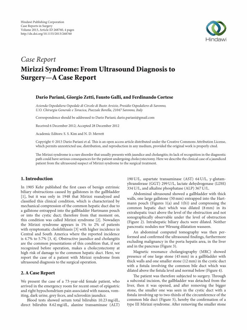
Abdominal ultrasound showed a gallbladder with thickwalls, one large gallstone (50mm) entrapped into the Hart-mann pouch (Figures 1(a) and 1(b)) and compressing thecommon hepatic duct which was dilated (8mm) in itsextrahepatic tract above the level of the obstruction and notsonographically observable under the level of obstruction(Figure 2). Intrahepatic biliary ducts were dilated. Neitherpancreatic nodules nor Wirsung dilatation wasseen.
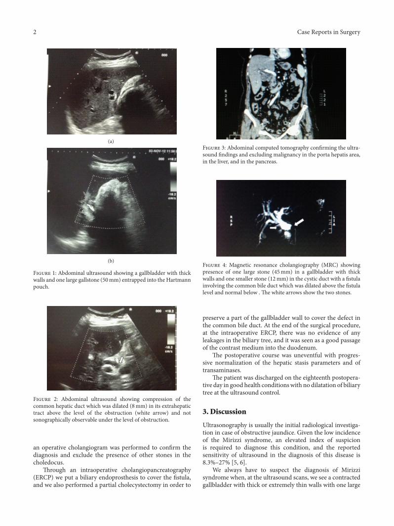
An abdominal computed tomography was then per-formed and con�rmed the ultrasound �ndings, furthermoreexcluding malignancy in the porta hepatis area, in the liverand in the pancreas (Figure 3).
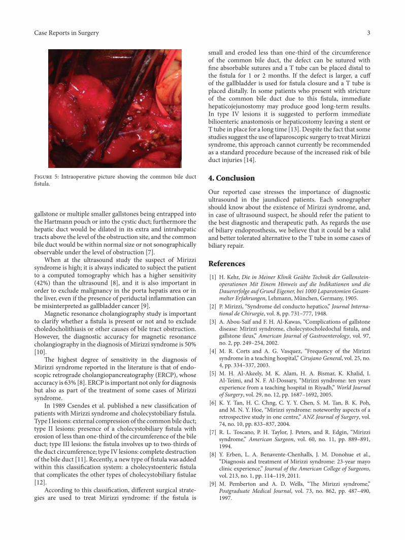
Magnetic resonance cholangiography (MRC) showedpresence of one large stone (45mm) in a gallbladder withthick walls and one smaller stone (12mm) in the cystic ductwith a �stula involving the common bile duct which wasdilated above the �stula level and normal below (Figure 4).
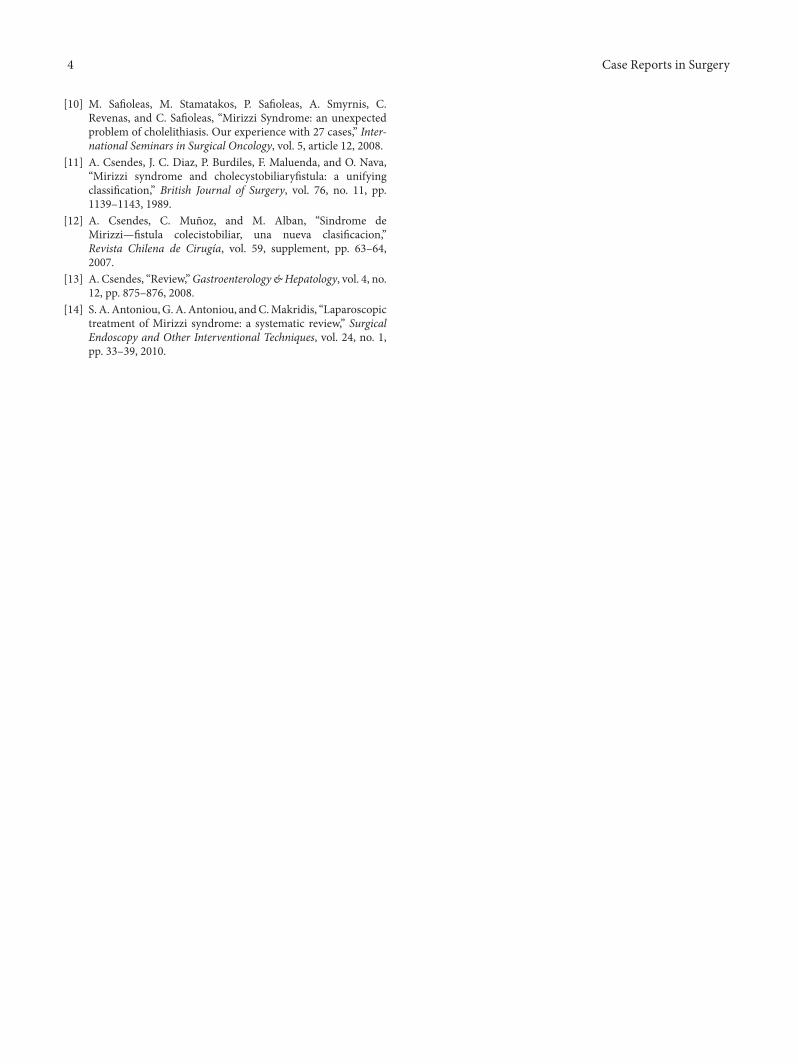
e patient was therefore subjected to surgery. rougha subcostal incision, the gallbladder was detached from theliver, then it was opened, and aer removing the biggerstone, the smaller one was seen in the cystic duct with a�stula involving up to two-thirds of the circumference of thecommon bile duct (Figure 5), hereby the con�rmation of atype III Mirizzi syndrome. Aer removing the smaller stone
2 Case Reports in Surgery
(a)
(b)
F 1: Abdominal ultrasound showing a gallbladder with thickwalls and one large gallstone (50mm) entrapped into the Hartmannpouch.
F 2: Abdominal ultrasound showing compression of thecommon hepatic duct which was dilated (8mm) in its extrahepatictract above the level of the obstruction (white arrow) and notsonographically observable under the level of obstruction.
an operative cholangiogram was performed to con�rm thediagnosis and exclude the presence of other stones in thecholedocus.
rough an intraoperative cholangiopancreatography(ERCP) we put a biliary endoprosthesis to cover the �stula,and we also performed a partial cholecystectomy in order to
F 3: Abdominal computed tomography con�rming the ultra-sound �ndings and excluding malignancy in the porta hepatis area,in the liver, and in the pancreas.
F 4: Magnetic resonance cholangiography (MRC) showingpresence of one large stone (45mm) in a gallbladder with thickwalls and one smaller stone (12mm) in the cystic duct with a �stulainvolving the common bile duct which was dilated above the �stulalevel and normal below . e white arrows show the two stones.
preserve a part of the gallbladder wall to cover the defect inthe common bile duct. At the end of the surgical procedure,at the intraoperative ERCP, there was no evidence of anyleakages in the biliary tree, and it was seen as a good passageof the contrast medium into the duodenum.
e postoperative course was uneventful with progres-sive normalization of the hepatic stasis parameters and oftransaminases.
e patient was discharged on the eighteenth postopera-tive day in good health conditionswith no dilatation of biliarytree at the ultrasound control.
3. Discussion
Ultrasonography is usually the initial radiological investiga-tion in case of obstructive jaundice. Given the low incidenceof the Mirizzi syndrome, an elevated index of suspicionis required to diagnose this condition, and the reportedsensitivity of ultrasound in the diagnosis of this disease is8.3%–27% [5, 6].
We always have to suspect the diagnosis of Mirizzisyndrome when, at the ultrasound scans, we see a contractedgallbladder with thick or extremely thin walls with one large
Case Reports in Surgery 3
F 5: Intraoperative picture showing the common bile duct�stula.
gallstone or multiple smaller gallstones being entrapped intothe Hartmann pouch or into the cystic duct; furthermore thehepatic duct would be dilated in its extra and intrahepatictracts above the level of the obstruction site, and the commonbile duct would be within normal size or not sonographicallyobservable under the level of obstruction [7].
When at the ultrasound study the suspect of Mirizzisyndrome is high; it is always indicated to subject the patientto a computed tomography which has a higher sensitivity(42%) than the ultrasound [8], and it is also important inorder to exclude malignancy in the porta hepatis area or inthe liver, even if the presence of periductal in�ammation canbe misinterpreted as gallbladder cancer [9].
Magnetic resonance cholangiography study is importantto clarify whether a �stula is present or not and to excludecholedocholithiasis or other causes of bile tract obstruction.However, the diagnostic accuracy for magnetic resonancecholangiography in the diagnosis of Mirizzi syndrome is 50%[10].
e highest degree of sensitivity in the diagnosis ofMirizzi syndrome reported in the literature is that of endo-scopic retrograde cholangiopancreatography (ERCP), whoseaccuracy is 63% [8]. ERCP is important not only for diagnosisbut also as part of the treatment of some cases of Mirizzisyndrome.
In 1989 Csendes et al. published a new classi�cation ofpatients with Mirizzi syndrome and cholecystobiliary �stula.Type I lesions: external compression of the commonbile duct;type II lesions: presence of a cholecystobiliary �stula witherosion of less than one-third of the circumference of the bileduct; type III lesions: the �stula involves up to two-thirds ofthe duct circumference; type IV lesions: complete destructionof the bile duct [11]. Recently, a new type of �stula was addedwithin this classi�cation system: a cholecystoenteric �stulathat complicates the other types of cholecystobiliary �stulae[12].
According to this classi�cation, di�erent surgical strate-gies are used to treat Mirizzi syndrome: if the �stula is
small and eroded less than one-third of the circumferenceof the common bile duct, the defect can be sutured with�ne absorbable sutures and a T tube can be placed distal tothe �stula for 1 or 2 months. If the defect is larger, a cu�of the gallbladder is used for �stula closure and a T tube isplaced distally. In some patients who present with strictureof the common bile duct due to this �stula, immediatehepaticojejunostomy may produce good long-term results.In type IV lesions it is suggested to perform immediatebilioenteric anastomosis or hepaticostomy leaving a stent orT tube in place for a long time [13]. Despite the fact that somestudies suggest the use of laparoscopic surgery to treatMirizzisyndrome, this approach cannot currently be recommendedas a standard procedure because of the increased risk of bileduct injuries [14].
4. Conclusion
Our reported case stresses the importance of diagnosticultrasound in the jaundiced patients. Each sonographershould know about the existence of Mirizzi syndrome, and,in case of ultrasound suspect, he should refer the patient tothe best diagnostic and therapeutic path. As regards the useof biliary endoprosthesis, we believe that it could be a validand better tolerated alternative to the T tube in some cases ofbiliary repair.
References
[1] H. Kehr, Die in Meiner Klinik Geübte Technik der Gallenstein-operationen Mit Einem Hinweis auf die Indikationen und dieDauererfolge auf Grund Eigener, bei 1000 Laparotomien Gesam-melter Erfahrungen, Lehmann, München, Germany, 1905.
[2] P. Mirizzi, “Syndrome del conducto hepatico,” Journal Interna-tional de Chirurgie, vol. 8, pp. 731–777, 1948.
[3] A. Abou-Saif and F. H. Al-Kawas, “Complications of gallstonedisease: Mirizzi syndrome, cholecystocholedochal �stula, andgallstone ileus,” American Journal of Gastroenterology, vol. 97,no. 2, pp. 249–254, 2002.
[4] M. R. Corts and A. G. Vasquez, “Frequency of the Mirizzisyndrome in a teaching hospital,” Cirujano General, vol. 25, no.4, pp. 334–337, 2003.
[5] M. H. Al-Akeely, M. K. Alam, H. A. Bismar, K. Khalid, I.Al-Teimi, and N. F. Al-Dossary, “Mirizzi syndrome: ten yearsexperience from a teaching hospital in Riyadh,” World Journalof Surgery, vol. 29, no. 12, pp. 1687–1692, 2005.
[6] K. Y. Tan, H. C. Chng, C. Y. Y. Chen, S. M. Tan, B. K. Poh,and M. N. Y. Hoe, “Mirizzi syndrome: noteworthy aspects of aretrospective study in one centre,” ANZ Journal of Surgery, vol.74, no. 10, pp. 833–837, 2004.
[7] R. L. Toscano, P. H. Taylor, J. Peters, and R. Edgin, “Mirizzisyndrome,” American Surgeon, vol. 60, no. 11, pp. 889–891,1994.
[8] Y. Erben, L. A. Benavente-Chenhalls, J. M. Donohue et al.,“Diagnosis and treatment of Mirizzi syndrome: 23-year mayoclinic experience,” Journal of the American College of Surgeons,vol. 213, no. 1, pp. 114–119, 2011.
[9] M. Pemberton and A. D. Wells, “e Mirizzi syndrome,”Postgraduate Medical Journal, vol. 73, no. 862, pp. 487–490,1997.
4 Case Reports in Surgery
[10] M. Sa�oleas, M. Stamatakos, P. Sa�oleas, A. Smyrnis, C.Revenas, and C. Sa�oleas, “Mirizzi Syndrome: an unexpectedproblem of cholelithiasis. Our experience with 27 cases,” Inter-national Seminars in Surgical Oncology, vol. 5, article 12, 2008.
[11] A. Csendes, J. C. Diaz, P. Burdiles, F. Maluenda, and O. Nava,“Mirizzi syndrome and cholecystobiliary�stula: a unifyingclassi�cation,” British Journal of Surgery, vol. 76, no. 11, pp.1139–1143, 1989.
[12] A. Csendes, C. Muñoz, and M. Alban, “Sindrome deMirizzi��stula colecistobiliar, una nueva clasi�cacion,”Revista Chilena de Cirugía, vol. 59, supplement, pp. 63–64,2007.
[13] A. Csendes, “Review,”Gastroenterology&Hepatology, vol. 4, no.12, pp. 875–876, 2008.
[14] S. A.Antoniou,G.A.Antoniou, andC.Makridis, “Laparoscopictreatment of Mirizzi syndrome: a systematic review,” SurgicalEndoscopy and Other Interventional Techniques, vol. 24, no. 1,pp. 33–39, 2010.

Submit your manuscripts athttp://www.hindawi.com
Stem CellsInternational
Hindawi Publishing Corporationhttp://www.hindawi.com Volume 2014
Hindawi Publishing Corporationhttp://www.hindawi.com Volume 2014
MEDIATORSINFLAMMATION
of
Hindawi Publishing Corporationhttp://www.hindawi.com Volume 2014
Behavioural Neurology
EndocrinologyInternational Journal of
Hindawi Publishing Corporationhttp://www.hindawi.com Volume 2014
Hindawi Publishing Corporationhttp://www.hindawi.com Volume 2014
Disease Markers
Hindawi Publishing Corporationhttp://www.hindawi.com Volume 2014
BioMed Research International
OncologyJournal of
Hindawi Publishing Corporationhttp://www.hindawi.com Volume 2014
Hindawi Publishing Corporationhttp://www.hindawi.com Volume 2014
Oxidative Medicine and Cellular Longevity
Hindawi Publishing Corporationhttp://www.hindawi.com Volume 2014
PPAR Research
The Scientific World JournalHindawi Publishing Corporation http://www.hindawi.com Volume 2014
Immunology ResearchHindawi Publishing Corporationhttp://www.hindawi.com Volume 2014
Journal of
ObesityJournal of
Hindawi Publishing Corporationhttp://www.hindawi.com Volume 2014
Hindawi Publishing Corporationhttp://www.hindawi.com Volume 2014
Computational and Mathematical Methods in Medicine
OphthalmologyJournal of
Hindawi Publishing Corporationhttp://www.hindawi.com Volume 2014
Diabetes ResearchJournal of
Hindawi Publishing Corporationhttp://www.hindawi.com Volume 2014
Hindawi Publishing Corporationhttp://www.hindawi.com Volume 2014
Research and TreatmentAIDS
Hindawi Publishing Corporationhttp://www.hindawi.com Volume 2014
Gastroenterology Research and Practice
Hindawi Publishing Corporationhttp://www.hindawi.com Volume 2014
Parkinson’s Disease
Evidence-Based Complementary and Alternative Medicine
Volume 2014Hindawi Publishing Corporationhttp://www.hindawi.com