built? a. moves bones and substances ( blood, waste, nutrients)
TRANSCRIPT


Built?
A. Moves bones and substances (blood, waste, nutrients)

A. SkeletalB. Smooth (hollow
organs)
C. Cardiac (heart wall only)

A. Skeletal (40% body mass)
1.Longest muscle fiber, multi nucleate, striations

B. Smooth

C. Cardiac


A. Microscopic Muscle cell or fiber

1. Each muscle fiber (cell) contains 2 myofibrils, actin and myosin (elastic filaments)
6 actins to 1 myosin

2. Outside of muscle cellEach compartment here is a muscle cell or muscle fiber

a. Myofibrils, Myosin and Actin

B. Parts of neuromuscular junction


1. Location2. Shape3. Relative Size4. Direction of Fibers5. Number & Location of Origins6. Action of Muscle

B. Four Functional roles
1.Prime mover-agonist primary movement
2.Antagonist- opposes prime mover
3.Synergist – assists prime mover
4.Fixator- holds muscle at origin

C. Gross Skeletal Muscle composition, Layers

1. Fascia is outside Epimysium. It allows space for blood vessels and nerves and forms part of tendon.
2. Epimysium is areolar connective 3. Fascicle is surrounded by a
dense connective tissue sheath of perimysuim.

4. Each muscle fiber (cell) is surrounded by an reticular connective sheath of endomysium

D. Individual Muscles
1. Face a. SCM b. masseteur c. frontalis d. occipitalis e. temporalis d. obicularis oris & occuli e. zygomaticus Facial expression attached to skin

2. neck

3. Shoulder arm (front), scapula, Lats

4. Abdomen

5. Back

6. Lower arm
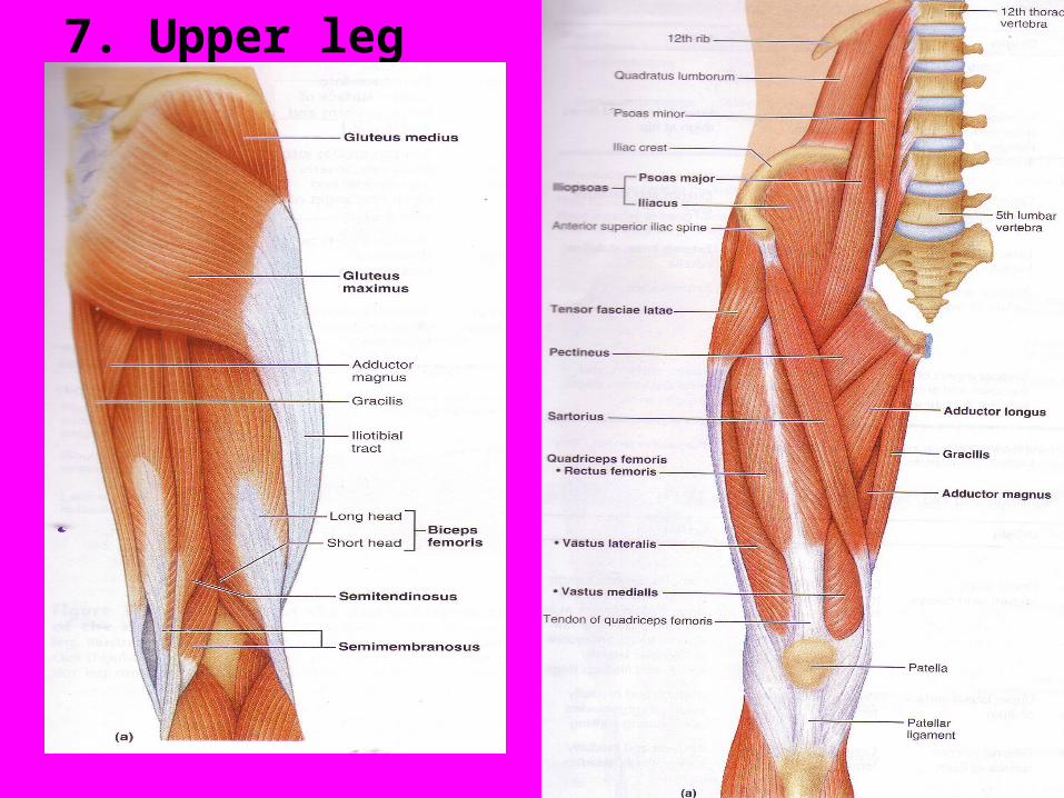
7. Upper leg

8. Lower leg


E. Origins and Insertions1. Origin is where a muscle begins, its
origin. It is the less moveable end.2. Insertion is where the muscle inserts
or leads to from its origin. It is the moveable end.
3. They will be studied for major muscles.
4. They are easier to remember seeing a picture of what the muscle looks like

Muscle Origin Insertion Movement a.Masseter zygomatic mandibular Close mouth arch ramus
b.SCM clavicle& manubrium flex neck, rotate mastoid process head
c.Biceps coracoid P, radial tuberosity flex forearm capsule of shoulder joint d.Triceps glenoid cavity olecron process extend forearm pos. humerus pos. humerus radial groove
e.Deltoid 1/3 clavicle, deltoid tuberosity abduction of arm acromion P of humerus scapular spine

Muscle Origin Insertion Movementf.Pec major clavicle, intertubercular shoulders sternum ribs groove of forward 1-6 humerusg.Trapezius occipital lat 1/3 clavicle shrug shoulders C7- T12 acromion spinous pro of scapula
h.Latissimus spinous floor of chop wood P of T6-L5 intertubercular 8-12 ribs, illiac groove of crest humerus
i.Rectus inferior illiac tibial tuberostiy extend knee,Femoris spine patella flex thigh

Muscle Origin Insertion Movement
j. Biceps Ischial tuberosity lateral condyl extend thigh
Femoris linea aspera of tibia flex knee
distal femur head of fibula
k.Gluteus dorsal ilium IT tract abduct thigh
Maximus sacrum, gluteal tuberosity climb stairs
coccyx of femur
l.Gastroc medial & lateral calcaneal tendon plantar
condyles of femur flexion