burden of prostate cancer: addressing issues of diagnosis
TRANSCRIPT

BURDEN OF PROSTATE CANCER: ADDRESSING ISSUES OF DIAGNOSIS AND OVERTREATMENT
by
PALAK KRUNAL KUMAR PATEL
(Under the Direction of Randall L. Tackett)
ABSTRACT
Objective: Increased detection of clinically insignificant prostate cancer with routinely used
diagnostic tests, as well as uncertainties in the available treatments to manage the low risk
disease, are expected to increase the future burden of the prostate cancer substantially. This
study aims to address three major areas in the field of prostate cancer including economic
burden, diagnostics, and treatment for low risk disease.
Methods: The economic burden of prostate cancer was assessed retrospectively using a
population based database. A novel imaging technique such as multiparametric magnetic
resonance imaging (MP-MRI) assisted transrectal ultrasound (TRUS) guided biopsy in prostate
cancer diagnosis was assessed and compared with the conventional 12-core TRUS guided biopsy
by performing a cost-effectiveness analysis. The Surveillance Epidemiology and End Results-
Medicare database was used to compare toxicity profiles among localized prostate cancer
patients who receive either conservative management or immediate treatment.
Results: An annual average total of $5.6 billion was spent on prostate cancer related conditions
in 2010 in the United States. Use of chemotherapy and ultrasound increased the expenditure
related to outpatient visits significantly; whereas use of ultrasound and x-ray increased office-
based visit costs significantly. The MP-MRI strategy was found to be cost-effective compared to
conventional TRUS guided biopsy assuming a threshold to pay for is $1781.60. Conservative
management was found to have lower odds of urinary, rectal, and erectile complications
without compromising the survival within a 5 year time period than the immediate treatment.
Conclusion: Routinely used TRUS guided biopsy is associated with a higher economic burden on
society. There is a need for tests that can diagnose prostate cancer accurately. MP-MRI/TRUS
fusion guided biopsy can characterize prostate cancer accurately and was found to be cost-
effective compared to TRUS guided biopsy provided the threshold to pay for this technology is
at least $1781.60. To avoid overtreatment among low risk prostate cancer patients, a
conservative management approach was found to be a better option because patients can delay
or avoid treatment related side effects without compromising prostate cancer specific survival
within a 5 year time period.
Keywords: Prostate cancer, economic burden, cost-effectiveness, multiparametric magnetic
resonance imaging, conservative management, comparative effectiveness analysis

BURDEN OF PROSTATE CANCER: ADDRESSING ISSUES OF DIAGNOSIS AND OVERTREATMENT
by
PALAK KRUNAL KUMAR PATEL
B.Pharm, The Maharaja Sayajirao University of Vadodara, India, 2003
M.Pharm, The Maharaja Sayajirao University of Vadodara, India, 2006
M.S., The Ohio State University, 2010
A Dissertation Submitted to the Graduate Faculty of The University of Georgia in Partial
Fulfillment of the Requirements for the Degree
DOCTOR OF PHILOSOPHY
ATHENS, GEORGIA
2014

© 2014
Palak Patel
All Rights Reserved

BURDEN OF PROSTATE CANCER: ADDRESSING ISSUES OF DIAGNOSIS AND OVERTREATMENT
by
PALAK KRUNAL KUMAR PATEL
Major Professor: Randall L. Tackett
Committee: Matthew Perri III
Brian S. Cummings
Robert D. Arnold
Electronic Version Approved:
Julie Coffield
Interim Dean of the Graduate School
The University of Georgia
August 2014

IV
DEDICATION
I dedicate this dissertation to my husband, Krunal, my two kids Milit and Shanaya, and my
parents. Without your support, guidance, sacrifice, and blessings, I wouldn’t have accomplished
my dreams.
V
ACKNOWLEDGEMENTS
I would like to express my deepest gratitude to my advisor and committee chair, Dr. Randall
Tackett, for his support, encouragement, and guidance throughout my stay in University of
Georgia. I am thankful to Dr. Tackett for providing me funding for SEER-Medicare dataset. My
words cannot express enough appreciation for Dr. Tackett for being there for me and keeping
faith in me throughout the course of research. I would also like to thank all my committee
members (Drs. Perri, Cummings, and Arnold) for their thought provoking suggestions,
educational support, and general collegiality that each of them offered to me.
This research would not have been possible to finish in a timely manner without support
from Ms. Anita Soni (AHRQ), Ms. Elaine Yanisko (EMS Inc.), and Mr. Vincent Marshall
(statistician, University of Michigan). I would like to express my sincere thanks to Ms. Anita for
helping with SAS codes and providing me MEPS dataset training workshop material for
estimating disease related expenditure. I am also very thankful to Ms. Elaine Yanisko for making
application process of SEER-Medicare dataset very smooth and quick. I would like to recognize
Mr. Vincent Marshall for helping me with model diagnostic inference and Charlson Comorbidity
Index calculation.
I would like to extend my thanks to all faculties, staff and students in Clinical and
Administrative Pharmacy Department, College of Pharmacy, University of Georgia. A special
thanks to Ms. Annelie Klein and Ms. Joanne Mauro for keeping me up to date with deadlines or

VI
forms and putting extra efforts for me to make sure everything is on the right track and all my
questions are answered. I am also thankful to all my colleagues and friends Surbhi, Samah,
Shada, Rian, Shardul, Ming-Yi, Heath, Rachel, Ramya, Rahat, and Ahmed. Without your
friendship and support, it would have been difficult for me to achieve what I have now.
Last but not the least, I would like to thank my family. First, I would like to thank my parents,
Kishorchandra Dave (father) and Hemlata Dave (mother) for providing me their continuous
support, empowerment, and encouragement over the years to help me reach at this career
stage. I cannot thank enough my husband, Krunal, for helping me reach my goals and believing
in me during tough time. His sacrifice made throughout is immeasurable. I would also like to
thank my parent in-laws for their support. Without their support, it would have been very
difficult to continue my PhD with two kids.

VII
TABLE OF CONTENTS
Page
ACKNOWLEDGEMENTS………………………………………………………………………………………………………………… V
LIST OF TABLES ................................................................................................................................... X
LIST OF FIGURES ................................................................................................................................. XII
CHAPTER ............................................................................................................................................
1 INTRODUCTION AND LITERATURE REVIEW .................................................................... 1
Prostate Cancer ........................................................................................................ 1
Prostate Cancer Screening ....................................................................................... 2
Transrectal Ultrasound (TRUS) Guided Biopsy ........................................................ 3
Current Trend of Repeat Biopsy .............................................................................. 4
Multiparametric Magnetic Resonance Imaging (MP-MRI) ...................................... 5
Localized Prostate Cancer (Low-risk or Intermediate-risk) ..................................... 8
Evidence on Comparative Clinical Effectiveness of Conservative Management vs
Immediate Treatment .............................................................................................. 15
Economic Burden ..................................................................................................... 18
REFERENCES ............................................................................................................. 21
2 RATIONALE AND HYPOTHESIS ........................................................................................ 30
Specific Aims ............................................................................................................ 32
REFERENCES ............................................................................................................. 34

VIII
3 DIRECT MEDICAL EXPENDITURE AND PREDICTORS ASSOCIATED WITH PROSTATE
CANCER FOR THE U.S. ADULT POPULATION: ESTIMATES FROM MEDICAL
EXPENDITURE PANEL SURVEY (2010) ............................................................................. 36
Introduction ............................................................................................................. 38
Methods ................................................................................................................... 39
Results ...................................................................................................................... 41
Discussion ................................................................................................................ 44
Limitations ............................................................................................................... 47
Conclusion ................................................................................................................ 48
REFERENCES ............................................................................................................. 52
4 SYSTEMATIC REVIEW ON ROLE OF MAGNETIC RESONANCE IMAGING IN PROSTATE
CANCER DIAGNOSIS........................................................................................................ 55
Introduction ............................................................................................................. 57
Methods ................................................................................................................... 58
Multiparametric Magnetic Resonance Imaging (MP-MRI) ...................................... 58
Magnetic Resonance Spectroscopy (MRS) based Imaging ...................................... 59
Dynamic Contrast Material-Enhanced (DCE) based Imaging ................................... 60
Diffusion-Weighted Imaging (DWI) .......................................................................... 61
Prebiopsy MRI for Men at Risk ................................................................................ 62
Cost-Effectiveness Analysis of Prebiopsy MRI Use .................................................. 63
Conclusion ................................................................................................................ 65
REFERENCES ............................................................................................................. 67

IX
5 COST-EFFECTIVENESS OF MULTIPARAMETRIC MAGNETIC RESONANCE IMAGING
FOLLOWED BY FUSION GUIDED BIOPSY IN PROSTATE CANCER DIAGNOSIS: AN
ECONOMIC ANALYSIS ..................................................................................................... 71
Introduction ............................................................................................................. 73
Methods ................................................................................................................... 74
Results ...................................................................................................................... 76
Discussion ................................................................................................................ 78
Conclusion ................................................................................................................ 81
REFERENCES ............................................................................................................. 88
6 COMPARATIVE ANALYSIS OF HARMS ASSOCIATED WITH CONSERVATIVE
MANAGEMENT AND IMMEDIATE TREATMENT AMONG LOW RISK LOCALIZED
PROSTATE CANCER PATIENTS: A POPULATION BASED STUDY ...................................... 91
Introduction ............................................................................................................. 93
Methods ................................................................................................................... 94
Results ...................................................................................................................... 97
Discussion ................................................................................................................ 99
Conclusion ................................................................................................................ 102
REFERENCES ............................................................................................................. 109
7 CONCLUSIONS ................................................................................................................ 112
X
LIST OF TABLES
Page
Table 1.1: Tumor staging system ..................................................................................................... 8
Table 1.2: Institution based inclusion criteria for active surveillance ........................................... 14
Table 1.3: Recommendation to healthcare professional regarding patient counseling ............... 17
Table 3.1: Descriptive statistics of weighted sample of patients with prostate cancer ................ 49
Table 3.2: Health care use and costs among patients with prostate cancer ................................. 49
Table 3.3: Predictors of prostate cancer related total health care costs (R2=0.1233, F=49.22, p
value <0.001).................................................................................................................................. 50
Table 3.4: Predictors of outpatient visit costs associated with prostate cancer (R2=0.2424,
F=58.60, p value <0.0001) .............................................................................................................. 50
Table 3.5: Predictors of office-based visit costs associated with prostate cancer (R2=0.1208,
F=39.37, p value <0.0001) .............................................................................................................. 50
Table 5.1: Probabilities used in the decision analytic model (test accuracy input data)............... 82
Table 5.2: Risks of biopsy complication ......................................................................................... 82
Table 5.3: Resources and cost input .............................................................................................. 83
Table 5.4: Economic model base case, number of correct diagnoses ........................................... 83
Table 5.5: Number of false positive and false negative cases avoided and associated costs ....... 84
Table 6.1: ICD-9 and CPT/HCPCS codes used to identify treatment modality............................. 104
Table 6.2: ICD-9 and CPT/HCPCS codes used to identify complications ...................................... 104
Table 6.3: Clinical and demographic characteristics of patients with clinically localized
Prostate cancer ............................................................................................................................ 105
XI
Table 6.4: Crude rates for complication diagnoses and invasive procedures ............................. 106
Table 6.5: Logistic regression analysis of urinary complications and predictors using
propensity score .......................................................................................................................... 106
Table 6.6: Logistic regression analysis of rectal complications and predictors using
propensity score .......................................................................................................................... 107
Table 6.7: Logistic regression analysis of erectile complications and predictors using
propensity score .......................................................................................................................... 107
Table 6.8: Cox proportional hazard model to assess hazard of dying due to prostate
cancer adjusted with propensity scores ...................................................................................... 108

XII
LIST OF FIGURES
Page
Figure 1.1: Sextant biopsy scheme. ................................................................................................. 3
Figure 1.2: Twelve core biopsy scheme ........................................................................................... 4
Figure 1.3: Gleason grading of prostate carcinoma ....................................................................... 10
Figure 3.1: Distribution of annual prostate cancer related direct medical costs .......................... 51
Figure 5.1: Decision analytical model comparing conventional 12-core TRUS guided biopsy with
MP-MRI followed by MRI/TRUS fusion guided biopsy .................................................................. 84
Figure 5.2: One-way sensitivity analysis varying positive predictive value of 12-core TRUS
biopsy ............................................................................................................................................. 85
Figure 5.3: One-way sensitivity analysis varying cost of MP-MRI.................................................. 85
Figure 5.4: One-way sensitivity analysis varying negative predictive value of MP-MRI ................ 86
Figure 5.5: One-way sensitivity analysis varying positive predictive value of MRI/TRUS fusion
guided biopsy ................................................................................................................................. 86
Figure 5.6: Three-way sensitivity analysis evaluating the impact of varying positive predictive
value of 12-core TRUS guided biopsy, negative predictive value of MP-MRI and positive
predictive value of MRI/TRUS fusion guided biopsy ...................................................................... 87
1
CHAPTER 1
INTRODUCTION AND LITERATURE REVIEW
Prostate Cancer
Prostate cancer is the second leading cause of cancer deaths among men in the United
States.1 Statistics related to prostate cancer diagnosis and death suggest that prostate cancer is
the most frequently diagnosed non-cutaneous cancer affecting one in every six men.2 The
American Cancer Society estimated that there were approximately 238,590 new cases and
29,720 deaths due to prostate cancer in 2013.1 Annual expenditures associated with prostate
cancer in 2006 was $9.862 billion in the U.S.3 Costs associated with prostate cancer are expected
to rise due to increased diagnosis, diagnosis at an earlier stage and increased survival.3
Prostate cancer is considered a disease of aging.4 Men under 50 years have a low risk
of being diagnosed with prostate cancer. The probability of developing prostate cancer rises
from one in 14 in those aged 60-69 to one in seven above the age of 70 years.4 The majority of
men diagnosed with localized prostate cancer are older than 65 years. The median age at
diagnosis is 72 years and many patients, especially those with localized tumors, may die of other
illnesses without ever having suffered significant disability from the cancer.5 The approach to
treatment is influenced by age and coexisting medical problems. The risk of prostate cancer
increases in black Caribbean and black African men in comparison with Caucasians.6

2
Prostate Cancer Screening
Currently, prostate cancer screening is based on the assessment of the serum level of
prostate specific antigen (PSA) and digital rectal examination (DRE). Both tests are limited in
accurately diagnosing prostate cancer.7 The PSA test has been widely criticized due to its low
specificity. A PSA level of 4 ng/mL is used as the cut off value.8 If PSA values rise above 4 ng/mL,
patients are considered to have a higher risk of prostate cancer. However, a PSA elevation above
the threshold of 4 ng/mL has a low specificity for prostate cancer.7 Mild elevations above 4
ng/mL may be caused by benign conditions such as benign prostatic hyperplasia (BPH) or
prostatitis. This may result in a false-positive PSA test and subsequently an unnecessary biopsy.
At least 15% of biopsy-proven prostate cancers occur in patients with PSA levels below 4
ng/mL.8 However, only 33% of patients with PSA values between 4 and 10 ng/ml actually have
cancer.8 In addition to the total PSA level, several additional PSA variant tests have been used
clinically to stratify patients. PSA velocity is the increase in the total PSA level over time. A PSA
velocity greater than 0.35 to 0.75 ng/mL/year is commonly considered suspicious for the
presence of prostate cancer.7 For men with PSA values less than 4 ng/mL, the threshold value of
PSA velocity is 0.35 ng/mL/year. In men with a PSA greater than 4 ng/ML, a PSA velocity of 0.75
ng/mL/year is reported to be suspicious for the presence of cancer.9,10
The ratio of free to total PSA has been found to improve the specificity of an elevated
total PSA level. In benign processes of the prostate, the percentage of free PSA tends to be
higher in comparison to the total PSA. If a ratio is below 10%, there is a 30% chance of being
diagnosed with prostate cancer.7 Another PSA variant test is the PSA density, which is defined as
the total PSA level divided by the prostate volume. Prostate volume is assessed by TRUS guided
biopsy by measuring three gland dimensions: the maximum length, the maximum height
3
orthogonal to the length, and the maximum width. Generally, a PSA density greater than or
equal to 0.15 has been proposed as the threshold for biopsy in men with PSA levels of 4 to 10
ng/mL. With this cut off value, approximately 40% of prostate cancers were missed.7
Transrectal Ultrasound (TRUS) Guided Biopsy
TRUS guided biopsy is considered the gold standard technique for diagnosing prostate
cancer with more than 1.2 million biopsies performed annually in the US.11 Ultrasound cannot
differentiate normal prostate gland from cancerous tissue.11 As a result, biopsies are not target
specific. Instead, a nontargeted, systematic, sextant biopsy schemata is used for cancer
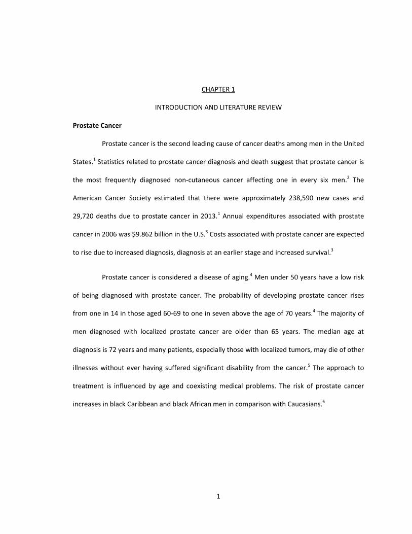
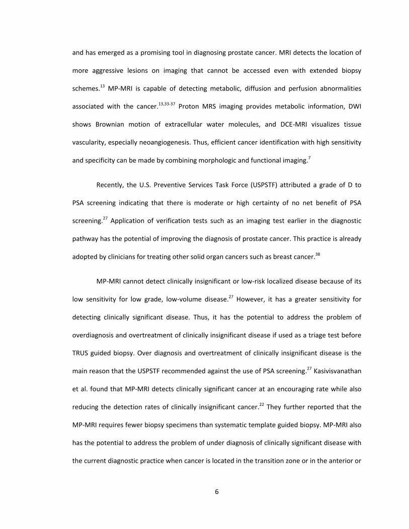
detection and characterization (Figure 1.1).11 Taking cores in an organized way is referred to as a
systematic biopsy.
Figure 1.1: Sextant biopsy scheme. A: site of biopsy cores on base, midline, and apex of each lobe of the prostate. B: Transverse view of the prostate. Figure is adapted from Mian, 2004.12
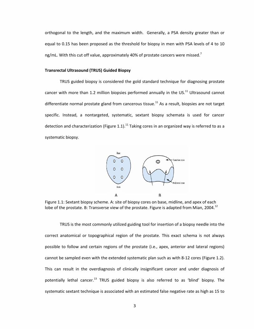
TRUS is the most commonly utilized guiding tool for insertion of a biopsy needle into the
correct anatomical or topographical region of the prostate. This exact schema is not always
possible to follow and certain regions of the prostate (i.e., apex, anterior and lateral regions)
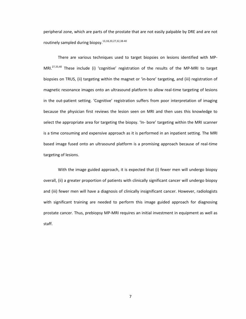
cannot be sampled even with the extended systematic plan such as with 8-12 cores (Figure 1.2).
This can result in the overdiagnosis of clinically insignificant cancer and under diagnosis of
potentially lethal cancer.13 TRUS guided biopsy is also referred to as ‘blind’ biopsy. The
systematic sextant technique is associated with an estimated false negative rate as high as 15 to
A B
4
34%.14 Thus, it requires numerous repeat biopsies, which has a cancer detection rate between
20 and 35%.15
Figure 1.2: Twelve core biopsy scheme. Figure is adapted from Mian, 2014.
To improve diagnostic performance, various biopsy strategies have been proposed in
prostate cancer detection. This includes sampling of visually abnormal areas, more lateral
placement of biopsies, anterior biopsies, and obtaining an increased number of cores, with up to
45 biopsy cores.16 Loch reported that there are centers where up to 143 biopsy specimens are
taken in one session.17 However, the efficiency of this practice is debated because of its
invasiveness and patient morbidity. Further, it leads to overdiagnosis of insignificant prostate
cancer. Alternatively, several studies have shown that systematic biopsy still misses a
considerable number of prostate cancers.18-21 Therefore, a diagnostic strategy that can improve
the quality of the investigation instead of raising the quantity of biopsies is clearly needed.
Current Trend of Repeat Biopsy
For patients with the suspicion of having prostate cancer but with more than one
negative biopsy, a repeat biopsy has been shown to be positive 10 to 35% of the time.19 When
considering a repeat biopsy, the adequacy of the initial biopsy should be considered. After an
initial extended biopsy, prostate cancer has been detected in 18%, 17% and 14% of second, third
and fourth saturation biopsies, respectively.19 If a patient has a precancerous condition, e.g.,
5
having atypical small acinar proliferation, repeat biopsy should be considered.22 A repeat biopsy
can create significant anxiety for the patient and his family because of fear of the procedure,
positive diagnosis of prostate cancer and also the risk of biopsy induced complications.19,22,23 A
large proportion of patients refuse a repeat biopsy because of fear of complications and/or
discomfort.19
Multiparametric Magnetic Resonance Imaging (MP-MRI)
In recent years, there has been considerable interest in the use of functional MRI in
prostate cancer diagnosis. Functional imaging or MP-MRI uses at least one of the following:
dynamic contrast enhancement, diffusion weighted imaging and magnetic resonance
spectroscopy together with T2-weighted imaging (T2-WI).16 On T2-WI (based on the transverse
relaxation time of tissue content upon magnetization), the peripheral zone of the normal
prostate shows high signal intensity due to high water content whereas central and transitional
zones show lower signal intensity.24 If prostate cancer is present, it shows lower signal intensity
on T2-WI. MRI technology has undergone a significant advancement and more consistent and
accurate results have been reported with its use.7,13,21,25-29 The capability of combining MRI with
techniques to simultaneously perform a targeted biopsy of the prostate is of particular interest
to urologists. MP-MRI used in conjunction with a MRI-ultrasound fusion guided biopsy platform
has demonstrated improved prostate cancer detection and localization.14,30
Conventional MRI at 1.5 or 3.0 Tesla (T) provides morphological information such as the
prostate’s zonal anatomy, seminal vesicles and the prostatic capsule using T2-WI.22,31,32 T1
weighted imaging (T1-WI) has been used to detect post-biopsy hemorrhage, lymph nodes, and
bone metastasis.16 T1-WI is based on longitudinal relaxation time of tissues upon magnetization.
Conventionally, MRI has been used in clinical practice for determining prostate cancer stage16
6
and has emerged as a promising tool in diagnosing prostate cancer. MRI detects the location of
more aggressive lesions on imaging that cannot be accessed even with extended biopsy
schemes.13 MP-MRI is capable of detecting metabolic, diffusion and perfusion abnormalities
associated with the cancer.13,33-37 Proton MRS imaging provides metabolic information, DWI
shows Brownian motion of extracellular water molecules, and DCE-MRI visualizes tissue
vascularity, especially neoangiogenesis. Thus, efficient cancer identification with high sensitivity
and specificity can be made by combining morphologic and functional imaging.7
Recently, the U.S. Preventive Services Task Force (USPSTF) attributed a grade of D to
PSA screening indicating that there is moderate or high certainty of no net benefit of PSA
screening.27 Application of verification tests such as an imaging test earlier in the diagnostic
pathway has the potential of improving the diagnosis of prostate cancer. This practice is already
adopted by clinicians for treating other solid organ cancers such as breast cancer.38
MP-MRI cannot detect clinically insignificant or low-risk localized disease because of its
low sensitivity for low grade, low-volume disease.27 However, it has a greater sensitivity for
detecting clinically significant disease. Thus, it has the potential to address the problem of
overdiagnosis and overtreatment of clinically insignificant disease if used as a triage test before
TRUS guided biopsy. Over diagnosis and overtreatment of clinically insignificant disease is the
main reason that the USPSTF recommended against the use of PSA screening.27 Kasivisvanathan
et al. found that MP-MRI detects clinically significant cancer at an encouraging rate while also
reducing the detection rates of clinically insignificant cancer.22 They further reported that the
MP-MRI requires fewer biopsy specimens than systematic template guided biopsy. MP-MRI also
has the potential to address the problem of under diagnosis of clinically significant disease with
the current diagnostic practice when cancer is located in the transition zone or in the anterior or
7
peripheral zone, which are parts of the prostate that are not easily palpable by DRE and are not
routinely sampled during biopsy.13,18,20,27,32,38-40
There are various techniques used to target biopsies on lesions identified with MP-
MRI.27,33,40 These include (i) ‘cognitive’ registration of the results of the MP-MRI to target
biopsies on TRUS, (ii) targeting within the magnet or ‘in-bore’ targeting, and (iii) registration of
magnetic resonance images onto an ultrasound platform to allow real-time targeting of lesions
in the out-patient setting. ‘Cognitive’ registration suffers from poor interpretation of imaging
because the physician first reviews the lesion seen on MRI and then uses this knowledge to
select the appropriate area for targeting the biopsy. ‘In- bore’ targeting within the MRI scanner
is a time consuming and expensive approach as it is performed in an inpatient setting. The MRI
based image fused onto an ultrasound platform is a promising approach because of real-time
targeting of lesions.
With the image guided approach, it is expected that (i) fewer men will undergo biopsy
overall, (ii) a greater proportion of patients with clinically significant cancer will undergo biopsy
and (iii) fewer men will have a diagnosis of clinically insignificant cancer. However, radiologists
with significant training are needed to perform this image guided approach for diagnosing
prostate cancer. Thus, prebiopsy MP-MRI requires an initial investment in equipment as well as
staff.

8
Localized Prostate Cancer (Low-risk or intermediate-risk)
a. Low-risk prostate cancer and associated treatments
Fifty percent of patients diagnosed with prostate cancer have low-risk localized
one.10,41,42 Low-risk prostate cancers are also labeled as clinically insignificant or indolent
disease. Clinical stages of cancer are presented in Table 1.1. T indicates clinical stage of the
tumor.
Table 1.1: Tumor staging system41
T Categories Description
T1
T1a T1b T1c
Non-palpable tumor
Cancer found incidentally during transurethral resection of the prostate (TURP) and less than 5% of the tissue removed is cancer and more than 95% benign. Cancers found during TURP and more than 5% of the removed tissue has cancer.
Cancer found by biopsy upon abnormal PSA blood test result.
T2
T2a
T2b
T2c
Palpable tumor confined to the prostate
Cancer is in only one side and covers half or less of the side of the prostate.
Cancer is in only one side of the prostate, but is in more than half of that side.
Cancers are found in both sides of the prostate.
T
T3 T3a T3b
Cancer has spread beyond the capsule of the prostate into the connective tissue next to the prostate and/or the seminal vesicles and/or the bladder neck
Cancer is growing outside the prostate but has not spread to the seminal vesicles.
Cancer has spread to the seminal vesicles.
T4 Metastasis
9
As per National Comprehensive Cancer Network guidelines, clinically localized low-risk
prostate cancer can be defined as10:
Low-risk prostate cancer
T1-T2a
Gleason score ≤ 6
PSA <10 ng/mL
Very low-risk prostate cancer
T1c
Gleason score ≤6
PSA <10 ng/mL
Fewer than 3 prostate biopsy cores
positive, ≤50% cancer in any core
PSA density <0.15 ng/mL/g
Note: Gleason score indicates tumor aggressiveness. Gleason score of ≤ 6 indicates that tumor is well differentiated.
The category of very low-risk prostate cancer differs from low-risk cancer and includes
criteria suggested by Epstein et al. among patients with stage T1c disease.43 Stage T1c includes
patients with impalpable tumors found by biopsy after abnormal PSA test results.44 PSA density
is included in very low-risk disease which has been confirmed to be a significant predictor of
higher risk disease.43
Early low-risk prostate cancer is usually asymptomatic.45 However, advanced prostate
cancer can cause symptoms such as slow or weak urinary stream, nocturia, hematuria,
impotence, pain in the hips, back, chest or other areas where the cancer has spread to bones,
and weakness or numbness in the legs or feet.45 Appropriate management of screen-detected,
early stage low-risk prostate cancer is an important public health issue. Treatment depends on
the patient’s age, life expectancy, tumor stage, tumor grade, existing co-morbidities and other
patient specific risk factors. A tumor stage of T1 (impalpable) and stage T2 (palpable but limited
to the prostate) are considered to have localized disease with no lymph node involvement and
10
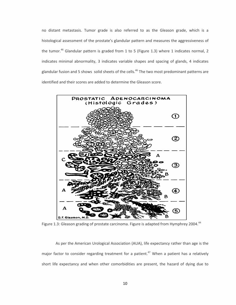
no distant metastasis. Tumor grade is also referred to as the Gleason grade, which is a
histological assessment of the prostate’s glandular pattern and measures the aggressiveness of
the tumor.46 Glandular pattern is graded from 1 to 5 (Figure 1.3) where 1 indicates normal, 2
indicates minimal abnormality, 3 indicates variable shapes and spacing of glands, 4 indicates
glandular fusion and 5 shows solid sheets of the cells.46 The two most predominant patterns are
identified and their scores are added to determine the Gleason score.
Figure 1.3: Gleason grading of prostate carcinoma. Figure is adapted from Hymphrey 2004.44
As per the American Urological Association (AUA), life expectancy rather than age is the
major factor to consider regarding treatment for a patient.47 When a patient has a relatively
short life expectancy and when other comorbidities are present, the hazard of dying due to
11
prostate cancer is reduced. However, if the patient has a relatively longer life expectancy,
localized prostate cancer can lead to morbidity or even mortality.
Conventional treatment options for localized early stage prostate cancer include: radical
prostatectomy, cryosurgery, radiation therapy, or conservative management. Currently there
are a lot of uncertainties regarding the relative efficacies of these treatments and there is no
consensus regarding which treatment to choose under what circumstances among providers. As
a result, this has led to marked variation in practice patterns.43
(i) Radical Prostatectomy
Radical prostatectomy has been the standard treatment for localized prostate cancer for
more than 25 years because of two common assumptions: (i) cure is achieved upon organ
removal, and (ii) the patient is fully recovered after organ removal.4,48 However, there is
evidence of PSA progression due to tumor recurrence after prostatectomy.48 Prostatectomy may
involve removal of the prostate with or without the neurovascular bundles running alongside of
the prostate. These surgeries are associated with urinary, rectal, and erectile complications.49
Modern applications of prostatectomy such as laparoscopic radical prostatectomy or robot-
assisted laparoscopic prostatectomy involve nerve sparing techniques to preserve post-surgical
erectile function.50
(ii) Radiation Therapy
Radiation therapy consists of administration of ionizing radiation by means of various
techniques such as external beam radiation, intensity modulated radiation therapy, proton
therapy, and brachytherapy. Radiation therapy is less invasive than prostatectomy and has
outcomes comparable to those of prostatectomy.4 Common side effects associated with

12
radiation therapy include nocturia, urinary frequency, impotence, and radiation proctitis.4,49
Intensity modulated radiation therapy and proton therapy are relatively advanced techniques of
administering radiation into the body and are expected to reduce urinary and rectal toxicity.51
Both of these techniques provide comparable outcomes.51 Brachytherapy involves radioactive
seed implantation such as iodine-125 or palladium-103 and is recommended as a
monotherapy.52 However, radiation therapy is combined with neoadjuvant and adjuvant
androgen deprivation treatment in many cases.
(iii) Cryosurgery
Cryosurgery uses liquid nitrogen to freeze and destroy abnormal tissue.53 Long-term
outcomes of cryosurgery are not known and this technique is relatively less well established.
(iv) Hormone ablation therapy
Hormonal therapy such as androgen deprivation treatment can be used along with
radiation therapy or prostatectomy as the initial treatment for patients who have a high risk of
cancer recurrence.1 Most of the time, hormonal therapy is reserved for those whose cancer has
already spread beyond the prostate gland or in men with limited life expectancy who are not
candidates for surgery or radiation. Possible side effects associated with hormone therapy
include reduced or absent libido, impotence, hot flashes, breast tenderness, osteoporosis,
anemia, decreased mental sharpness, loss of muscle mass, weight gain, fatigue, and
depression.1
Both prostatectomy and radiation therapy are reported to reduce mortality among men
with high risk tumors.54 However, it is reported that prostatectomy among younger patients
with relatively low-risk prostate cancer is associated with moderate overtreatment (the number
13
needed to treat to prevent one prostate cancer death ranges from 7 to 15) and prolonged side
effects.55 In one trial performed in US Department of Veterans Affairs hospitals, radical
prostatectomy did not show any mortality benefit over conservative management.56 Despite
these results, the majority of patients diagnosed with low-risk prostate cancer receive
immediate aggressive treatment.
(v) Conservative management
Conservative management of early stage low-risk prostate cancer includes two
observational strategies: (i) watchful waiting, and (ii) active surveillance. Watchful waiting
involves relatively passive patient follow-up and is palliative.57 It is reserved for relatively older
patients who cannot tolerate the aggressive treatments. Upon cancer progression, androgen
deprivation treatment is initiated. Active surveillance has emerged as a treatment option for
relatively younger individuals who are closely monitored by frequent PSA testing and imaging.57
However, there is no strict criterion regarding the patient’s age as older men may also opt for
surveillance despite some high risk features.58 The NCCN favors active surveillance in patients
with very low risk disease and a life expectancy of less than 20 years or in those with low-risk
disease and less than 10 years of life expectancy.10 Patients with very low risk disease who are in
active surveillance are less likely to have adverse pathology at the time of radical prostatectomy
during the course of progression compared to those who have low risk disease.43 Also patients
with very low risk disease are less likely to experience biochemical recurrence upon switching to
curative treatment.43
Different institutions have suggested different inclusion criteria for active surveillance.
These criteria mainly include identifying clinically insignificant, low-risk tumors based on biopsy
and other clinical data (Table 1.2).

14
Table 1.2: Institution based inclusion criteria for active surveillance
Institution Clinical stage
PSA Gleason grade
Total positive cores
Single core positivity
Other
Johns Hopkins ≤T2a - ≤3 + 3 ≤2 ≤50% PSA DT
≤0.15
University of Toronto NS ≤10 ≤3 + 3 NR NR -
UCSF ≤T2a ≤10 ≤3 + 3 ≤33% ≤50% -
ERSPC (PRIAS criteria) ≤T2a ≤10 ≤3 + 3 ≤2 NR PSA DT ≤0.2
Royal Marsden Hospital ≤T2a ≤15 ≤3 + 4 ≤50% NR -
MSKCC ≤T2a ≤10 ≤3 + 3 ≤3 ≤50% -
University of Miami ≤T2a ≤10 ≤3 + 3 ≤2 ≤20% -
Note: PSA DT = PSA doubling time; NS = Not stated; NR = Not recorded; UCSF = University of California, San Francisco; MSKCC = Memorial Sloan-Kettering Cancer Center; ERSPC = European Randomized Study of Screening for Prostate Cancer; PRIAS = Prostate Cancer Research International Active Surveillance. Table is adapted from Dall’Era et al., 2012
58
The threshold to trigger treatment in patients who are on active surveillance are not
standardized.59 Some institutions use PSA velocity as an indicator to measure disease
progression whereas some use PSA doubling time within 3-4 years.59 Some institutions rely only
on the results of repeat biopsy.59 The NCCN guidelines have recommended that patients in an
active surveillance program should have PSA measurement as often as every 3 months but at
least every 6 months, DRE performed as often as every 6 months but at least every 12 months,
and a needle biopsy may be repeated within 6 months of diagnosis if the initial biopsy included
fewer than 10 cores.10
As prostate cancer is a slowly growing tumor, it makes observational strategies more
appealing to patients who have low-risk localized disease. Further, there is a high level of
evidence that older men with low risk disease are over treated and observational strategies are
underutilized in this group of patients.43 However, patients who undergo observational strategy
15
have anxiety of tumor progression and not being treated. Additional barriers to the adoption of
active surveillance include potential disease misclassification issues due to inaccurate diagnoses
described earlier.60 As a result, both clinicians and patients lack confidence in prostate biopsy
results. Further, there remain uncertainties regarding the long-term all-cause or disease-specific
mortality, optimal patient selection, surveillance strategies, and triggers for intervention.
b. Intermediate risk prostate cancer and associated treatment
The NCCN guideline has defined intermediate risk prostate cancer as one with T2b-T2c disease,
a Gleason score of 7 or PSA of 10-20 ng/mL.10 T2b indicates that the cancer covers more than
half of only one side of the prostate whereas T2c indicates that the cancer is present in both
sides of the prostate.44 Treatment for localized intermediate risk can be categorized based on
life expectancy. If the patient has a life expectancy lower than 10 years, then treatment options
include10: (1) Conservative management: Watchful waiting or active surveillance; (2) Radiation
therapy alone or in combination with androgen deprivation or brachytherapy; or (3)
Brachytherapy as a monotherapy.
If the patient has a life expectancy greater than 10 years then treatment options include10: (1)
Radical prostatectomy; or (2) Radiation therapy alone or in combination with androgen
deprivation or brachytherapy.
Evidence on Comparative Clinical Effectiveness of Conservative Management vs Immediate Treatment
Two large randomized controlled trials, the Prostate Testing for Cancer and Treatment
(ProtectT) trial in the United Kingdom and the Surveillance Therapy Against Radical Treatment
(START) trial in North America, are currently ongoing to compare active surveillance versus
16
treatment with radiation or surgery. Results of these trials will not be available in the near
future.
A study was performed using the Surveillance Epidemiology End Results (SEER)-
Medicare database to measure survival among patients diagnosed with localized prostate
cancer who did not receive initial definitive treatment within 6 months of diagnosis.61 Low-risk
patients aged 66 to 74 years with comorbidity scores of 0,1, and 2+ had 10- year overall and
prostate cancer specific mortality rates of 29% and 4.8%, 51% and 2%, and 83% and 5.3%
respectively. This study indicates that fewer men older than 65 years of age die due to prostate
cancer within 10 years of diagnosis.61 Abdollah et al compared radiotherapy with observation
within the SEER database.62 They found radiotherapy provided a 10-year cancer-specific
mortality benefit among elderly men (75-80 years) compared with observation.62 However, the
benefit of radiation therapy was not seen in men with low to intermediate risk disease.62
The greatest advantage of active surveillance is to maximize and maintain quality of
life since all immediate treatment strategies have risk of urinary, bowel, and sexual side effects.
As active surveillance is a relatively new management strategy, studies comparing the quality of
life by measuring long term urinary, bowel and erectile toxicity among patients receiving active
surveillance versus immediate treatment are very few. Recently, Hayes et al performed a
decision analysis that compared active surveillance with brachytherapy, intensity-modulated
radiation therapy, and radical prostatectomy in terms of quality adjusted life expectancy among
65-year old men with low-risk prostate cancer.42 Active surveillance had the longest quality-
adjusted life expectancy compared with the rest. However, it is important to note that this study
was limited to those 65-year olds and cannot be generalized to other age categories. Liu et al
compared active surveillance with radical prostatectomy among low-risk prostate cancer

17
patients and found that older men in worse health have better quality adjusted life expectancy
in active surveillance than radical prostatectomy.63 Fujita et al studied approximately 150
patients on active surveillance who underwent a mean of 2.3 biopsies and reported that
patients’ urinary complications such as lower urinary tract symptoms in active surveillance did
not decrease over a period of approximately 3 years.64
Patients who are on active surveillance have to undergo frequent repeat biopsies.
Frequent biopsy associated long-term adverse outcomes among active surveillance patients are
not known. Biopsies frequently lead to bleeding and lower urinary tract infections. Both of these
side effects are transient. Recent evidence suggests there is an increased risk of erectile
dysfunction among patients who undergo serial prostate biopsies.64 Clinical guidelines have
suggested some key recommendations to healthcare professionals regarding patient counseling
and choosing treatment options or observation.52 Table 1.3 depicts these recommendations.
Table 1.3: Recommendation to healthcare professionals regarding patient counseling
Clinical recommendation Evidence rating
Immediate curative treatment should be recommended for patients having localized higher risk tumor. Risk can be estimated based on cancer stage and grade, PSA level, and comorbidity adjusted life expectancy Counseling that surgery and external beam radiation therapy are almost equal should be provided to patients. Brachytherapy can be recommended as a monotherapy in low-risk patients. Active surveillance is a reasonable approach for low risk and very low risk patients.
B B B B
Note: Evidence A = consistent, good-quality patient-oriented evidence; B= inconsistent or limited-quality patient-oriented evidence; C = consensus, disease-oriented evidence, usual practice, expert opinion, or case series. This Table is adapted from Mohan et al., 2011.
52
18
There is a need for research that can identify the long-term side effects of the active
surveillance protocol. Patients diagnosed with low-risk localized disease need to know
treatment effects so that they can make an informed decision regarding what treatment
strategy to pursue. Multiple factors influence a patient’s treatment decisions; however,
perceptions of treatment efficacy and side effects (either from the physician’s description or
from experience of family and friends) have been reported to be the most influential.65
Economic Burden
The five year survival rate for patients diagnosed with localized prostate cancer is 100%
irrespective of the type of treatment received.1 Health care costs have been rising for several
years and represent a significant national issue in health care reform. Increased detection of
clinically insignificant prostate cancer due to PSA testing, frequent repeat biopsies to rule out
the possibility of the presence of lethal prostate cancer, and uncertainties in the available
treatments to manage the low-risk disease are expected to substantially increase the future
fiscal burden of the disease.
In the United States, the prevalence based total costs of prostate cancer were estimated
to be almost $10 billion in 2006.66 Stokes et al estimated the average per person prostate cancer
specific life-time cost to be approximately $34,000 for patients diagnosed between 1991 and
2002.66 However, it is important to note that these costs vary by cancer stage and its estimates
were shown to vary from $26, 078 (stage III) to $39, 182 (stage I).66 There is a report of racial
and ethnic disparities in health care utilization and associated costs of prostate cancer but these
studies are dated.67
In health care cost analysis, costs are generally divided into three categories: direct,
indirect, and intangible costs.68 Direct health care costs include all costs related to drugs, tests,
19
health care professionals, and medical facilities. Direct non-health care costs include cost related
to transportation to medical facilities, child care cost or cost resulting due to accommodation at
home or home care givers. Indirect costs include costs due to productivity loss. This may include
costs due to loss or impaired ability to work due to the disease condition or morbidity.
Intangible costs cannot be measured directly and may include costs due to limitation in leisure
activities. Previous studies have reported treatment specific short-term and long-term costs.
Hormonal therapy has been reported to cost the highest ($26,896) within 5 years of cancer
diagnosis followed by hormonal + radiation treatment ($25,097), surgery ($19,214), radiation
therapy only ($15,589) and watchful waiting ($9130) among the older population.69 However,
these estimates are found to vary widely among the relatively younger population. Watchful
waiting strategy is found to cost approximately $24, 809 per patient for a period of 2 years due
to multiple follow-ups and close monitoring.2
Recently, Cooperberg et al. conducted a comprehensive lifetime cost-utility analysis
among hypothetical men with clinically localized prostate cancer having low, intermediate or
high risk of disease.70 In this hypothetical scenario, patients received relatively advanced and
conventional treatments such as open radical prostatectomy, laparoscopic-assisted radical
prostatectomy, robot-assisted radical prostatectomy, three-dimensional conformal radiation
therapy, intensity modulated radiation therapy, brachytherapy, or a combination of intensity
modulated radiation therapy and brachytherapy in 2009.70 Further, they assumed that low,
intermediate, and high risk patients are 75%, 50%, and 25% likely to receive salvage radiation
therapy respectively. The remainder receives androgen deprivation treatment. Surgical methods
such as open radical prostatectomy, laparoscopic-assisted radical prostatectomy, or robot-
assisted radical prostatectomy were found to cost around $20,000, $28,500, and $35,500
20
respectively, for low, intermediate, and high risk patients.70 Brachytherapy was found to cost
around $25,066, $32,553 and $43, 952 for low, intermediate, and high risk patients.70
In each risk stratum, all advanced radiation therapies were found more costly while less
effective. On the other hand, surgeries were less costly and more effective compared to all
radiation therapies. This study did not include active surveillance in modeling.70 In view of the
new and emerging diagnostic technologies and increasing aging population, there is a need for
more recent overall national estimate of the economic burden associated with prostate cancer.

21
REFERENCES
1. American Cancer Society. Prostate cancer. 2013; Retrieved in 2014 from
http://www.cancer.org/cancer/prostatecancer/detailedguide/prostate-cancer-key-
statistics.
2. Crawford ED, Black L, Eaddy M, Kruep EJ. A retrospective analysis illustrating the
substantial clinical and economic burden of prostate cancer. Prostate Cancer and
Prostatic Diseases. Jun 2010;13(2):162-167.
3. Roehrborn CG, Black LK. The economic burden of prostate cancer. BJU International. Sep
2011;108(6):806-813.
4. Mohile SG, Lachs M, Dale W. Management of prostate cancer in the older man.
Seminars in Oncology. Dec 2008;35(6):597-617.
5. National Cancer Institute. General information about prostate cancer. 2014. Retrieved in
2014 from
http://www.cancer.gov/cancertopics/pdq/treatment/prostate/HealthProfessional/page
1
6. Pal RP, Maitra NU, Mellon JK, Khan MA. Defining prostate cancer risk before prostate
biopsy. Urologic Oncology. Nov 2013;31(8):1408-1418.
7. Bonekamp D, Jacobs MA, El-Khouli R, Stoianovici D, Macura KJ. Advancements in MR
imaging of the prostate: From diagnosis to interventions. Radiographics.
2011;31(3):677-703.
8. Umbehr M, Bachmann LM, Held U, et al. Combined magnetic resonance imaging and
magnetic resonance spectroscopy imaging in the diagnosis of prostate cancer: A
systematic review and meta-analysis. European Urology. 2009;55(3):575-590.

22
9. American Urological Association. Prostate cancer: Screening and management. etrieved
in 2014 from http://www.auanet.org/education/prostate-cancer-psa.cfm.
10. National Comprehensive Cancer Network. The NCCN guidelines for prostate cancer early
detection. 2013; Retrieved in 2014 from
http://www.nccn.org/professionals/physician_gls/f_guidelines.asp.
11. Stoianovici D. Technology advances for prostate biopsy and needle therapies. The
Journal of Urology. 2012 Oct;188(4):1074-5. doi: 10.1016/j.juro.2012.06.127. Epub 2012
Aug 15.
12. Mian BM. Prostate biopsy strategies: Current state of the art. Journal of the National
Comprehensive Cancer Network : JNCCN. May 2004;2(3):213-222.
13. Dianat SS, Carter HB, Macura KJ. Performance of multiparametric magnetic resonance
imaging in the evaluation and management of clinically low-risk prostate cancer.
Urologic Oncology. 2013;17(13):002.
14. Rais-Bahrami S, Siddiqui MM, Turkbey B, et al. Utility of Multiparametric MRI Suspicion
Levels in Detecting Prostate Cancer. The Journal of Urology. 2013;29(13):04417-04410.
15. Engehausen DG, Engelhard K, Schwab SA, et al. Magnetic resonance image-guided
biopsies with a high detection rate of prostate cancer. Scientific World Journal.
2012;975971(10):12.
16. Pinto F, Totaro A, Calarco A, et al. Imaging in prostate cancer diagnosis: Present role and
future perspectives. Urologia Internationalis. 2011;86(4):373-382.
17. Loch T. Urologic imaging for localized prostate cancer in 2007. World Journal of Urology.
2007;25(2):121-129.

23
18. Haffner J, Lemaitre L, Puech P, et al. Role of magnetic resonance imaging before initial
biopsy: comparison of magnetic resonance imaging-targeted and systematic biopsy for
significant prostate cancer detection. BJU International. 2011;108(8 Pt 2):22.
19. Kirby R, Fitzpatrick JM. Optimising repeat prostate biopsy decisions and procedures. BJU
International. 2012;109(12):1750-1754.
20. Komai Y, Numao N, Yoshida S, et al. High Diagnostic Ability of Multiparametric Magnetic
Resonance Imaging to Detect Anterior Prostate Cancer Missed by Transrectal 12-Core
Biopsy. The Journal of Urology. 2013;28(13):03871-03878.
21. Nix JW, Turkbey B, Hoang A, et al. Very distal apical prostate tumours: Identification on
multiparametric MRI at 3 Tesla. BJU International. 2012;110(11 Pt B):4.
22. Mowatt G, Scotland G, Boachie C, et al. The diagnostic accuracy and cost-effectiveness
of magnetic resonance spectroscopy and enhanced magnetic resonance imaging
techniques in aiding the localisation of prostate abnormalities for biopsy: Asystematic
review and economic evaluation. Health Technology Assessment. 2013;17(20):1-281.
23. Stravodimos KG, Haritopoulos KN, Alamanis C, Anastasiou I, Constantinides C. Local
anesthesia during transrectal ultrasonography-guided prostate biopsy: Does it have any
effect on sexual function? International Urology and Nephrology. 2007;39(3):893-896.
24. Abdellaoui A, Iyengar S, Freeman S. Imaging in prostate cancer. Future Oncology
(London, England). May 2011;7(5):679-691.
25. Perdona S, Di Lorenzo G, Autorino R, et al. Combined magnetic resonance spectroscopy
and dynamic contrast-enhanced imaging for prostate cancer detection. Urologic
Oncology. 2013;31(6):761-765.
26. Carey BM. Imaging for prostate cancer. Clinical Oncology. 2005;17(7):553-559.

24
27. Dickinson L, Ahmed HU, Allen C, et al. Clinical applications of multiparametric MRI
within the prostate cancer diagnostic pathway. Urologic Oncology. 2013;31(3):281-284.
28. Goris Gbenou MC, Peltier A, Addla SK, et al. Localising prostate cancer: comparison of
endorectal magnetic resonance (MR) imaging and 3D-MR spectroscopic imaging with
transrectal ultrasound-guided biopsy. Urologia Internationalis. 2012;88(1):12-17.
29. Hambrock T, Hoeks C, Hulsbergen-van de Kaa C, et al. Prospective assessment of
prostate cancer aggressiveness using 3-T diffusion-weighted magnetic resonance
imaging-guided biopsies versus a systematic 10-core transrectal ultrasound prostate
biopsy cohort. European Urology. 2012;61(1):177-184.
30. Fiard G, Hohn N, Descotes JL, Rambeaud JJ, Troccaz J, Long JA. Targeted MRI-guided
prostate biopsies for the detection of prostate cancer: Initial clinical experience with
real-time 3-dimensional transrectal ultrasound guidance and magnetic
resonance/transrectal ultrasound image fusion. Urology. 2013;81(6):1372-1378.
31. Kaplan I, Oldenburg NE, Meskell P, Blake M, Church P, Holupka EJ. Real time MRI-
ultrasound image guided stereotactic prostate biopsy. Magnetic Resonance Imaging.
2002;20(3):295-299.
32. Puech P, Sufana Iancu A, Renard B, Villers A, Lemaitre L. Detecting prostate cancer with
MRI - why and how. Diagnostic and Interventional Imaging. 2012;93(4):268-278.
33. Moore CM, Robertson NL, Arsanious N, et al. Image-guided prostate biopsy using
magnetic resonance imaging-derived targets: A systematic review. European Urology.
2013;63(1):125-140.
34. Park BK, Lee HM, Kim CK, Choi HY, Park JW. Lesion localization in patients with a
previous negative transrectal ultrasound biopsy and persistently elevated prostate
25
specific antigen level using diffusion-weighted imaging at three Tesla before rebiopsy.
Investigative Radiology. 2008;43(11):789-793.
35. Hara N, Okuizumi M, Koike H, Kawaguchi M, Bilim V. Dynamic contrast-enhanced
magnetic resonance imaging (DCE-MRI) is a useful modality for the precise detection
and staging of early prostate cancer. Prostate. 2005;62(2):140-147.
36. Nagarajan R, Margolis D, Raman S, et al. MR spectroscopic imaging and diffusion-
weighted imaging of prostate cancer with Gleason scores. Journal of Magnetic
Resonance Imaging. 2012;36(3):697-703.
37. Sciarra A, Panebianco V, Ciccariello M, et al. Value of magnetic resonance spectroscopy
imaging and dynamic contrast-enhanced imaging for detecting prostate cancer foci in
men with prior negative biopsy. Clinical Cancer Research. 2010;16(6):1875-1883.
38. Villers A, Marliere F, Ouzzane A, Puech P, Lemaitre L. MRI in addition to or as a
substitute for prostate biopsy: The clinician's point of view. Diagnostic and
Interventional Imaging. 2012;93(4):262-267.
39. Sciarra A, Barentsz J, Bjartell A, et al. Advances in magnetic resonance imaging: How
they are changing the management of prostate cancer. European Urology.
2011;59(6):962-977.
40. Ouzzane A, Puech P, Lemaitre L, et al. Combined multiparametric MRI and targeted
biopsies improve anterior prostate cancer detection, staging, and grading. Urology.
2011;78(6):1356-1362.
41. Lu-Yao GL, Albertsen PC, Moore DF, et al. Outcomes of localized prostate cancer
following conservative management. JAMA : the Journal of the American Medical
Association. Sep 16 2009;302(11):1202-1209.

26
42. Hayes JH, Ollendorf DA, Pearson SD, et al. Active surveillance compared with initial
treatment for men with low-risk prostate cancer: A decision analysis. JAMA : the Journal
of the American Medical Association. Dec 1 2010;304(21):2373-2380.
43. Carter HB. Management of low (favourable)-risk prostate cancer. BJU International. Dec
2011;108(11):1684-1695.
44. Roswell Park Cancer Institute. Prostate Cancer Staging.
https://www.roswellpark.org/cancer/prostate/diagnosis/staging.
45. Mayo Clinic. Prostate Cancer Symtoms. http://www.mayoclinic.org/diseases-
conditions/prostate-cancer/basics/symptoms/con-20029597.
46. Humphrey PA. Gleason grading and prognostic factors in carcinoma of the prostate.
Mod Pathol. 02/13/online 2004;17(3):292-306.
47. American Urological Association. The Management of Localized prostate cancer Patient
guide. 2008. https://www.auanet.org/common/pdf/education/clinical-
guidance/Prostate-Cancer-PatientGuide.pdf.
48. Weissbach L, Altwein J. Active surveillance or active treatment in localized prostate
cancer? Deutsches Arzteblatt International. May 2009;106(22):371-376.
49. Michaelson MD, Cotter SE, Gargollo PC, Zietman AL, Dahl DM, Smith MR. Management
of complications of prostate cancer treatment. CA: A Cancer Journal for Clinicians. Jul-
Aug 2008;58(4):196-213.
50. American Cancer Society. Surgery for prostate cancer. 2013.
http://www.cancer.org/cancer/prostatecancer/detailedguide/prostate-cancer-treating-
surgery.

27
51. Hoppe BS, Michalski JM, Mendenhall NP, et al. Comparative effectiveness study of
patient-reported outcomes after proton therapy or intensity-modulated radiotherapy
for prostate cancer. Cancer. Apr 1 2014;120(7):1076-1082.
52. Mohan R, Schellhammer PF. Treatment options for localized prostate cancer. American
Family Physician. Aug 15 2011;84(4):413-420.
53. National Cancer Institute. Cryosurgery in cancer treatment: Questions and answers.
2003.
54. Thompson IM, Tangen CM. Prostate Cancer — Uncertainty and a Way Forward. New
England Journal of Medicine. 2012;367(3):270-271.
55. Sandhu GS, Andriole GL. Active surveillance for prostate cancer: Barriers to widespread
adoption. European Urology. Dec 2012;62(6):984-985.
56. Wilt TJ, Brawer MK, Jones KM, et al. Radical Prostatectomy versus Observation for
Localized Prostate Cancer. New England Journal of Medicine. 2012;367(3):203-213.
57. Klotz L. Active surveillance for prostate cancer: patient selection and management.
Current Oncology (Toronto, Ont.). Sep 2010;17 Suppl 2:S11-17.
58. Dall'Era MA, Albertsen PC, Bangma C, et al. Active surveillance for prostate cancer: a
systematic review of the literature. European Urology. Dec 2012;62(6):976-983.
59. Whitson JM, Carroll PR. Active surveillance for early-stage prostate cancer: defining the
triggers for intervention. Journal of Clinical Oncology : Official Journal of the American
Society of Clinical Oncology. Jun 10 2010;28(17):2807-2809.
60. Singer EA, Kaushal A, Turkbey B, Couvillon A, Pinto PA, Parnes HL. Active surveillance for
prostate cancer: Past, present and future. Current Opinion in Oncology. May
2012;24(3):243-250.
28
61. Albertsen PC, Moore DF, Shih W, Lin Y, Li H, Lu-Yao GL. Impact of comorbidity on
survival among men with localized prostate cancer. Journal of Clinical Oncology : Official
Journal of the American Society of Clinical Oncology. Apr 1 2011;29(10):1335-1341.
62. Abdollah F, Sun M, Schmitges J, et al. Competing-risks mortality after radiotherapy vs.
observation for localized prostate cancer: A population-based study. International
Journal of Radiation Oncology, Biology, Physics. Sep 1 2012;84(1):95-103.
63. Liu D, Lehmann HP, Frick KD, Carter HB. Active surveillance versus surgery for low risk
prostate cancer: a clinical decision analysis. The Journal of Urology. Apr
2012;187(4):1241-1246.
64. Fujita K, Landis P, McNeil BK, Pavlovich CP. Serial prostate biopsies are associated with
an increased risk of erectile dysfunction in men with prostate cancer on active
surveillance. The Journal of urology. Dec 2009;182(6):2664-2669.
65. Xu J, Dailey RK, Eggly S, Neale AV, Schwartz KL. Men's perspectives on selecting their
prostate cancer treatment. Journal of the National Medical Association. Jun
2011;103(6):468-478.
66. Stokes ME, Ishak J, Proskorovsky I, Black LK, Huang Y. Lifetime economic burden of
prostate cancer. BMC Health Services Research. 2011;11:349.
67. Jayadevappa R, Malkowicz SB, Chhatre S, Gallo J, Schwartz JS. Racial and ethnic variation
in health resource use and cost for prostate cancer. BJU International. Sep
2010;106(6):801-808.
68. Gold MR. Cost-effectiveness in health and medicine. New York: Oxford University Press;
1996.
29
69. Snyder CF, Frick KD, Blackford AL, et al. How does initial treatment choice affect short-
term and long-term costs for clinically localized prostate cancer? Cancer.
2010;116(23):5391-5399.
70. Cooperberg MR, Ramakrishna NR, Duff SB, et al. Primary treatments for clinically
localised prostate cancer: A comprehensive lifetime cost-utility analysis. BJU
International. Mar 2013;111(3):437-450.

30
CHAPTER 2
RATIONALE AND HYPOTHESIS
This work focuses on three broad areas in the realm of prostate cancer outcomes
research. The first part of the dissertation focuses on assessing healthcare utilization and
associated expenditures among prostate cancer patients. The economic burden of the disease is
commonly estimated using cost of illness studies.1 Most cost of illness studies associated with
prostate cancer are dated.2-4 As overdiagnosis of prostate cancer has imposed a greater burden
on the healthcare system, there is a need for more recent cost studies that determine the
average cost per patient as well as provide estimates on utilization of different health services in
different delivery settings. This study is important for policy makers and health researchers to
determine how healthcare resources are expended on prostate cancer and the magnitude of the
impact of prostate cancer on society.
The second part of the dissertation focuses on determining cost-effectiveness of
multiparametric magnetic resonance imaging (MP-MRI) followed by fusion guided biopsy in
prostate cancer diagnosis. Accurate prostate cancer diagnosis is limited by poor specificity and
the sensitivity of PSA and TRUS guided biopsy. Further, to rule out the presence of prostate
cancer, many patients undergo additional biopsies and are diagnosed with clinically insignificant
cancers.5 These biopsies are not without complications and are associated with hematuria,
hematospermia, rectal bleeding and urinary retention.6 To avoid these repeated biopsies, new
diagnostic strategies are needed that can identify patients with clinically significant cancer only.

31
MP-MRI is one of the promising modalities for prostate imaging. MP-MRI involves the
use of different MRI based techniques such as T1-weighted, T2-weighted, diffusion weighted
imaging, dynamic contrast enhanced imaging, and/or magnetic resonance spectroscopy in one
or more combinations.7 MRI/TRUS fusion guided biopsy is an outpatient procedure, in which
prebiopsy MRI of the prostate is fused with real-time TRUS.8 Currently, a clinical trial is being
conducted to determine whether MRI/TRUS fusion guided biopsy is superior to conventional
biopsy alone in diagnosing subjects with prostate cancer.9 However, the results of this study will
not be available until April 2015. Economic modelling would be an appropriate approach to
compare the relative performance of these diagnostic technologies.
The third part of the dissertation focuses on the comparative analysis of harms
associated with conservative management and immediate treatment among low-risk prostate
cancer patients. Conservative management, especially watchful waiting of low-risk prostate
cancer, is not new. However, active surveillance is a new emerging term that focuses on
individuals who are relatively healthy and have longer life expectancies rather than the sicker
older population. Active surveillance has been recommended as a treatment option in many
guidelines but is not practiced clinically because of uncertainties associated with clinical
outcomes.10 The Institute of Medicine’s Committee on Comparative Effectiveness Research has
identified treatment for localized prostate cancer as a high-priority research area.11 This study
focuses on the urinary, rectal, and erectile complications, and survival associated with
conservative management and immediate treatment approach within 5 years of diagnosis.
This part of the study aims to reduce uncertainties such as the probability of developing
adverse effects associated with two broad treatment regimens. The relative effectiveness of
treatment alternatives for localized prostate cancer is uncertain and costs are variable. More
32
specifically, patients need predictions of not only effectiveness but also the various long-term
outcomes such as survival, impotence, incontinence, and bowel problems associated with
treatment choice. Clinical decision regarding which treatment to choose is a multidimensional
and complex process in which patients consider their personal preferences and the advice of
their health care practitioners. In this decision process, patients give subsequent emphasis of
treatment related side effects. Our study makes this decision process easier for patients by
providing long-term outcomes associated with a conservative management option.
Specific Aims
The three specific aims of this project are to:
Aim 1: Estimate the annual average prostate cancer cost in different health delivery settings in
the United States and to determine the predictors of higher annual costs of prostate cancer.
We hypothesized that higher expenditures may be associated with chemotherapy, advanced
diagnostic strategies such as magnetic resonance imaging, radiation treatment, and surgery.
Aim 2: Determine the cost-effectiveness of MP-MRI followed by MRI/TRUS fusion guided biopsy
compared with the conventional TRUS guided biopsy among biopsy naïve patients in prostate
cancer diagnosis.
We hypothesized that the MP-MRI approach is more cost-effective than the conventional TRUS
guided biopsy because MP-MRI may reduce the number of many false positive and false
negative cases and thus avoid costs related to unncecessary repeated biopsies and
overdiagnosis.

33
Aim 3: Compare side effect profile and survival among patients diagnosed with low to
intermediate risk prostate cancer undergoing either conservative management or immediate
treatments.
We hypothesized that patients who undergo conservative management options such as
watchful waiting or active surveillance may delay or avoid toxicities associated with immediate
treatment as prostate cancer is a slowly growing tumor and many patients diagnosed with low-
risk prostate cancer may not require treatment for long time.
34
REFERENCES
1. Segel J.E. Cost-of-Illness Studies - A primer. RTI International; 2006.
2. Roehrborn CG, Black LK. The economic burden of prostate cancer. BJU International
2011;108:806-13.
3. Stokes ME, Ishak J, Proskorovsky I, Black LK, Huang Y. Lifetime economic burden of
prostate cancer. BMC Health Services Eesearch 2011;11:349.
4. Stokes ME, Black L, Benedict A, Roehrborn CG, Albertsen P. Long-term medical-care costs
related to prostate cancer: Estimates from linked SEER-Medicare data. Prostate cancer
and Prostatic Diseases 2010;13:278-84.
5. Shariat SF, Roehrborn CG. Using biopsy to detect prostate cancer. Reviews in Urology
2008;10:262-80.
6. Loeb S, Vellekoop A, Ahmed HU, et al. Systematic review of complications of prostate
biopsy. European Urology 2013;64:876-92.
7. Pinto F, Totaro A, Calarco A, et al. Imaging in prostate cancer diagnosis: Present role and
future perspectives. Urologia Internationalis 2011;86:373-82.
8. Penzkofer T, Tempany-Afdhal CM. Prostate cancer detection and diagnosis: the role of
MR and its comparison with other diagnostic modalities - A radiologist's perspective.
NMR in Biomedicine 2014;27:3-15.
9. ClinicalTrials. MRI/TRUS Fusion Guided Prostate Biopsy- An Improved Way to Detect and
Quantify Prostate Cancer. 2013.
10. Weissbach L, Altwein J. Active surveillance or active treatment in localized prostate
cancer? Deutsches Arzteblatt International 2009;106:371-6.

35
11. Perlroth DJ, Bhattacharya J, Goldman DP, Garber AM. An economic analysis of
conservative management versus active treatment for men with localized prostate
cancer. Journal of the National Cancer Institute Monographs 2012;2012:250-7.

36
CHAPTER 3
DIRECT MEDICAL EXPENDITURE AND PREDICTORS ASSOCIATED WITH PROSTATE CANCER FOR
THE U.S. ADULT POPULATION: ESTIMATES FROM MEDICAL EXPENDITURE PANEL SURVEY (2010)
____________________________
1Patel P, Perri M, Tackett R. To be submitted to Journal of Pharmacy and Pharmacology.
37
Purpose: The purpose of this study was to explore the annual average prostate cancer cost in
different health delivery settings in 2010 and to determine significant predictors of the annual
cost of prostate cancer. Methods: Using the Medical Expenditure Panel Survey (MEPS), a
retrospective study was conducted assessing healthcare utilization and associated expenditures
among patients having prostate cancer. Information on patient demographics, health care
service utilization and cost estimates were derived from the database representing 1,422,218
patients with prostate cancer and related conditions. To predict the effects of demographics,
testing procedures, and types of services used on annual prostate cancer related costs, an
ordinary least squares model with logarithmic form of expenditure was used. Results: An annual
average total of $5.6 billion was spent on the treatment of prostate cancer related conditions in
2010 in the United States. Outpatient and inpatient visits were found to contribute significantly
to the increased total prostate cancer related expenditure. Weighted multiple linear regression
analyses revealed that the use of chemotherapy (p value=0.0004) and ultrasound (p
value<0.0001) were found to increase the expenditures related to outpatient visits significantly.
Use of ultrasound (p value<0.0001) and x-ray (p value <0.0001) were found to increase office-
based visit costs significantly. Conclusion: Results of our study indicate that the expenditure
related to prostate cancer absorbs a significant portion of healthcare resources. Ultrasound, the
most commonly used diagnostic technique to guide biopsy, was found to be associated with
higher prostate cancer expenditure. Lately, there is advancement in chemotherapy drugs that
have improved cancer care but these modern medicines are expensive. The current study found
chemotherapy to be associated with higher prostate cancer expenditure. Cost estimates derived
from this study may be used by healthcare decision makers in developing disease management
strategies with efficient allocation of resources.
Keywords: Prostate cancer expenditure, service utilization, Medical Expenditure Panel Survey

38
Introduction
Prostate cancer is the second leading cause of non-cutaneous cancer deaths among men
in the United States.1 Statistics related to prostate cancer diagnosis and death suggest that
prostate cancer is one of the most frequently diagnosed cancers affecting one in every six men.2
The American Cancer Society estimated that there were approximately 238,590 new cases and
29,720 deaths due to prostate cancer in 2013.1 Annual expenditures associated with prostate
cancer in 2006 was $9.862 billion in the U.S.3 Costs associated with prostate cancer are expected
to rise due to increased diagnosis, diagnosis at an earlier stage and increased survival.3 With the
advent of Prostate Specific Antigen (PSA) screening as the most common method of detection in
the United States, the ability to diagnose nonlethal prostate cancers has further increased.4
Thus, increased detection combined with increased survival has imposed a greater burden from
prostate cancer on the healthcare system.
Most cost of illness studies associated with prostate cancer are dated.3,5,6 Thus, there is
a need for more recent cost studies that determine the average cost per patient. In addition,
we need more current information on the utilization of different health services in different
delivery settings associated with prostate cancer. The incidence of prostate cancer has been
reported to vary according to race with black men experiencing a higher incidence and more
advanced anatomic stage of disease.7 Additionally, current diagnostic procedures associated
with prostate cancer result in frequent unnecessary biopsy procedures and cause a financial
burden.8 Few studies in the literature have determined the factors that document the high costs
associated with prostate cancer. It is important for policy makers and health researchers to
determine how resources are expended for prostate cancer. Increased attention must be paid to
the patient’s average cost of care and to the factors that predict those costs.

39
The purpose of this study was to explore the annual average prostate cancer cost in
different health delivery settings in 2010 and to determine the significant predictors of the
annual cost of prostate cancer. From the perspective of policymakers and researchers, this study
is important as it will provide reliable estimates of prostate cancer-specific medical care costs.
These estimates can be used to help describe the overall economic burden associated with
cancer morbidity and mortality and to understand the magnitude of financial resources that
must be mobilized to effectively care for cancer patients.
Methods
Data source: Using the Medical Expenditure Panel Survey (MEPS), a retrospective study was
conducted assessing healthcare utilization and associated expenditures among patients having
prostate cancer. Administered by the Agency for Healthcare Research and Quality, the MEPS is a
nationally representative survey of health care use, expenditures, sources of payment, and
insurance coverage for the US civilian, non-institutionalized population.9 The Health Component
is the core survey and collects detailed data at both the personal and house-hold levels, using an
overlapping panel design with 5 rounds of computer-assisted personal interviews over a 2.5-
year period.9 In the MEPS database, one respondent per household is interviewed about the
medical events of household members, including health status, health care utilization, and
health insurance. Surveys of the household’s medical providers, employers, and health insurers
verify and supplement the interview data.9 In particular, medical providers, such as office based
physicians, hospital, home health agencies, and pharmacies, serve as the primary source for
expenditure information, including out-of-pocket payments by the family.

40
Subjects: The subjects for this study were identified using the clinical classification system code
of 29, which indicates that the patient has prostate cancer related conditions. Patients of all
ages and ethnicities were included in this study.
Measures: The primary outcome measures were prostate cancer related health care services
use and associated costs. Services included outpatient visits, emergency department visits,
inpatient visits, office-based visits, prescriptions, and home health visit days. Costs consisted of
total as well as component costs, such as outpatient, emergency department, home health,
inpatient, and prescriptions. Health care service utilization and cost estimates were derived
from the 2010 MEPS Full Year Consolidated File, Medical Conditions File, Office-Based Medical
Provider Visit File, Outpatient Visits File, Emergency Room Visit File, Hospital Inpatient Stays File,
Home Health File, and Prescribed Medicine File.
The effect of various demographic characteristics, comorbidities, types of diagnostic
services, chemotherapy, radiation, and laboratory tests performed on prostate cancer related
expenditures was evaluated. Demographic characteristics such as age, race/ethnicity and
insurance status were determined through the full year consolidated data file. Comorbidity was
measured using the Charlson comorbidity index.10 The Charlson comorbidity index assigns
weights ranging from 0 to 6 based on the severity of a number of major conditions. The index
severity score was calculated for each patient by adding the assigned weight for each patient’s
comorbid conditions, with higher scores representing greater severity of comorbidity. Patients
were categorized based on the comorbidity level. A Charlson score of 0 indicates no
comorbidity; a score of 1 to 2 indicates moderate comorbidity; and a score greater than or equal
to 3 indicates severe comorbidity. Total expenses for an “event” were defined as the sum of

41
direct payments by households, private insurance, Medicare, Medicaid, and other sources to
providers of the care.
Statistical analysis: Estimates presented in the tables and texts were statistically weighted to
reflect national population totals. The weights, which were provided by the data collection
agency, are equal to the inverse of the sampling probability for each case, adjusted for
nonresponse. As medical expenditure data are not normally distributed, an ordinary least
squares model with logarithmic form of expenditure was used to predict the effects of
demographics, testing procedures, and types of services used on annual prostate cancer related
costs.11 All analyses were conducted using SAS (version 9.3, SAS Institute, Carey NC).
Results
Sample characteristics:
Data for 1,422,218 patients with prostate cancer (weighted sample size) were analyzed
from the 2010 MEPS dataset. Demographic characteristics of the study population (Table 3.1)
show that the mean age of the patients with prostate cancer was approximately 70 years and
the majority of patients were between 65 and 75 years old. The majority of patients were white
(≈85%) with moderate comorbid conditions (Charlson comorbidity index of ≈ 2) and had some
form of private insurance (≈69%).
Patterns of Health care use and expenditure associated with prostate cancer:
Results related to health care services used and the associated expenditures are
presented in Table 3.2. As a group, patients with prostate cancer related conditions had a total
of 5.2 million office-based visits, 1.1 million outpatient visits, and 0.2 million in-patient visits.
Patients with prostate cancer received a total of 2.5 million prescriptions in 2010.
42
An annual average total of $5.6 billion was spent on treatment of prostate cancer
related conditions in 2010 in the US. Almost half of the expenditure was spent on office based
and outpatient visits. Approximately $1.7 billion was spent on inpatient services such as surgery
or diagnostic testing. Due to the smaller number of patients who visited hospitals and stayed
overnight in our sample, the expenditure related to inpatient services should be interpreted
with caution. There were no patients in the study sample who visited the emergency
department due to prostate cancer. Prescription expenditures for treatment of prostate cancer
amounted to $0.3 billion.
Among persons with any expense for prostate cancer, the average annual expenditure
was $3,977.78 in 2010. The mean expense per adult for office-based visits was $410.68, $1272.3
for outpatient visits, and $110.67 for prescriptions. The average per person amount spent on
hospital stays was $10,502. The frequency distribution of prostate cancer related direct medical
costs is presented in Figure 3.1.
Predictors of Health care expenditure related to prostate cancer:
We examined the influences of demographic characteristics and types of health service
utilization on annual per capital health care expenditures associated with prostate cancer. An
ordinary least squares model with logarithmic form of expenditure was employed incorporating
survey design strategies such as stratum, primary sampling unit, and sampling weight
information. The results of the multivariate analysis for predictors of prostate cancer specific
healthcare costs are presented in Table 3.3.
The overall model with log transformed cost data was found to be statistically significant
(Table 3.3). After controlling for other variables, outpatient and inpatient visits were found to be
statistically significant predictors for the expenditure related to prostate cancer. In order to

43
eliminate bias associated with retransformation of log transformed cost data back into natural
form, a smearing estimator was calculated. This estimator is the mean of the antilog of the
residuals and represents the estimate of the untransformed scale free from the distribution
assumptions on the error distribution. Compared to office based visits, patients with outpatient
visits increased the prostate cancer expenditure annually on average by 18.62 fold (antilog of
estimate*smearing estimator). Similarly inpatient visits were found to increase expenditures by
20.5 fold on average compared to office based visits. Age or prescriptions were not statistically
significantly associated with prostate cancer related expenditures.
Predictors of outpatient visit costs associated with prostate cancer:
As outpatient visits were found to be statistically significant predictors of prostate
cancer expenditure, a separate regression analysis was performed to assess the predictors of
total annual expenditures associated with prostate cancer related outpatient visits (Table 3.4).
We also analyzed office based visit data to estimate the influence of various predictors on office
based visit expenditures (Table 3.5). We could not analyze predictors of inpatient costs because
of the limited number of patients who had inpatient visits in our sample.
Multiple linear regression analysis of outpatient visit costs revealed chemotherapy,
ultrasound, and x-ray as the statistical significant predictors of the cost. Chemotherapy and
ultrasound were found to increase the expenditures related to outpatient visits whereas x-ray
cost was found to decrease it while holding other variables constant.
Predictors of office-based visit costs associated with prostate cancer:
Multiple regression analysis examining predictors of office-based visit explained
approximately 25% of the variance in the model (Table 3.5). Ultrasound and x-ray were
44
associated with the higher office-based visit costs, whereas age was found to be associated with
lower office-based visit costs.
Discussion
This study used a nationally representative database and provides the most
comprehensive and recent estimates of direct medical costs of prostate cancer related
conditions among adults in the US. Using estimates from this study, the annual direct medical
expenditures associated with prostate cancer was found to be $5.6 billion in 2010 US dollars. A
recent study by Roehrborn et al. estimated the cost of prostate cancer to be $9.862 billion for
2006 using published data and internet sources.3 There are only a few recent studies in the
literature that provide estimates on different health service utilization and expenditure
associated with prostate cancer. Our study is comprehensive in terms of providing estimates of
total expenditure, expenditure related to specific health service used, and predictors of
expenditures.
With respect to cost categories, outpatient and inpatient visits were found to be the
major driver of healthcare costs related to prostate cancer. Inpatient and outpatient visits
accounted for approximately 30% and 25% of the overall costs, respectively. Our findings are
consistent with the known healthcare utilization patterns and treatment seeking behaviors of
individuals with prostate cancer.12 Higher costs found with the inpatient visits can be explained
by the fact that the majority of patients diagnosed with localized prostate cancer undergo
surgery in an inpatient setting. Most of the treatments other than surgery occur in the
outpatient setting explaining higher costs associated with the outpatient setting.
This study found strong associations that are of great interest to policy makers.
Individual regression analysis for outpatient visit cost found chemotherapy and ultrasound as
45
the major source of higher expenditure related to outpatient expenditure. As ultrasound is
cheaper than some advanced imaging technologies, it is the most commonly preferred
technique to guide biopsy for the diagnostic confirmation of prostate cancer.13 Although
ultrasound is cheaper, it is not as reliable a technique and leads to many false negative test
results.14 As a result, patients undergo repeated biopsy techniques for further confirmation
leading to increased costs due to additional ultrasound biopsy techniques performed as well as
treatment of biopsy induced complications. There is a need for imaging technologies that can
diagnose prostate cancer accurately and avoid repeated testing resulting in cost saving.
Ultrasound guided biopsy is usually performed either in the medical provider’s office or in
outpatient settings. However, it’s a fact that if the biopsy is performed in an outpatient setting,
it is more expensive due to overhead costs.
Chemotherapy is administered at late or an advanced stage of prostate cancer mainly in
a hospital setting.15 Chemotherapy use has increased markedly for almost all cancers.16 The
most commonly used chemo drug for prostate cancer is docetaxel, combined with prednisone.17
Cabazitaxel was approved as a second line choice in June 2010 for patients among whom
docetaxel does not work.18 Both treatments have been reported to have improved survival
outcomes among patients with metastatic prostate cancer and thus, are the preferred choice.
Prescription drugs in this study were not found to be statistical significant predictors of higher
expenditure associated with prostate cancer in 2010. However, it is important to note that the
prescription drug file in MEPS data does not collect information on prescription drugs obtained
during hospital visits. Advanced treatments for castrate resistant prostate cancer such as
sipuleucel-t vaccination and abiraterone acetate were approved in April 2010 and April 2011,
respectively.19 These recently approved treatment options are very expensive and are expected
to increase prostate cancer expenditure with improved survival benefits in the near future.
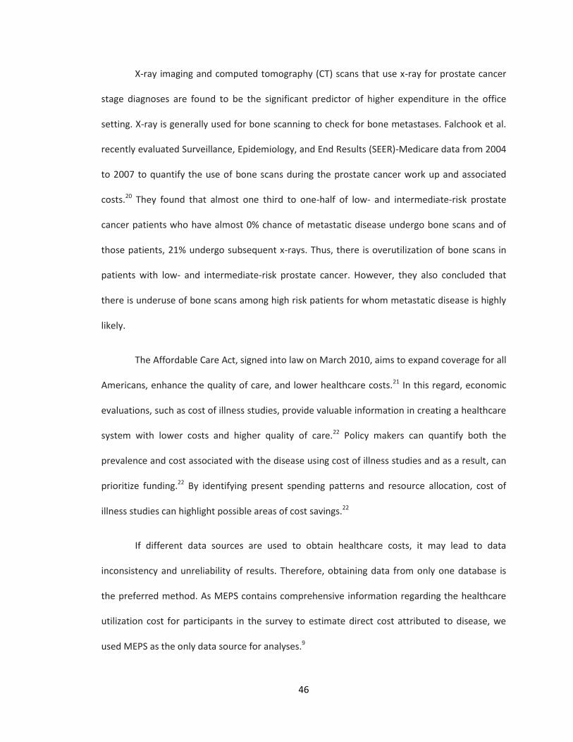
46
X-ray imaging and computed tomography (CT) scans that use x-ray for prostate cancer
stage diagnoses are found to be the significant predictor of higher expenditure in the office
setting. X-ray is generally used for bone scanning to check for bone metastases. Falchook et al.
recently evaluated Surveillance, Epidemiology, and End Results (SEER)-Medicare data from 2004
to 2007 to quantify the use of bone scans during the prostate cancer work up and associated
costs.20 They found that almost one third to one-half of low- and intermediate-risk prostate
cancer patients who have almost 0% chance of metastatic disease undergo bone scans and of
those patients, 21% undergo subsequent x-rays. Thus, there is overutilization of bone scans in
patients with low- and intermediate-risk prostate cancer. However, they also concluded that
there is underuse of bone scans among high risk patients for whom metastatic disease is highly
likely.
The Affordable Care Act, signed into law on March 2010, aims to expand coverage for all
Americans, enhance the quality of care, and lower healthcare costs.21 In this regard, economic
evaluations, such as cost of illness studies, provide valuable information in creating a healthcare
system with lower costs and higher quality of care.22 Policy makers can quantify both the
prevalence and cost associated with the disease using cost of illness studies and as a result, can
prioritize funding.22 By identifying present spending patterns and resource allocation, cost of
illness studies can highlight possible areas of cost savings.22
If different data sources are used to obtain healthcare costs, it may lead to data
inconsistency and unreliability of results. Therefore, obtaining data from only one database is
the preferred method. As MEPS contains comprehensive information regarding the healthcare
utilization cost for participants in the survey to estimate direct cost attributed to disease, we
used MEPS as the only data source for analyses.9

47
The confidence in the current findings is also derived from the adaptation of a robust
statistical analysis technique. In modeling healthcare cost data, bias was reduced in estimates by
employing statistical techniques that considered the skewed nature of such data.
Limitations
This study has several limitations. Most importantly, our findings might have under-
estimated the direct medical expenditures associated with prostate cancer. Patients with
prostate cancer conditions were identified as those who reported being diagnosed with these
conditions. So prevalence of prostate cancer might have been under-reported in MEPS. As such,
our findings should be interpreted with caution. Results from this study, due to potential under-
representation of prostate cancer in MEPS, should be interpreted as a conservative estimate of
the total direct medical costs of prostate cancer related conditions. Some covariates such as
history and severity of illness were not included in the analysis, due to unavailability of this
information in MEPS. We have studied the direct medical costs of prostate cancer related
conditions. A more comprehensive study capturing indirect costs such as morbidity and
mortality costs, as well as direct medical costs, would provide a more precise estimate of the
economic burden of these conditions. We could not study the predictors of inpatient service
cost due to the limited number of patients in our sample who had overnight hospitalization. The
household component of MEPS consists of non-institutionalized, community-dwelling residents.
Prostate cancer patients living in supported living facilities, nursing homes, institutions, and
prisons were not included. Homeless people and undocumented immigrants were also not
included. Thus, the studied population is not representative of all prostate cancer patients in the
U.S. We accounted for prostate-related costs only but it is sometimes difficult to separate

48
prostate-related and unrelated costs, especially at later time periods in the disease and in those
patients with multiple comorbidities.
Conclusion
The current study demonstrates conclusively that prostate cancer conditions, cost
around $ 5.6 billion annually in 2010 US dollars. In effectively reducing the economic burden of a
disease, one needs to know the overall cost of illness, as well as the distribution of costs in
different health delivery settings. The latter provides more insight as to which category of
healthcare services requires more costs, and therefore would benefit the most from disease
management programs. Our results revealed that office-based visits account for the highest
proportion of the overall cost of prostate cancer followed by outpatient costs. Further,
chemotherapy and ultrasound are the main drivers of increased outpatient setting costs,
whereas ultrasound and x-ray are responsible for increased office based costs. Our cost
estimates may also be used by healthcare decision makers in developing disease management
strategies.

49
TABLES
Table 3.1: Descriptive statistics of weighted sample of patients with prostate cancer
Variable Mean (SE)*
Age (years) 69.9 (±0.82)
45-65 (%) 29.3 (±5.16)
65-75 (%) 40.1 (±5.17)
75-90 (%) 30.6 (±4.79)
Sample with private insurance (%) 68.7 (±4.54)
Race
White (%) 84.5 (±2.85)
Black (%) 13.3 (±2.65)
Others (%) 2.20 (±0.99)
Charlson comorbidity index score 1.99 (±0.01)
Table 3.2: Health care use and costs among patients with prostate cancer
Health care use and costs Mean (SEM) Weighted Total (Std)
Service use
Office-based visits 57.83 (%) (±2.19) 5,179,228 (±211,478)
Outpatient visits 12.7 (%) ( ±1.62) 1,137,294 (± 151,501)
Inpatient visits 1.86 (%) (±0.72) 166,262 (± 64,857)
Prescription 27.62 (%) (±1.94) 2,473,224 (± 179,131)
Expenditure, $
Office-based visits 410.68 (± 73.92) 2,127,010,643 (± 412,287,455)
Outpatient visits 1272.30 (±147.62) 1,446,979,485 (±30,756,549)
Inpatient visits 10502 1,746,053,092
Prescription 110.67 (±20.49) 273,723,923 (± 97,234,123)
Total 3,977.78 (± 1,004.81) 5,657,264,402 (± 1,598,361,118)
50
Table 3.3: Predictors of prostate cancer related total health care costs (R2=0.1233, F=49.22, p value <0.001)
Parameters Estimate Antilog(estimate) Standard error
T statistics
P value
Intercept 8.751 6317 1.65 5.32 < 0.0001*
Age -0.037 0.96 0.02 -1.62 0.1101
Type of service
Prescription 0.965 2.62 0.5 1.92 0.0582
Outpatient 1.511 4.53 0.51 2.96 0.0042*
Inpatient 1.607 4.99 0.55 2.91 0.0047*
Office visit
* indicates significant at alpha of 0.05; smearing factor=4.11; Cost data converted to log(cost); office visit was used as a reference for type of services.
Table 3.4: Predictors of outpatient visit costs associated with prostate cancer (R2=0.2424, F=58.60, p value <0.0001)
Parameters Estimate Antilog(estimate) Standard error
T statistics
P value
Intercept 5.7 298.87 0.33 17.3 < 0.0001*
Chemotherapy 1.12 3.06 0.27 4.12 0.0004*
Ultrasound 2.84 17.11 0.38 7.47 <0.0001*
Xray -1.73 -0.177 0.48 -3.56 0.0016*
Labtests -0.98 -0.375 0.64 -1.54 0.1375
* indicates significant at alpha of 0.05; smearing estimator=3.92; cost is in log(cost) form
Table 3.5: Predictors of office-based visit costs associated with prostate cancer (R2=0.1208, F=39.37, p value <0.0001)
Parameters Estimate Antilog(estimate) Standard error T statistics
P value
Intercept 7.78 2392.27 1.3 6 < 0.0001*
Age -0.04 0.96 0.02 -2.2 0.03*
Ultrasound 2.02 7.54 0.48 4.22 <0.0001*
Xray 1.31 3.71 0.23 5.77 <0.0001*
* indicates significant at alpha of 0.05; smearing estimator=3.05; cost is in log(cost) form
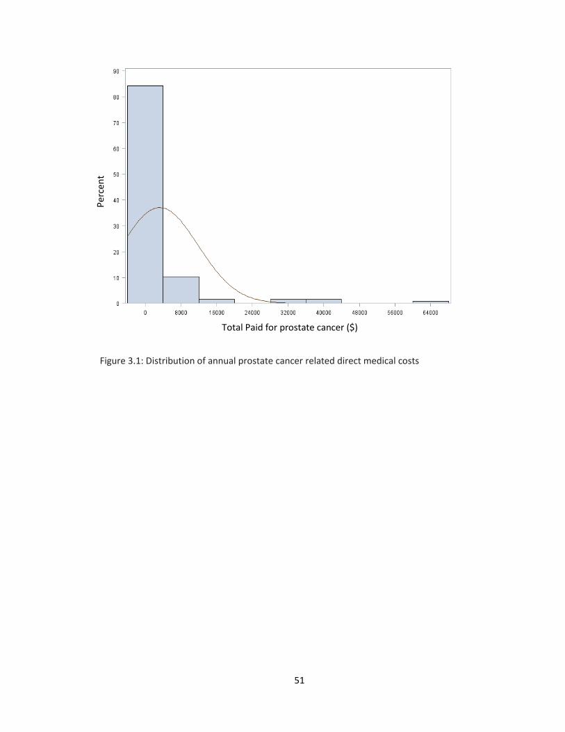
51
Figure 3.1: Distribution of annual prostate cancer related direct medical costs
Total Paid for prostate cancer ($)
Per
cen
t

52
REFERENCES
1. Prostate cancer. 2013. 2014, at
http://www.cancer.org/cancer/prostatecancer/detailedguide/prostate-cancer-key-
statistics.)
2. Crawford ED, Black L, Eaddy M, Kruep EJ. A retrospective analysis illustrating the substantial
clinical and economic burden of prostate cancer. Prostate Cancer and Prostatic Diseases
2010;13:162-7.
3. Roehrborn CG, Black LK. The economic burden of prostate cancer. BJU International
2011;108:806-13.
4. National Cancer Institute. (Accessed December, 4th, 2013, at
http://www.cancer.gov/cancertopics/pdq/treatment/prostate/healthprofessional.)
5. Stokes ME, Black L, Benedict A, Roehrborn CG, Albertsen P. Long-term medical-care costs
related to prostate cancer: Estimates from linked SEER-Medicare data. Prostate Cancer and
Prostatic Diseases 2010;13:278-84.
6. Stokes ME, Ishak J, Proskorovsky I, Black LK, Huang Y. Lifetime economic burden of prostate
cancer. BMC Health Services Research 2011;11:349.
7. National Cancer Institute. SEER Stat Fact Sheets: Prostate Cancer.Retrieved in 2014 from
http://seer.cancer.gov/statfacts/html/prost.html
8. Voigt JD, Zappala SM, Vaughan ED, Wein AJ. The Kallikrein Panel for prostate cancer
screening: Its economic impact. The Prostate 2014;74:250-9.
9. Agency for Healthcare Research and Quality. Medical Expenditure Panel Survey. 2009.
10. Charlson ME, Pompei P, Ales KL, MacKenzie CR. A new method of classifying prognostic
comorbidity in longitudinal studies: Development and validation. Journal of Chronic
Diseases 1987;40:373-83.
53
11. Trogdon JG, Finkelstein EA, Hoerger TJ. Use of econometric models to estimate expenditure
shares. Health Services Research 2008;43:1442-52.
12. Wilson LS, Tesoro R, Elkin EP, et al. Cumulative cost pattern comparison of prostate cancer
treatments. Cancer 2007;109:518-27.
13. Shariat SF, Roehrborn CG. Using biopsy to detect prostate cancer. Review of Urology
2008;10:262-80.
14. Rais-Bahrami S, Siddiqui MM, Turkbey B, et al. Utility of multiparametric magnetic
resonance imaging suspicion levels for detecting prostate cancer. The Journal of Urology
2013;190:1721-7.
15. Krahn MD, Zagorski B, Laporte A, et al. Healthcare costs associated with prostate cancer:
Estimates from a population-based study. BJU International 2010;105:338-46.
16. Warren JL, Yabroff KR, Meekins A, Topor M, Lamont EB, Brown ML. Evaluation of trends in
the cost of initial cancer treatment. Journal of National Cancer Institute 2008;100:888-97.
17. Chemotherapy for prostate cancer. 2014. at
http://www.cancer.org/cancer/prostatecancer/detailedguide/prostate-cancer-treating-
chemotherapy.)
18. Paller CJ, Antonarakis ES. Cabazitaxel: A novel second-line treatment for metastatic
castration-resistant prostate cancer. Drug Design, Development and Therapy 2011;5:117-
24.
19. FDA News & Events. U.S. Food and Drug Administration. Retrieved in 2014 from
http://www.fda.gov/NewsEvents/.
20. Falchook AD, Salloum RG, Hendrix LH, Chen RC. Use of bone scan during initial prostate
cancer workup, downstream procedures, and associated medicare costs. International
Journal of Radiation Oncology,Biology,Physics 2013; 89:243-48

54
21. Affordable Care Act. Medicaid. Retrieved in 2014 from
http://www.medicaid.gov/AffordableCareAct/Affordable-Care-Act.html.
22. Cost-of-Illness Studies: A Primer. RTI International, 2006. 2014, at
http://www.rti.org/publications/abstract.cfm?pubid=6042.)

55
CHAPTER 4
SYSTERMATIC REVIEW ON ROLE OF MAGNETIC RESONANCE IMAGING IN PROSTATE CANCER
DIAGNOSIS
____________________________
1Patel P, Perri M, Griffin S, Tackett R. To be submitted to Magnetic Resonance Imaging.

56
Prostate cancer is the most frequently diagnosed and the second leading cause of non-
cutaneous cancer related deaths among men in the US. Routinely used prostate cancer
screening and diagnostic tests are associated with low sensitivity and specificity. As a result,
these tests are more likely to identify smaller and more indolent tumors compared to larger and
more aggressive ones among patients having localized disease. An imaging technology is needed
that can accurately detect and differentiate clinical significant tumors from nonsignificant
tumors. Multiparametric magnetic resonance imaging (MP-MRI), a promising technology has
been proposed to be used before biopsy to make an accurate diagnosis. This article reviews the
current clinical role of MP-MRI and its potential role in the detection of prostate cancer using a
systematic literature research. Results of this study suggest that MP-MRI has the potential to
detect clinically significant disease while avoiding detection of clinically insignificant disease. In
prebiopsy settings, MP-MRI is expected to cause financial and physical burdens. However,
prebiopsy MRI is also reported to yield possible savings from fewer biopsies, the avoidance of
unnecessary biopsies and treatment, and better risk stratification. Performance of cost-
effectiveness analysis of prebiopsy use of MP-MRI is absolutely needed.
Key words: Multiparametric magnetic resonance imaging, Prostate specific antigen, Digital rectal
examination, Transrectal ultrasound guided biopsy, Prostate cancer, magnetic resonance
spectroscopy, dynamic contrast enhanced imaging, diffusion weighted imaging, MRI-TRUS
fusion, cost-effectiveness
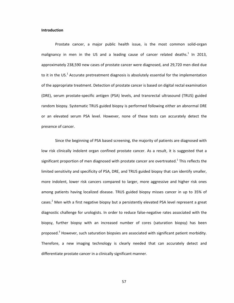
57
Introduction
Prostate cancer, a major public health issue, is the most common solid-organ
malignancy in men in the US and a leading cause of cancer related deaths.1 In 2013,
approximately 238,590 new cases of prostate cancer were diagnosed, and 29,720 men died due
to it in the US.1 Accurate pretreatment diagnosis is absolutely essential for the implementation
of the appropriate treatment. Detection of prostate cancer is based on digital rectal examination
(DRE), serum prostate-specific antigen (PSA) levels, and transrectal ultrasound (TRUS) guided
random biopsy. Systematic TRUS guided biopsy is performed following either an abnormal DRE
or an elevated serum PSA level. However, none of these tests can accurately detect the
presence of cancer.
Since the beginning of PSA based screening, the majority of patients are diagnosed with
low risk clinically indolent organ confined prostate cancer. As a result, it is suggested that a
significant proportion of men diagnosed with prostate cancer are overtreated.1 This reflects the
limited sensitivity and specificity of PSA, DRE, and TRUS guided biopsy that can identify smaller,
more indolent, lower risk cancers compared to larger, more aggressive and higher risk ones
among patients having localized disease. TRUS guided biopsy misses cancer in up to 35% of
cases.2 Men with a first negative biopsy but a persistently elevated PSA level represent a great
diagnostic challenge for urologists. In order to reduce false-negative rates associated with the
biopsy, further biopsy with an increased number of cores (saturation biopsy) has been
proposed.3 However, such saturation biopsies are associated with significant patient morbidity.
Therefore, a new imaging technology is clearly needed that can accurately detect and
differentiate prostate cancer in a clinically significant manner.
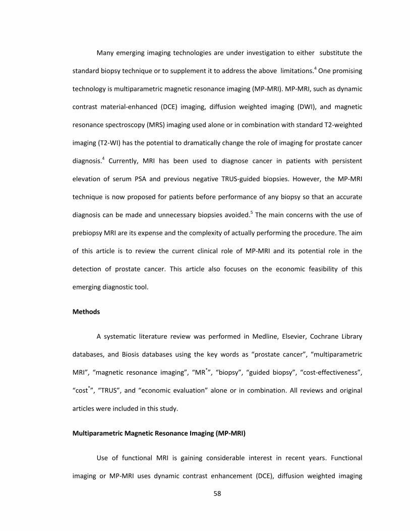
58
Many emerging imaging technologies are under investigation to either substitute the
standard biopsy technique or to supplement it to address the above limitations.4 One promising
technology is multiparametric magnetic resonance imaging (MP-MRI). MP-MRI, such as dynamic
contrast material-enhanced (DCE) imaging, diffusion weighted imaging (DWI), and magnetic
resonance spectroscopy (MRS) imaging used alone or in combination with standard T2-weighted
imaging (T2-WI) has the potential to dramatically change the role of imaging for prostate cancer
diagnosis.4 Currently, MRI has been used to diagnose cancer in patients with persistent
elevation of serum PSA and previous negative TRUS-guided biopsies. However, the MP-MRI
technique is now proposed for patients before performance of any biopsy so that an accurate
diagnosis can be made and unnecessary biopsies avoided.5 The main concerns with the use of
prebiopsy MRI are its expense and the complexity of actually performing the procedure. The aim
of this article is to review the current clinical role of MP-MRI and its potential role in the
detection of prostate cancer. This article also focuses on the economic feasibility of this
emerging diagnostic tool.
Methods
A systematic literature review was performed in Medline, Elsevier, Cochrane Library
databases, and Biosis databases using the key words as “prostate cancer”, “multiparametric
MRI”, “magnetic resonance imaging”, “MR*”, “biopsy”, “guided biopsy”, “cost-effectiveness”,
“cost*”, “TRUS”, and “economic evaluation” alone or in combination. All reviews and original
articles were included in this study.
Multiparametric Magnetic Resonance Imaging (MP-MRI)
Use of functional MRI is gaining considerable interest in recent years. Functional
imaging or MP-MRI uses dynamic contrast enhancement (DCE), diffusion weighted imaging
59
(DWI) or magnetic resonance spectroscopy (MRS) together with T2-weighted imaging (T2-WI) in
one or more combinations.4 MRI technology has undergone a significant advancement and more
consistent and accurate results have been reported with its use.6-13 The capability of combining
MRI with techniques to simultaneously perform a targeted biopsy of the prostate is of particular
interest to urologists. MP-MRI used in conjunction with a MRI-ultrasound fusion guided biopsy
platform has demonstrated improved prostate cancer detection and localization.1,14
MRI has an unparalleled ability to provide detailed information about the prostate due
to excellent soft tissue contrast.8 Conventional MRI at 1.5 or 3.0 Tesla (T) provides
morphological information such as the prostate’s zonal anatomy, seminal vesicles and the
prostatic capsule using T2-WI.15-17 T1-weighted imaging (T1-WI) has been used to detect post-
biopsy hemorrhage, lymph nodes, and bone metastasis.4 Conventionally, MRI has been used in
clinical practice for determining prostate cancer stage4 and has emerged as a promising tool in
diagnosing prostate cancer. MRI detects the location of more aggressive lesions on imaging that
cannot be accessed through even with extended biopsy schemes.8 MP-MRI is capable of
detecting metabolic, diffusion and perfusion abnormalities associated with the cancer.8,18-22
Proton MRS imaging provides metabolic information, DWI shows Brownian motion of
extracellular water molecules, and DCE-MRI visualizes tissue vascularity, especially
neoangiogenesis. Thus, efficient cancer identification with high sensitivity and specificity can be
made by combining morphologic and functional imaging.7
Magnetic Resonance Spectroscopy (MRS) based Imaging
MRS imaging also known as chemical shift imaging assesses the level of metabolites in
tissue.1,4 The metabolites measured by MRS include citrate (reflecting glandular composition) 8,
creatine and choline (a composite of phospholipid membranes).17 Generally, prostate cancer
60
shows a high level of choline and a low level of citrate relative to the normal peripheral zone.
An increase in the choline-to-citrate ratio is used as a marker in prostate cancer and increases
the specificity of diagnosis. Scheidler et al. showed a sensitivity and specificity for prostate
cancer detection of 95% and 91%, respectively, for combined MRS and MRI, but 61% to 77% and
46% to 81% for MRI alone and 75% and 63% for MRS alone.4 MRS together with MRI has been
found to significantly improve localization of prostate cancer.4
MRS suffers from long acquisition time, possible variability in results dependent on post-
processing or shimming, no direct visualization of the periprostatic anatomy and its expense.4
Some of these limitations of MRSI might be improved by new technical developments and the
use of higher magnetic fields (3.0 T).
Dynamic Contrast Material-Enhanced (DCE) based Imaging
Cancerous tissue is generally marked by an increased number of vessels as well as
greater vascular permeability. DCE MRI measures the passing of an intravenously administered
contrast agent through prostate on T1-weighted imaging.4,7,8 Cancerous tissues show higher
contrast enhancement parameters, such as mean transit time, blood flow, permeability surface
area and interstitial volume compared to normal tissue, and therefore allow differentiation
between benign and malignant tissue.
Several studies comparing the results of DCE-MRI with surgical pathology as the
reference standard have reported sensitivity, specificity, and accuracy levels ranging from 69%
to 95%, from 80% to 96.2%, and from 77.5% to 92%, respectively.4 Addition of DCE-MRI in
conjunction with T2-WI of MRI has been shown to increase sensitivity from 69% to 95% and
specificity from 80% to 93%.4 Moreover, combined DCE-MRI and MRSI significantly improve the

61
accuracy in prostate cancer localization. The limitation of DCE MRI is primarily an unsatisfactory
depiction of transitional zone cancer in patients with hypervascular BPH.4
Diffusion-Weighted Imaging (DWI)
The amount of interstitial free water and permeability determines the diffusion
properties of the tissues. DWI provides information on the functional environment of water in
tissue by measuring the Brownian motion or diffusion coefficient of water molecules.7,8 Thus,
based on the motion of water molecules, differentiation between malignant and normal tissues
can be made. Cancer tissues have higher cell densities and extracellular disorganization.
Therefore, cancer tissues tend to have more restricted water diffusion than normal tissue.
Without administration of any exogenous contrast material, these images can be acquired
quickly.4
Addition of DWI to conventional T2-WI imaging significantly improves tumor detection
compared to conventional MRI alone. Combined DWI with MRI has been shown to have
sensitivity, specificity, positive predictive value, and negative predictive value of 86%, 84%, 90%
and 79%, respectively.4 In addition to detection of cancer, DWI has also been shown to provide
information about tumor aggressiveness with good correlation between diffusion coefficient
values and the Gleason score.17 The main strengths of DWI are shorter acquisition time and high
contrast resolution between tumors and normal tissue.4 However, this technique suffers from
poor spatial resolution and susceptibility-induced distortions.4
Imaging algorithm suggested in patients:
If hemorrhage is present in the patient, DWI should be included in the MR diagnosis.7
With post hemorrhage detection of prostate cancer, MRS together with DCE is recommended.
62
Use of 3.0 T strength and an endorectal coil is preferred in general but use of 3.0 T without a
endorectal coil is an option among patients who do not prefer to use a endorectal coil.7 For
patients with a low glomerular filtration rate, DCE should be avoided. For assessment of
transition zone cancer in patients with advanced age, DWI is recommended.7
Prebiopsy MRI for Men at Risk
Application of verification tests such as an imaging test earlier in the diagnostic pathway
has the potential of improving the diagnosis of prostate cancer diagnosis. This practice is already
adopted by clinicians for treating other solid organ cancers such as breast cancer.5 MP-MRI
cannot detect clinically insignificant disease because of its low sensitivity for low grade, low-
volume disease.10 However, it has a greater sensitivity for detecting clinically significant disease.
Thus, it has the potential to address the problem of overdiagnosis and overtreatment of
clinically insignificant disease if used as a triage test before TRUS guided biopsy.
Overdiagnosis and overtreatment of clinically insignificant disease is the main reason
that the United States Preventive Services Task Force (USPSTF) recommended against the use of
PSA screening.10 Kasivisvanathan et al. found that multiparametric MRI detects clinically
significant cancer at an encouraging rate while also reduces the detection rates of clinically
insignificant cancer.17 They further reported that the MP-MRI requires fewer biopsy specimens
than systematic template guided biopsy. MP-MRI also has the potential to address the problem
of underdiagnosis of clinically significant disease with the current diagnostic practice when
cancer is located in the transition zone or in the anterior or peripheral zone, which are parts of
the prostate that are not easily palpable by DRE and are not routinely targeted during
biopsy.3,5,8,10,16,23-25
63
Pinto et al reported MP-MRI outcomes on patients with low, moderate or high suspicion
lesions that were subsequently targeted via an MRI/ultrasound fusion biopsy platform.4 They
found prostate cancer detection rates to be 27%, 66% and 89% for low, moderate and high
suspicion lesions, respectively. With MP-MRI, more accurate classification of patients into
observational strategies such as active surveillance can be made with greater
confidence.5,8,10,16,21,26,27 Further, if upgrading of disease occurs in patients undergoing active
surveillance, MP-MRI can accurately detect, localize and characterize tumors. Thus, MP-MRI can
act as an accurate monitoring tool for prostate cancer progression in those undergoing active
surveillance.
Techniques used to target biopsies on lesions identified with MP-MRI include10,18,25: (i)
‘cognitive’ registration of the results of the MP-MRI to target biopsies on TRUS, (ii) targeting
within the magnet or ‘in-bore’ targeting, and (iii) registration of magnetic resonance images
onto an ultrasound platform to allow real-time targeting of lesion in out-patient setting.
‘Cognitive’ registration suffers from poor interpretation of imaging because the physician first
reviews the lesion seen on MRI and then uses this knowledge to select the appropriate area for
targeting biopsy. ‘In- bore’ targeting within the MRI scanner is a time consuming and expensive
approach as it is performed in an inpatient setting. The MRI based image fused onto an
ultrasound platform is a promising approach because of real-time targeting of lesions.
Cost-Effectiveness Analysis of Prebiopsy MRI Use
Cost-effectiveness analysis allows one to understand the probable health outcomes and
fiscal expenditures associated with a particular health policy or program.28 Jager et al.
performed a cost effectiveness study to determine role of preoperative MRI staging.29 They
64
concluded that MR staging was cost-effective for men with moderate or high prior probability of
extracapsular disease.
Recently, Mowatt et al. carried out a systematic review of the literature to assess the
diagnostic accuracy of MP-MRI techniques and the clinical effectiveness and cost-effectiveness
of these techniques in the localization of prostate abnormalities for biopsy in patients with a
prior negative biopsy.17 In their study, they included costs associated with the final diagnosis,
management of biopsy complications, cancer staging, cancer treatment, and management of
complications resulting from cancer treatment. They concluded that T2-MRI may be cost-
effective compared to systematic TRUS under certain circumstances. They further reported that
MRS and DWI could be cost-effective if they can be shown to have a high sensitivity for
detecting moderate to high risk cancer while ignoring disease in patients with no cancer or low
risk disease. They recognized that there were not enough reliable studies so further studies
were recommended. Their main approach focused on those patients having prior negative
biopsy. Further, they did not report any results using the MRI-TRUS fusion platform.
Use of MRI before performing any biopsy can reduce unnecessary biopsies as well as
treatments. The MRI-TRUS fusion approach to guide biopsies is an attractive alternative to MRI
guided biopsy because it significantly reduces the procedure time as well as cost and can be
performed in an outpatient setting.30 A cost-effectiveness study of prebiopsy use of MP-MRI as a
screening test is needed. There is concern about its financial and physical burdens, at the initial
biopsy setting. However, it is expected that the initial biopsy cost of prebiopsy MP-MRI will be
compensated by the possible savings that would result from fewer biopsies, the avoidance of
unnecessary biopsies and treatment, and better risk stratification. Unnecessary biopsies cost
much more than MRI.31 Performance of a cost-effectiveness analysis of prebiopsy use of MP-
65
MRI is absolutely needed in order to relieve concerns of clinicians adopting it as a routine
practice.
Conclusion
Urologists and other physicians who manage patients who may have prostate cancer are
frustrated with the current standards of care, due to their limitations, especially in patients for
whom a first set of biopsies was negative. It is possible to miss cancer because of the limitation
of biopsy schemes, volume of biopsy specimen sampled and the multifocal nature of prostate
cancer. Extended biopsy schemes (8-12 cores) or saturation biopsy schemes (greater than12
cores) have been proposed to solve this problem. This trend has led to performance of many
unnecessary biopsies and treatments as well as a financial burden on patients. It also creates
emotional stress for patients who may be facing a series of repeat biopsies. There is a risk of the
diagnosis being delayed, possibly leading to disease progression, and increased morbidity due to
the biopsy as well as the disease itself.17 Many older men who present for biopsy are taking
anticoagulants or antiplatelet drugs.32 In this group of patients, biopsy complications could
become life threatening. The number of prostate biopsies should be limited to reduce potential
and serious complications. A promising imaging technology is needed that can detect prostate
cancer accurately at a lower cost. MP-MRI used in conjunction with a MR/US fusion guided
biopsy platform has demonstrated improved prostate cancer detection and localization. MP-
MRI has a negative predictive value ranging from 90 to 100% in identifying significant cancer.
Thus, it can be used as a screening tool to avoid biopsy if it does not reveal any abnormalities on
MR images. Its cost-effectiveness in prebiopsy setting has not been evaluated yet.
In the prebiopsy setting, MP-MRI can diagnose lesions that cannot be diagnosed with
even an extended systematic biopsy scheme. Additionally, it can improve targeting of biopsy for

66
more precise tumor characterization such as size and histological grade of the cancer. MP-MRI
represents a reasonable approach alternative to extended systematic biopsies to relieve
tremendous psychological stresses in a group of patients by improving the accuracy in
diagnosing significant prostate cancer.

67
REFERENCES
1. Rais-Bahrami S, Siddiqui MM, Turkbey B, et al. Utility of Multiparametric MRI Suspicion
Levels in detecting prostate cancer. The Journal of Urology 2013;29:04417-0.
2. Roethke M, Anastasiadis AG, Lichy M, et al. MRI-guided prostate biopsy detects clinically
significant cancer: Analysis of a cohort of 100 patients after previous negative TRUS
biopsy. World Journal of Urology 2012;30:213-8.
3. Sciarra A, Barentsz J, Bjartell A, et al. Advances in magnetic resonance imaging: How
they are changing the management of prostate cancer. European Urology 2011;59:962-
77.
4. Pinto F, Totaro A, Calarco A, et al. Imaging in prostate cancer diagnosis: Present role and
future perspectives. Urologia Internationalis 2011;86:373-82.
5. Villers A, Marliere F, Ouzzane A, Puech P, Lemaitre L. MRI in addition to or as a
substitute for prostate biopsy: The clinician's point of view. Diagnostic and
Interventional Imaging 2012;93:262-7.
6. Perdona S, Di Lorenzo G, Autorino R, et al. Combined magnetic resonance spectroscopy
and dynamic contrast-enhanced imaging for prostate cancer detection. Urologic
Oncology 2013;31:761-5.
7. Bonekamp D, Jacobs MA, El-Khouli R, Stoianovici D, Macura KJ. Advancements in MR
imaging of the prostate: From diagnosis to interventions. Radiographics 2011;31:677-
703.
8. Dianat SS, Carter HB, Macura KJ. Performance of multiparametric magnetic resonance
imaging in the evaluation and management of clinically low-risk prostate cancer.
Urologic Oncology 2013;17:002.
9. Carey BM. Imaging for prostate cancer. Clinical Oncology 2005;17:553-9.
68
10. Dickinson L, Ahmed HU, Allen C, et al. Clinical applications of multiparametric MRI
within the prostate cancer diagnostic pathway. Urologic Oncology 2013;31:281-4.
11. Goris Gbenou MC, Peltier A, Addla SK, et al. Localising prostate cancer: Comparison of
endorectal magnetic resonance (MR) imaging and 3D-MR spectroscopic imaging with
transrectal ultrasound-guided biopsy. Urologia Internationalis 2012;88:12-7.
12. Hambrock T, Hoeks C, Hulsbergen-van de Kaa C, et al. Prospective assessment of
prostate cancer aggressiveness using 3-T diffusion-weighted magnetic resonance
imaging-guided biopsies versus a systematic 10-core transrectal ultrasound prostate
biopsy cohort. European Urology 2012;61:177-84.
13. Nix JW, Turkbey B, Hoang A, et al. Very distal apical prostate tumours: Identification on
multiparametric MRI at 3 Tesla. BJU International 2012;110:4.
14. Fiard G, Hohn N, Descotes JL, Rambeaud JJ, Troccaz J, Long JA. Targeted MRI-guided
prostate biopsies for the detection of prostate cancer: Initial clinical experience with
real-time 3-dimensional transrectal ultrasound guidance and magnetic
resonance/transrectal ultrasound image fusion. Urology 2013;81:1372-8.
15. Kaplan I, Oldenburg NE, Meskell P, Blake M, Church P, Holupka EJ. Real time MRI-
ultrasound image guided stereotactic prostate biopsy. Magnetic Resonance Imaging
2002;20:295-9.
16. Puech P, Sufana Iancu A, Renard B, Villers A, Lemaitre L. Detecting prostate cancer with
MRI - why and how. Diagnostic and Interventional Imaging 2012;93:268-78.
17. Mowatt G, Scotland G, Boachie C, et al. The diagnostic accuracy and cost-effectiveness
of magnetic resonance spectroscopy and enhanced magnetic resonance imaging
techniques in aiding the localisation of prostate abnormalities for biopsy: A systematic
review and economic evaluation. Health Technology Assessment 2013;17:1-281.

69
18. Moore CM, Robertson NL, Arsanious N, et al. Image-guided prostate biopsy using
magnetic resonance imaging-derived targets: A systematic review. European Urology
2013;63:125-40.
19. Park BK, Lee HM, Kim CK, Choi HY, Park JW. Lesion localization in patients with a
previous negative transrectal ultrasound biopsy and persistently elevated prostate
specific antigen level using diffusion-weighted imaging at three Tesla before rebiopsy.
Investigative Radiology 2008;43:789-93.
20. Hara N, Okuizumi M, Koike H, Kawaguchi M, Bilim V. Dynamic contrast-enhanced
magnetic resonance imaging (DCE-MRI) is a useful modality for the precise detection
and staging of early prostate cancer. Prostate 2005;62:140-7.
21. Nagarajan R, Margolis D, Raman S, et al. MR spectroscopic imaging and diffusion-
weighted imaging of prostate cancer with Gleason scores. Journal of Magnetic
Resonance Imaging 2012;36:697-703.
22. Sciarra A, Panebianco V, Ciccariello M, et al. Value of magnetic resonance spectroscopy
imaging and dynamic contrast-enhanced imaging for detecting prostate cancer foci in
men with prior negative biopsy. Clinical Cancer Research 2010;16:1875-83.
23. Komai Y, Numao N, Yoshida S, et al. High diagnostic ability of multiparametric magnetic
resonance imaging to detect anterior prostate cancer missed by transrectal 12-Core
biopsy. The Journal of Urology 2013;28:03871-
24. Haffner J, Lemaitre L, Puech P, et al. Role of magnetic resonance imaging before initial
biopsy: Comparison of magnetic resonance imaging-targeted and systematic biopsy for
significant prostate cancer detection. BJU International 2011;108:22.
70
25. Ouzzane A, Puech P, Lemaitre L, et al. Combined multiparametric MRI and targeted
biopsies improve anterior prostate cancer detection, staging, and grading. Urology
2011;78:1356-62.
26. Kuru TH, Roethke MC, Seidenader J, et al. Critical evaluation of magnetic resonance
imaging targeted, transrectal ultrasound guided transperineal fusion biopsy for
detection of prostate cancer. The Journal of Urology 2013;19:04093-7.
27. Kirkham AP, Emberton M, Allen C. How good is MRI at detecting and characterising
cancer within the prostate? European Urology 2006;50:1163-74.
28. Saigal CS, Litwin MS. The economic costs of early stage prostate cancer.
Pharmacoeconomics 2002;20:869-78.
29. Jager GJ, Severens JL, Thornbury JR, de la Rosette JJMCH, Ruijs SHJ, Barentsz JO.
Prostate cancer staging: Should MR imaging be used?—A Decision analytic approach.
Radiology 2000;215:445-51.
30. Sonn GA, Natarajan S, Margolis DJA, et al. Targeted biopsy in the detection of prostate
cancer using an office based magnetic resonance ultrasound fusion device. The Journal
of Urology 2013;189:86-92.
31. Park BK, Park JW, Park SY, et al. Prospective evaluation of 3-T MRI performed before
initial transrectal ultrasound-guided prostate biopsy in patients with high prostate-
specific antigen and no previous biopsy. American Journal of Roentgenology 2011;197.
32. Philip J, Manikandan R, Javle P, Foster CS. Prostate cancer diagnosis: Should patients
with prostate specific antigen >10ng/mL have stratified prostate biopsy protocols?
Cancer Detection and Prevention 2009;32:314-8.

71
CHAPTER 5
COST-EFFECTIVENESS OF MULTIPARAMETRIC MAGNETIC RESONANCE IMAGING FOLLOWED BY
FUSION GUIDED BIOPSY IN PROSTATE CANCER DIAGNOSIS: AN ECONOMIC ANALYSIS
___________________________
1Patel P, Perri M, Griffin S, Tackett R. To be submitted to Journal of the National Comprehensive
Cancer Network.

72
Purpose: The purpose of this study was to determine the cost-effectiveness of multiparametric
magnetic resonance imaging (MP-MRI) followed by MRI/Transrectal ultrasound (TRUS) fusion
guided biopsy compared with the standard TRUS guided biopsy among biopsy naïve men in
diagnosing prostate cancer. Methods: A decision analytic model was developed to evaluate the
costs and outcomes for conventional TRUS guided biopsy and experimental MP-MRI followed by
MRI/TRUS guided biopsy. Input data for the model such as sensitivity, specificity, and risks of
complications associated with diagnostic strategies were derived through a systematic literature
search. Costs associated with diagnostic strategies were based on 2013 Medicare
reimbursement rates. To test the robustness of the results in model parameters, n-way
sensitivity analyses were conducted by varying values of different parameters such as the cost
of MP-MRI and MRI/TRUS fusion guided biopsy, positive predictive value of TRUS guided biopsy,
positive and negative predictive values of MRI/TRUS fusion guided biopsy. Results: Use of MP-
MRI strategy was found to be more costly than TRUS guided biopsy ($1249.65 vs $860.05);
however, it was more effective (no. of correct diagnoses 0.90 vs 0.68). The MP-MRI strategy was
found to be cost-effective even at a threshold of $10,000. Conclusion: MRI assisted TRUS fusion
guided biopsy was found to be cost-effective compared with conventional 12-core TRUS guided
biopsy in symptomatic patients with suspected prostate cancer. As MRI/TRUS fusion is a new
strategy, there is little data on its effectiveness. Further studies demonstrating the effectiveness
of MRI/TRUS fusion are required.
Keywords: Multiparametric magnetic resonance imaging, Transrectal ultrasound, biopsy, cost-
effectiveness
73
Introduction
Prostate cancer is a commonly diagnosed malignancy in men in the United States.1
Despite its very high incidence, a small percentage of men will die from the disease.2 Six to 12-
core transrectal ultrasound (TRUS) guided biopsy has been the standard technique for
diagnosing prostate cancer followed by elevated prostate specific antigen (PSA) and/or digital
rectal examination (DRE).3 Although TRUS biopsy is simple, relatively easy, and cheaper, it is
limited by its poor sensitivity.4,5 As a result, many patients undergo additional biopsies. Most of
the biopsies are well tolerated but some may experience complications such as hematuria,
hematospermia, rectal bleeding and urinary retention.6 Rarely, infectious complications can also
result.6 Further, TRUS guided biopsy detects many clinically insignificant cancers.7 With the
increased recognition of over-diagnosis and overtreatment, prostate cancer has substantial
psychological and economic impacts on society.8 There is a need for new diagnostic strategies
that can identify patients with clinically significant cancer.
Multiparametric magnetic resonance imaging (MP-MRI) is one of the promising
modalities for prostate imaging. MP-MRI involves the use of different MRI based techniques
such as T1-weighted, T2-weighted, diffusion weighted imaging, dynamic contrast enhanced
imaging, and/or magnetic resonance spectroscopy in one or more combinations.2 MP-MRI can
easily detect lesions in areas that are poorly sampled by the TRUS biopsy.9 Targeted biopsy of
suspicious areas identified through MP-MRI has the potential to improve diagnosis. MRI guided
biopsy can be performed either ‘in-bore’ or ‘out-of-bore’.2 The ‘in-bore’ or direct MRI guided
biopsy is time consuming and expensive.2 The ‘out-of-bore’ procedure involves MRI/TRUS fusion
guided biopsy where patients first undergo prebiopsy MP-MRI and then images obtained are
fused with real-time TRUS to guide biopsy.2 As MRI/TRUS fusion guided biopsy is performed in
an outpatient setting, it is expected to cost less compared to the direct MRI guided biopsy.
74
One concern regarding MP-MRI followed by MRI/TRUS fusion guided biopsy in biopsy
naïve patients is the cost effectiveness compared to the conventional less expensive TRUS
guided biopsy. There is a clinical trial in progress to determine whether MRI/TRUS fusion guided
biopsy is superior to conventional biopsy alone in diagnosing subjects with prostate cancer.10
However, the results of this study will not be available until April 2015. Further, this trial does
not mention that whether it aims to determine the cost-effectiveness of MRI/TRUS fusion
guided biopsy or not. The objective of this study is to determine the cost-effectiveness of the
MP-MRI followed by MRI/TRUS fusion guided biopsy compared with the standard TRUS guided
biopsy among biopsy naïve men in diagnosing prostate cancer.
Methods
We developed a decision analytic model to evaluate the costs and outcomes for
conventional TRUS guided biopsy and experimental MP-MRI followed by MRI/TRUS guided
biopsy. Strategies were compared based on costs and number of correct diagnoses, false-
positive results avoided, and false-negative results avoided among patients with an elevated
PSA (>4 ng/mL) and/or abnormal DRE. The model was a decision tree. Based on clinical
guidelines, a typical clinical setting was created. The first strategy evaluated the current
standard of care, where an elevated serum PSA is followed by systematic 12-core TRUS guided
biopsy. The second strategy was the experimental strategy in which an elevated serum PSA is
followed by MP-MRI including T2-weighted, dynamic contrast-enhanced, diffusion weighted
imaging, and magnetic resonance spectroscopy sequences performed on a 3.0 T MRI scanner.
The second strategy assumed that patients underwent a 5-core targeted biopsy only when a
tumor suspicious area was identified on MP-MRI. The decision tree was not extrapolated to
future outcomes as there is not enough information on the long term outcomes associated with

75
MP-MRI guided biopsy (Figure 5.1) in the literature. Further, it was assumed that if differences
in the initial outcomes between two diagnostic strategies are observed, their future outcomes
can be predicted.
Clinical Model Input Parameters: A systematic literature search was performed in PubMed,
Elsevier, Cochrane Library databases, and Biosis to obtain sensitivity, specificity, and risks of
complications associated with 12-core TRUS guided biopsy, MP-MRI, and MRI/TRUS fusion
guided biopsy (Table 5.1 & Table 5.2).
Cost information: We used a payer’s perspective. Costs associated with diagnostic strategies
were based on 2013 Medicare reimbursement rates. Costs of hospitalization due to biopsy
induced complications were obtained from a study conducted by Adibi et al.11 Table 5.3 outlines
the resources and costs used in the economic model.
The total direct cost per patient for the conventional 12-core TRUS biopsy was
estimated using the following formula:
Cost of TRUS biopsy + cost of histopathological analysis of 12-core + (probability of moderate or
severe post biopsy complications * probability of hospitalization * total cost of hospital
admission). The total direct cost per patient for the MRI/TRUS fusion guided biopsy was
estimated using the following formula:
Cost of MP-MRI + cost of MRI/TRUS fusion + cost of histopathological analysis of 5 cores
obtained through targeted biopsy + (probability of moderate or severe post biopsy
complications * probability of hospitalization * total cost of hospital admission).
Cost-effectiveness analyses were performed using TreeAge Pro Suite 2014. Three
different incremental cost-effectiveness ratios (ICERs) were calculated by dividing the estimated
76
difference in costs by three different outcome measures which include the difference in the
number of false positives avoided, number of false negatives avoided, or number of correct
diagnoses.
Sensitivity Analysis: To test the robustness of the results in model parameters, n-way sensitivity
analyses were conducted. The following model parameters were varied: the cost of MP-MRI
and the MR/TRUS fusion guided biopsy, positive predictive value (PPV) of TRUS guided biopsy,
PPV and negative predictive value (NPV) of MRI/TRUS fusion guided biopsy.
Results
Cost-effectiveness of MP-MRI followed by MR/TRUS fusion guided biopsy compared with TRUS
guided biopsy:
Expected costs and the number of correct diagnoses made using two diagnostic
strategies are presented in Table 5.4. Use of MP-MRI strategy was found to be more costly than
TRUS guided biopsy ($1249.65 vs $860.05); however, it was more effective (number of correct
diagnoses 0.90 vs 0.68). The MP-MRI strategy was found to cost approximately $1781.60 per
one additional correct diagnosis compared to 12-core TRUS guided biopsy.
Table 5.5 shows the number of misdiagnoses such as false positive and false negative
tests avoided for each diagnostic strategy. MRI strategy was found to cost around $2238.69 per
additional false positive case avoided compared to the 12-core TRUS biopsy. The numbers of
false negative cases avoided did not differ much between the two strategies. MRI strategy was
found to cost around $8725.74 per additional false negative case avoided compared to the 12-
core TRUS biopsy.
77
Sensitivity analysis:
A one-way sensitivity analysis was conducted on key model parameters (Figure 5.2 –
Figure 5.5). The sensitivity analysis revealed that the model results were sensitive to the PPVs of
the 12-core TRUS guided biopsy and MRI/TRUS fusion guided biopsy respectively. If the PPV of
12-core TRUS guided biopsy is as high as 0.99, ICER is approximately $18,000 per one correct
diagnosis for the MRI strategy compared with 12-core TRUS guided biopsy (Figure 5.2).
Similarly, if the PPV of MRI/TRUS fusion guided biopsy is as low as 0.53 or below, ICER
per one correct diagnosis is around $13,000 for the MRI strategy compared to the 12-core TRUS
guided biopsy (Figure 5.5). As there are uncertainties regarding cost of MP-MRI and MRI/TRUS
fusion guided biopsy, two way sensitivity analysis was performed varying the cost of MP-MRI
(from $200 to $900) and the cost of MRI/TRUS fusion guided biopsy (from $200 to $700), MRI
strategy was found to be cost-effective at a threshold value of $4,000.
A three-way sensitivity analysis was carried out in which all three parameters were
varied: PPV of 12-core TRUS biopsy, NPV of MP-MRI, and PPV of MRI/TRUS fusion guided biopsy.
Figure 5.6 shows that result with varying values of PPV of 12-core TRUS guided biopsy and NPV
of MP-MRI while holding positive predictive value of MRI/TRUS fusion constant at 0.875. This
result indicates that 12-core TRUS guided biopsy is favored when NPV of MP-MRI is very low
whereas, PPV of 12-core is very high considering PPV of MRI/TRUS fusion guided biopsy is 0.875.
However, the literature suggests that the NPV of MP-MRI is quite high (0.91) and PPV of 12-core
TRUS is not so high (0.59).

78
Discussion
The results of our model suggest that the MRI strategy is cost-effective in diagnosing
prostate cancer compared with the TRUS guided biopsy assuming a threshold to pay per correct
diagnosis is at least $1781.60. In our study, decision analysis was used to compare the cost-
effectiveness of MP-MRI followed by MRI/TRUS fusion guided biopsy with the conventional 12-
core TRUS guided biopsy. As health care expenses are rapidly rising, cost-effectiveness analyses
are increasingly being advocated as techniques to compare alternative strategies to optimize
delivery of healthcare. Due to several limitations associated with these techniques, they should
not be used as the only basis for a decision. In spite of the limitations, they can aid in making
decisions, give structure to the problem, allow consideration of relevant effects of a decision
and also identify key assumptions. Limitations associated with decision tree analysis are
presented in a separate section. Thus, one can use decision-analytic methods and sensitivity
analysis to address issues for which randomized controlled trials have not been performed or
are not feasible.
Approximately 1.3 million prostate biopsies are performed each year in the United
States.12 It has been estimated that only 25% of men who undergo a prostate biopsy due to
elevated PSA actually have prostate cancer indicating that many unnecessary biopsies are
performed.12 Unnecessary biopsies are estimated to cost around $2 billion per year.13 In our
baseline model, assuming MP-MRI to have a sensitivity and specificity of 94% and 28%
respectively14, it can avoid biopsy in 17% of patients who enter the current diagnostic pathway.
Further, MP-MRI followed by MRI/TRUS fusion guided biopsy can avoid 17% of more false
positive cases compared to conventional 12-core TRUS guided biopsy. The cost of unnecessary
treatment among those who have indolent or insignificant disease is estimated to be around
79
$1.6 billion.13 Thus, use of MP-MRI is expected to save costs by avoiding unnecessary biopsies
and reducing treatment costs resulting from fewer false positives and a better estimation of
tumor aggressiveness.
Sensitivity analysis found that our model was sensitive to the PPVs of 12-core TRUS and
MRI/TRUS fusion guided biopsy. With the extended biopsy scheme such as 12-core TRUS guided
biopsy, there is an increased chance of detecting smaller-volume tumors of little clinical
relevance.15 Some studies have reported that a higher number of biopsy cores are associated
with smaller tumor volumes at radical prostatectomy.15 Thus, the risks of detecting insignificant
tumors and missing significant ones should be balanced with the extended biopsy scheme. The
current study used a PPV of 0.59 for 12-core TRUS guided biopsy.16 In order to make 12-core
TRUS guided biopsy more cost-effective at threshold of $4,000, it has to have a PPV higher than
approximately 0.8. Advancement in imaging technology such as MP-MRI has improved the
diagnosis of significant cancer. Fusion of MR images of suspicious lesions with real-time TRUS
biopsy techniques in an office/outpatient setting can guide biopsy needles to be inserted into
suspicious areas. Targeted biopsy using MRI/TRUS fusion has greater ability to detect
intermediate and high risk prostate cancer with greater than 85% positive predictive value.16
Our model suggests that the positive predictive value of MRI/TRUS should be greater than 0.53
to make it cost-effective.
To our knowledge, this is the first study comparing the cost-effectiveness of the
MRI/TRUS fusion strategy with the TRUS guided biopsy in United States. Recently, de Rooji et al.
published a paper on the cost-effectiveness of MRI and MR-guided targeted biopsy versus
systematic TRUS-guided biopsy in diagnosing prostate cancer.17 Their results show that a MRI
strategy leads to the reduction of overdiagnosis and overtreatment due to reduced false

80
positives with improvements in quality of life at almost equal cost to the conventional care.17
Although the conclusions are in line with this paper, their study was conducted in the
Netherlands.17 Further, they used MRI guided biopsy but did not clarify whether they used a
MRI/TRUS fusion approach, which is cheaper and a more convenient option than the MRI in-
gantry biopsy.17
We acknowledge several limitations of the present study. First, we could not find
current procedural terminology (CPT) codes to estimate the reimbursement rates of MP-MRI
and MRI/TRUS fusion guided biopsy. Therefore, we used other relevant codes. For example, the
CPT code associated with MRI guidance for needle placement was used to estimate the
reimbursement rate of MRI assisted TRUS fusion guided biopsy. Similarly, cost associated with
MP-MRI was estimated using a CPT code which determines the reimbursement rate of MRI of
the pelvis with and without contrast material followed by further sequences. However, we
varied costs of both MP-MRI and MRI/TRUS fusion in sensitivity analyses to see their influence
on incremental cost-effectiveness ratio. Our model was found to be robust and did not change
our conclusion of MRI strategy being more cost-effective at threshold of $10,000. Our model
used cost associated with hospital admission due to biopsy induced complications from a study
previously done in one institution.15 This analysis was performed from the perspective of the
payer and considered only direct costs, without including other economic healthcare concerns
such as costs due to productivity loss and reduced quality of life due to hospital admissions.
Decision tree analysis simplifies the real life situation and thus ignores the complexity. Finally,
this study did not construct a long term Markov model and results of this model are based on
intermediate patient outcomes.
81
Conclusion
MRI assisted TRUS fusion guided biopsy was found to be cost-effective compared with
conventional 12-core TRUS guided biopsy in symptomatic patients with suspected prostate
cancer provided threshold to pay for MRI/TRUS fusion biopsy is at least $1781.60. As MRI/TRUS
fusion guided biopsy represents targeted biopsy, it is less invasive due to the reduced number of
cores required to diagnose compared to conventional 12-core TRUS guided biopsy. However,
due to the paucity of data on effectiveness of MRI/TRUS fusion guided biopsy, further studies
demonstrating their effectiveness are required.
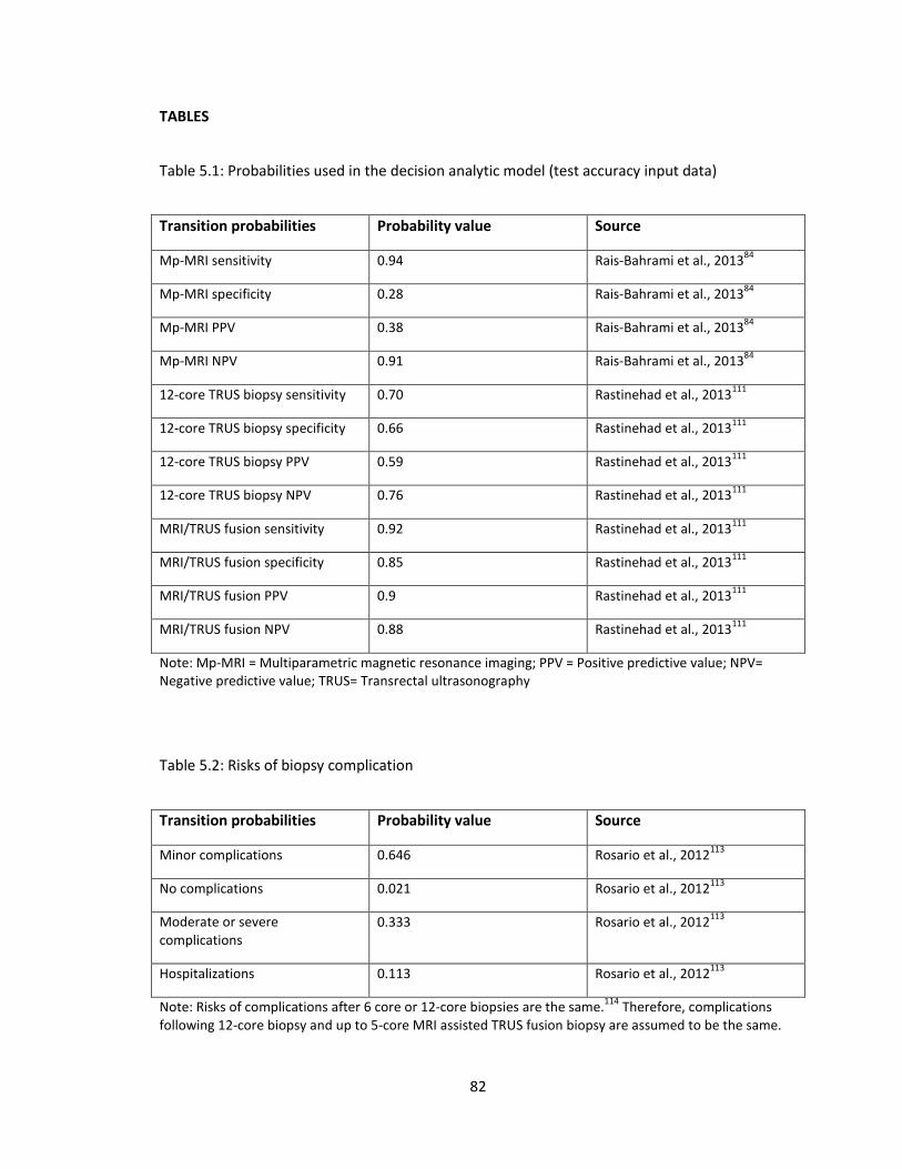
82
TABLES
Table 5.1: Probabilities used in the decision analytic model (test accuracy input data)
Transition probabilities Probability value Source
Mp-MRI sensitivity 0.94 Rais-Bahrami et al., 201384
Mp-MRI specificity 0.28 Rais-Bahrami et al., 201384
Mp-MRI PPV 0.38 Rais-Bahrami et al., 201384
Mp-MRI NPV 0.91 Rais-Bahrami et al., 201384
12-core TRUS biopsy sensitivity 0.70 Rastinehad et al., 2013111
12-core TRUS biopsy specificity 0.66 Rastinehad et al., 2013111
12-core TRUS biopsy PPV 0.59 Rastinehad et al., 2013111
12-core TRUS biopsy NPV 0.76 Rastinehad et al., 2013111
MRI/TRUS fusion sensitivity 0.92 Rastinehad et al., 2013111
MRI/TRUS fusion specificity 0.85 Rastinehad et al., 2013111
MRI/TRUS fusion PPV 0.9 Rastinehad et al., 2013111
MRI/TRUS fusion NPV 0.88 Rastinehad et al., 2013111
Note: Mp-MRI = Multiparametric magnetic resonance imaging; PPV = Positive predictive value; NPV= Negative predictive value; TRUS= Transrectal ultrasonography
Table 5.2: Risks of biopsy complication
Transition probabilities Probability value Source
Minor complications 0.646 Rosario et al., 2012113
No complications 0.021 Rosario et al., 2012113
Moderate or severe complications
0.333 Rosario et al., 2012113
Hospitalizations 0.113 Rosario et al., 2012113
Note: Risks of complications after 6 core or 12-core biopsies are the same.114
Therefore, complications following 12-core biopsy and up to 5-core MRI assisted TRUS fusion biopsy are assumed to be the same.
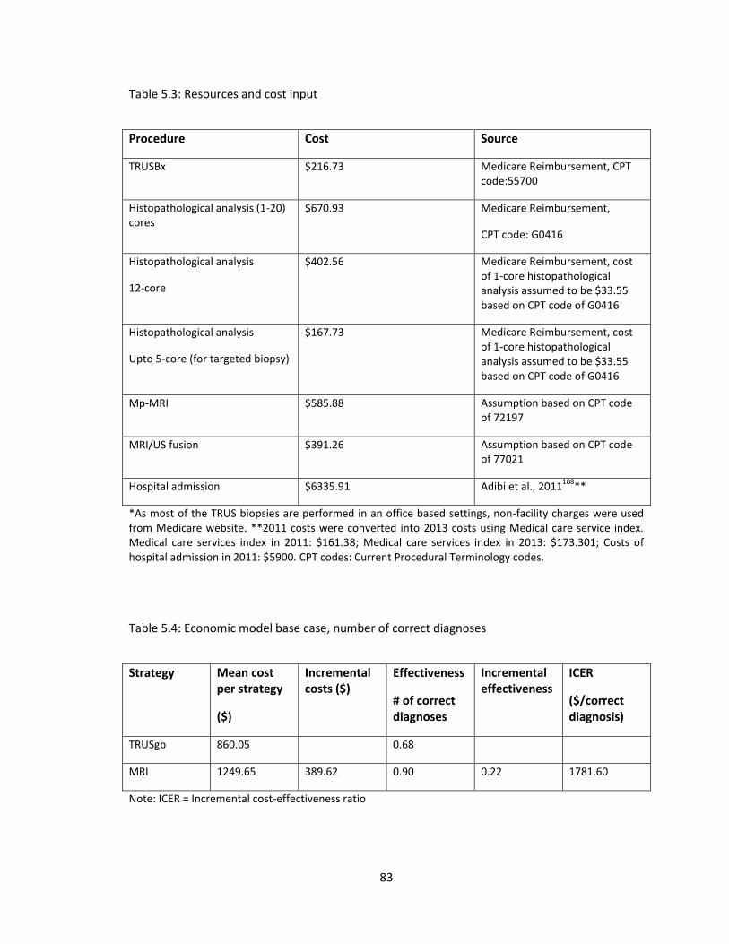
83
Table 5.3: Resources and cost input
Procedure Cost Source
TRUSBx $216.73 Medicare Reimbursement, CPT code:55700
Histopathological analysis (1-20) cores
$670.93 Medicare Reimbursement,
CPT code: G0416
Histopathological analysis
12-core
$402.56 Medicare Reimbursement, cost of 1-core histopathological analysis assumed to be $33.55 based on CPT code of G0416
Histopathological analysis
Upto 5-core (for targeted biopsy)
$167.73 Medicare Reimbursement, cost of 1-core histopathological analysis assumed to be $33.55 based on CPT code of G0416
Mp-MRI $585.88 Assumption based on CPT code of 72197
MRI/US fusion $391.26 Assumption based on CPT code of 77021
Hospital admission $6335.91 Adibi et al., 2011108
**
*As most of the TRUS biopsies are performed in an office based settings, non-facility charges were used from Medicare website. **2011 costs were converted into 2013 costs using Medical care service index. Medical care services index in 2011: $161.38; Medical care services index in 2013: $173.301; Costs of hospital admission in 2011: $5900. CPT codes: Current Procedural Terminology codes.
Table 5.4: Economic model base case, number of correct diagnoses
Strategy Mean cost per strategy
($)
Incremental costs ($)
Effectiveness
# of correct diagnoses
Incremental effectiveness
ICER
($/correct diagnosis)
TRUSgb 860.05 0.68
MRI 1249.65 389.62 0.90 0.22 1781.60
Note: ICER = Incremental cost-effectiveness ratio
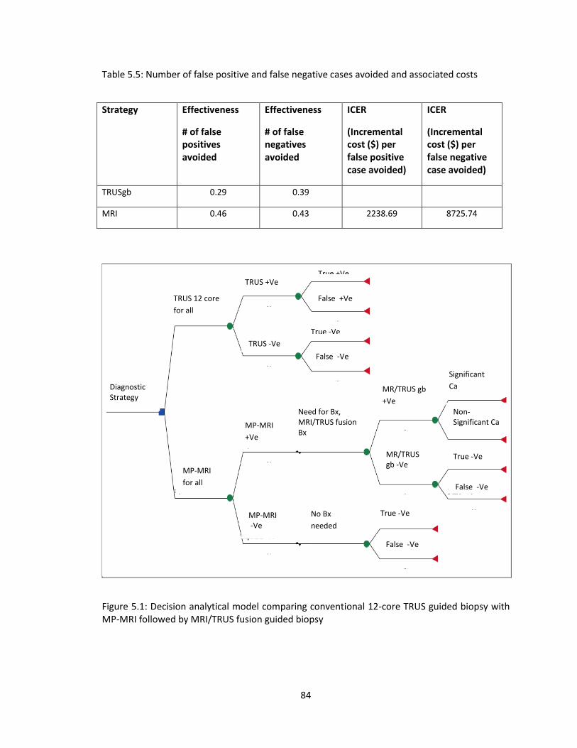
84
Table 5.5: Number of false positive and false negative cases avoided and associated costs
Strategy Effectiveness
# of false positives avoided
Effectiveness
# of false negatives avoided
ICER
(Incremental cost ($) per false positive case avoided)
ICER
(Incremental cost ($) per false negative case avoided)
TRUSgb 0.29 0.39
MRI 0.46 0.43 2238.69 8725.74
Figure 5.1: Decision analytical model comparing conventional 12-core TRUS guided biopsy with MP-MRI followed by MRI/TRUS fusion guided biopsy
Diagnostic Strategy
TRUS 12 core
for all
MP-MRI
for all
MP-MRI -Ve
MP-MRI
+Ve
No Bx
needed
Need for Bx, MRI/TRUS fusion Bx
MR/TRUS gb -Ve
MR/TRUS gb
+Ve
TRUS -Ve
TRUS +Ve
True -Ve
Strategy
Significant
Ca
Non-Significant Ca
False -Ve
Strategy True -Ve
Strategy
False -Ve
Strategy
False -Ve
Strategy
False +Ve
Strategy
True +Ve
Strategy
True -Ve
Strategy
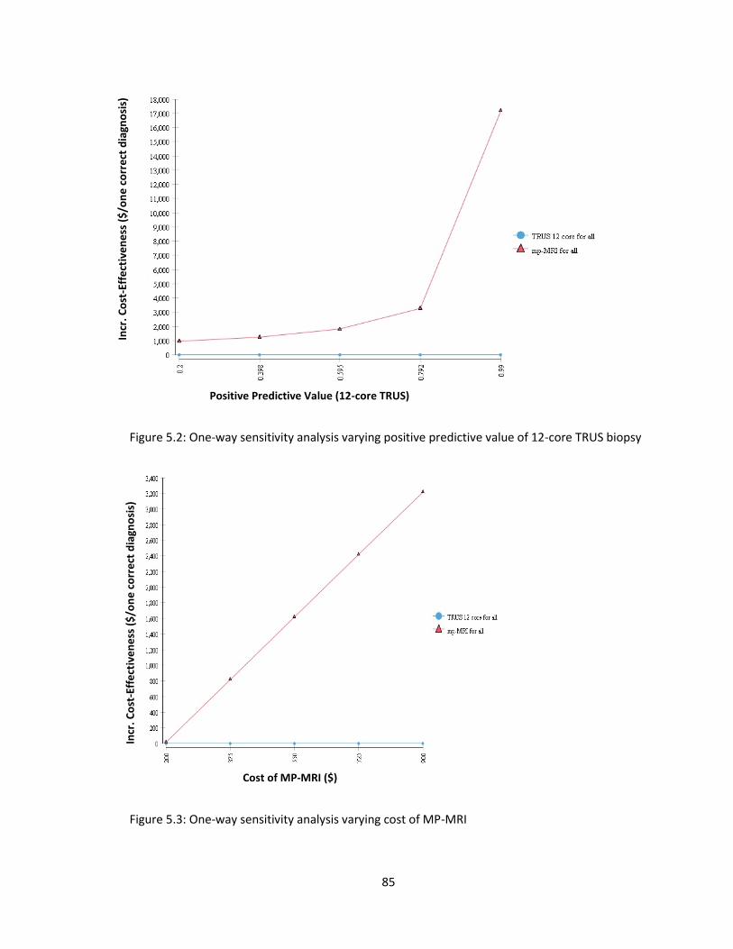
85
Figure 5.2: One-way sensitivity analysis varying positive predictive value of 12-core TRUS biopsy
Figure 5.3: One-way sensitivity analysis varying cost of MP-MRI
Cost of MP-MRI ($)
Incr
. Co
st-E
ffe
ctiv
en
ess
($
/on
e c
orr
ect
dia
gno
sis)
Positive Predictive Value (12-core TRUS)
Incr
. Co
st-E
ffe
ctiv
en
ess
($
/on
e c
orr
ect
dia
gno
sis)
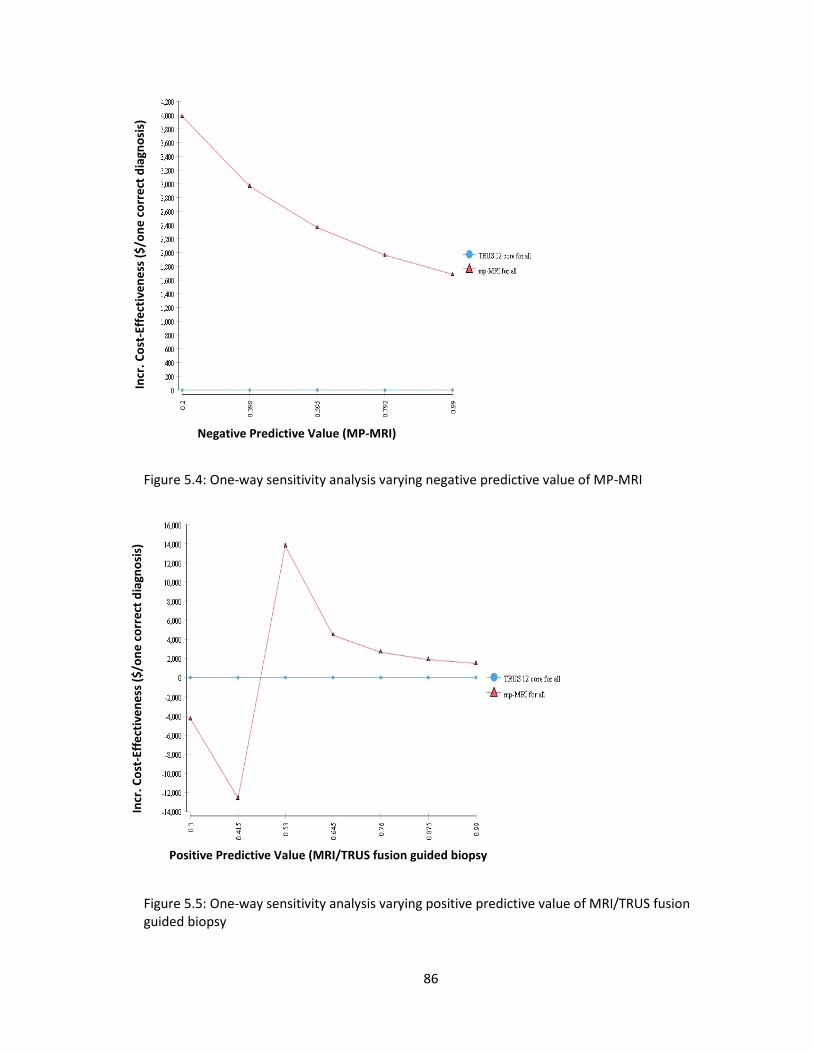
86
Figure 5.4: One-way sensitivity analysis varying negative predictive value of MP-MRI
Figure 5.5: One-way sensitivity analysis varying positive predictive value of MRI/TRUS fusion guided biopsy
Negative Predictive Value (MP-MRI)
Incr
. Co
st-E
ffe
ctiv
en
ess
($
/on
e c
orr
ect
dia
gno
sis)
In
cr. C
ost
-Eff
ect
ive
ne
ss (
$/o
ne
co
rre
ct d
iagn
osi
s)
Positive Predictive Value (MRI/TRUS fusion guided biopsy
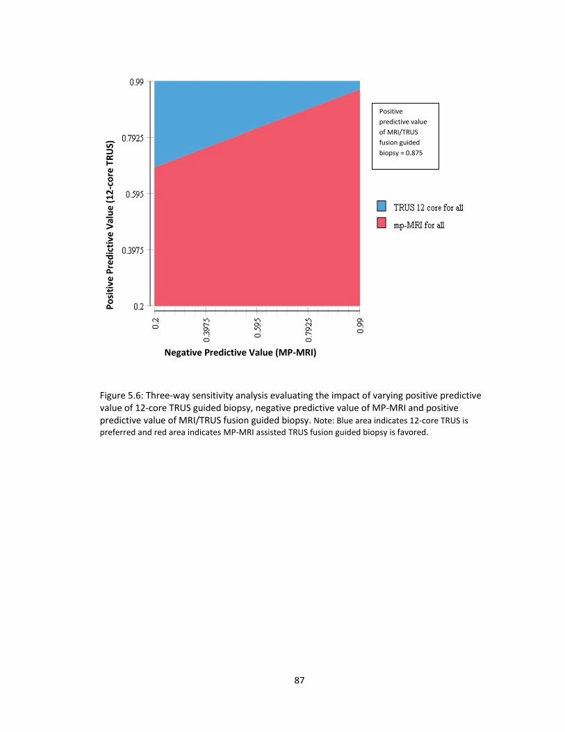
87
Figure 5.6: Three-way sensitivity analysis evaluating the impact of varying positive predictive value of 12-core TRUS guided biopsy, negative predictive value of MP-MRI and positive predictive value of MRI/TRUS fusion guided biopsy. Note: Blue area indicates 12-core TRUS is
preferred and red area indicates MP-MRI assisted TRUS fusion guided biopsy is favored.
Positive
predictive value
of MRI/TRUS
fusion guided
biopsy = 0.875
Negative Predictive Value (MP-MRI)
P
osi
tive
Pre
dic
tive
Val
ue
(12
-co
re T
RU
S)
88
REFERENCES
1. Prostate Cancer 2014. (Accessed 2013, at
http://www.cancer.org/cancer/prostatecancer/detailedguide/prostate-cancer-diagnosis.)
2. Penzkofer T, Tempany-Afdhal CM. Prostate cancer detection and diagnosis: The role of MR
and its comparison with other diagnostic modalities – a radiologist's perspective. NMR in
Biomedicine 2014;27:3-15.
3. AUA/Optimal techniques of prostate biopsy & specimen handling. (Accessed 2014, at
https://www.auanet.org/common/pdf/education/clinical-guidance/Prostate-Biopsy-
WhitePaper.pdf.)
4. Xu S, Kruecker J, Turkbey B, et al. Real-time MRI-TRUS fusion for guidance of targeted
prostate biopsies. Computer Aided Surgery 2008;13:255-64.
5. Fiard G, Hohn N, Descotes JL, Rambeaud JJ, Troccaz J, Long JA. Targeted MRI-guided prostate
biopsies for the detection of prostate cancer: Initial clinical experience with real-time 3-
dimensional transrectal ultrasound guidance and magnetic resonance/transrectal ultrasound
image fusion. Urology 2013;81:1372-8.
6. Satyanarayana R, Parekh D. Prevention and treatment of biopsy-related complications.
Current Urology Reports 2014;15:013-0381.
7. Siddiqui MM, Rais-Bahrami S, Truong H, et al. Magnetic resonance imaging/ultrasound-fusion
biopsy significantly upgrades prostate cancer versus systematic 12-core transrectal ultrasound
biopsy. European Urology 2013;64:713-9.
8. Haffner J, Lemaitre L, Puech P, et al. Role of magnetic resonance imaging before initial biopsy:
comparison of magnetic resonance imaging-targeted and systematic biopsy for significant
prostate cancer detection. BJU International 2011;108:22.
89
9. Puech P, Sufana Iancu A, Renard B, Villers A, Lemaitre L. Detecting prostate cancer with MRI -
why and how. Diagnostic and Interventional Imaging 2012;93:268-78.
10. ClinicalTrials. MRI/TRUS Fusion Guided Prostate Biopsy- An Improved Way to Detect and
Quantify Prostate Cancer. 2013.
11. Rais-Bahrami S, Siddiqui MM, Turkbey B, et al. Utility of multiparametric magnetic resonance
imaging suspicion levels for detecting prostate cancer. The Journal of Urology 2013;190:1721-7.
12. Rastinehad AR, Turkbey B, Salami SS, et al. Improving detection of clinically significant
prostate cancer: Magnetic resonance imaging/transrectal ultrasound fusion guided prostate
biopsy. The Journal of Urology 2013;12:06089-8.
13. Rosario DJ, Lane JA, Metcalfe C, et al. Short term outcomes of prostate biopsy in men tested
for cancer by prostate specific antigen: prospective evaluation within ProtecT study. British
Medical Journal 2012; 344:d7894.
14. Naughton CK, Ornstein DK, Smith DS, Catalona WJ. Pain and morbidity of transrectal
ultrasound guided prostate biopsy: A prospective randomized trial of 6 versus 12 cores. The
Journal of Urology 2000;163:168-71.
15. Adibi M, Pearle MS, Lotan Y. Cost-effectiveness of standard vs intensive antibiotic regimens
for transrectal ultrasonography (TRUS)-guided prostate biopsy prophylaxis. BJU International
2011;110:E86-91.
16. Crawford ED, Ventii K, Shore ND. New biomarkers in prostate cancer. Oncology 2014;28:135-
42.
17. Ahmed HU, Kirkham A, Arya M, et al. Is it time to consider a role for MRI before prostate
biopsy? Nature Reviews Clinical Oncology 2009;6:197-206.
18. Shariat SF, Roehrborn CG. Using biopsy to detect prostate cancer. Reviews in Urology
2008;10:262-80.

90
19. de Rooij M, Crienen S, Witjes JA, Barentsz JO, Rovers MM, Grutters JP. Cost-effectiveness of
magnetic resonance (MR) imaging and MR-guided targeted biopsy versus systematic transrectal
ultrasound-guided biopsy in diagnosing prostate cancer: A modelling study from a health care
perspective. European Urology 2013;21:012.

91
CHAPTER 6
COMPARATIVE ANALYSIS OF HARMS ASSOCIATED WITH CONSERVATIVE MANAGEMENT AND
IMMEDIATE TREATMENT AMONG LOW RISK LOCALIZED PROSTATE CANCER PATIENTS: A
POPULATION BASED STUDY
____________________________
1Patel P, Perri M, Griffin S, Tackett R. To be submitted to Journal of the National Comprehensive
Cancer Network.
92
Purpose: The purpose of this study was to examine urinary, rectal, erectile side effects, and
cancer specific survival in localized prostate cancer patients who were treated with immediate
treatment or conservative management. Methods: Using the Surveillance Epidemiology and End
Results Medicare-linked database, a total of 6,868 patients ≥ 66 years of age with localized low
risk prostate cancer were identified (2004 and 2005). Patients who received either immediate
treatment or delayed treatment (> 6 months after diagnosis) were followed for a period of 5
years to determine toxicities and survival. Propensity score matching was used to adjust for
selection bias associated with treatment type received. The presence of toxicity in each cohort
was determined using logistic regression. The Cox proportional hazard model was used to
estimate prostate cancer specific survival rates. Results: Overall, 735 patients received delayed
treatment and 6,133 patients received immediate treatment. Multivariate logistic regression
analysis showed that the conservative management group was found to have lower odds for
urinary complications (odds ratio: 0.824, p value<0.0001), rectal complications (odds ratio:
0.770, p value<0.0001) and erectile toxicities (odds ratio: 0.636, p value < 0.0001) compared to
the immediate treatment group within 5 years of diagnosis. The results of survival analysis
showed that there was no additional hazard of dying due to prostate cancer in conservative
management within a 5 year time period among studied patients than those in the immediate
treatment group (Hazard ratio: 0.736, p value: 0.2696). Conclusion: Patients ≥ 66 years of age
diagnosed with low risk prostate cancer are not at additional risk of dying due to prostate cancer
within a 5 year time period if kept on conservative management or delayed treatment. The
results of this study should be interpreted with caution because we could not differentiate
active surveillance from watchful waiting group from the database.
Keywords: Prostate cancer, treatment strategies, survival analysis, comparative effectiveness

93
Introduction
Prostate cancer is the second leading cause of non-cutaneous cancer related deaths
among men in the United States.1 The National Cancer Institute has estimated that there will be
approximately 233,000 new cases of prostate cancer and approximately 29,480 will die of it in
2014.2 Prostate cancer leads in terms of costs as well. The overall direct cost of prostate cancer
in the United States in 2010 was estimated to be more than $12 billion in annual costs.3 In 2020,
the direct cost of prostate cancer is projected to be $19 billion.3 Currently, most prostate
cancers are detected by a blood test that measures prostate specific antigen (PSA), and digital
rectal examination.4 More than half of cancers detected with PSA screening are localized, not
aggressive at diagnosis, and unlikely to become life threatening.5 However, 90% of patients
receive immediate treatment for prostate cancer such as surgery or radiation therapy resulting
into tremendous overtreatments.5,6 In many patients, these overtreatments have substantial
short- and long-term effects without any clinical benefit.
Appropriate management of screen detected, early-stage, low to intermediate risk
prostate cancer is an important public health issue given the number of men affected and the
risk for adverse outcomes, such as diminished sexual function and loss of urinary control.
Potential strategies to eliminate overtreatment include more widespread implementation of
observational therapies. Currently, clinicians rely on two observational strategies as alternative
to immediate treatment of early-stage prostate cancer: watchful waiting and active surveillance.
Watchful waiting involves relatively passive patient follow-up, with palliative interventions when
any symptoms develop. Active surveillance typically involves proactive patient follow-up in
which PSA levels are closely monitored, prostate biopsies may be repeated, and eventual
treatment is anticipated.

94
As prostate cancer often has an indolent natural history, it makes observational
management strategies more appealing.7 The life time risk of being diagnosed with prostate
cancer is about 17%, while the corresponding risk of dying of this disease is 3%.8,9 This evidence
suggests that conservative management may be an important treatment consideration of the
sizable majority of men diagnosed with localized prostate cancer.
Watchful waiting in low-risk prostate cancer is not new to the field. However, active
surveillance is a new emerging strategy that focuses on relatively young individuals rather than
the sicker older population. Despite its potential as a reasonable treatment choice active
surveillance has been used in only about 10% of the patients, perhaps because of a limited
understanding of and contemporary data on the anticipated course and outcomes of this
approach. Long term outcomes and effects on quality of life have not been well
characterized.8,10 The Institute of Medicine’s Committee on Comparative Effectiveness Research
has identified treatment for localized prostate cancer as a high-priority research area.11
The objective of the current study was to compare conservative management with the
immediate treatment based on long term clinical outcomes mainly disease and treatment
related toxicities. The central hypothesis of the study is that conservative management has a
better toxicity profile compared to the group of immediate treatment. The rationale behind this
study is that it may provide substantial evidence to choose an appropriate regime that may
reduce the patient burden and healthcare costs associated with prostate cancer.
Methods
Data Sources: Data for this study was obtained from the National Cancer Institute’s Surveillance
Epidemiology Ends Results (SEER) program database linked to Medicare administrative claims
from 2003 to 2009. The SEER program captures clinical, demographic and survival information of
95
approximately 28% of the US population and is 98% complete for case ascertainment.12 The
Medicare program covers approximately 97% of the persons aged 65 years and older.13 This
study was approved by the University of Georgia’s Institutional Review Board as well as by the
SEER-Medicare for Data Use Agreement with National Cancer Institute. As the data did not
contain personal identifiers, informed consent was not requested by the Institutional Review
Board.
Cancer related information such as cancer stage, grade, tumor extension, and tumor
size was obtained from the SEER’s Patient Entitlement and Diagnosis Summary File (PEDSF). Well
differentiated cancers were characterized by a Gleason score of 2 to 4; moderately
differentiated, 5 to 6, and poorly differentiated, 7 to 10. Treatment related information was
obtained from both SEER and Medicare files. A Charlson comorbidity score was derived from
Medicare claims during the year prior to prostate cancer diagnosis using a validated algorithm.
Race was self-determined by the patients.
Study Participants: Study participants were men that were mainly 66 years or older SEER
residents and diagnosed with stage T1 or T2a between 2004 and 2005 (ICD-O-3 site code C619)
and followed for 5 years. The current study utilized only newly diagnosed cases to understand
the outcomes of the treatment strategies from the identification of the disease. Patients were
excluded from the study if they (i) did not survive the first 6 months after the diagnosis, (ii) had a
personal history of malignant neoplasm of prostate, (iii) were enrolled in HMO, (iv) did not have
both Medicare Part A and Part B, and (v) had end-stage renal disease. Eligible identified patients
were categorized into two cohorts: 1) conservative management and 2) immediate treatment.
Patients who were in the immediate treatment group were identified as patients undergoing
radical prostatectomy, radiation, or brachytherapy immediately after diagnosis. ICD-9 codes and

96
Healthcare Common Procedure Coding System (HCPCS) codes used in identifying patients are
presented in Table 6.1. Patients who were in the conservative management group were
identified as those who did not receive any immediate treatment within 6 months of diagnosis
of localized prostate cancer.
Outcomes Assessment: Both incident and prevalent cases of urinary, rectal, and erectile
complications were assessed separately in both cohorts. Urinary complications were defined as
having incontinence, obstruction, irradiation cystitis, bladder hemorrhage, urinary fistulas, or
urinary tract infections. Rectal complications were defined as having rectal hemorrhage, ulcers,
fistulas or bowel incontinence. Erectile complications were defined as having impotence. These
complications were identified using appropriate ICD-9 diagnoses codes as well as based on
Common Procedure Terminology (CPT) /HCPCS codes of invasive procedures performed to
repair these complications. The medical codes for diagnoses and procedures performed for the
complications are provided in Table 6.2. Patient’s dates of diagnosis and death were obtained
from the SEER’s PEDSF file.
Statistical Analyses: Demographic and clinical variables across study cohorts were compared. A
chi-square test was used to compare categorical variables and a t-test was used to compare
continuous variables. As the aim of this study was to compare treatment outcomes, propensity
score matching was used to address the issue of selection bias and generate comparable study
arms. In this two-step procedure, the probability of receiving treatment (conservative
management vs. immediate treatment) was first calculated based on multivariate logistic
regression that included the patient’s demographic information such as age, race, income, and
tumor related information (e.g., grade, tumor extension and tumor size) as predictors of
receiving treatment. The obtained probability scores were then used in analyzing outcomes. Risk

97
of complications such as urinary, rectal, and sexual dysfunction was estimated using logistic
regression. A Cox proportional hazard model was used to estimate the prostate cancer specific
survival rate in both the cohorts. All analyses were performed using SAS statistical software
(version 9.3, SAS Institute, Cary, NC).
Results
The study population included 6,868 patients who were diagnosed with incident prostate
cancer between 2004 and 2005, and fit into the eligibility criteria. Conservative management
and immediate treatment cohorts consisted of 735 and 6,133 patients respectively. Table 6.3
describes the baseline characteristics of the study cohorts. The majority of eligible patients were
aged 66 to 74 years and white in both the cohorts. Approximately 97% of the patients in both
cohorts had either a moderately or intermediately differentiated tumor grade. All patients had
localized prostate cancer. Tumor size was not recorded in more than 85% of patients in both
cohorts. There was no nodal involvement in all the eligible patients. In terms of comorbidity
burden, the majority of patients had either 0 or 1 comorbid condition. There were more married
individuals in the immediate treatment group than the conservative management cohort. There
was no significant difference in the proportion of patients who had T1 or T2a staging in both the
treatment arms.
Risk of adverse outcomes:
Patients diagnosed with prostate cancer were followed for a 5 year time period to
measure adverse events in both cohorts. Adverse events studied included urinary, rectal and
erectile complications. Rates of urinary, rectal and erectile complications were 55.51%, 20.27%
and 6.12%, respectively, for conservative management patients. Immediate treatment patients
were found to have urinary, rectal and erectile rates of 57.26%, 25.04% and 10.75%
98
respectively. Table 6.4 presents crude rates of complication diagnoses and invasive procedures
performed for both cohorts.
Table 6.5 shows the results of the logistic regression predicting urinary complications
based on the treatment arm, demographic, and tumor related variables adjusted with
propensity scores. Odds of urinary complications were statistically significantly lower in the
conservative management than the immediate treatment group (odds ratio: 0.824, p< 0.0001).
Patients aged from 66 to 79 years were found to have significantly lower odds of
urinary complications than those above 80 years old. Patients with null or moderate comorbidity
were found to have reduced odds of urinary complications than those with more than one
comorbidity. Black patients compared to whites had reduced odds of urinary complications.
Single patients were found to have higher odds of having urinary complications than married
patients. Results related to rectal complications are presented in Table 6.6. Conservative
management was found to have a lower odds of rectal complications compared to the
immediate treatment group (odds ratio: 0.770, p<0.0001). Compared to whites, black and
patients with other ethnicities were found to have reduced odds of rectal complications. Those
with null or moderate comorbidity were found to have lower odds of getting rectal
complications than those with higher comorbidity.
Factors associated with rates of erectile complications are presented in Table 6.7.
Conservative management was found to be less likely to have erectile complications than the
immediate treatment group (odds ratio: 0.636, p<0.0001). There was not a statistically
significant difference between blacks and whites regarding erectile complications. However,
patients with other ethnicities (other than black) were found to have reduced odds of having
erectile complications than whites. Patients aged from 66 to 79 years were found to have higher
99
odds of erectile complications than those above 79 years of age. Patients with null or one
comorbidity had higher odds of having erectile complications than those with higher
comorbidity.
Survival analysis:
Data for survival analysis are presented in Table 6.8. These data represent up to a 5-year
follow up period from time of diagnosis. Patients who were in the conservative management
group did not differ significantly from those in the immediate treatment group in terms of
prostate cancer specific mortality at any point in time (Hazard ratio: 0.736, p value: 0.2696)
within the 5 year time period. However, our study found that black patients compared to whites
had higher hazard of dying due to prostate cancer within 5 year time period (Hazard ratio:
2.537, p value: 0.0112). Patients aged from 66 to 79 years were found to have better survival
experience than those aged above 79 years within the study time period. We also found that
patients with null or one comorbidity had a reduced hazard of dying due to prostate cancer than
those with greater than one comorbidity.
Discussion
Our study of prostate cancer patients diagnosed between 2004 and 2005, with a 5-year
follow up, has focused on whether urinary, rectal, or erectile complication rates differ between
conservative management and immediate treatment groups. Further, this study assessed the
survival experiences of both treatment groups. Prostate cancer is considered a disease of older
men and the median age at diagnosis reported is 72 years.14 Thus, the SEER-Medicare
population is representative of the population of interest. Results of the current study suggest
that urinary, rectal and erectile complications are more likely to be present in the immediate
treatment group than in the conservative management group within a 5 year time period
100
among patients aged above 65 years. Results of survival analysis indicate that patients who opt
for conservative management have no additional higher hazard of dying due to prostate cancer
within 5 years than those who undergo immediate treatment.
Results of this study are important to patients as well as health care practitioners.
Patients who are in the conservative group can delay or avoid urinary, rectal, and erectile
complications by delaying immediate treatment strategies such as radical prostatectomy or
radiation therapy. Radical prostatectomy and radiation therapy affect a patient’s bowel, urinary
and sexual function tremendously and thereby affect the patient’s quality of life.15 Men in the
intermediate risk category face the most challenging decisions regarding treatment and
physicians recommend working backward from the known side effects associated with each
treatment option.15 Prostate cancer itself can affect bladder and sexual function.16 Urinary
incontinence is the most common symptom of prostate cancer and its severity depends on the
type of the disease. Tumor growth can also damage the nerves that control the erection and
thus leave a patient unable to engage in sexual activity.
As patients live longer with low-risk localized prostate cancer, they live with sequalae of
the treatments they receive. Thus, it is important that both patients and clinicians understand
the long-term consequences of various treatments. Demographic characteristics and tumor
grades also were found to affect the complication rates in the current study. Relatively younger
individuals, healthy, or black patients were less likely to experience urinary complications. On
the other hand, younger individuals were more likely to experience erectile dysfunction than
older people. We found this difference because younger individuals were more likely to receive
both non-surgical and surgical treatments to repair erectile dysfunction.17 Older individuals may
not seek surgical treatments but prefer to take medication.18 As we did not study Medicare part

101
D data, we could not find any claims related to drugs such as sildenafil citrate, tadalafil citrate or
vardenafil to treat erectile dysfunction. Patient preferences for outcomes among competing
treatment strategies may be an important factor that drives treatment decisions.
There are patients who want to avoid therapy induced distressful symptoms even when
faced with a reduced prospect of survival. Some men give full priority to survival even though
the survival gain may be very small. Our study found no significant difference in hazards of dying
due to prostate cancer between the conservative management group and the immediate
treatment. This result is consistent with the literature. Recently, Bill-Axelson et al. presented
extended follow up results of the Scandinavian Prostate Cancer Group Study Number 4 (SPCG-
4).19 This trial randomized patients to watchful waiting or radical prostatectomy between 1989
and 1999. They found a substantial reduction in mortality after the radical prostatectomy group
was followed for up to 23.2 years among men younger than 65 years of age. They did not find a
significant difference in mortality rate between radical prostatectomy and watchful waiting
among low risk localized prostate cancer patients. However, our study is different than SPCG-4
trial or other studies that have compared immediate treatment options with watchful waiting
program.9,20 We combined different immediate treatment options into one category. Further,
we had a conservative management group that included both watchful waiting and active
surveillance options. Acceptance of active surveillance or watchful waiting depends on a
patient’s physical and psychological well-being. Both strategies offer the opportunity to delay
treatment. However, watchful waiting is reserved for those who cannot tolerate aggressive
treatment and are offered hormonal therapy upon cancer progression. On the other hand,
active surveillance involves curative treatment upon cancer progression. It is difficult to separate
patients who received watchful waiting versus active surveillance from the claims database as
102
both groups receive frequent PSA screening. As a result, both treatment options in this study are
combined as a conservative management approach.
Several potential limitations of this study should be considered when interpreting these
results. Large administrative data sets such as Medicare data contain data originally intended for
billing purposes. Procedures or treatments that do not incur any costs are not reported as there
is no financial incentive to document them. Further, complication rates obtained using these
data represent underestimates as not all patients are likely to receive treatments for
complications. The treatment modalities were not randomized in the study. However, the issue
of selection bias was addressed using propensity score matching. We could not differentiate
patients who had watchful waiting or active surveillance as an observational strategy. This may
bias the results because patients in watchful waiting are more likely to receive hormonal
treatment and are at higher risk of having serious side effects or even death. Finally, the study
was limited to Medicare eligible patients aged 66 years or older and those receiving care
through the traditional, fee-for-service system, limiting the utility of the finding to older patients
not enrolled in managed care programs.
Conclusion
Treatment options for patients diagnosed with low to intermediate risk prostate cancer
include i) immediate treatment with either radical prostatectomy or radiation therapy and ii)
observational strategies such as active surveillance or watchful waiting. In summary, our study
suggests that the conservative management approach is associated with lower urinary, rectal,
and erectile complications than immediate treatment within 5 years of diagnosis. Prostate
cancer specific mortality does not differ significantly among patients who receive either
immediate treatment or observation treatment. Choosing the appropriate treatment regimen

103
for disease management is critical and should account for (i) the patient’s tumor characteristics
such as its grade or aggressiveness, (ii) patient age, overall health, and remaining life
expectancy, and (iii) patient preferences for the potential side effects of treatment options.

104
TABLES
Table 6.1: ICD-9 and CPT/HCPCS codes used to identify treatment modality
Treatment ICD-9 codes HCPCS
Immediate Radiation treatment Prostatectomy Chemotherapy
V58.0, V66.1, V67.1, 92.21-92.29 60.5, 60.2, 60.21-60.29, 60.3-60.6, 60.61, 60.62, 60.69, 60.9 V58.1, V66.2, V67.2, 99.25
77401-77499, 77750-77799, 77014, 77334, 77336, 77520, 77522-77525 55812-55845, 55866, 55810, 55899, 55867-55880 96401-96549
Table 6.2: ICD-9 and CPT/HCPCS codes used to identify complications
Complications Diagnoses ICD-9
CPT/HCPCS
Urinary 788.3X, 595.85, 596.7, 599.0, 596.0, 598.X, 599.6, 788.2X, 596.1, 596.2, 599.1
52275, 52276, 52281, 52510, 53010, 53400, 53405, 53410, 52415, 53420, 53425, 53600, 53601, 53605, 53620, 53621, 52252, 53440, 51840, 51841, 53442, 53443
Rectal 558.0, 558.1-558.4, 558.9, 569.0-569.4, 569.41-569.44, 569.49, 569.81, 565.0, 562.10-562.12, 578.1, 787.6, 787.60-787.63, 455.7, 455.8
45800, 45805, 45820, 45825
Erectile 607.84 54400-54402, 54405, 54407-54411, 54415,-54417, C1007, C1813, C2622, C3500 C8514, C8516, L7900, 54231, 54235, J0270, J0275, J2440, J2760
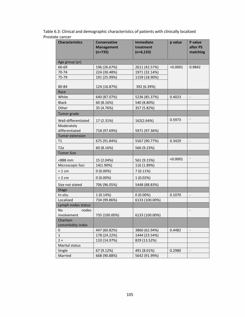
105
Table 6.3: Clinical and demographic characteristics of patients with clinically localized Prostate cancer
Characteristics Conservative Management (n=735)
Immediate treatment (n=6,133)
p value P value after PS matching
Age group (yr)
66-69 196 (26.67%) 2611 (42.57%) <0.0001
0.9842
70-74 224 (30.48%) 1971 (32.14%)
75-79 191 (25.99%) 1159 (18.90%)
80-84 124 (16.87%) 392 (6.39%)
Race
White 640 (87.07%) 5236 (85.37%) 0.4023 -
Black 60 (8.16%) 540 (8.80%)
Other 35 (4.76%) 357 (5.82%)
Tumor grade
Well-differentiated 17 (2.31%) 162(2.64%) 0.5973 -
Moderately differentiated 718 (97.69%) 5971 (97.36%)
Tumor extension
T1 675 (91.84%) 5567 (90.77%) 0.3429 -
T2a 60 (8.16%) 566 (9.23%)
Tumor Size
<888 mm 15 (2.04%) 561 (9.15%) <0.0001 -
Microscopic foci 14(1.90%) 116 (1.89%)
< 1 cm 0 (0.00%) 7 (0.11%)
< 2 cm 0 (0.00%) 1 (0.02%)
Size not stated 706 (96.05%) 5448 (88.83%)
Stage
In-situ 1 (0.14%) 0 (0.00%) 0.1070 -
Localized 734 (99.86%) 6133 (100.00%)
Lymph nodes status
No nodes involvement 735 (100.00%) 6133 (100.00%)
-
Charlson comorbidity index
0 447 (60.82%) 3860 (62.94%) 0.4482 -
1 178 (24.22%) 1444 (23.54%)
2 + 110 (14.97%) 829 (13.52%)
Marital status
Single 67 (9.12%) 491 (8.01%) 0.2980 -
Married 668 (90.88%) 5642 (91.99%)
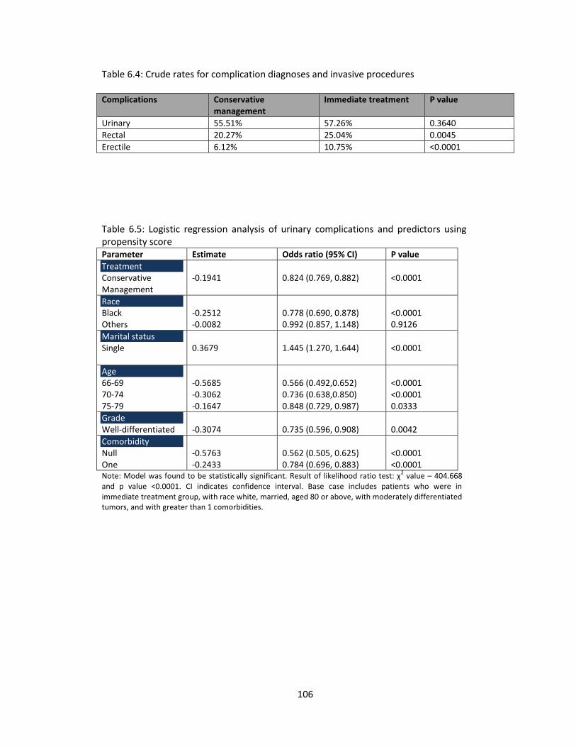
106
Table 6.4: Crude rates for complication diagnoses and invasive procedures
Complications Conservative management
Immediate treatment P value
Urinary 55.51% 57.26% 0.3640
Rectal 20.27% 25.04% 0.0045
Erectile 6.12% 10.75% <0.0001
Table 6.5: Logistic regression analysis of urinary complications and predictors using propensity score Parameter Estimate Odds ratio (95% CI) P value
Treatment Conservative Management
-0.1941
0.824 (0.769, 0.882)
<0.0001
Race Black Others
-0.2512 -0.0082
0.778 (0.690, 0.878) 0.992 (0.857, 1.148)
<0.0001 0.9126
Marital status Single
0.3679
1.445 (1.270, 1.644)
<0.0001
Age 66-69 70-74 75-79
-0.5685 -0.3062 -0.1647
0.566 (0.492,0.652) 0.736 (0.638,0.850) 0.848 (0.729, 0.987)
<0.0001 <0.0001 0.0333
Grade Well-differentiated
-0.3074
0.735 (0.596, 0.908)
0.0042
Comorbidity Null One
-0.5763 -0.2433
0.562 (0.505, 0.625) 0.784 (0.696, 0.883)
<0.0001 <0.0001
Note: Model was found to be statistically significant. Result of likelihood ratio test: χ2 value – 404.668
and p value <0.0001. CI indicates confidence interval. Base case includes patients who were in immediate treatment group, with race white, married, aged 80 or above, with moderately differentiated tumors, and with greater than 1 comorbidities.

107
Table 6.6: Logistic regression analysis of rectal complications and predictors using propensity score Parameter Estimate Odds ratio (95% CI) P value
Treatment Conservative Management
-0.2620
0.770 (0.710, 0.834)
<0.0001
Race Black Others
-0.1925 -0.5486
0.825 (0.712, 0.956) 0.578 (0.474, 0.705)
0.0104 <0.0001
Marital status Single
-0.3301
0.719 (0.612, 0.845)
<0.0001
Age 66-69 70-74 75-79
0.1219 0.0902 0.1218
1.130 (0.959, 1.331) 1.094 (0.927, 1.292) 1.130 (0.949, 1.345)
0.1453 0.2861 0.1704
Grade Well-differentiated
-0.1525
0.859 (0.664, 1.111)
0.2459
Comorbidity Null One
-0.4056 -0.1563
0.667 (0.594, 0.748) 0.855 (0.752, 0.973)
<0.0001 0.0172
Note: Model was found to be statistically significant. Result of likelihood ratio test: χ2 value – 156.787
and p value <0.0001. CI indicates confidence interval. Base case includes patients who were in immediate treatment group, with race white, married, aged 80 or above, with moderately differentiated tumors, and with greater than 1 comorbidities.
Table 6.7: Logistic regression analysis of erectile complications and predictors using propensity score Parameter Estimate Odds ratio (95% CI) P value
Treatment Conservative Management
-0.4524
0.636 (0.564, 0.718)
<0.0001
Race Black Others
-0.0691 -0.7625
0.933 (0.758, 1.149) 0.467 (0.330, 0.660)
0.5142 <0.0001
Marital status Single
-0.1877
0.829 (0.656, 1.048)
0.1168
Age 66-69 70-74 75-79
1.6555 1.1695 0.8049
5.236 (3.521, 7.785) 3.220 (2.154, 4.816) 2.236 (1.469, 3.404)
<0.0001 <0.0001 0.0002
Grade Well-differentiated
0.2031
1.225 (0.857, 1.751)
0.2652
Comorbidity Null One
0.2799 0.4896
1.323 (1.074, 1.630) 1.632 (1.302, 2.045)
0.0085 <0.0001
Note: Model was found to be statistically significant. Result of likelihood ratio test: χ2 value – 281.549
and p value <0.0001. CI indicates confidence interval. Base case includes patients who were in immediate treatment group, with race white, married, aged 80 or above, with moderately differentiated tumors, and with greater than 1 comorbidities.
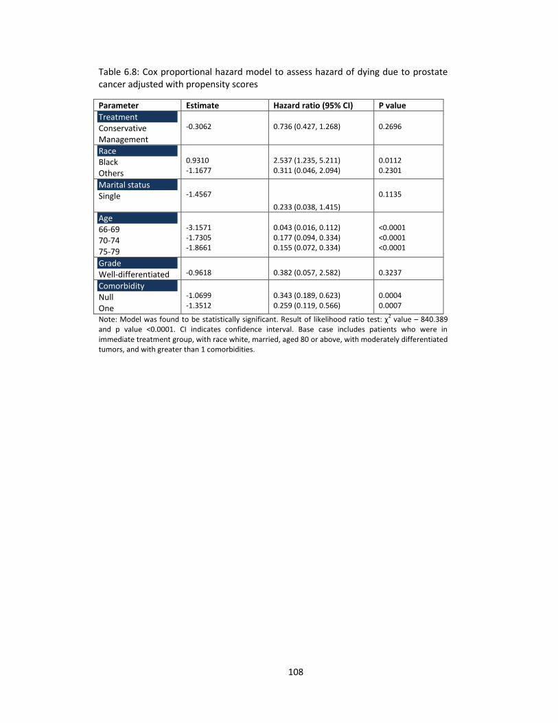
108
Table 6.8: Cox proportional hazard model to assess hazard of dying due to prostate cancer adjusted with propensity scores
Parameter Estimate Hazard ratio (95% CI) P value
Treatment Conservative Management
-0.3062
0.736 (0.427, 1.268)
0.2696
Race Black Others
0.9310 -1.1677
2.537 (1.235, 5.211) 0.311 (0.046, 2.094)
0.0112 0.2301
Marital status Single
-1.4567
0.233 (0.038, 1.415)
0.1135
Age 66-69 70-74 75-79
-3.1571 -1.7305 -1.8661
0.043 (0.016, 0.112) 0.177 (0.094, 0.334) 0.155 (0.072, 0.334)
<0.0001 <0.0001 <0.0001
Grade Well-differentiated
-0.9618
0.382 (0.057, 2.582)
0.3237
Comorbidity Null One
-1.0699 -1.3512
0.343 (0.189, 0.623) 0.259 (0.119, 0.566)
0.0004 0.0007
Note: Model was found to be statistically significant. Result of likelihood ratio test: χ2 value – 840.389
and p value <0.0001. CI indicates confidence interval. Base case includes patients who were in immediate treatment group, with race white, married, aged 80 or above, with moderately differentiated tumors, and with greater than 1 comorbidities.
109
REFERENCES
1. Wilt TJ, Brawer MK, Barry MJ, et al. The Prostate cancer Intervention Versus
Observation Trial:VA/NCI/AHRQ Cooperative Studies Program #407 (PIVOT): design and
baseline results of a randomized controlled trial comparing radical prostatectomy to
watchful waiting for men with clinically localized prostate cancer. Contemporary Clinical
Trials. 2009;30(1):81-87.
2. National Cancer Institute. Prostate Cancer. 2014.
3. Hodges JC, Lotan Y, Boike TP, Benton R, Barrier A, Timmerman RD. Cost-effectiveness
analysis of stereotactic body radiation therapy versus intensity-modulated radiation
therapy: An emerging initial radiation treatment option for organ-confined prostate
cancer. Journal of Oncology Practice. May 1, 2012 2012;8(3S):e31s-e37s.
4. American Urological association. www.auanet.org.
5. Wong YN, Mitra N, Hudes G, et al. Survival associated with treatment vs observation of
localized prostate cancer in elderly men. The Journal of Medical Association.
2006;296(22):2683-2693.
6. van den Bergh RC, Steyerberg EW, Khatami A, et al. Is delayed radical prostatectomy in
men with low-risk screen-detected prostate cancer associated with a higher risk of
unfavorable outcomes? Cancer. 2010;116(5):1281-1290.
7. Klotz L. Active surveillance for prostate cancer: Patient selection and management.
Current Oncology. 2010;17(2):S11-17.
8. Lu-Yao GL, Albertsen PC, Moore DF, et al. Outcomes of localized prostate cancer
following conservative management. The Journal of Medical Association. Sep 16
2009;302(11):1202-1209.
110
9. Wilt TJ, Brawer MK, Jones KM, et al. Radical prostatectomy versus observation for
localized prostate cancer. New England Journal of Medicine. 2012;367(3):203-213.
10. Hayes JH, Ollendorf DA, Pearson SD, et al. Active surveillance compared with initial
treatment for men with low-risk prostate cancer: A decision analysis. The Journal of
Medical Association. Dec 1 2010;304(21):2373-2380.
11. Institute of Medicine. Initial National Priorities for Comparative Effectiveness Research.
2009;
http://www.iom.edu/reports/2009/comparativeeffectivenessresearchpriorities.aspx.
12. Surveillance E, and End Results: Program Turning Cancer Data Into Discovery,. Overview
of the SEER program. In: Institute NC, ed 2014.
13. Engels EA, Pfeiffer RM, Ricker W, Wheeler W, Parsons R, Warren JL. Use of surveillance,
epidemiology, and end results-medicare data to conduct case-control studies of cancer
among the US elderly. American Journal of Epidemiology. 2011;174(7):860-870.
14. National Cancer Institute. General information about prostate cancer. 2014.
15. Potosky AL, Davis WW, Hoffman RM, et al. Five-year outcomes after prostatectomy or
radiotherapy for prostate cancer: The Prostate Cancer Outcomes Study. Journal of the
National Cancer Institute. September 15, 2004 2004;96(18):1358-1367.
16. Mayo Clinic. Prostate Cancer. 2013; http://www.mayoclinic.org/diseases-
conditions/prostate-cancer/basics/symptoms/con-20029597, 2014.
17. Tal R, Jacks LM, Elkin E, Mulhall JP. Penile implant utilization following treatment for
prostate cancer: Analysis of the SEER-Medicare database. Journal of Sex Medicine.
2011;8(6):1797-1804.
111
18. Prasad MM, Prasad SM, Hevelone ND, et al. Utilization of pharmacotherapy for erectile
dysfunction following treatment for prostate cancer. Journal of Sex Medicine.
2010;7(3):1062-1073.
19. Bill-Axelson A, Holmberg L, Garmo H, et al. Radical prostatectomy or watchful waiting in
early prostate cancer. New England Journal of Medicine. 2014;370(10):932-942.
20. Hoffman RM, Barry MJ, Stanford JL, Hamilton AS, Hunt WC, Collins MM. Health
outcomes in older men with localized prostate cancer: Results from the Prostate Cancer
Outcomes Study. American Journal of Medicine. 2006;119(5):418-425.
112
CHAPTER 7
CONCLUSIONS
The current study has addressed three major areas in the field of prostate cancer
outcomes research. The first part of the study addressed the issue of healthcare utilization and
associated expenditures among prostate cancer patients in United States. This study provides
the most recent cost estimate as well as associated predictors of higher costs among patients
with prostate cancer compared to other previously conducted studies. Using a nationally
representative database, the annual direct medical expenditures associated with prostate
cancer was found to be $5.6 billion in 2010 US dollars. Both outpatient and inpatient settings
are associated with greater costs as the majority of prostate cancer related treatments are
performed in those settings.
Our study found chemotherapy and ultrasound as the significant predictors of higher
outpatient expenditure. This result requires further attention from policymakers. Ultrasound is
the most commonly preferred technique to guide biopsy for diagnostic confirmation of prostate
cancer. However, ultrasound is not a reliable technique and physicians and patients lack
confidence in ultrasound guided biopsy results. Frequent repetitive biopsies are performed in
many patients to rule out the possibility of false results. As a result, ultrasound is associated
with increased costs. X-ray imaging and computed tomography (CT) scans were found to be the
significant predictors of higher expenditure in office-based visits. X-rays and CT scans are
generally used to check for bony metastases and expenditure related to x-ray imaging and CT
scans may be avoided in certain cases that are at very low risk of having metastases but
routinely undergo bone scans.
113
The second part of the study addressed the issue of diagnosis especially that of transrectal
ultrasound (TRUS) guided biopsy in prostate cancer diagnosis. Conventional TRUS guided biopsy
has been reported to miss 30-40% of cancers. Likewise, conventional biopsy may detect
clinically insignificant tumors as well. MRI/TRUS fusion guided biopsy is performed out-of-bore
where tumor vascularity and anatomical data provided by multiparametric MRI is electronically
delivered to a fusion device that allows urologists to use the detail provided during MRI to guide
live, real-time ultrasound scanning. This study performed cost-effectiveness analysis of relatively
advanced and more accurate MRI/TRUS fusion guided biopsy compared with 12-core TRUS
guided biopsy. Our study found MRI assisted TRUS fusion guided biopsy to be cost-effective
compared with 12-core TRUS guided biopsy. MRI/TRUS fusion guided biopsy has better ability to
identify and characterize prostate cancer. However, further studies demonstrating the
effectiveness of MRI/TRUS fusion approach are required.
The third part of the study addressed the issue of overtreatment especially among low-
to intermediate-risk prostate cancer patients who have very low risk of having metastasis. This
study focused on incidence as well as prevalence based urinary, rectal, and erectile
complications, and survival associated with conservative management and immediate
treatment approach within 5 years of diagnosis. Results of our study indicate that patients in
conservative management can delay or avoid urinary, rectal, and erectile dysfunction
significantly compared to the immediate treatment with radical prostatectomy or radiation
therapy. Patients older than 65 years in conservative management do not have a higher risk of
mortality due to prostate cancer within a 5 year time period. Results of our study indicate that
patients with conservative management can have a better quality of life without compromising
survival in 5 year time frame. Younger individuals are more likely to have better survival
compared to older individuals irrespective of the treatment received. Similarly, patients with

114
one or no comorbidity have better survival experience than those with more than one
comorbidity irrespective of the treatment received. Treatment selection for patients diagnosed
with low to intermediate risk is very crucial and should account for the patient’s tumor
characteristics, age, life-expectancy, comorbidities, and patient’s preference for treatment side
effects.
The results of this study indicate that there is a need for more accurate cost-effective
technology in prostate cancer diagnoses. Routinely used TRUS guided biopsy is associated with
higher economic and psychological burden on society due to numerous inaccurate diagnoses
associated with it. Mp-MRI/TRUS fusion guided biopsy which can characterize prostate cancer
accurately is found to be cost-effective compared to TRUS guided biopsy at a threshold to pay
minimum of $1781.60. Observational strategies among low risk localized prostate cancer can
avoid treatment related side effects especially rectal and erectile complications significantly
within 5 years of diagnosis without compromising with survival. Thus, observational strategies
represent one of the best approaches in reducing over treatment among patients diagnosed
with low risk localized prostate cancer.