butterfly effects: novel functional materials inspired from the wings scales
TRANSCRIPT

PCCPPhysical Chemistry Chemical Physicswww.rsc.org/pccp
ISSN 1463-9076
PERSPECTIVEWang Zhang, Di Zhang et al.Butterfl y eff ects: novel functional materials inspired from the wings scales
Volume 16 Number 37 7 October 2014 Pages 19749–20252

This journal is© the Owner Societies 2014 Phys. Chem. Chem. Phys., 2014, 16, 19767--19780 | 19767
Cite this:Phys.Chem.Chem.Phys.,
2014, 16, 19767
Butterfly effects: novel functional materialsinspired from the wings scales
Wang Zhang,* Jiajun Gu, Qinglei Liu, Huilan Su, Tongxiang Fan and Di Zhang*
Through millions of years of evolutionary selection, nature has created biological materials with various
functional properties for survival. Many complex natural architectures, such as shells, bones, and
honeycombs, have been studied and imitated in the design and fabrication of materials with enhanced
hardness and stiffness. Recently, more and more researchers have started to research the wings of
butterflies, mostly because of their dazzling colors. It was found that most of these iridescent colors are
caused by periodic photonic structures on the scales that make up the surfaces of these wings. These
materials have recently become a focus of multidiscipline research because of their promising
applications in the display of structural colors, and in advanced sensors, photonic crystals, and solar
cells. This paper review aims to provide a perspective overview of the research inspired by these wing
structures in recent years.
1. Introduction
Lepidoptera area huge family of insects with nearly 180 000species,1 including butterflies and moths. These species(mostly butterflies) have evolved many different kinds of wingsto meet their survival or courtship needs.1 Butterfly phylogenyhas been shown to be related to the optical properties of thesewing structures and to their evolutionary development.2 Morethan twenty features can be used to characterize butterfly ormoth species, with one of the most apparent characteristicsbeing the distinctive color of the scales that cover their wingsand bodies. In butterfly wings, the colorization effects caninvolve pigment color and structural color, and combinationsof the two. The colors of the butterfly scales are produced bypigments that selectively absorb lights of certain wavelengths.These pigments are embedded (some are called pigment beads,shown in Fig. 1 S1) in the scale structures. These structuresreflect and scatter light that has not been absorbed by thepigments.3 However, structural color comes from the hierarch-ical structures in the wing scales. The brilliant colors generatedat the sub-micrometer level by the complex architectures of thescales have attracted much interest from scientists from biology,physics, and the materials science fields.4
In the last decades, many sagacious reviews and books havefocused on the structural colors of butterfly wing scales.H. Ghiradella published the first prospective overview andcomprehensive classification of the development of the preciseand elaborate structures of butterfly wings, including hairs,
bristles, and scales, with studies dating back to the 1970s.5
Different research aspects of wing colors (especially structuralcolor), such as characterization, identification, and colormechanisms were discussed by S. Berthier in the year 2000.6
An intensive study classifying largely Morphinae and Papiliobutterfly wing scales conducted by P. Vukusic was publishedeven more recently.7 Research on the optical properties andsimulation fundamentals of the structural colors in Lepidopterasub-families was summarized by S. Kinoshita in chapter 3 of hisbook.8 The tree-like structures in Morphinae wings, the mostpopular butterfly (and which is very pleasing to the eye), havereceived much more attention, especially on their photonicproperties.9 A. R. Parker summarized studies on the diversityand evolution of the photonic structures in butterflies, includingmany of the early unpublished works.10 In summary, theseoverviews5,7,8,10–14 have supplied a theoretical foundation forsubsequent biomimetic fabrication.
Since the introduction of the concept of modern biomimeticmaterials in 1992,15 many research groups have devoted attentionto the fabrication of biomimetic materials inspired by naturalmicrostructures. Several excellent reviews and books on bioin-spired and biomimetic materials have been published in the pasttwo decades.16–20 In these studies, research into the biomimeticsand biotemplates of butterfly wings is considered to be growingdue to the various complex and hierarchical structures in thesewings. Many novel applications related to the microstructures ofthese bioinspired materials have been explored, includingadvanced structured colors,21 new generation solar cells,22 anti-counterfeiting labels,23 gas sensors,24 and high-speed infraredimaging.25 Some of these have attracted considerable interestfrom government and industry.26 The present work will present
State Key Lab of Metal matrix Composites, Shanghai Jiaotong University,
800 Dongchuan Rd., Shanghai 200240, P. R. China. E-mail: [email protected]
Received 8th April 2014,Accepted 17th July 2014
DOI: 10.1039/c4cp01513d
www.rsc.org/pccp
PCCP
PERSPECTIVE
Publ
ishe
d on
18
July
201
4. D
ownl
oade
d by
Ast
on U
nive
rsity
on
04/0
9/20
14 1
4:53
:09.
View Article OnlineView Journal | View Issue

19768 | Phys. Chem. Chem. Phys., 2014, 16, 19767--19780 This journal is© the Owner Societies 2014
an overview of the most recent developments in this growingfield. Since the characterization and simulation of butterfly wingscale microstructures have been summarized in previous studies,biomimetic and biotemplate fabrications inspired by these wingscales have not been discussed thoroughly since the preliminaryattempt by L. P. Biro.27 Here, this review will present an intensiveoverview of research into the up-to-date fabrication methods ofbioinspired artificial wing-like structures.
2. Brief introduction of thecharacterization and simulation ofbutterfly wing structures2.1 Classification of butterfly wing structures
Butterflies may be the most conspicuous insects in the world,and have attracted a great deal of scientific and aestheticinterest. Butterflies take advantage of their wings singularityoptical effects to achieve different strategies, such as camou-flage, courtship, communication, and sending warnings topredators.28–30 Moreover, several studies have evaluated theenhancement effects of these microstructures on hydrophobi-city and aerodynamic properties.31,32
Study into the relationship between color and the hierarchicalmicrostructures in the butterfly wing scales has a long history,which is roughly simultaneous with the development of themicroscope. Ever since R. Hooke invented the optical micro-scope in 1665,33 many pioneers have studied those colorfulbutterflies. Research took a leap forward with the developmentof the electron microscope, which established remarkably accu-rate physical structural models of butterfly wings.34,35 The mosttypical example is the characterization of the blue wings of theMorpho butterfly. Early inquiries showed that the metallic bluecolor was attributed to a very thin surface layer but that the lusterand brilliancy of the colors were inconsistent with the thicknessof the structure.34 More than twenty years later, more carefulcharacterization and preliminary simulation and modeling of
the cause of the brilliant blue color of these scales was provided,although the modeling used in their work was later shown to beinaccurate.36 Seventeen years later, microstructures, especially thecross-sections of the iridescent scales of Urania moths, were finallyobserved under electron microscopes and discussed in greatdetail.37 Due to the continual improvement in the resolution ofelectron microscopes,38 much more complicated structures havebeen observed in each tiny scale of the butterfly wings. Researchand the understanding of butterfly wing structures have thusadvanced from the micrometer to nanometer level.
Both structural colors and hydrophobic properties arederived from the cuticle structures of butterfly wing scales. Inmost species, the arrangement of the scales on the wingresembles that of shingles on a roof, each of the scales evolvefrom a single epidermal cell. A typical scale may be thought ofas a flattened sac approximately 75 mm in width, 200 mm inlength, and 5 mm in thickness. The walls of the sac become theupper and lower surfaces, or laminae, of the scale. The lowersurface, which faces the membrane, is usually featureless, andthe upper surface, which faces the outside, has complicatedshapes, such as gratings with longitudinal ridges joined atintervals by cross-ribs. Different parts of the scales structureare labeled in Fig. 1 S1. These structure groups are mainlybased on the specialized regions in the scales, and are shown inFig. 1 S1, which includes typical unspecialized wing scales. Theupper surfaces of these scales have longitudinal ridges roofedwith overlapping lamellae. These cover a series of folds (themicroribs) perpendicular to the lamellae. The longitudinalridges are connected by cross-ribs. These form so-calledwindows to the interior of the scale. The other four groups(S2–S5) of the structures are arranged based on which regions ofthe scale are modified. What is more, the relationships betweenthe different structures groups are marked on the scheme. TheS2 type scales have highly specialized ridges, mainly developedfrom the upper structure (ridges and lamellae) of the scales.Through the evolution of the lower structure, including cross-ribs and trabeculae, the S3 type scales have porous windows
Fig. 1 Five microstructure groups of butterfly wing scales. Ridges, cross-ribs (or horizontal struts), lamellae (or sub-ribs), trabeculae (or vertical struts),and pigment beads, all marked in S1.5
Perspective PCCP
Publ
ishe
d on
18
July
201
4. D
ownl
oade
d by
Ast
on U
nive
rsity
on
04/0
9/20
14 1
4:53
:09.
View Article Online
This journal is© the Owner Societies 2014 Phys. Chem. Chem. Phys., 2014, 16, 19767--19780 | 19769
between the ridges. Once both the upper and lower structure arespecialized, the S4 type can form a body-laminae structure likemultilayer reflectors. Further cross-linking of the pores in S3,leads to 3D photonic crystal structures developing in the scales.All the five structure groups and their characterization are shownin Fig. 1 and listed in Table 1 (not an exhaustive list).
2.2 Modeling and simulation of butterfly wing structures
In addition to the aerodynamic properties and surface hydro-phobicity, the major focus of current research is the opticalrelated properties of these wing microstructures. Many differentmathematical algorithms have been developed and used toexplain the corresponding optical properties using Maxwellequations. Most of the size parameters of these microstructurescan be measured carefully from traditional SEM, TEM, oradvanced HIM images.38 For the original butterfly wing scales,the most important parameter is the refractive index (RI) ofchitin, the main component of the wings. Early in 1927, the RIwas estimated to be about 1.55,35 which is still close to recentdata39 (real component 1.56 � 0.01 and imaginary component0.06 � 0.01) from index-matching techniques.
It is relatively difficult to find an index that precisely matchesan immersion fluid. Several new experimental procedures havebeen developed and applied. For instance, using polarizationreflectance spectra, the anisotropic refractive index of the chitinwas investigated within a range of 350–800 nm.40 The RI values(1.53–1.76) were shown to be consistent with those observedthough the liquid index match technique in the 450–500 nmspectral range. Recently, the wavelength dependence of therefractive index of each immersion fluid was derived based onthe Cauchy equation (n(l) = A + B/l2).41 Based on these para-meters, intensive studies were conducted on S2,42–45 S423,46–48
and S549,50 type structures, and which will guild the furtherfabrication and optimization design of novel functional materi-als inspired by butterfly wings.
3. Progress in the fabrication ofhierarchical functional materialsinspired by butterfly wings
The special functions of butterfly wings, especially in optics andhydrophobicity, have consistently inspired material scientists.Modeling and simulation results have improved the community’sunderstanding of the subtle structures that guide the design andfabrication of advanced functional materials. In this review, wewill be more concerned on the recent progress in the fabricationof artificial functional materials both through direct templatingusing butterfly wings and through biomimetic approachesinspired by these wing scales. Table 1 summarizes the currentstate of the characterization, fabrication, and applications of novelmaterials inspired by different butterfly wing structure groups.
3.1 Processes involved in fabrication methods that usebutterfly wings as biotemplates
The wing scales are composed of trace proteins, pigments(mostly melanin), inorganic salts, and chitin (more than83%).108 Chitin, specifically a-chitin, is the main componentof butterfly wings, and hence most biotemplating processes arebased on the chitin/chitosan chemistry.109 Chitin is made ofunits of N-acetylglucosamine and it forms covalent b-1,4linkages similar to the linkages between the glucose units thatform cellulose. In this way, chitin may be described as acellulose in which one hydroxyl group on each monomer has
Table 1 Functional materials inspired by different butterfly wing structures
Wings scales
Characterization
Fabrication
ApplicationsGroup Subgroup Biotemplating Biomimetic
I S11 Unspecialized structure Cathodoluminescence andiridescence51–54
— —
S12 S11 studded withpigment beads
— — Enhancement of scattering;enhancement of white color
II S21 Parallel ridges Most studied group, especiallyfor color display technology21,55–79
Challenging to mimic thedetails of the structure80–84
—S22 Inclined ridge-lamellae
structuresFabrication of biomimeticmorpho-blue dye and fibers;highly selective and sensitivegas sensors
III S31 Quasi-honeycombstructures
Light-harvestingproperties22,63,71,85–90
Honeycomb structure bybreath figure method91–101
Enhancement of theefficiency of light harvesting
IV S41 Parallellaminaestructures
Adjustment of color display bycontrolling the external field102
— pH and micro-electricitysensors
S42 Concave multi-layerstructures
Templating method is difficultfor the microstructurereplication76,85,86,103,104
Combination of layerdeposition techniques tomimic the color mixingeffect23,105
Anti-counterfeiting andcolored paint
V S5 3D photonic crystalmicrostructures
Successful fabrication of 3Dphotonic crystal (PC) gyroidstructure106,107
— Fabrication of 3D photoniccrystals with full band gap
PCCP Perspective
Publ
ishe
d on
18
July
201
4. D
ownl
oade
d by
Ast
on U
nive
rsity
on
04/0
9/20
14 1
4:53
:09.
View Article Online
19770 | Phys. Chem. Chem. Phys., 2014, 16, 19767--19780 This journal is© the Owner Societies 2014
been replaced with an acetyl amine group. These free aminogroups78,110 facilitate the biotemplating fabrication process. Toexpose more active reaction groups, such as amino groups,deacetylation of the wing templates is necessary before thetemplating process in an alkaline environment, such as NaOHsolution.111
3.1.1 Sol–gel process. The sol–gel process is widely used inthe fields of biotemplating and bioinspired fabrication. Sols arecolloidal dispersions of particles in liquid. Colloids are solidparticles with diameters of 1–100 nm. A gel is an interconnected,rigid network with pores of sub-micrometer dimensions andpolymeric chains whose average length is greater than amicrometer. The sol–gel processing is beneficial for controllingthe structure of the material on the nanometer scale from theearliest stage of processing.112 Several different approaches havebeen developed to synthesize these ordered materials templatedfrom natural materials based on sol–gel chemistry.113 This iswell suited to the fabrication of the sorts of microstructuresfound in butterfly wings, because of its flexibility with respect towhich chemicals are used, its facile shape control, and mildreaction conditions.
In 2005, while searching for ways to fabricate three-dimensional (3D) photonic band gap (PBG) structures in largequantities Y2O3:Eu and TiO2:Eu phosphor precursor solutionswere used, i.e., air stable supersaturated europium-dopedyttrium nitrate and air sensitive europium-doped titaniumethoxide respectively.64 Negative replicas of the wings ofM. peleides were successfully synthesized using this method.Though the filled-in samples (negative cast) did not show thephotonic properties of the original butterfly scales, they didshow that it was possible to reproduce fine details in thesecasts. The structural features of these replicas had dimensionsof approximately 100 nm, but they also exhibited cathodolumi-nescence properties different from those of nanocasts.114 Fromthe air stable supersaturated europium-doped yttrium nitratesolution previously developed for 3D PBG crystal templateinfilling,115 further wing scale casts were fabricated fromSericinus montelus specimens and where FESEM studiesconfirmed the nano-replication down to 20 nm.116 Because ofthe super-hydrophobic surfaces of most butterfly wings,117
precursors with strong impregnation that could homoge-neously soak into wing templates are not easy to produce.Ethanol solutions of different metal nitrates were preparedand used to replicate butterfly wings with different microstruc-tures.52–54 The results showed that the fabrication of wings outof ZnO instead of chitin greatly enhanced the intensity of theoptical reflectance of visible light, especially with semi-transparent wing replicas based on the wings (S11) of Ideopsissimilis.52 Our later study evaluated the room temperaturecathodoluminescence spectra of these ZnO replicas templatedfrom two wing scales with different structures (S31&S42) fromPapilio paris forewings and hindwings.86 Both spectra showed asimilar sharp emission near the band-edge, but they showeddifferent green emissions which may due to the differentmicrostructures of the ZnO replicas. In addition to mono-component functional oxides, some complex composites and
silicon carbide can also be obtained through the optimized sol–gel processes.103,118,119
Later, a conformal and continuous nanocrystalline rutileTiO2-based coating was generated on Morpho butterfly wingsusing the computer controlled surface sol–gel coatingprocess.118 After 40 cycles of layer-by-layer deposition andsubsequent 450 1C firing, a replica showing the intricate andhierarchical 3D morphology of a Morpho wing, including thenanocrystalline rutile, was produced and characterized. Thisprocess can also be expanded to the fabrication of othernanocrystalline multicomponent oxides.62
Most of the works introduced above focused on fabricationtechnology, but now increasing numbers of studies exploringnew properties related to the specific hierarchical microstruc-tures of the wings have been reported. Novel titania photo-anodes with different butterfly wings for dye-sensitized solarcells were fabricated using modified titanium(IV) sulfide sol–gelas a precursor.22 Analysis of absorption spectra measurementsin visible light indicated that photoanodes with quasi-honeycomb structures copied from Papilio paris black wingscales harvested light more efficiently than normal titaniaphotoanodes, due to the templated structure. This can increasethe overall efficiency of solar cell systems and sunlight-basedwater-splitting catalysts121,122 and may be benefit the colloidalphotonic crystals research.123 The mechanism and the bestphotocatalytic activity in both photocatalytic degradation andO2 evolution from water splitting (ca. 800 mmol L�1) is carefullydiscussed and shown in Fig. 2.120
Besides ethanol solutions, it is also possible to use an aqueoussol–gel process to replicate butterfly wings by pretreatingoriginal wing templates with ethanol. With the help of ethanolpre-treatment, it is possible to use an aqueous sol–gel process toreplicate butterfly wings. This process was later developed into anaqueous sol–gel process assisted by ethanol-wetting to minimizethe surface hydrophobicity.65,66,124 The SnO2 replicas of Euploeamulciber, which had coatings of different thicknesses, showedcontrollable sensitivity to ethanol and formaldehyde. This demon-strated that such structures could not only enhance the gasresponse but also provide good selectivity. It was also found thatthe gas response could be significantly affected by membranousstructures. This mechanism was different from the optical gassensitive response of the original Morpho butterfly wings,24 whichshowed a high selectivity vapor response.24,122 The linear relation-ship between the effective refractive index and the liquid quantitywas later observed and is simulated later.125
Optical properties, especially selective or angle-dependentreflective properties, are a major research interest in biotem-plate materials research. The enhanced, or modified, opticalproperties of the original wings were thus studied. In earlierwork, the whole wings were used as the biotemplates, so large-area (about 3 � 4 cm2) optical inorganic wings were synthe-sized.52,67,126 These replicas could reflect visible or ultravioletlight strongly because of the relatively large refractive index offunctional oxides. Other oxides, such as ZnO, SnO2, and SiO2
etc. were also fabricated using the sol–gel process with ultra-sonication.56,87 Different reflective spectra of these replicas
Perspective PCCP
Publ
ishe
d on
18
July
201
4. D
ownl
oade
d by
Ast
on U
nive
rsity
on
04/0
9/20
14 1
4:53
:09.
View Article Online

This journal is© the Owner Societies 2014 Phys. Chem. Chem. Phys., 2014, 16, 19767--19780 | 19771
showed the pronounced influence of the effective refractiveindex on the optical properties.
To distinguish single scale and multi scales arrangementinfluence on the optical performance, one study evaluatedisolated scales using different scales with static needles earlyin 1999.39 Single scales with different types were selected as
templates.57,68 The spatial optical anisotropy of ZnO (shown inFig. 3) and ZrO2 were templated from isolated scales ofM. menelaus and M. didius. Because there can be as many as600 individual scales per square millimeter of wing surface, thiswork presents a potential route for the large-scale productionof small, complex photonic devices using these wing scales as
Fig. 3 Schematic illustration of the reflectance measurement setup and corresponding spectrum results. (I) Angle dependent reflectance measurementsetup in this work. The SEM image was taken from an original butterfly wing scale. (II) Angle-dependent reflectance spectra (j = 451, y moves from 01 to3601). (III) Optical microscope images of measured SWSs with different y. Scale bar = 100 mm.57
Fig. 2 Background: the mechanism proposed for illustrating the role of hierarchical structures from butterfly wings and carbon in the photocatalyticperformance of [email protected] (C-doped BiVO4). (a) The photocatalytic degradation of MB at an initial concentration of 20 mg L�1 (50 mL) of BVOP andBVOB@xC under visible light irradiation (l > 420 nm). The amount was kept the same for all the samples. The degree of MB photodegradation wasobtained by calculating the change of concentration (C/C0) from the variation of absorbance (A/A0) at 665 nm, and (b) Photocatalytic O2 evolution of thesamples from aqueous AgNO3 solutions (0.05 mol L�1, 90 mL) under visible light irradiation (l > 420 nm).120
PCCP Perspective
Publ
ishe
d on
18
July
201
4. D
ownl
oade
d by
Ast
on U
nive
rsity
on
04/0
9/20
14 1
4:53
:09.
View Article Online
19772 | Phys. Chem. Chem. Phys., 2014, 16, 19767--19780 This journal is© the Owner Societies 2014
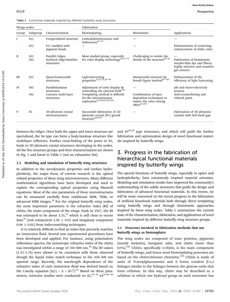
building blocks. This approach may also offer many differentmodel systems for 3D photonic crystal (PC) research.69 As thestrength of an external magnetic field increased, the reflectancespectra of the Fe3O4 replica of the wings was red-shifted. Thistype of tunability opens up an avenue for the creation of newmagneto-optical devices and may inspire theoretical studies inthe field.70 Inorganic (SiO2 and TiO2) chiral 3-D PC with abicontinuous gyroid structure (S5) were replicated from C. rubiwing scales using a similar sol–gel process106,107 (shown inFig. 4). The replica has an equivalent unit cell, an increased fillfraction, and a decreased dielectric constant relative to itschitin template. Thus, the change in the dielectric contrast isthe main factor for the observed blue shift upon replication.The 3DSiO2 and TiO2 photonic crystals replicated in C. Mille’swork have nearly a complete overlapping of the partial bandgaps, strongly suggesting that materials with full photonicband gaps are experimentally within reach.
Metals, including Co, Ni, Cu, Pd, Ag, Pt, and Au, which areshown in Fig. 5 II, can also be reduced using the electrolessdeposition methods in the sol–gel solution.77,78 The Au, Ag, andCu replicas of butterfly wing scales were found to have mark-edly more pronounced Raman scattering (SERS) effects thanreplicas made of other materials. The pronounced Ramanenhancement mainly originates from the 3D sub-micrometerperiodic rib-structures located on the main ridges of Cu scales,
rather than from the morphological or size effects of Cu NPsthemselves.79 Similar to the way the more floors a building has,the more residents it can contain, more periodically arrangedrib-layers per unit square result in more piled-up hotspots,leading to better SERS performance. This is because of theaccumulation of hotspots, which are located at sites where thelocal electromagnetic fields are markedly enhanced. Thesehotspots are produced by the intrinsic 3D sub-micrometerstructures of the metals. SERS has attracted a great deal ofattention due to its ability to facilitate the fast detection of traceamounts of chemicals. Metallic SERS substrates can enhance theweak Raman signals of analytes adsorbed on them by severalorders of magnitude. However, the low reproducibility and highcost of SERS substrates as consumables restrict their use. Incomparison, the sensitivity of the system’s ability to detectRhodamine 6G (R6G) on Au (10�13 M) is ten times higher thanthat of its ability to detect R6G on Klarites (10�12 M), which is thecommercial SERS substrate. Considering the high cost and thesensitivity of detection on metallic butterfly wings, this strategymay render affordable and stable SERS substrates accessibleto laboratories across the world. By combining well-developedtuning methods with these naturally designed 3D scaffolds, themorphology of the nanometal particles and the formation ofstructures with multiple components can be controlled.127 Evenhigher SERS performance can also be expected.
Fig. 4 SEM images of a cross-section of original C. rubi wing scales (a) and SiO2 (b), TiO2 (c) scale templated from (a). Insets of (a), (b), (c) represent theoptical image of relative scales. Scale bars are all 1 mm for (a), (b) and (c). (d) Is the partial band gap map for titania as a function of solid volume fractionfeaturing the partial band gaps, shown as colored regions. Note the shift to lower wavelengths with decreasing volume fraction. The region denoted bythe rectangle centered near the 50% volume fraction is a zone of reasonable fit to the observed spectral data. The three primary bands correspond to theGG–N, G–P, and G–H directions, i.e., the h110i, h111i and h100i. Crystallographic directions are represented in light green, blue, and purple, respectively.(e) Representative reflectivity spectra of C. rubi and silica and titania replicas, with the position of the reflection maxima centered at 540 nm, 500 nm, and505 nm, respectively. Note that the maximum reflectivity of silica (y25%) is below that of the original butterfly scale. The titania replica shows a reflectivitymaximum of 90%. (f) Comparison of the spectra obtained from titania photonic crystals calcined between 400 1C and 900 1C.106,107
Perspective PCCP
Publ
ishe
d on
18
July
201
4. D
ownl
oade
d by
Ast
on U
nive
rsity
on
04/0
9/20
14 1
4:53
:09.
View Article Online

This journal is© the Owner Societies 2014 Phys. Chem. Chem. Phys., 2014, 16, 19767--19780 | 19773
3.1.2 Nanoimprintating process. Nanoimprint lithography(NIL) shows considerable promise due to its low cost, highthroughput capacity, and ability to imprint large areas. It createspatterns using the mechanical deformation of an imprint resistand subsequent processes.128 The imprint resist is typically amonomer or polymer formulation that is cured by heat or UVlight during the imprinting. Adhesion between the resist andthe template is controlled to allow proper demoulding. Thisprocedure usually requires the fabrication of master-molds byconventional microelectronics techniques, such as lithographyand etching. These are time-consuming and limited in scale. Thepreparation of stamps that maintain high resolution over largeareas is key to NIL. Many efforts have been made to developstamps with different alignment structures, especially bionano-structures, such as those of leaves and insect wings, to resolvethe problem of micro- and nanostructure imprinting.129,130
Using these techniques, some research groups have used naturalbutterfly wings as master-molds to cast elastomeric templates,both negative and positive structures of the original wings. Thissimplified the fabrication process and allowed the reproductionof the original complex patterns on the wings.
The wing of P. ulysses has concave microstructures andmultilayers of about five to ten periods. This wing was chosenfor replication using flexible polydimethylsiloxane (PDMS).104
Because of the low Young modulus of the PDMS (around0.8 MPa), the as-synthesized templates can replicate theconcave curvature of the surfaces of the butterfly wings. Thismakes them suitable for use in imprint spin-coated resistantfilms at low pressures. Methyltriethoxysilane (MTEOS) sol–gelfilms were prepared for the imprint at a pressure of under 2 bars.
The temperature was kept between 80 1C and 150 1C, and theprocess lasted about 20 min. This technique produces a multi-layered structure, and it is well suited to producing the tree-likestructures of Morpho wings. The results of absorptivity, reflectivity,and the fluorescent characteristics of the replicas showed that themicrostructural and optical characteristics of the replicatedwing were qualitatively consistent with those of actual wings.82
Researchers believed that this technique may represent a viableapproach to the mass production of artificial PC structuressuitable for a variety of commercial applications. First, electricfield and pH sensors were produced by filling the multilayerstructures (C. rhipheus, S4) with electric-field-sensitive orpH-sensitive hydrogels.10,21,31 The visible reflectance of theimmobilized wing scales was found to be responsive to electricfield and pH conditions (shown in Fig. 6). This was attributableto the inner microstructural changes induced by the changes inthe volume of the embedded polymer during the swelling–de-swelling process. The pH-induced color change was detected byreflectance spectra, as well as by optical observation. A distinct Utransition with pH was observed, demonstrating PMAA content-dependent properties. These works set up a strategy for thedesign and fabrication of tunable photonic crystals with hier-archical structures, which provides a route for combining func-tional polymers with biotemplates for wide potential use inmany fields.110,131
3.1.3 Vapor phase deposition. Vapor phase deposition is aprocess in which a gas phase transforms into a solid phase. It iswildly used in thin-film coating. Two major vapor phasedeposition techniques, chemical vapor deposition (CVD) andatomic layer deposition (ALD), are based on the sequential use
Fig. 5 (I) Fabrication of Ag-replicas of butterfly wings.77 (II) Pseudo-color SEM images of seven metallic wing-scale replicas.78 (III) (a) SERS spectra of a10�3 M R6G solution on substrate I (a smooth, thin Ag film), substrate II (Ag nanoparticles ground from the biotemplated Ag replicas using an agatemortar), substrate III (Ag replica with the quasi-periodic submicrostructures prepared using P. paris as a template), and substrate IV (Ag replica with theperiodic submicrostructures replicated from E. mulciber). (b) Comparison of the 612 cm�1 (left column) and 1364 cm�1 (right column) SERS signal bandareas of R6G for the four types of substrates in (a). The intensities of the R6G Raman signals on substrate IV were approximately 15, 6, and 2 times higherthan those of substrates I–III, respectively.77
PCCP Perspective
Publ
ishe
d on
18
July
201
4. D
ownl
oade
d by
Ast
on U
nive
rsity
on
04/0
9/20
14 1
4:53
:09.
View Article Online

19774 | Phys. Chem. Chem. Phys., 2014, 16, 19767--19780 This journal is© the Owner Societies 2014
of a gas phase chemical process. ALD is chemically similar toCVD, except that the ALD reaction breaks the CVD reaction intotwo half-reactions, keeping the precursor materials separatedduring the reaction. Because its reactions are self-limiting andtake place on the surface of the substrate, ALD makes atomicscale deposition control possible.132,133 Compared to CVD, ALDcan provide a simple and accurate thickness control as it can becontrolled with every cycle. Furthermore, ALD offers faciledoping and large area uniformity, and thus a straightforwardscale-up.133 The advantage is obvious, as this method can beapplied to most materials which can be deposited by ALD, e.g.,on the soft and fragile butterfly wing templates.21
Early in 2003, the technique involving the controlled vapor-phase oxidation of silanes on the surface of peacock butterflywings was used to produce exact inorganic oxide replicas ofstructures observed in nature.51 The primary silica clusters hadextraordinary flow properties and were found capable of creepinginto the smallest gaps within the wings. In this way, this methodshowed promise for the replication of these intricate hierarchicalstructures. However, incompatibility between the butterfly wingsand the silane coating can cause catastrophic cracking of thecoating during calcination, as observed under SEM.
Therefore, to fabricate high-fidelity replicas of differentbiotemplates, another film deposition process, conformalevaporated film by rotation (CEFR), was developed.89,90 TheCEFR technique is particularly well suited to biomimetization,because the temperatures involved are sufficiently low and thereplication process occurs in a noncorrosive environment,which prevents damage and distortion of the C-, H-, andO-based organic skeletons. By controlling the current andvapor flux, a 0.5 mm coating was placed on the wings. Becausethe morphology of the butterfly wing makes it a very efficientdiffuser of light, the chalcogenide glass (Ge28Sb12Se60) replicaof Battus philenor, which has a large index of refraction withinthe visible and infrared spectra, can be used as an antireflec-tion structure for increased photon trapping and opticaldiffusers.
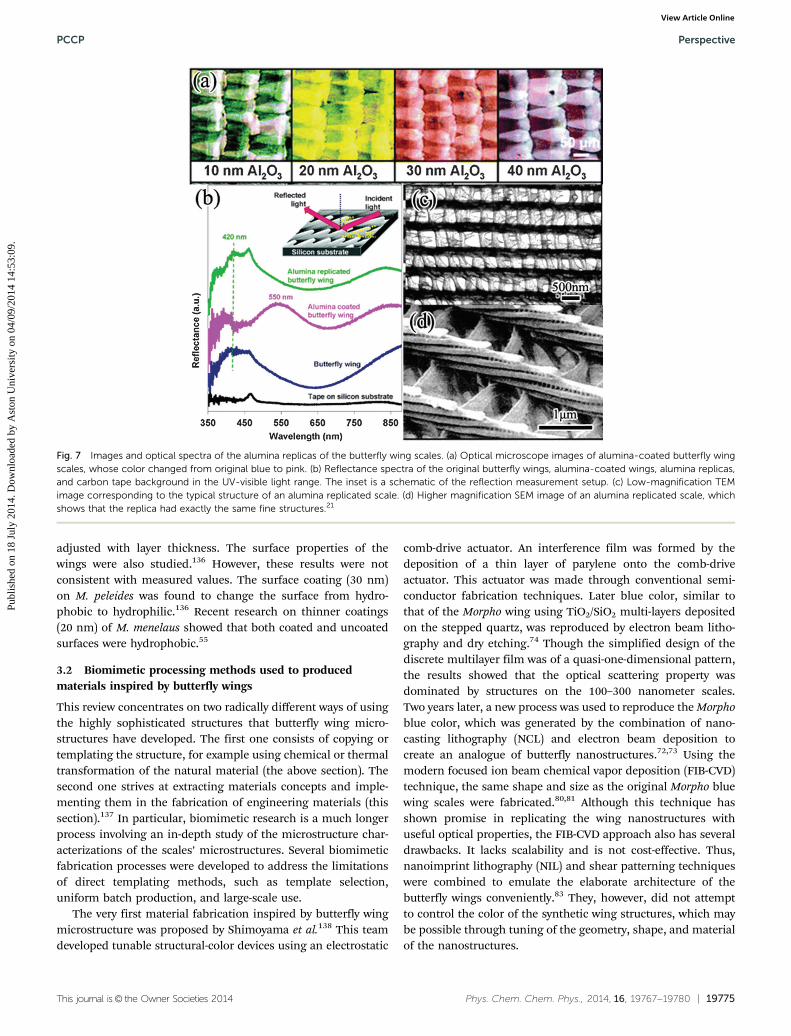
The thickness of the coating was not very easy to adjustthrough the amount of silane precursor and the reaction timeof the CVD technique or the current and flux of CEFR. In orderto overcome these problems, the atomic layer deposition (ALD)technique was used in the replication of butterfly wings.21 ALDis a self-limiting, sequential surface chemical technique thatdeposits thin, conformal films of materials onto substrates ofvarying compositions.134 This mechanism facilitates the growthof conformal thin films of specific thicknesses on large areas.By keeping the precursors separate throughout the coatingprocess, control of the growth of atomic films can be renderedas fine as E0.1 Å (100 pm) per cycle. These advantages makethe ALD method suitable for the more exact replication ofnatural materials with 3D hierarchical structures. Z. L. Wang’set al. were the first to use the atomic layer deposition approachto replicate and coat the photonic structures of M. peleides.21
The growth rate was 1 Å per cycle, which allowed exact controlof the thickness of alumina replicas of the wings (Fig. 7(a)).The alumina replicas also exhibited optical properties thatfacilitated applications in waveguides and beam splitters.Further research into the influence of layer thickness on theoptical properties was performed using ALD on the iridescentgreen scales (S42) of P. blumei.135 Studies have shown that,depending on the structural integrity of the initially sealedscale, it is possible to replicate not only the outer but also theinner surfaces of the structure, which may be even morecomplex than the outer surfaces. Each of these replicationprocesses produces distinct multicolor optical properties, asshown by experimental and theoretical data. One recent studyevaluated the influence of layer thickness on the reflectanceproperties and the corresponding angle-resolved optical perfor-mance of alumina wing replicas using the higher aspect ratiomicrostructure of M. menelaus.55 The ALD replication processmakes it possible to use these precise replicas as building blocksfor photonic integrated circuit systems with higher reproduci-bility and lower fabrication costs than traditional lithographytechniques. Results showed that the optical properties could be
Fig. 6 The upper column: (a) overall synthesis of pH-responsive PCs from Morpho butterfly wings. Lower column: representative spectral responses of(b) PMAA-PC-1, (c) PMAA-PC-2, and (d) PMAA-PC-3 to different pH values, with representative optical images obtained at typical pH values.110
Perspective PCCP
Publ
ishe
d on
18
July
201
4. D
ownl
oade
d by
Ast
on U
nive
rsity
on
04/0
9/20
14 1
4:53
:09.
View Article Online
This journal is© the Owner Societies 2014 Phys. Chem. Chem. Phys., 2014, 16, 19767--19780 | 19775
adjusted with layer thickness. The surface properties of thewings were also studied.136 However, these results were notconsistent with measured values. The surface coating (30 nm)on M. peleides was found to change the surface from hydro-phobic to hydrophilic.136 Recent research on thinner coatings(20 nm) of M. menelaus showed that both coated and uncoatedsurfaces were hydrophobic.55
3.2 Biomimetic processing methods used to producedmaterials inspired by butterfly wings
This review concentrates on two radically different ways of usingthe highly sophisticated structures that butterfly wing micro-structures have developed. The first one consists of copying ortemplating the structure, for example using chemical or thermaltransformation of the natural material (the above section). Thesecond one strives at extracting materials concepts and imple-menting them in the fabrication of engineering materials (thissection).137 In particular, biomimetic research is a much longerprocess involving an in-depth study of the microstructure char-acterizations of the scales’ microstructures. Several biomimeticfabrication processes were developed to address the limitationsof direct templating methods, such as template selection,uniform batch production, and large-scale use.
The very first material fabrication inspired by butterfly wingmicrostructure was proposed by Shimoyama et al.138 This teamdeveloped tunable structural-color devices using an electrostatic
comb-drive actuator. An interference film was formed by thedeposition of a thin layer of parylene onto the comb-driveactuator. This actuator was made through conventional semi-conductor fabrication techniques. Later blue color, similar tothat of the Morpho wing using TiO2/SiO2 multi-layers depositedon the stepped quartz, was reproduced by electron beam litho-graphy and dry etching.74 Though the simplified design of thediscrete multilayer film was of a quasi-one-dimensional pattern,the results showed that the optical scattering property wasdominated by structures on the 100–300 nanometer scales.Two years later, a new process was used to reproduce the Morphoblue color, which was generated by the combination of nano-casting lithography (NCL) and electron beam deposition tocreate an analogue of butterfly nanostructures.72,73 Using themodern focused ion beam chemical vapor deposition (FIB-CVD)technique, the same shape and size as the original Morpho bluewing scales were fabricated.80,81 Although this technique hasshown promise in replicating the wing nanostructures withuseful optical properties, the FIB-CVD approach also has severaldrawbacks. It lacks scalability and is not cost-effective. Thus,nanoimprint lithography (NIL) and shear patterning techniqueswere combined to emulate the elaborate architecture of thebutterfly wings conveniently.83 They, however, did not attemptto control the color of the synthetic wing structures, which maybe possible through tuning of the geometry, shape, and materialof the nanostructures.
Fig. 7 Images and optical spectra of the alumina replicas of the butterfly wing scales. (a) Optical microscope images of alumina-coated butterfly wingscales, whose color changed from original blue to pink. (b) Reflectance spectra of the original butterfly wings, alumina-coated wings, alumina replicas,and carbon tape background in the UV-visible light range. The inset is a schematic of the reflection measurement setup. (c) Low-magnification TEMimage corresponding to the typical structure of an alumina replicated scale. (d) Higher magnification SEM image of an alumina replicated scale, whichshows that the replica had exactly the same fine structures.21
PCCP Perspective
Publ
ishe
d on
18
July
201
4. D
ownl
oade
d by
Ast
on U
nive
rsity
on
04/0
9/20
14 1
4:53
:09.
View Article Online
19776 | Phys. Chem. Chem. Phys., 2014, 16, 19767--19780 This journal is© the Owner Societies 2014
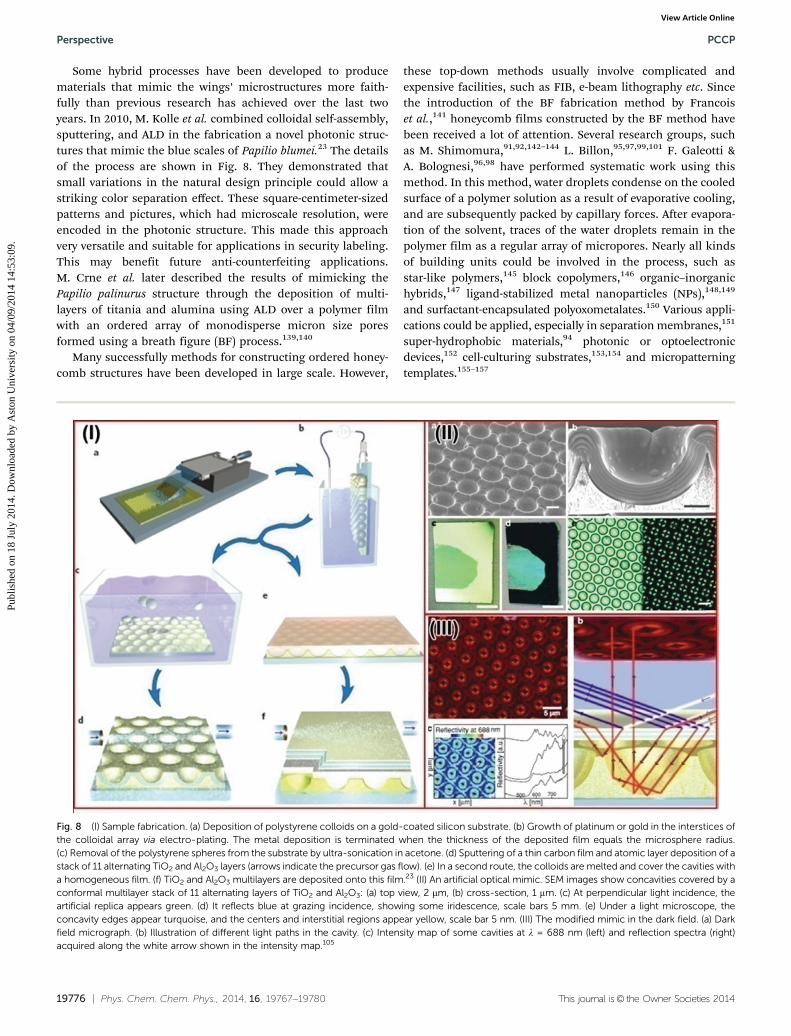
Some hybrid processes have been developed to producematerials that mimic the wings’ microstructures more faith-fully than previous research has achieved over the last twoyears. In 2010, M. Kolle et al. combined colloidal self-assembly,sputtering, and ALD in the fabrication a novel photonic struc-tures that mimic the blue scales of Papilio blumei.23 The detailsof the process are shown in Fig. 8. They demonstrated thatsmall variations in the natural design principle could allow astriking color separation effect. These square-centimeter-sizedpatterns and pictures, which had microscale resolution, wereencoded in the photonic structure. This made this approachvery versatile and suitable for applications in security labeling.This may benefit future anti-counterfeiting applications.M. Crne et al. later described the results of mimicking thePapilio palinurus structure through the deposition of multi-layers of titania and alumina using ALD over a polymer filmwith an ordered array of monodisperse micron size poresformed using a breath figure (BF) process.139,140
Many successfully methods for constructing ordered honey-comb structures have been developed in large scale. However,
these top-down methods usually involve complicated andexpensive facilities, such as FIB, e-beam lithography etc. Sincethe introduction of the BF fabrication method by Francoiset al.,141 honeycomb films constructed by the BF method havebeen received a lot of attention. Several research groups, suchas M. Shimomura,91,92,142–144 L. Billon,95,97,99,101 F. Galeotti &A. Bolognesi,96,98 have performed systematic work using thismethod. In this method, water droplets condense on the cooledsurface of a polymer solution as a result of evaporative cooling,and are subsequently packed by capillary forces. After evapora-tion of the solvent, traces of the water droplets remain in thepolymer film as a regular array of micropores. Nearly all kindsof building units could be involved in the process, such asstar-like polymers,145 block copolymers,146 organic–inorganichybrids,147 ligand-stabilized metal nanoparticles (NPs),148,149
and surfactant-encapsulated polyoxometalates.150 Various appli-cations could be applied, especially in separation membranes,151
super-hydrophobic materials,94 photonic or optoelectronicdevices,152 cell-culturing substrates,153,154 and micropatterningtemplates.155–157
Fig. 8 (I) Sample fabrication. (a) Deposition of polystyrene colloids on a gold-coated silicon substrate. (b) Growth of platinum or gold in the interstices ofthe colloidal array via electro-plating. The metal deposition is terminated when the thickness of the deposited film equals the microsphere radius.(c) Removal of the polystyrene spheres from the substrate by ultra-sonication in acetone. (d) Sputtering of a thin carbon film and atomic layer deposition of astack of 11 alternating TiO2 and Al2O3 layers (arrows indicate the precursor gas flow). (e) In a second route, the colloids are melted and cover the cavities witha homogeneous film. (f) TiO2 and Al2O3 multilayers are deposited onto this film.23 (II) An artificial optical mimic. SEM images show concavities covered by aconformal multilayer stack of 11 alternating layers of TiO2 and Al2O3: (a) top view, 2 mm, (b) cross-section, 1 mm. (c) At perpendicular light incidence, theartificial replica appears green. (d) It reflects blue at grazing incidence, showing some iridescence, scale bars 5 mm. (e) Under a light microscope, theconcavity edges appear turquoise, and the centers and interstitial regions appear yellow, scale bar 5 nm. (III) The modified mimic in the dark field. (a) Darkfield micrograph. (b) Illustration of different light paths in the cavity. (c) Intensity map of some cavities at l = 688 nm (left) and reflection spectra (right)acquired along the white arrow shown in the intensity map.105
Perspective PCCP
Publ
ishe
d on
18
July
201
4. D
ownl
oade
d by
Ast
on U
nive
rsity
on
04/0
9/20
14 1
4:53
:09.
View Article Online

This journal is© the Owner Societies 2014 Phys. Chem. Chem. Phys., 2014, 16, 19767--19780 | 19777
4. Conclusion and future outlook
Attracted by the fascinating subtle structures of butterfly wingscales, more and more interest has been raised in their studyover the past decade. For the re-production of these structuresin the laboratory, an exact and convenient technique is stillrequired that would allow an excellent replication of theoriginal wing scales microstructures. Biotemplating makes itpossible, even easy, to maintain the complexity of the originalstructures, but is not suitable for batch fabrication. Biomimeticmethods can be used to produce uniform materials but theycannot readily be used to mimic the original hierarchicalmicrostructures of the butterfly wings. Herein, through thecomparison and evaluation of various preparation methods, apotential method combining both biotemplating and biomi-metic concepts has been developed. In this process, Fe@carbonreplicas of wing scales was used as a second imprintingtemplate, and massive biomimetic butterfly wing scales struc-tures could be reproduced.158 This is the first step to combineboth the benefits of biotemplating and biomimetics, thoughthe second imprinting microstructure is not as perfect as theoriginal scale. Also, more simulations and design researchmust be performed before biomimetic fabrication can becomea reality on any practical scale. The optimization of properstructures and extraction of the key parameters with corres-ponding properties may guide and simplify the synthesis ofmaterials.45,159 The ultimate aim of the current work is tofacilely, accurately and reproducibly design and optimize var-ious functional materials based on the different structureseffected by the butterfly wings.
Acknowledgements
This work was supported by the National Natural Science Founda-tion of China (no. 51202145, no. 51171110, no. 51271116 and no.91333115), the National Basic Research Program of China (973Program, no. 2011cb922200). Research Fund for the DoctoralProgram of Higher Education of China (20120073120006 and20120073130001), and Scientific Research Foundation forReturned Scholars.
References
1 R. G. Foottit and P. H. Adler, Insect biodiversity: science andsociety, Blackwell Pub, 2009.
2 S. Wickham, M. Large, L. Poladian and L. Jermiin, J. R. Soc.Interface, 2006, 3, 99.
3 D. G. Stavenga, M. A. Giraldo and H. L. Leertouwer, J. Exp.Biol., 2010, 213, 1731–1739.
4 S. Berthier, Iridescences: the physical colors of insects,Springer, New York, NY, 2007.
5 H. Ghiradella, Microscopic anatomy of invertebrates, 1998,vol. 11, pp. 257–287.
6 S. Berthier, La couleur des papillons ou l’imperative beaute:proprietes optiques des ailes de papillons, Springer, 2000.
7 P. Vukusic, R. Sambles, Physics, Theory, and Applications ofPeriodic Structures in Optics, 2001, vol. 4438, pp. 85–95.
8 S. Kinoshita, Structural colors in the realm of nature, WorldScientific Pub Co Inc, 2008.
9 S. Berthier, Photonique des Morphos, Springer, 2010.10 A. L. Ingram and A. R. Parker, Philos. Trans. R. Soc. London,
Ser. B, 2008, 363, 2465–2480.11 H. Ghiradella and W. Radigan, J. Morphol., 1976, 150,
279–297.12 H. Ghiradella, Insect cuticular surface modifications: scales
and other structural formations, 2010.13 P. Vukusic, Functional Surfaces in Biology, 2009, pp. 237–258.14 S. Kinoshita and S. Yoshioka, ChemPhysChem, 2005, 6,
1442–1459.15 A. Heuer, D. Fink, V. Laraia, J. Arias, P. Calvert, K. Kendall,
G. Messing, J. Blackwell, P. Rieke and D. Thompson,Science, 1992, 255, 1098.
16 S. Sotiropoulou, Y. Sierra-Sastre, S. Mark and C. Batt,Chem. Mater., 2008, 20, 821–834.
17 K. Liu and L. Jiang, ACS Nano, 2011, 5, 6786–6790.18 M. H. Bartl, J. W. Galusha and M. R. Jorgensen, Oxide-
Based Photonic Crystals from Biological Templates, inFunctional Metal Oxide Nanostructures, ed. J. Wu, J. Cao,W.-Q. Han, A. Janotti and H.-C. Kim, Springer, New York,2012, vol. 149, pp. 175–207.
19 Y. Liu, J. Goebl and Y. Yin, Chem. Soc. Rev., 2013, 42,2610–2653, DOI: 10.1039/C2CS35369E.
20 F. P. Barrows and M. H. Bart, Nanomater. Nanotechnol.,2014, 4, DOI: 10.5772/58289.
21 J. Huang, X. Wang and Z. Wang, Nano Lett., 2006, 6,2325–2331.
22 W. Zhang, D. Zhang, T. Fan, J. Gu, J. Ding, H. Wang,Q. Guo and H. Ogawa, Chem. Mater., 2009, 21, 33–40.
23 M. Kolle, P. M. Salgard-Cunha, M. R. J. Scherer, F. Huang,P. Vukusic, S. Mahajan, J. J. Baumberg and U. Steiner, Nat.Nanotechnol., 2010, 5, 511–515.
24 R. A. Potyrailo, H. Ghiradella, A. Vertiatchikh,K. Dovidenko, J. R. Cournoyer and E. Olson, Nat. Photonics,2007, 1, 123–128.
25 A. D. Pris, Y. Utturkar, C. Surman, W. G. Morris, A. Vert,S. Zalyubovskiy, T. Deng, H. T. Ghiradella andR. A. Potyrailo, Nat. Photonics, 2012, 6, 195–200.
26 Butterfly tech team: DARPA nod ‘detonates’ status quo,http://www.gereports.com/butterfly-tech-team-darpa-nod-detonates-status-quo/.
27 L. P. Biro and J. P. Vigneron, Laser Photonics Rev., 2011, 5,27–51.
28 A. Sweeney, C. Jiggins and S. Johnsen, Nature, 2003, 423,31–32.
29 S. M. Doucet and M. G. Meadows, J. R. Soc. Interface, 2009,6, S115–S132.
30 D. J. Kemp, Proc. R. Soc. B, 2007, 274, 1043–1047.31 N. D. Wanasekara and V. B. Chalivendra, Soft Matter, 2011,
7, 373–379.32 Y. Fang, G. Sun, Q. Cong, G. H. Chen and L. Q. Ren,
J. Bionic Eng., 2008, 5, 127–133.
PCCP Perspective
Publ
ishe
d on
18
July
201
4. D
ownl
oade
d by
Ast
on U
nive
rsity
on
04/0
9/20
14 1
4:53
:09.
View Article Online

19778 | Phys. Chem. Chem. Phys., 2014, 16, 19767--19780 This journal is© the Owner Societies 2014
33 R. Hooke, Micrographia: or some physiological descriptionsof minute bodies, made by magnifying glasses with observa-tions and inquiries thereupon, James Allestry, 1665.
34 C. W. Mason, J. Phys. Chem., 1926, 30, 383–395.35 C. W. Mason, J. Phys. Chem., 1927, 31, 321–354.36 T. F. Anderson and A. G. Richards, J. Appl. Phys., 1942, 13,
748–758.37 W. Lippert and K. Gentil, Zoomorphology, 1959, 48,
115–122.38 S. A. Boden, A. Asadollahbaik, H. N. Rutt and D. M. Bagnall,
Scanning, 2012, 34, 107–120, DOI: 10.1002/sca.20267.39 P. Vukusic, J. R. Sambles, C. R. Lawrence and
R. J. Wootton, Proc. R. Soc. B, 1999, 266, 1403–1411.40 S. Berthier, E. Charron and A. D. Silva, Opt. Commun., 2003,
228, 349–356.41 H. L. Leertouwer, B. D. Wilts and D. G. Stavenga, Opt.
Express, 2011, 19, 24061–24066.42 A. Mejdoubi, C. Andraud, S. Berthier, J. Lafait,
J. Boulenguez and E. Richalot, Phys. Rev. E: Stat., Nonlinear,Soft Matter Phys., 2013, 87, 022705.
43 R. Lee, PhD thesis, Georgia Institute of Technology, 2009.44 R. Lee and G. Smith, Appl. Opt., 2009, 48, 4177–4190.45 W. Wang, W. Zhang, W. Chen, J. Gu, Q. Liu, T. Deng and
D. Zhang, Opt. Lett., 2013, 38, 169–171.46 P. Vukusic, J. Sambles and C. Lawrence, Nature, 2000,
404, 457.47 P. Vukusic, R. Sambles, C. Lawrence and G. Wakely, Appl.
Opt., 2001, 40, 1116–1125.48 J. P. Vigneron, M. Ouedraogo, J.-F. Colomer and
M. Rassart, Phys. Rev. E: Stat., Nonlinear, Soft Matter Phys.,2009, 79, 021907.
49 M. Saba, M. Thiel, M. D. Turner, S. T. Hyde, M. Gu,K. Grosse-Brauckmann, D. N. Neshev, K. Mecke andG. E. Schroder-Turk, Phys. Rev. Lett., 2011, 106, 103902.
50 B. D. Wilts, K. Michielsen, H. De Raedt and D. G. Stavenga,Interface Focus, 2012, 2, 681–687.
51 G. Cook, P. Timms and C Gltner-Spickermann, Angew.Chem., Int. Ed., 2003, 42, 557–559.
52 W. Zhang, D. Zhang, T. X. Fan, J. Ding, J. J. Gu, Q. X. Guoand H. Ogawa, Bioinspiration Biomimetics, 2006, 1, 89–95.
53 W. Zhang, D. Zhang, T. X. Fan, J. Ding, Q. X. Gu andH. Ogawa, Nanotechnology, 2006, 17, 840–844.
54 W. Zhang, D. Zhang, T. X. Fan, J. Ding, Q. X. Guo andH. Ogawa, Microporous Mesoporous Mater., 2006, 92,227–233.
55 F. Liu, Y. P. Liu, L. Huang, X. H. Hu, B. Q. Dong, W. Z. Shi,Y. Q. Xie and X. A. Ye, Opt. Commun., 2011, 284, 2376–2381.
56 S. Zhu, D. Zhang, Z. Chen, J. Gu, W. Li, H. Jiang andG. Zhou, Nanotechnology, 2009, 20, 315303.
57 Y. Chen, X. N. Zang, J. J. Gu, S. M. Zhu, H. L. Su, D. Zhang,X. B. Hu, Q. L. Liu, W. Zhang and D. X. Liu, J. Mater. Chem.,2011, 21, 6140–6143.
58 M. R. Weatherspoon, Y. Cai, M. Crne, M. Srinivasarao andK. H. Sandhage, Angew. Chem., Int. Ed., 2008, 47, 7921–7923.
59 X. Y. Liu, S. M. Zhu, D. Zhang and Z. X. Chen, Mater. Lett.,2010, 64, 2745–2747.
60 O. Sato, S. Kubo and Z. Z. Gu, Acc. Chem. Res., 2009, 42,1–10.
61 W. Zhang, D. Zhang, T. Fan, J. Ding, Q. Guo and H. Ogawa,Nanotechnology, 2006, 17, 840–844.
62 J. P. Vernon, Y. N. Fang, Y. Cai and K. H. Sandhage, Angew.Chem., Int. Ed., 2010, 49, 7765–7768.
63 H. H. Liu, QibinZhao, H. Zhou, J. Ding, D. Zhang,H. X. Zhu and T. X. Fan, Phys. Chem. Chem. Phys., 2011,13, 10872–10876.
64 J. Silver, R. Withnall, T. G. Ireland and G. R. Fern, J. Mod.Opt., 2005, 52, 999–1007.
65 F. Song, H. Su, J. Han, J. Xu and D. Zhang, Sens. Actuators,B, 2010, 145, 39–45.
66 F. Song, H. Su, J. Han, D. Zhang and Z. Chen, Nanotechno-logy, 2009, 20, 495502.
67 Y. Chen, J. Gu, S. Zhu, T. Fan and Q. Guo, Appl. Phys. Lett.,2009, 94, 053901.
68 Y. Chen, J. Gu, D. Zhang, S. Zhu, H. Su, X. Hu, C. Feng,W. Zhang, Q. Liu and A. R. Parker, J. Mater. Chem., 2011,21, 15237.
69 W. Peng, X. Hu and D. Zhang, J. Magn. Magn. Mater., 2011,323, 2064–2069.
70 W. Peng, S. Zhu, W. Wang, W. Zhang, J. Gu, X. Hu,D. Zhang and Z. Chen, Adv. Funct. Mater., 2012, 22, 2071.
71 S. Zhu, X. Liu, Z. Chen, C. Liu, C. Feng, J. Gu, Q. Liu andD. Zhang, J. Mater. Chem., 2010, 20, 9126–9132.
72 A. Saito, Y. Miyamura, M. Nakajima, Y. Ishikawa, K. Sogo,Y. Kuwahara and Y. Hirai, J. Vac. Sci. Technol., B: Micro-electron. Nanometer Struct.–Process., Meas., Phenom., 2006,24, 3248–3251.
73 A. Saito, M. Nakajima, Y. Miyamura, K. Sogo, Y. Ishikawaand Y. Hirai, Nanoengineering: Fabrication, Properties,Optics, and Devices III, 2006, vol. 6327, p. Z3270.
74 A. Saito, S. Yoshioka and S. Kinoshita, presented in part atthe Proceedings of SPIE, Optical Systems Degradation,Contamination, and Stray Light: Effects, Measurements,and Control, 2004.
75 Q. Zhao, T. Fan, J. Ding, D. Zhang, Q. Guo and M. Kamada,Carbon, 2011, 49, 877–883.
76 J. Han, H. L. Su, F. Song, D. Zhang and Z. X. Chen,Nanoscale, 2010, 2, 2203–2208.
77 Y. Tan, X. Zang, J. Gu, D. Liu, S. Zhu, H. Su, C. Feng, Q. Liu,W. M. Lau, W.-J. Moon and D. Zhang, Langmuir, 2011, 27,11742–11746.
78 Y. Tan, J. Gu, X. Zang, W. Xu, K. Shi, L. Xu and D. Zhang,Angew. Chem., Int. Ed., 2011, 50, 1–6.
79 Y. W. Tan, J. J. Gu, L. H. Xu, X. N. Zang, D. Liu, W. Zhang,Q. L. Liu, S. M. Zhu, H. L. Su, C. L. Feng, G. L. Fan andD. Zhang, Adv. Funct. Mater., 2012, 22, 1578–1585.
80 K. Watanabe, T. Hoshino, K. Kanda, Y. Haruyama, T. Kaitoand S. Matsui, J. Vac. Sci. Technol., B: Microelectron. Nano-meter Struct.–Process., Meas., Phenom., 2005, 23, 570–574.
81 K. Watanabe, T. Hoshino, K. Kanda, Y. Haruyama andS. Matsui, Jpn. J. Appl. Phys., 2005, 44, L48–L50.
82 S. H. Kang, T. Y. Tai and T. H. Fang, Curr. Appl. Phys., 2010,10, 625–630.
Perspective PCCP
Publ
ishe
d on
18
July
201
4. D
ownl
oade
d by
Ast
on U
nive
rsity
on
04/0
9/20
14 1
4:53
:09.
View Article Online

This journal is© the Owner Societies 2014 Phys. Chem. Chem. Phys., 2014, 16, 19767--19780 | 19779
83 T. S. Kustandi, H. Y. Low, J. H. Teng, I. Rodriguez andR. Yin, Small, 2009, 5, 574–578.
84 R. Coath, MPhil thesis, University of Southampton, 2007.85 Z. Xu, K. Yu, B. Li, R. Huang, P. Wu, H. Mao, N. Liao and
Z. Zhu, Nano Res., 2011, 4, 737–745.86 W. Zhang, D. Zhang, T. X. Fan, J. Ding, J. J. Gu, Q. Guo and
H. Ogawa, Mater. Sci. Eng., C, 2009, 29, 92–96.87 S. Zhu, D. Zhang, Z. Li, H. Furukawa and Z. Chen, Lang-
muir, 2008, 24, 6292–6299.88 A. R. Maddocks and A. T. Harris, Mater. Lett., 2009, 63,
748–750.89 R. J. Martın-Palma, C. G. Pantano and A. Lakhtakia, Appl.
Phys. Lett., 2008, 93, 083901.90 A. Lakhtakia, R. Martin-Palma, M. Motyka and C. Pantano,
Bioinspiration Biomimetics, 2009, 4, 034001.91 T. Kawano, M. Sato, H. Yabu and M. Shimomura, Biomater.
Sci., 2014, 2, 52–56.92 Y. Saito, M. Shimomura and H. Yabu, Chem. Commun.,
2013, 49, 6081–6083.93 H. Yabu, R. Jia, Y. Matsuo, K. Ijiro, S.-a. Yamamoto,
F. Nishino, T. Takaki, M. Kuwahara and M. Shimomura,Adv. Mater., 2008, 20, 4200–4204.
94 H. Yabu, M. Takebayashi, M. Tanaka and M. Shimomura,Langmuir, 2005, 21, 3235–3237.
95 S. Chen, M.-H. Alves, M. Save and L. Billon, Polym. Chem.,2014, DOI: 10.1039/C4PY00390J.
96 F. Galeotti, W. Mroz, G. Scavia and C. Botta, Org. Electron.,2013, 14, 212–218.
97 A.-C. Courbaron Gilbert, C. Derail, N. E. El Bounia andL. Billon, Polym. Chem., 2012, 3, 415–420.
98 F. Galeotti, V. Calabrese, M. Cavazzini, S. Quici,C. Poleunis, S. Yunus and A. Bolognesi, Chem. Mater.,2010, 22, 2764–2769.
99 C. Deleuze, M. H. Delville, V. Pellerin, C. Derail andL. Billon, Macromolecules, 2009, 42, 5303–5309.
100 L. Billon, M. Manguian, V. Pellerin, M. Joubert,O. Eterradossi and H. Garay, Macromolecules, 2008, 42,345–356.
101 L. Ghannam, M. Manguian, J. Francois and L. Billon, SoftMatter, 2007, 3, 1492–1499.
102 X. Zang, Y. Ge, J. Gu, S. Zhu, H. Su, C. Feng, W. Zhang, Q. Liuand D. Zhang, J. Mater. Chem., 2011, 21, 13913–13919.
103 B. Li, J. Zhou, R. Zong, M. Fu, Y. Bai, L. Li and Q. Li, J. Am.Ceram. Soc., 2006, 89, 2298–2300.
104 T. Saison, C. Peroz, V. Chauveau, S. Berthier, E. Sondergardand H. Arribart, Bioinspiration Biomimetics, 2008,3, 046004.
105 M. Kolle, Photonic structures inspired by nature, not avail,2010.
106 C. Mille, E. C. Tyrode and R. W. Corkery, Chem. Commun.,2011, 47, 9873–9875.
107 C. Mille, E. C. Tyrode and R. W. Corkery, RSC Adv., 2013, 3,3109–3117.
108 A. G. Richards, Ann. Entomol. Soc. Am., 1947, 40, 227–240.109 J. D. Schiffman and C. L. Schauer, Mater. Sci. Eng., C, 2009,
29, 1370–1374.
110 R. Lakes, Nature, 1993, 361, 511–515.111 Z. Mu, X. Zhao, Z. Xie, Y. Zhao, Q. Zhong, L. Bo and Z. Gu,
J. Mater. Chem. B, 2013, 1, 1607–1613.112 L. L. Hench and J. K. West, Chem. Rev., 1990, 90, 33–72.113 S. Mann, S. L. Burkett, S. A. Davis, C. E. Fowler,
N. H. Mendelson, S. D. Sims, D. Walsh and N. T. Whilton,Chem. Mater., 1997, 9, 2300–2310.
114 R. Withnall, J. Silver, T. G. Ireland, S. Zhang and G. R. Fern,Opt. Laser Technol., 2011, 43, 401–409.
115 J. Silver, T. Ireland and R. Withnall, J. Mater. Res., 2004, 19,1656–1661.
116 J. Silver, R. Withnall, T. Ireland, G. Fern and S. Zhang,Nanotechnology, 2008, 19, 095302.
117 Y. Zheng, X. Gao and L. Jiang, Soft Matter, 2007, 3,178–182.
118 M. R. Weatherspoon, Y. Cai, M. Crne, M. Srinivasarao andK. H. Sandhage, Angew. Chem., Int. Ed., 2008, 47, 7921–7923.
119 F. Yao, Q. Q. Yang, C. Yin, S. M. Zhu, D. Zhang, W. J. Moonand Y. S. Kim, Mater. Lett., 2012, 77, 21–24.
120 C. Yin, S. Zhu, Z. Chen, W. Zhang, J. Gu and D. Zhang,J. Mater. Chem. A, 2013, 1, 8367–8378.
121 S. Lou, X. Guo, T. Fan and D. Zhang, Energy Environ. Sci.,2012, 5, 9195–9216.
122 L. P. Biro, K. Kertesz, Z. Vertesy and Z. Balint, Photonicnanoarchitectures occurring in butterfly scales as selectivegas/vapor sensors, 2008.
123 L. Gonzalez-Urbina, K. Baert, B. Kolaric, J. Perez-Morenoand K. Clays, Chem. Rev., 2012, 112, 2268–2285.
124 W. Zhang, J. Tian, Y. Wang, X. Fang, W. Chen, Q. Liu andD. Zhang, J. Mater. Chem. A, 2014, 2, 4543–4550.
125 S. Mouchet, O. Deparis and J. P. Vigneron, Unexplainedhigh sensitivity of the reflectance of porous natural photonicstructures to the presence of gases and vapours in the atmo-sphere, 2012.
126 F. Song, H. Su, J. Chen, D. Zhang and W. J. Moon, Appl.Phys. Lett., 2011, 99, 163705.
127 B. Liu, W. Zhang, H. Lv, D. Zhang and X. Gong, Mater. Lett.,2012, 74, 43–45.
128 L. J. Guo, Adv. Mater., 2007, 19, 495–513.129 M. Sun, C. Luo, L. Xu, H. Ji, Q. Ouyang, D. Yu and Y. Chen,
Langmuir, 2005, 21, 8978–8981.130 G. M. Zhang, J. Zhang, G. Y. Xie, Z. F. Liu and H. B. Shao,
Small, 2006, 2, 1440–1443.131 X. N. Zang, Y. W. Tan, Z. Lv, J. J. Gu and D. Zhang, Sens.
Actuators, B, 2012, 166, 824–828.132 C. Marichy, M. Bechelany and N. Pinna, Adv. Mater., 2012,
24, 1017–1032.133 M. Knez, K. Niesch and L. Niinisto, Adv. Mater., 2007, 19,
3425–3438.134 S. M. George, Chem. Rev., 2010, 110, 111–131.135 D. Gaillot, O. Deparis, V. Welch, B. Wagner, J. Vigneron
and C. Summers, Phys. Rev.E: Stat., Nonlinear, Soft MatterPhys., 2008, 78, 31922.
136 Y. Ding, S. Xu, Y. Zhang, A. C. Wang, M. H. Wang, Y. Xiu,C. P. Wong and Z. L. Wang, Nanotechnology, 2008,19, 355708.
PCCP Perspective
Publ
ishe
d on
18
July
201
4. D
ownl
oade
d by
Ast
on U
nive
rsity
on
04/0
9/20
14 1
4:53
:09.
View Article Online
19780 | Phys. Chem. Chem. Phys., 2014, 16, 19767--19780 This journal is© the Owner Societies 2014
137 O. Paris, I. Burgert and P. Fratzl, MRS Bull., 2010, 35,219–225.
138 E. Iwase, K. Matsumoto and I. Shimoyama, Proc. – IEEEMicro Electro Mech. Syst., 2004, 105–108.
139 M. Crne, V. Sharma, J. Blair, J. O. Park, C. J. Summers andM. Srinivasarao, EPL, 2011, 93, 14001.
140 J. Christopher, D. Gaillot, C. Matija, J. Blair, O. Jung,M. Srinivasarao, O. Deparis, V. Welch and J. Vigneron,J. Nonlinear Opt. Phys. Mater., 2010, 19, 489–501.
141 G. Widawski, M. Rawiso and B. Francois, Nature, 1994,387–389.
142 T. Kawano, Y. Nakamichi, S. Fujinami, K. Nakajima, H. Yabuand M. Shimomura, Biomacromolecules, 2013, 14, 1208–1213.
143 H. Yabu, K. Inoue and M. Shimomura, Colloids Surf., A,2006, 284, 301–304.
144 S. I. Matsushita, N. Kurono, T. Sawadaishi andM. Shimomura, Synth. Met., 2004, 147, 237–240.
145 M. H. Stenzel-Rosenbaum, T. P. Davis, A. G. Fane andV. Chen, Angew. Chem., Int. Ed., 2001, 113, 3536–3540.
146 C. X. Cheng, Y. Tian, Y. Q. Shi, R. P. Tang and F. Xi,Langmuir, 2005, 21, 6576–6581.
147 N. Maruyama, T. Koito, J. Nishida, T. Sawadaishi,X. Cieren, K. Ijiro, O. Karthaus and M. Shimomura, ThinSolid Films, 1998, 327, 854–856.
148 P. S. Shah, M. B. Sigman, C. A. Stowell, K. T. Lim, K. P. Johnstonand B. A. Korgel, Adv. Mater., 2003, 15, 971–974.
149 H. M. Ma and J. C. Hao, Chem. – Eur. J., 2010, 16, 655–660.150 W. F. Bu, H. L. Li, H. Sun, S. Y. Yin and L. X. Wu, J. Am.
Chem. Soc., 2005, 127, 8016–8017.151 M. Tanaka, M. Takebayashi, M. Miyama, J. Nishida and
M. Shimomura, Bio-Med. Mater. Eng., 2004, 14, 439–446.152 A. E. Saunders, P. S. Shah, M. B. Sigman, T. Hanrath,
H. S. Hwang, K. T. Lim, K. P. Johnston and B. A. Korgel,Nano Lett., 2004, 4, 1943–1948.
153 T. Nishikawa, J. Nishida, R. Ookura, S. I. Nishimura,S. Wada, T. Karino and M. Shimomura, Mater. Sci. Eng.,C, 1999, 8–9, 495–500.
154 M. Hernandez-Guerrero, E. Min, C. Barner-Kowollik,A. H. E. Muller and M. H. Stenzel, J. Mater. Chem., 2008,18, 4718–4730.
155 B. de Boer, U. Stalmach, H. Nijland and G. Hadziioannou,Adv. Mater., 2000, 12, 1581–1583.
156 H. Yabu and M. Shimomura, Langmuir, 2005, 21,1709–1711.
157 L. Li, Y. W. Zhong, C. Y. Ma, J. Li, C. K. Chen, A. J. Zhang,D. L. Tang, S. Y. Xie and Z. Ma, Chem. Mater., 2009, 21,4977–4983.
158 Z. He, W. Zhang, W. Wang, M. Tassin, J. Gu, Q. Liu, S. Zhu,H. Su, C. Feng and D. Zhang, J. Mater. Chem. B, 2013, 1,1673–1677.
159 W. Wang, W. Zhang, J. Gu, Q. Liu, T. Deng, D. Zhang andH.-Q. Lin, Sci. Rep., 2013, 3, 3427.
Perspective PCCP
Publ
ishe
d on
18
July
201
4. D
ownl
oade
d by
Ast
on U
nive
rsity
on
04/0
9/20
14 1
4:53
:09.
View Article Online