bwmk1, a rice mitogen-activated protein kinase, …...bwmk1, a rice mitogen-activated protein...
TRANSCRIPT
BWMK1, a Rice Mitogen-Activated Protein Kinase, Locatesin the Nucleus and Mediates Pathogenesis-Related GeneExpression by Activation of a Transcription Factor1
Yong Hwa Cheong2, Byeong Cheol Moon2, Jong Kyong Kim, Cha Young Kim, Min Chul Kim,Ihn Hyoung Kim, Chan Young Park, Jong Cheol Kim, Byung Ouk Park, Seong Cheol Koo,Hae Won Yoon, Woo Sik Chung, Chae Oh Lim, Sang Yeol Lee, and Moo Je Cho*
Division of Applied Life Science (BK21 Program) and Plant Molecular Biology and Biotechnology ResearchCenter, Gyeongsang National University, Jinju 660–701, Korea
Mitogen-activated protein kinase (MAPK) cascades are known to transduce plant defense signals, but the downstreamcomponents of the MAPK have as yet not been elucidated. Here, we report an MAPK from rice (Oryza sativa), BWMK1, anda transcription factor, OsEREBP1, phosphorylated by the kinase. The MAPK carries a TDY phosphorylation motif instead ofthe more common TEY motif in its kinase domain and has an unusually extended C-terminal domain that is essential to itskinase activity and translocation to the nucleus. The MAPK phosphorylates OsEREBP1 that binds to the GCC box element(AGCCGCC) of the several basic pathogenesis-related gene promoters, which in turn enhances DNA-binding activity of thefactor to the cis element in vitro. Transient co-expression of the BWMK1 and OsEREBP1 in Arabidopsis protoplasts elevatesthe expression of the �-glucuronidase reporter gene driven by the GCC box element. Furthermore, transgenic tobacco(Nicotiana tabacum) plants overexpressing BWMK1 expressed many pathogenesis-related genes at higher levels thanwild-type plants with an enhanced resistance to pathogens. These findings suggest that MAPKs contribute to plant defensesignal transduction by phosphorylating one or more transcription factors.
Mitogen-activated protein kinase (MAPK) cascadesare known to play essential roles in the signal trans-duction pathways involved in numerous eukaryoticcellular processes from cell division to cell death(Davis, 2000; Ligterink and Hirt, 2001). In the last fewyears, it has become apparent that MAPK cascadesalso play vital roles in signal transduction pathwaysof plants, including plant defense signaling (Innes,2001; Tena et al., 2001; Zhang and Klessig, 2001). TheArabidopsis genome sequence has revealed the pres-ence of 23 MAPK genes in the genome, which sug-gests that the MAPK cascades in plants may be quitecomplex.
Accumulating lines of evidence indicate that plantsrapidly activate MAPKs when exposed to a variety ofabiotic and biotic stress stimuli (Ligterink et al., 1997;Zhang et al., 1998; Seo et al., 1999; Cardinale et al.,2000; Ichimura et al., 2000). These include pathogens,pathogen-derived elicitors, and defense-related sec-ond messengers. In tobacco (Nicotiana tabacum), two
MAPKs, SIPK and WIPK, are activated by both var-ious pathogen-related signals and diverse abioticstresses, indicating that pathogen defense signalingis part of an integrated stress-signaling network inplants. Orthologs of SIPK and WIPK in Arabidopsis(AtMPK6 and AtMAPK3, respectively) and alfalfa(Medicago sativa; SIMK and SAMK, respectively) arealso activated by both biotic and abiotic stresses (Seoet al., 1995; Zhang and Klessig, 1997; Nuhse et al.,2000). Recently, the MAPKK, NtMEK2, was identi-fied to operate in the cascade upstream of SIPK andWIPK because a constitutively active NtMEK2 acti-vates endogenous SIPK and WIPK molecules in tran-siently transformed tobacco cells. Furthermore, theconstitutively active NtMEK2 induces hypersensitivecell death and the expression of defense genes (Yanget al., 2001; Zhang and Liu, 2001). However, otherconstitutively active tobacco MAPKKs neither acti-vate SIPK or WIPK nor induce defense responses.This suggests that these two MAPKs are involvedspecifically in the defense response of tobacco plants(Yang et al., 2001). Another type of MAPK that playsnegative regulatory roles in plant defense has beenidentified in Arabidopsis, namely MAPK4 (AtMPK4;Petersen et al., 2000). Thus, several groups haveclearly shown that MAPK cascades are involved inplant defense mechanisms. However, less well de-fined are the downstream components of the MAPKsignaling pathway associated with plant defense re-sponses, including the substrates that are directlyphosphorylated by MAPK.
1 This work was supported by Korea Science and EngineeringFoundation (grant no. 2000 –2–20900 – 001–1), by Crop FunctionalGenomic Center (grant no. CG1512), by National Research Labo-ratory (grant no. 2000 –N–NL– 01–C–236), and by the BK21 pro-gram from the Ministry of Education to M.J.C.
2 These authors contributed equally to the paper.* Corresponding author; e-mail [email protected]; fax
82–55–759 –9363.Article, publication date, and citation information can be found
at http://www.plantphysiol.org/cgi/doi/10.1104/pp.103.023176.
Plant Physiology, August 2003, Vol. 132, pp. 1961–1972, www.plantphysiol.org © 2003 American Society of Plant Biologists 1961 www.plantphysiol.orgon February 23, 2020 - Published by Downloaded from Copyright © 2003 American Society of Plant Biologists. All rights reserved.
Understanding the transcriptional regulation ofdefense-associated genes is important in helping im-prove the disease resistance mechanisms in plants(Dangl and Jones, 2001). It has been reported thatpotential substrates for protein kinases in response tofungal elicitors include DNA-binding proteins be-cause a novel basic Leu zipper DNA-binding protein,G/HBF-1, shows enhanced DNA-binding activity tothe Chs15 (chalcone synthetase15) promoter after be-ing phosphorylated in vitro (Droge-Laser et al.,1997). In addition, elicitor-induced phosphorylationof the nuclear factor PBF-1 is required before it canbind to the promoter of the pathogenesis-related (PR)gene, PR10a (Despres et al., 1995). Furthermore, theresistance gene product Pto, a Ser/Thr protein kinasein tomato (Lycopersicon esculentum) plants, phosphor-ylates and thereby activates Pti4, a transcription fac-tor that binds to GCC box elements of PR genes(although whether Pti is the true in vivo Pto kinasetarget involved in pathogen resistance remains to beelucidated; Thara et al., 1999; Gu et al., 2000). More-over, an elicitor-responsive MAPK (ERMK) in pars-ley (Petroselinum crispum) is activated and trans-ported to the nucleus upon stimulation with apathogen-derived elicitor, which suggests that it mayparticipate in the transcriptional activation ofdefense-responsive genes (Ligterink et al., 1997).Thus, several lines of evidence suggest that proteinphosphorylation, particularly the phosphorylation oftranscription factors, is involved in the regulation ofplant defense responses. Recently, Asai et al. (2002)reported that MAP kinase cascade (MEKK1, MKK4/MKK5, and MPK3/MPK6) and WRKY22/WRKY29transcription factors in Arabidopsis may functiondownstream of the flagellin receptor-like kinase(FLS2).
To date, some MAPKs have been identified andcharacterized from rice (Oryza sativa; He et al., 1999;Xiong et al., 2001; Agrawal et al., 2002; Huang et al.,2002; Song and Goodman, 2002; Wen et al., 2002).Despite these observations, however, there is no directevidence that plant MAPKs phosphorylate transcrip-tion factor(s) and that this leads to the transcriptionalactivation of defense-responsive genes, although thisis common in mammals.
Here, we report our molecular and functional anal-ysis of a rice MAPK, BWMK1. This protein phosphor-ylates the rice transcription factor OsEREBP1 (riceethylene-responsive element-binding protein 1; ac-cession no. AF193803). Such EREBPs are known tobind to the GCC box DNA motif (AGCCGCC) that islocated in the promoter of several PR genes. BWMK1is localized in the nucleus and is activated by a fungalelicitor. In vitro phosphorylation of OsEREBP1 byBWMK1 enhanced its ability to bind to the GCC box.Transient co-expression of BWMK1 with OsEREBP1in Arabidopsis protoplasts improved the expressionof the �-glucuronidase (GUS) reporter gene drivenby the GCC box fused to a minimal 35S promoter.
Furthermore, ectopic expression of the BWMK1 intobacco plant induced the expression of a broad spec-trum of PR genes and was associated with increasedresistance to pathogens. These observations providenew insights into the MAPK signaling pathways in-volved in plant defense responses.
RESULTS
BWMK1 Belongs to a New Family of Plant MAPKs
To isolate the rice MAPKs that are induced by afungal elicitor, we screened the full-length cDNAfrom fungal elicitor-treated rice cDNA library usingthe MAP kinase fragment that resulted from reversetranscriptase-PCR using degenerate oligonucleotideprimers corresponding to highly conserved regionsfound in plant Ser/Thr protein kinases, namely,LREIKLCRM and DVWSVGCIF. The longest cDNAconsisted of 2,032 bp that contained full-lengthcDNA (EMBL/GenBank accession no. AF194415) en-coding a 55.7-kD protein. The deduced amino acidsequence of the protein contains MAPK motifs, andwe designated it as OsMAPK1 (rice MAPK isoform1). After we registered the OsMAPK1 clone, an iden-tical rice blast- and wounding-activated MAP kinase(BWMK1, accession no. AF177392) was reported byHe et al. (1999), although this study did not charac-terize the clone biochemically and functionally. Thus,we refer to this gene as BWMK1 for furthercharacterization.
BWMK1 is composed of an N-terminal kinase do-main (KD) and an unusually long C-terminal exten-sion domain (CD) that contains a putative Leu zippermotif (He et al., 1999). Unlike most other plantMAPKs, the KD region of BWMK1 carries a TDYphosphorylation motif instead of TEY, a sequencebelieved to be essential in MAPK activation. Thus,based on these common structural elements, we pro-pose that BWMK1 is a member of a new family ofplant MAPKs.
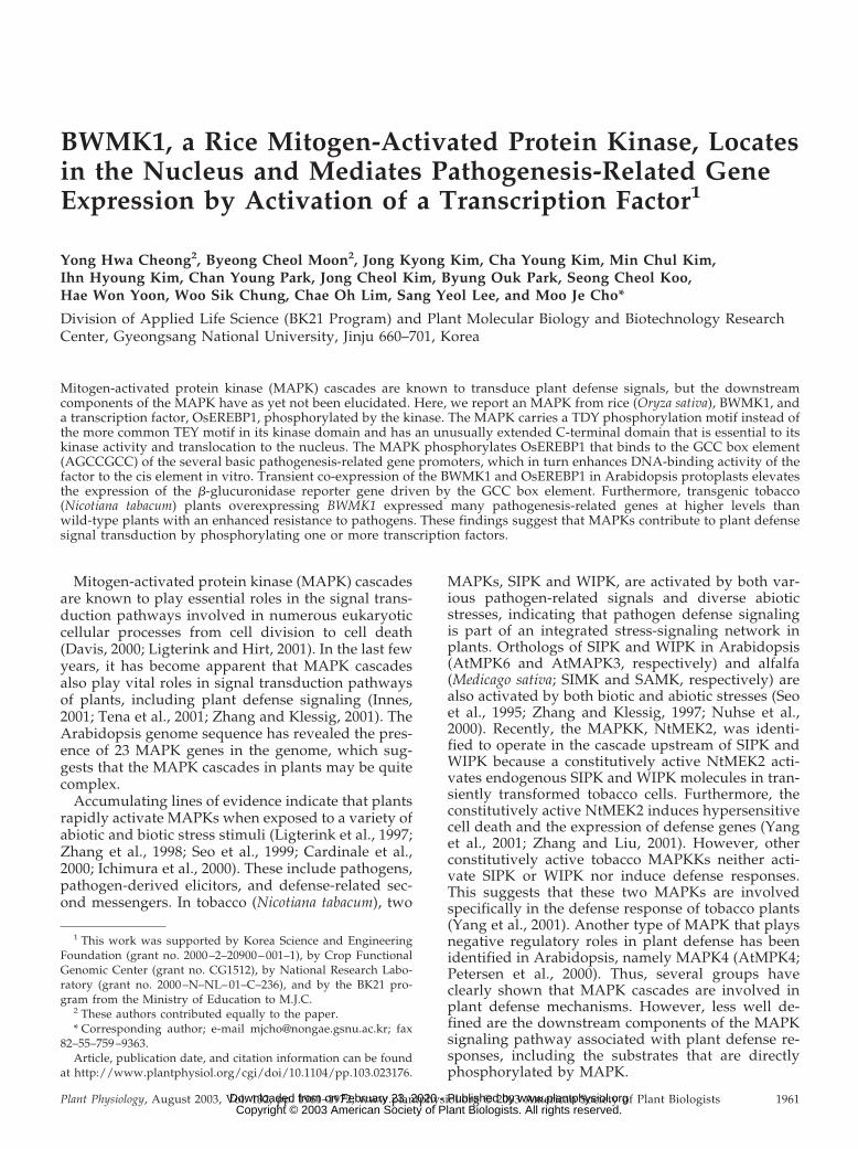
The phylogenetic tree resulting from comparisonsof deduced amino acid sequences indicates that plantMAPKs can be grouped into at least five distinctfamilies (Fig. 1). Among them, the MAPKs in familiesI and II are mostly involved in pathogen and abioticstress signalings, whereas some family III MAPKs areinvolved in cell cycle regulation (Zhang and Klessig,2001). Interestingly, AtMPK4, which is in family III,negatively regulates systemic acquired resistance(SAR; Petersen et al., 2000). BWMK1 belongs to familyV, which includes the AtMPK8, AtMPK9, and TDY1,which have the TDY motif instead of TEY in KD andalso have the CD. Recently, Schoenbeck et al. (1999)reported that the TDY1 gene is expressed in the leafmesophyll-surrounding areas of mechanical wound-ing and pathogen invasion, which suggests that it mayplay a role in wound signaling. Furthermore, BWMK1was also activated by wounding and rice blast fungus(He et al., 1999). Thus, the family V of MAPKs includ-
Cheong et al.
1962 Plant Physiol. Vol. 132, 2003 www.plantphysiol.orgon February 23, 2020 - Published by Downloaded from Copyright © 2003 American Society of Plant Biologists. All rights reserved.
ing BWMK1 is believed to be involved in pathogenand wounding signal transduction.
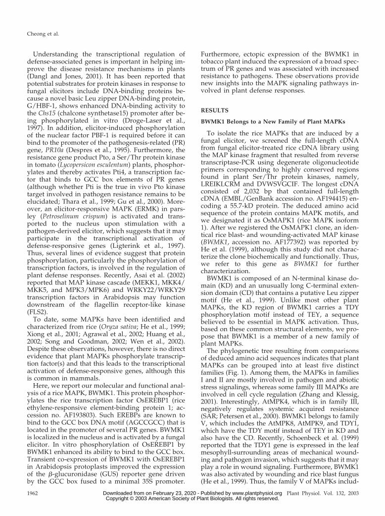
The CD of BWMK1 Is Essential for Kinase Activity andNuclear Localization
To characterize the BWMK1, we produced gluta-thione S-transferase (GST) fusion proteins containingfull-length, KD or CD in Escherichia coli and thenpurified the GST fusion proteins (Fig. 2A). Before weperformed the kinase activity assay with purifiedGST fusion proteins, amounts of purified proteinswas checked by loading with 10% (w/v) SDS-PAGE(data not shown). As shown in Figure 2B, the full-length protein was able to phosphorylate both MBP
and itself. However, both phosphorylation activitieswere completely destroyed when the CD was de-leted, indicating that it is essential for the kinaseactivity of BWMK1. Similar observations have beenmade for other protein kinases, such as Nlk (Brott etal., 1998) and TSL (Roe et al., 1996), which also havean extension domain containing a Leu zipper.
To determine the subcellular localization of theMAPK in vivo, the green fluorescent protein (smGFP)gene (Davis and Vierstra, 1998) was fused to theC-terminal end of either the full-length BWMK1(BWMK1-smGFP) or the KD (BWMK1 KD-smGFP),and these constructs were cotransfected with the redfluorescent protein gene fused to the nuclear local-ization signal peptide (NLS-RFP; Lee et al., 2001) as apositive control into Arabidopsis protoplasts. Asshown in Figure 2C, the full-length BWMK1-smGFPfusion protein localized exclusively in the nucleusshowing completely overlapping location with NLS-RFP originated from the simian virus 40 large Tantigen (Dingwall and Laskey, 1991). In contrast,KD-smGFP was not translocated to the nucleus.Thus, the CD of BWMK1, which contains a putativeLeu zipper motif, is believed to be essential for thekinase activities of the protein and its targeting to thenucleus.
BWMK1 Is Activated by Pathogen Signals
To determine whether BWMK1 is activated in re-sponse to pathogen signals, we performed immuno-complex kinase assays using Ab-pNBWMK1, an an-tibody that was raised against a synthetic peptiderepresenting the N-terminal 15 amino acids of
Figure 1. BWMK1 belongs to an MAPK family in plants. Phyloge-netic tree of plant MAPKs. Plant MAPKs included were alfalfaMMK1-4 (Jonak et al., 1995, 1996) and TDY1 (Schoenbeck et al.,1999); Arabidopsis AtMPK1-7 (Mizoguchi et al., 1993), AtMPK8(accession no. BAA92222), and AtMPK9 (accession no. BAA92223);parsley ERMK (Ligterink et al., 1997); tobacco NtF3, 4, and 6 (Wilsonet al., 1995), WIPK (Seo et al., 1995), and SIPK (Zhang and Klessig,1997); and rice BWMK1 (He et al., 1999). The phylogenic tree wascreated using ClustalW program (Thompson et al., 1994).
Figure 2. The CD of BWMK1 is essential forboth kinase activity and nuclear localization. A,Schematic diagram of the GST fusion constructsof BWMK1. Amino acid numbers of domainboundaries are indicated. B, Role of theC-terminal domain in autophosphorylation andmyelin basic protein (MBP) kinase activity. �,With MBP; �, without MBP. The arrows indi-cate the positions where GST-BWMK1 and MBPmigrated. The apparent molecular masses (kilo-daltons) are indicated at the left. C, Subcellularlocalization of BWMK1. Arabidopsis protoplastswere cotransfected with three sets of smGFP andRFP constructs, that is, smGFP and RFP (a, d, g,and j), smGFP-fused BWMK1 and RFP-fusedNLS (b, e, h, and k), and smGFP-fused KD andRFP-fused NLS (c, f, i, and l). Bars � 20 �m.
BWMK1 Mediates Plant Defense Responses
Plant Physiol. Vol. 132, 2003 1963 www.plantphysiol.orgon February 23, 2020 - Published by Downloaded from Copyright © 2003 American Society of Plant Biologists. All rights reserved.
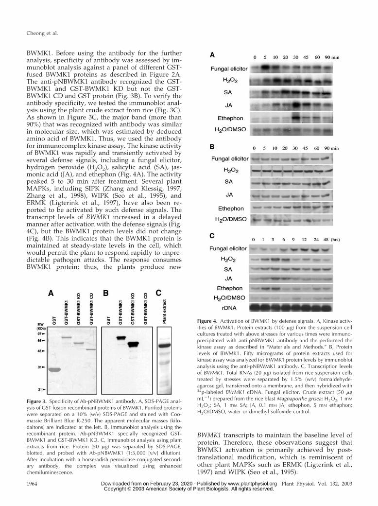
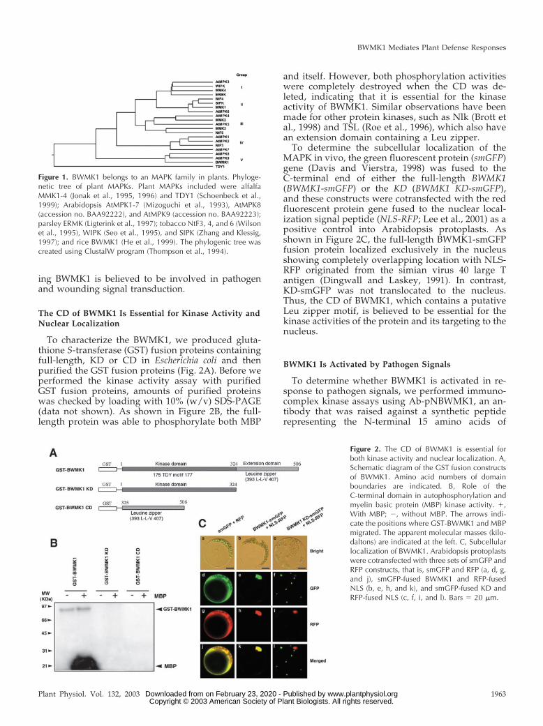
BWMK1. Before using the antibody for the furtheranalysis, specificity of antibody was assessed by im-munoblot analysis against a panel of different GST-fused BWMK1 proteins as described in Figure 2A.The anti-pNBWMK1 antibody recognized the GST-BWMK1 and GST-BWMK1 KD but not the GST-BWMK1 CD and GST protein (Fig. 3B). To verify theantibody specificity, we tested the immunoblot anal-ysis using the plant crude extract from rice (Fig. 3C).As shown in Figure 3C, the major band (more than90%) that was recognized with antibody was similarin molecular size, which was estimated by deducedamino acid of BWMK1. Thus, we used the antibodyfor immunocomplex kinase assay. The kinase activityof BWMK1 was rapidly and transiently activated byseveral defense signals, including a fungal elicitor,hydrogen peroxide (H2O2), salicylic acid (SA), jas-monic acid (JA), and ethephon (Fig. 4A). The activitypeaked 5 to 30 min after treatment. Several plantMAPKs, including SIPK (Zhang and Klessig, 1997;Zhang et al., 1998), WIPK (Seo et al., 1995), andERMK (Ligterink et al., 1997), have also been re-ported to be activated by such defense signals. Thetranscript levels of BWMK1 increased in a delayedmanner after activation with the defense signals (Fig.4C), but the BWMK1 protein levels did not change(Fig. 4B). This indicates that the BWMK1 protein ismaintained at steady-state levels in the cell, whichwould permit the plant to respond rapidly to unpre-dictable pathogen attacks. The response consumesBWMK1 protein; thus, the plants produce new
BWMK1 transcripts to maintain the baseline level ofprotein. Therefore, these observations suggest thatBWMK1 activation is primarily achieved by post-translational modification, which is reminiscent ofother plant MAPKs such as ERMK (Ligterink et al.,1997) and WIPK (Seo et al., 1995).
Figure 3. Specificity of Ab-pNBWMK1 antibody. A, SDS-PAGE anal-ysis of GST fusion recombinant proteins of BWMK1. Purified proteinswere separated on a 10% (w/v) SDS-PAGE and stained with Coo-massie Brilliant Blue R-250. The apparent molecular masses (kilo-daltons) are indicated at the left. B, Immunoblot analysis using therecombinant protein. Ab-pNBWMK1 specially recognized GST-BWMK1 and GST-BWMK1 KD. C, Immunoblot analysis using plantextracts from rice. Protein (50 �g) was separated by SDS-PAGE,blotted, and probed with Ab-pNBWMK1 (1:3,000 [v/v] dilution).After incubation with a horseradish peroxidase-conjugated second-ary antibody, the complex was visualized using enhancedchemiluminescence.
Figure 4. Activation of BWMK1 by defense signals. A, Kinase activ-ities of BWMK1. Protein extracts (100 �g) from the suspension cellcultures treated with above stresses for various times were immuno-precipitated with anti-pNBWMK1 antibody and the performed thekinase assay as described in “Materials and Methods.” B, Proteinlevels of BWMK1. Fifty micrograms of protein extracts used forkinase assay was analyzed for BWMK1 protein levels by immunoblotanalysis using the anti-pNBWMK1 antibody. C, Transcription levelsof BWMK1. Total RNAs (20 �g) isolated from rice suspension cellstreated by stresses were separated by 1.5% (w/v) formaldehyde-agarose gel, transferred onto a membrane, and then hybridized with32p-labeled BWMK1 cDNA. Fungal elicitor, Crude extract (50 �gmL�1) prepared from the rice blast Magnaporthe grisea; H2O2, 1 mM
H2O2; SA, 1 mM SA; JA, 0.1 mM JA; ethephon, 5 mM ethaphon;H2O/DMSO, water or dimethyl sulfoxide control.
Cheong et al.
1964 Plant Physiol. Vol. 132, 2003 www.plantphysiol.orgon February 23, 2020 - Published by Downloaded from Copyright © 2003 American Society of Plant Biologists. All rights reserved.
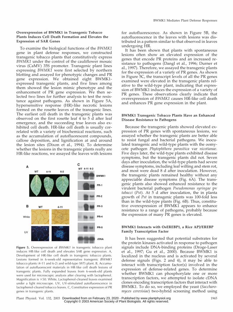
Overexpression of BWMK1 in Transgenic TobaccoPlants Induces Cell Death Formation and Elevates theExpression of SAR Genes
To examine the biological functions of the BWMK1gene in plant defense responses, we constructedtransgenic tobacco plants that constitutively expressBWMK1 under the control of the cauliflower mosaicvirus (CaMV) 35S promoter. Transgenic plant linesexpressing BWMK1 were first selected by northernblotting and assayed for phenotypic changes and PRgene expression. We obtained eight BWMK1-expressed transgenic plants, and five lines amongthem showed the lesion mimic phenotype and theenhancement of PR gene expression. We then se-lected two lines for further analysis to test the resis-tance against pathogens. As shown in Figure 5A,hypersensitive response (HR)-like necrotic lesionsformed on the rosette leaves of the transgenic lines.The earliest cell death in the transgenic plants wasobserved on the first rosette leaf 4 to 5 d after leafemergence, and the succeeding true leaves also ex-hibited cell death. HR-like cell death is usually cor-related with a variety of biochemical reactions, suchas the accumulation of autofluorescent compounds,callose deposition, and lignification at and aroundthe lesion sites (Dixon et al., 1994). To determinewhether the lesions in the transgenic plants really areHR-like reactions, we assayed the leaves with lesions
for autofluorescence. As shown in Figure 5B, theautofluorescence in the leaves with lesions was dis-tributed in a pattern similar to that observed in leavesundergoing HR.
It has been shown that plants with spontaneouslesions often show an elevated expression of thegenes that encode PR proteins and an increased re-sistance to pathogens (Dangl et al., 1996; Durner etal., 1997). Therefore, we assayed the transgenic plantsfor the expression of a variety of PR genes. As shownin Figure 5C, the transcript levels of all the PR genesexamined were elevated in the transgenic plants rel-ative to the wild-type plant, indicating that expres-sion of BWMK1 induces the expression of a variety ofPR genes. These observations clearly indicate thatoverexpression of BWMK1 causes HR-like cell deathand enhances PR gene expression in the plant.
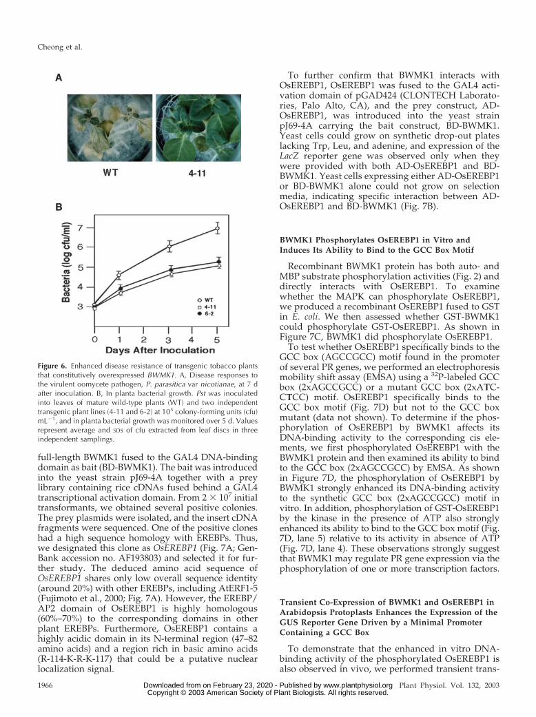
BWMK1 Transgenic Tobacco Plants Have an EnhancedDisease Resistance to Pathogens
Because the transgenic plants showed elevated ex-pression of PR genes with spontaneous lesions, weassayed whether the transgenic plants are better ableto resist fungal and bacterial pathogens. We inocu-lated transgenic and wild-type plants with the oomy-cete pathogen Phytophthora parasitica var nicotianae.Five days later, the wild-type plants exhibited diseasesymptoms, but the transgenic plants did not. Sevendays after inoculation, the wild-type plants had severedisease symptoms, including leaf wilting and stem rot,and most were dead 8 d after inoculation. However,the transgenic plants remained healthy without anyappreciable disease symptoms (Fig. 6A). The trans-genic plants also showed enhanced resistance to thevirulent bacterial pathogen Pseudomonas syringae pvtabacci (Pst). At 5 d after inoculation, the in plantagrowth of Pst in transgenic plants was 100-fold lessthan in the wild-type plants (Fig. 6B). Thus, constitu-tive overexpression of BWMK1 appears to enhanceresistance to a range of pathogens, probably becausethe expression of many PR genes is elevated.
BWMK1 Interacts with OsEREBP1, a Rice AP2/EREBPFamily Transcription Factor
It has been suggested that potential substrates forthe protein kinases activated in response to pathogensignals include DNA-binding proteins (Droge-Laseret al., 1997; Gu et al., 2000). Because BWMK1 islocalized in the nucleus and is activated by severaldefense signals (Figs. 2 and 4), it may be able tointeract with transcription factor(s) involved in theexpression of defense-related genes. To determinewhether BWMK1 can phosphorylate one or moretranscription factors, we attempted to isolate cDNAclones encoding transcription factors that interact withBWMK1. To do so, we employed the yeast (Saccharo-myces cerevisiae) two-hybrid screening method using
Figure 5. Overexpression of BWMK1 in transgenic tobacco plantinduces HR-like cell death and elevates SAR gene expression. A,Development of HR-like cell death in transgenic tobacco plants.Lesions formed in 6-week-old representative transgenic BWMK1tobacco plants (4-11 and 6-2) and wild-type (WT) plant. B, Accumu-lation of autofluorescent materials in HR-like cell death lesions oftransgenic plants. Fully expanded leaves from 6-week-old plantswere used for microscopic analysis after clearing with lactophenol.Magnification is �50. White, Lactophenol-cleaned leaves examinedunder a light microscope. UV, UV-stimulated autofluorescence inlactophenol-cleared tobacco leaves. C, Constitutive expression of PRgenes in transgenic plants.
BWMK1 Mediates Plant Defense Responses
Plant Physiol. Vol. 132, 2003 1965 www.plantphysiol.orgon February 23, 2020 - Published by Downloaded from Copyright © 2003 American Society of Plant Biologists. All rights reserved.
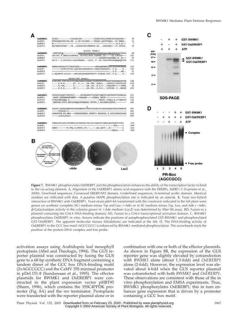
full-length BWMK1 fused to the GAL4 DNA-bindingdomain as bait (BD-BWMK1). The bait was introducedinto the yeast strain pJ69-4A together with a preylibrary containing rice cDNAs fused behind a GAL4transcriptional activation domain. From 2 � 107 initialtransformants, we obtained several positive colonies.The prey plasmids were isolated, and the insert cDNAfragments were sequenced. One of the positive cloneshad a high sequence homology with EREBPs. Thus,we designated this clone as OsEREBP1 (Fig. 7A; Gen-Bank accession no. AF193803) and selected it for fur-ther study. The deduced amino acid sequence ofOsEREBP1 shares only low overall sequence identity(around 20%) with other EREBPs, including AtERF1-5(Fujimoto et al., 2000; Fig. 7A). However, the EREBP/AP2 domain of OsEREBP1 is highly homologous(60%–70%) to the corresponding domains in otherplant EREBPs. Furthermore, OsEREBP1 contains ahighly acidic domain in its N-terminal region (47–82amino acids) and a region rich in basic amino acids(R-114-K-R-K-117) that could be a putative nuclearlocalization signal.
To further confirm that BWMK1 interacts withOsEREBP1, OsEREBP1 was fused to the GAL4 acti-vation domain of pGAD424 (CLONTECH Laborato-ries, Palo Alto, CA), and the prey construct, AD-OsEREBP1, was introduced into the yeast strainpJ69-4A carrying the bait construct, BD-BWMK1.Yeast cells could grow on synthetic drop-out plateslacking Trp, Leu, and adenine, and expression of theLacZ reporter gene was observed only when theywere provided with both AD-OsEREBP1 and BD-BWMK1. Yeast cells expressing either AD-OsEREBP1or BD-BWMK1 alone could not grow on selectionmedia, indicating specific interaction between AD-OsEREBP1 and BD-BWMK1 (Fig. 7B).
BWMK1 Phosphorylates OsEREBP1 in Vitro andInduces Its Ability to Bind to the GCC Box Motif
Recombinant BWMK1 protein has both auto- andMBP substrate phosphorylation activities (Fig. 2) anddirectly interacts with OsEREBP1. To examinewhether the MAPK can phosphorylate OsEREBP1,we produced a recombinant OsEREBP1 fused to GSTin E. coli. We then assessed whether GST-BWMK1could phosphorylate GST-OsEREBP1. As shown inFigure 7C, BWMK1 did phosphorylate OsEREBP1.
To test whether OsEREBP1 specifically binds to theGCC box (AGCCGCC) motif found in the promoterof several PR genes, we performed an electrophoresismobility shift assay (EMSA) using a 32P-labeled GCCbox (2xAGCCGCC) or a mutant GCC box (2xATC-CTCC) motif. OsEREBP1 specifically binds to theGCC box motif (Fig. 7D) but not to the GCC boxmutant (data not shown). To determine if the phos-phorylation of OsEREBP1 by BWMK1 affects itsDNA-binding activity to the corresponding cis ele-ments, we first phosphorylated OsEREBP1 with theBWMK1 protein and then examined its ability to bindto the GCC box (2xAGCCGCC) by EMSA. As shownin Figure 7D, the phosphorylation of OsEREBP1 byBWMK1 strongly enhanced its DNA-binding activityto the synthetic GCC box (2xAGCCGCC) motif invitro. In addition, phosphorylation of GST-OsEREBP1by the kinase in the presence of ATP also stronglyenhanced its ability to bind to the GCC box motif (Fig.7D, lane 5) relative to its activity in absence of ATP(Fig. 7D, lane 4). These observations strongly suggestthat BWMK1 may regulate PR gene expression via thephosphorylation of one or more transcription factors.
Transient Co-Expression of BWMK1 and OsEREBP1 inArabidopsis Protoplasts Enhances the Expression of theGUS Reporter Gene Driven by a Minimal PromoterContaining a GCC Box
To demonstrate that the enhanced in vitro DNA-binding activity of the phosphorylated OsEREBP1 isalso observed in vivo, we performed transient trans-
Figure 6. Enhanced disease resistance of transgenic tobacco plantsthat constitutively overexpressed BWMK1. A, Disease responses tothe virulent oomycete pathogen, P. parasitica var nicotianae, at 7 dafter inoculation. B, In planta bacterial growth. Pst was inoculatedinto leaves of mature wild-type plants (WT) and two independenttransgenic plant lines (4-11 and 6-2) at 105 colony-forming units (cfu)mL�1, and in planta bacterial growth was monitored over 5 d. Valuesrepresent average and SDs of cfu extracted from leaf discs in threeindependent samplings.
Cheong et al.
1966 Plant Physiol. Vol. 132, 2003 www.plantphysiol.orgon February 23, 2020 - Published by Downloaded from Copyright © 2003 American Society of Plant Biologists. All rights reserved.
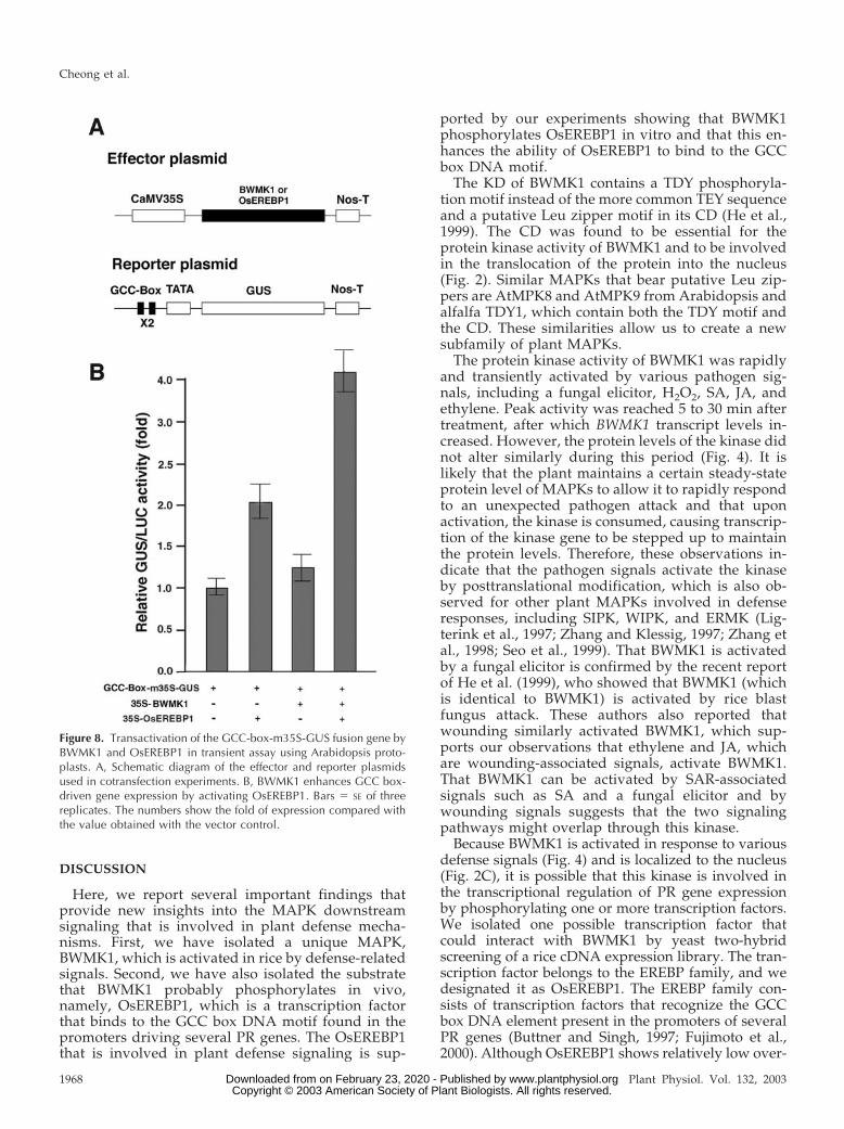
activation assays using Arabidopsis leaf mesophyllprotoplasts (Abel and Theologis, 1994). The GUS re-porter plasmid was constructed by fusing the GUSgene to a 68-bp synthetic DNA fragment containing atandem dimer of the GCC box DNA-binding motif(2xAGCCGCC) and the CaMV 35S minimal promoterin pDel.151-8 (Sundaresan et al., 1995). The effectorplasmids for BWMK1 and OsEREBP1 were con-structed in the plant expression vector pHBT95(Sheen, 1996), which contains the 35SC4PPDK pro-moter (Fig. 8A) and the nos terminator. Protoplastswere transfected with the reporter plasmid alone or in
combination with one or both of the effector plasmids.As shown in Figure 8B, the expression of the GUSreporter gene was slightly elevated by cotransfectionwith BWMK1 alone (about 1.3-fold) and OsEREBP1alone (2-fold). However, the expression level was ele-vated about 4-fold when the GUS reporter plasmidwas cotransfected with both BWMK1 and OsEREBP1.These observations are consistent with those of the invitro phosphorylation and EMSA experiments. Thus,BWMK1 phosphorylates OsEREBP1; this in turn en-hances gene expression that is driven by a promotercontaining a GCC box motif.
Figure 7. BWMK1 phosphorylates OsEREBP1 and the phosphorylation enhances the ability of the transcription factor to bindto the cis-acting element. A, Alignment of the OsEREBP1 amino acid sequence with the EREBPs, AtERF1-5 (Fujimoto et al.,2000). Overlined sequences, Conserved EREBP/AP2 domain. Underlined sequences, N-terminal acidic domain. Identicalresidues are indicated with dots. A putative MAPK phosphorylation site is indicated by an asterisk. B, Yeast two-hybridinteraction of BWMK1 with OsEREBP1. Yeast strain pJ69-4A transformed with the constructs indicated in the left plant weregrown on synthetic complete (SC) medium minus Trp and Leu (�Ade) or in SC medium minus Trp, Leu, and Ade (�Ade).�-Galactosidase activity in the colonies grown in �Ade medium (LacZ) was determined by filter-lift assay. BD, Fusion to aplasmid containing the GAL4 DNA-binding domain; AD, Fusion to a GAL4 transcriptional activation domain. C, BWMK1phosphorylates OsEREBP1 in vitro. Arrows indicate the positions of autophosphorylated GST-BWMK1 and phosphorylatedGST-OsEREBP1. The apparent molecular masses (kilodaltons) are indicated at the left. D, The DNA-binding activity ofOsEREBP1 to the GCC box motif (AGCCGCC) is enhanced by BWMK1-mediated phosphorylation. The arrowheads mark theposition of the protein-DNA complex and free probe.
BWMK1 Mediates Plant Defense Responses
Plant Physiol. Vol. 132, 2003 1967 www.plantphysiol.orgon February 23, 2020 - Published by Downloaded from Copyright © 2003 American Society of Plant Biologists. All rights reserved.
DISCUSSION
Here, we report several important findings thatprovide new insights into the MAPK downstreamsignaling that is involved in plant defense mecha-nisms. First, we have isolated a unique MAPK,BWMK1, which is activated in rice by defense-relatedsignals. Second, we have also isolated the substratethat BWMK1 probably phosphorylates in vivo,namely, OsEREBP1, which is a transcription factorthat binds to the GCC box DNA motif found in thepromoters driving several PR genes. The OsEREBP1that is involved in plant defense signaling is sup-
ported by our experiments showing that BWMK1phosphorylates OsEREBP1 in vitro and that this en-hances the ability of OsEREBP1 to bind to the GCCbox DNA motif.
The KD of BWMK1 contains a TDY phosphoryla-tion motif instead of the more common TEY sequenceand a putative Leu zipper motif in its CD (He et al.,1999). The CD was found to be essential for theprotein kinase activity of BWMK1 and to be involvedin the translocation of the protein into the nucleus(Fig. 2). Similar MAPKs that bear putative Leu zip-pers are AtMPK8 and AtMPK9 from Arabidopsis andalfalfa TDY1, which contain both the TDY motif andthe CD. These similarities allow us to create a newsubfamily of plant MAPKs.
The protein kinase activity of BWMK1 was rapidlyand transiently activated by various pathogen sig-nals, including a fungal elicitor, H2O2, SA, JA, andethylene. Peak activity was reached 5 to 30 min aftertreatment, after which BWMK1 transcript levels in-creased. However, the protein levels of the kinase didnot alter similarly during this period (Fig. 4). It islikely that the plant maintains a certain steady-stateprotein level of MAPKs to allow it to rapidly respondto an unexpected pathogen attack and that uponactivation, the kinase is consumed, causing transcrip-tion of the kinase gene to be stepped up to maintainthe protein levels. Therefore, these observations in-dicate that the pathogen signals activate the kinaseby posttranslational modification, which is also ob-served for other plant MAPKs involved in defenseresponses, including SIPK, WIPK, and ERMK (Lig-terink et al., 1997; Zhang and Klessig, 1997; Zhang etal., 1998; Seo et al., 1999). That BWMK1 is activatedby a fungal elicitor is confirmed by the recent reportof He et al. (1999), who showed that BWMK1 (whichis identical to BWMK1) is activated by rice blastfungus attack. These authors also reported thatwounding similarly activated BWMK1, which sup-ports our observations that ethylene and JA, whichare wounding-associated signals, activate BWMK1.That BWMK1 can be activated by SAR-associatedsignals such as SA and a fungal elicitor and bywounding signals suggests that the two signalingpathways might overlap through this kinase.
Because BWMK1 is activated in response to variousdefense signals (Fig. 4) and is localized to the nucleus(Fig. 2C), it is possible that this kinase is involved inthe transcriptional regulation of PR gene expressionby phosphorylating one or more transcription factors.We isolated one possible transcription factor thatcould interact with BWMK1 by yeast two-hybridscreening of a rice cDNA expression library. The tran-scription factor belongs to the EREBP family, and wedesignated it as OsEREBP1. The EREBP family con-sists of transcription factors that recognize the GCCbox DNA element present in the promoters of severalPR genes (Buttner and Singh, 1997; Fujimoto et al.,2000). Although OsEREBP1 shows relatively low over-
Figure 8. Transactivation of the GCC-box-m35S-GUS fusion gene byBWMK1 and OsEREBP1 in transient assay using Arabidopsis proto-plasts. A, Schematic diagram of the effector and reporter plasmidsused in cotransfection experiments. B, BWMK1 enhances GCC box-driven gene expression by activating OsEREBP1. Bars � SE of threereplicates. The numbers show the fold of expression compared withthe value obtained with the vector control.
Cheong et al.
1968 Plant Physiol. Vol. 132, 2003 www.plantphysiol.orgon February 23, 2020 - Published by Downloaded from Copyright © 2003 American Society of Plant Biologists. All rights reserved.
all amino acid sequence similarity to the other mem-bers of the EREBP family, it does recognize the syn-thetic GCC box motif (AGCCGCC) in vitro (Fig. 7D).Furthermore, BWMK1 could phosphorylate a recom-binant OsEREBP1 in vitro (Fig. 7C), and this phos-phorylation enhanced the ability of the transcriptionfactor to bind to the synthetic GCC box motif in vitro(Fig. 7D). These observations support the hypothesisthat BWMK1 transduces its signal(s) by phosphorylat-ing one or more transcription factor(s).
To further verify the relationship between BWMK1-mediated phosphorylation of the OsEREBP1 and PRgene expression, we transiently transfected Arabidop-sis leaf mesophyll protoplasts with a GUS reportergene fused to a synthetic tandem dimer of the GCCbox DNA motif and the minimal promoter pDel.151-8.Co-expression with the OsEREBP1 effector plasmiddriven by the CaMV 35S promoter transactivated GUSreporter gene expression about 2-fold. When the pro-toplasts were cotransfected with the reporter constructand both the OsEREBP1 and BWMK1 effector plas-mids, the reporter gene expression was further ele-vated by 4-fold. These observations strongly suggestthat BWMK1 enhances the expression of GCC box-driven PR genes by phosphorylating the OsEREBP1transcription factor.
The GCC box motif is present in the PR2 and PR5gene promoter that is stimulated by both ethylene andJA (Zhou et al., 1997). Because BWMK1 is activated bythe SAR-associated SA signal and by the ethylene andJA signals mainly associated with wounding, it cannotbe ruled out that BWMK1 could phosphorylate an-other transcription factor(s), one that is exclusivelyassociated with SA-mediated gene expression and thatthe ethylene/JA and SA-mediated defense signalingpathways may cross through this mechanism.
The Pto kinase in tomato plants phosphorylates thePti4 protein. The phosphorylation of Pti4 enhances itsability to bind to the GCC box contained in severalPR gene promoters (Gu et al., 2000). Although thissuggests that plant MAPKs phosphorylate transcrip-tion factor(s) and that this in turn triggers PR geneexpression, no direct evidence of this has been ob-tained thus far. To provide this evidence, we createdtransgenic tobacco plants that overexpress BWMK1under the control of the CaMV 35S promoter. Signif-icantly, the plants had higher levels of PR gene ex-pression and an enhanced resistance to virulent bac-terial and fungal pathogens (Figs. 5 and 6). Theseobservations strongly support the notion thatBWMK1 is involved in triggering PR gene expres-sion. Another observation made with the transgenictobacco plants that overexpress BWMK1 is that theold leaves of transgenic plants had HR-like lesions.Recently, Zhang and Liu (2001) and Yang et al. (2001)also reported that overexpression of SIPK and WIPKresulted in both plant defense gene activation andenhanced programmed cell death, suggesting thetwo processes are linked.
In summary, BWMK1, which can be classified intofamily V MAPK, is activated by SA-associated de-fense signals and by ethylene/JA-associated plantdefense signals. The MAPK phosphorylates theOsEREBP1, which in turn enhances the DNA-bindingactivity of the factor to the corresponding cis-actingelement, GCC box motif (AGCCGCC), in several ba-sic PR gene promoters. Transient cotransfection as-says using Arabidopsis leaf protoplast support the invitro DNA-binding result by elevating expressionlevel of the GUS reporter gene about 4-fold when thereporter plasmid was cotransfected with bothBWMK1 and OsEREBP1. Ectopic overexpression ofthe BWMK1 in transgenic tobacco plants causes HR-like cell death and increased resistance to pathogenswith elevated level of PR gene expression. Thus,these results suggest that BWMK1 is involved inMAPK cascades in plant defense signal transductionvia direct phosphorylation of a transcriptionfactor(s).
MATERIALS AND METHODS
Rice (Oryza sativa L. Milyang 117) Suspension CellCulture and Treatments
Suspension cell lines of rice were cultured and maintained as describedby Kyozuka et al. (1990). Treatment with an elicitor or chemicals wasperformed in the dark. The fungal elicitor was prepared from the riceblast-causing fungus Magnaporthe grisea as described (Simmons et al., 1992)and was added to the cell culture at a final concentration of 50 �g mL�1 interms of total reducing sugars. The chemicals used were SA (used at 1 mm),H2O2 (1 mm), ethephon (5 mm), and JA (0.1 mm). The cells were harvestedby filtration at various time periods after treatment, quickly frozen in liquidnitrogen, and stored at �80°C until analysis.
Yeast (Saccharomyces cerevisiae) Two-Hybrid Screening
The yeast two-hybrid screening method was employed to isolate tran-scription factors that interact with BWMK1. We digested the BWMK1 cDNAwith SmaI and BclI and ligated the fragments into the pGBT9 plasmid, whichcontains the Trp1 selection marker (CLONTECH). The prey library contain-ing cDNA from rice suspension cells was constructed in plasmid pAD-GAL4 (Stratagene, La Jolla, CA), which harbors the Leu2 selection marker.The yeast strain pJ69-4A (James et al., 1996) was used to express theseplasmids. The two-hybrid screenings and assays were performed as de-scribed in CLONTECH’s Yeast Protocols. Positive interactions were verifiedby the �-galactosidase assay.
Northern-Blot Analysis
Total RNA was isolated as described by Lee et al. (1995), and 20 �g of theRNA was subjected to electrophoresis through a 1.5% (w/v) formaldehyde-agarose gel, transferred onto a GeneScreen Plus membrane (New EnglandNuclear, Boston), and hybridized to random primer-labeled full-lengthBWMK1 cDNA or PR gene probes (Ryals et al., 1996) under the conditionsdescribed previously (Lee et al., 1995).
Expression of Fusion Proteins
For expression in bacteria, full-length BWMK1, the KD (BWMK1 KD),and the CD (BWMK1 CD) were fused to the C terminus of GST. TheGST-BWMK1 fusion construct was generated by digesting the full-lengthBWMK1 in pBluescript SK� with SmaI/BclI and inserting the excised frag-ment into the corresponding sites of the GST expression vector pGEX-2T(Amersham, Buckinghamshire, UK). An EcoRI fragment of BWMK1 in
BWMK1 Mediates Plant Defense Responses
Plant Physiol. Vol. 132, 2003 1969 www.plantphysiol.orgon February 23, 2020 - Published by Downloaded from Copyright © 2003 American Society of Plant Biologists. All rights reserved.
pBluescript SK� that encodes only the KD of BWMK1 was subcloned intothe GST expression vector to generate GST-BWMK1 KD, whereas an EcoRI-XhoI fragment encoding only the CD was subcloned into the same expres-sion vector to create GST-BWMK1 CD. The OsEREBP1 cDNA in pBluescriptSK� was also digested with SmaI/XhoI and ligated into the pGEX-5X vectorto generate GST-OsEREBP1. The resulting constructs were then introducedinto Escherichia coli strain BL21 (pLysS), and the GST fusion proteins wereexpressed and purified using glutathione-agarose beads according to themanufacturer’s instructions (Amersham).
In Vitro Kinase Assay
Autophosphorylation activities of the full-length, KD and CD of BWMK1were assayed at different protein concentrations in 20 �L of reaction buffer(20 mm Tris-HCl [pH 7.5], 1 mm dithiothreitol, 10 mm MgCl2, and 100 �mATP) containing 5 �Ci [�-32P] ATP (6,000 Ci mmol�1; Amersham). Thesubstrate kinase activity of GST-BWMK1 was assayed at room temperaturefor 20 min in a final volume of 20 �L using one of the substrates, MBP orGST-OsEREBP1, at a final protein concentration of 0.1 mg mL�1. Reactionswere terminated by the addition of 4� SDS sample buffer. The samples werethen analyzed by 12.5% (w/v) SDS-PAGE and subsequent autoradiography.
Antibody Production and Immunoblot Analysis
A polyclonal antibody recognizing BWMK1 (Ab-pNBWMK1) was raisedby immunizing rabbits with a synthetic peptide representing the NH2
terminus (MEFFTEYGEAASQYQ) of BWMK1. For immunoblot analysis, 50�g of total protein per lane was resolved by 10% (w/v) SDS-PAGE. Theseparated proteins were then transferred to a nitrocellulose membrane(Amersham) by semidry electroblotting. After blocking the membrane atroom temperature for 1 h in Tris-buffered saline containing 0.1% (v/v)Tween 20 buffer (20 mm Tris [pH 7.5], 150 mm NaCl, and 0.1% [v/v] Tween20) with 6% (w/v) nonfat dry milk (Carnation, Glendale, CA), the mem-brane was incubated with 0.2 �g mL�1 Ab-pNBWMK1 for 1 h. The blot wasthen washed four times in TTBS buffer, incubated with a horseradishperoxidase-conjugated secondary antibody (1:5,000 [v/v] dilution), and de-veloped by using an enhanced chemiluminescence kit (Amersham).
Preparation of Protein Extracts and ImmunocomplexKinase Assay
Preparation of protein extracts from rice suspension cells treated withfungal elicitor or chemicals and immunocomplex kinase assay were per-formed as described by Zhang and Klessig (1997). Protein extracts (100 �g)were incubated with Ab-pNBWMK1 (2 �g) in immunoprecipitation buffer(20 mm Tris [pH 7.5], 150 mm NaCl, 1 mm EDTA, 1 mm EGTA, 1 mmNa3VO4, 1 mm NaF, 10 mm �-gylcerophosphate, 5 �g mL�1 antipain, 5 �gmL�1 aprotinin, 5 �g mL�1 leupeptin, and 0.1% [v/v] Tween 20) at 4°C for4 h on a rocker. Approximately 20 �L packed volume of 50% (v/v) proteinA-agarose washed with immunoprecipitation buffer was added, and theincubation was continued for another 4 h. Agarose bead-protein complexeswere pelleted by brief centrifugation and washed three times with 1.5 mL ofimmunoprecipitation buffer, once with immunoprecipitation buffer plus 1m NaCl, and three times with 1 mL of kinase reaction buffer. Kinase activityin the complex was assayed at room temperature for 20 min in a finalvolume of 20 �L containing 0.1 mg mL�1 MBP and 100 �m ATP with 5 �Ciof [�-32P-ATP]. The reaction was stopped by the addition of SDS-PAGEsample loading buffer. After electrophoresis on 12.5% (w/v) SDS-PAGE, thephosphorylated MBP was visualized by autoradiography.
Construction of Transgenic Plants
For construction of transgenic tobacco (Nicotiana tabacum cv Xanthi-nc)plants, BWMK1 cDNA was ligated into the plant binary vector pGA643 (Anet al., 1988). The recombinant plasmids were introduced into Agrobacteriumtumefaciens EHA101, and transgenic tobacco plants were generated by astandard leaf disc transformation method (Horsch et al., 1988) and selectedby kanamycin resistance. T2 progeny of transgenic plants expressing highlevels of BWMK1 were grown at 25°C (day) and 20°C (night) temperatures,a 16-h photoperiod, and 65% relative humidity, and used for the assays.
Histochemistry and Microscopy
The cell death phenotype of tobacco leaves was photographed by usinga dissecting microscope. Autofluorescent materials and callos depositionwere detected using an UV epifluorescence microscope as described byDietrich et al. (1994)
Pathogen Infections and Resistance Assay
A bacterial pathogen (Pseudomonas syringae pv tabacci) was culturedovernight at 30°C in Kings B medium containing 50 �g mL�1 rifampicin and50 �g mL�1 kanamycin. The bacterial culture was washed twice with 10 mmMgCl2 and resuspended in 10 mm MgCl2. Bacterial density was determinedby absorbance at OD600 nm. Bacteria were diluted to the desired concentra-tions in 10 mm MgCl2 for inoculation. Six-week-old plants were inoculatedby vacuum infiltration, and the inoculated plants were kept in a greenhouse.Leaf discs were ground in 10 mm MgCl2 and plated on appropriate Kings Bplates. The number of bacteria in the leaves was calculated by counting cfu.Infection with Phytophthora parasitica pv nicotianae was performed as de-scribed by Mittler et al., (1995). The oomycete was subcultured at 23°C onone-quarter-strength potato dextrose agar. Conidia from 3-week-old cul-tures on potato dextrose agar were used for inoculation (Heo et al., 1999).
In Vitro DNA-Binding Assay
A synthetic GCC box (CAT AAG AGC CGC CAC TAA AAT AAG ACCGAT CAA ATA AGA GCC GCC AT) and GCC box mutant (CAT AAG ATCCTC CAC TAA AAT AAG ACC GAT CAA ATA AGA TCC TCC AT;Ohme-Takagi and Shinshi, 1995) were end labeled with 32P as describedpreviously (Cheong et al., 1998). The probe (4 fmol) was mixed with each ofthe purified GST fusion proteins in a buffer containing 2 �g of poly (dA-dT).(dA-dT), 25 mm HEPES (pH 7.5), 40 mm KCl, 0.1 mm EDTA, 10% (v/v)glycerol, and 1 mm dithiothreitol. After incubation at room temperature for20 min, the reaction mixtures were separated in a 5% (w/v) polyacrylamidegel using 0.5� Tris-borate/EDTA buffer. The gel was then dried and ex-posed to x-ray film.
Localization and Transient Expression Assay
To observe the cellular localization of BWMK1, we prepared BWMK1-smGFP or BWMK1 KD-smGFP fusion constructs. The BamHI (1–506 aminoacids) or EcoRI (1–324 amino acids) fragment of BWMK1 was fused to thecoding region of smGFP under the control of the CaMV 35S promoter. Thefusion construct of nuclear localization signal from simian virus 40 large Tantigen and red fluorescent protein (NLS-RFP) was used as a positivecontrol (Lee et al., 2001). Polyethylene glycol-mediated cotransfection wasperformed to introduce the constructs into Arabidopsis protoplasts (Abeland Theologis, 1994). Expression of the fusion constructs was monitored atvarious times after transfection by an Axioplan 2 fluorescence microscope(Zeiss, Jena, Germany), and images were captured with a Zeiss Axiocam HRcamera using XF116-2 (exciter, 475AF20; dichroic, 500DRLP; and emitter,510AF23) and XF33 (exciter, 535AF35; dichroic, 570DRLP; and emitter,605DF50) filter sets (Omega, Inc., Brattleboro, VT) for GFP and RFP, respec-tively. For transient expression, a GCC box dimer (2xAGCCGCC) was fusedto a minimal �46 CaMV 35S promoter-GUS reporter gene (GCC-box-m35S-GUS; Jefferson et al., 1987; Zhou et al., 1997). To construct the two effectorplasmids, full-length BWMK1 and OsEREBP1 cDNA were inserted into aplant expression vector (pHBT95) containing the 35SC4PPDK promoter andthe nos terminator (Sheen, 1996). Transient expression of these constructswas carried out in Arabidopsis mesophyll protoplasts as described by Abeland Theologis (1994). In each transfection, 5 � 105 protoplasts were trans-fected with 10 �g of reporter construct alone or together with 15 �g of eacheffector construct or a vector DNA control (pHBT95). The transfected pro-toplasts were incubated in W5 medium for 16 h in the dark. A constructcarrying the LUC (luciferase) gene driven by the 35S promoter was used asan internal control in each transfection. The GUS activity of the cell lysatewas divided by the corresponding LUC activity to standardize the data. Theresults are expressed as means of three independent replicate transfectionswith sds.
Cheong et al.
1970 Plant Physiol. Vol. 132, 2003 www.plantphysiol.orgon February 23, 2020 - Published by Downloaded from Copyright © 2003 American Society of Plant Biologists. All rights reserved.
ACKNOWLEDGMENTS
We thank Drs. Cris Lamb and John A. Ryals for generously providing usPR protein cDNAs. We also thank Dr. Jen Sheen for providing pHBT95plasmid and Dr. Inhwan Hwang for providing NLS-RFP plasmid.
Received March 6, 2003; returned for revision April 7, 2003; accepted May 9,2003.
LITERATURE CITED
Abel S, Theologis A (1994) Transient transformation of Arabidopsis leafprotoplasts: a versatile experimental system to study gene expression.Plant J 5: 421–427
Agrawal GK, Rakwal R, Iwahashi H (2002) Isolation of novel rice (Oryzasativa L.) multiple stress responsive MAP kinase gene, OsMSRMK2,whose mRNA accumulates rapidly in response to environmental cues.Biochem Biophys Res Commun 294: 1009–1016
An G, Prebert AM, Ha SB (1988) Binary vectors. In SB Gelvin, RA Schilp-eroort, eds, Plant Molecular Biology Manual. Academic Press, Dordrecht,The Netherlands, pp 1–19
Asai T, Tena G, Plotnikova J, Willmann MR, Chiu WL, Gomez-Gomez L,Boller T, Ausubel FM, Sheen J (2002) MAP kinase signalling cascade inArabidopsis innate immunity. Nature 415: 977–983
Brott BK, Pinsky BA, Erikson RL (1998) Nlk is a murine protein kinaserelated to Erk/MAP kinases and localized in the nucleus. Proc Natl AcadSci USA 95: 963–968
Buttner M, Singh KB (1997) Arabidopsis thaliana ethylene-responsive ele-ment binding protein (AtEBP), an ethylene-inducible, GCC box DNA-binding protein interacts with an ocs element binding protein. Proc NatlAcad Sci USA 94: 5961–5966
Cardinale F, Jonak C, Ligterink W, Niehaus K, Boller T, Hirt H (2000)Differential activation of four specific MAPK pathways by distinct elici-tors. J Biol Chem 275: 36734–36740
Cheong YH, Yoo CM, Park JM, Ryu GR, Goekjian VH, Nagao RT, Key JL,Cho MJ, Hong JC (1998) STF1 is a novel TGACG-binding factor with azinc-finger motif and a bZIP domain which heterodimerizes with GBFproteins. Plant J 15: 199–209
Dangl JL, Dietrich RA, Richberg MH (1996) Death don’t have no mercy:cell death programs in plant-microbe interactions. Plant Cell 8: 1793–1807
Dangl JL, Jones JDG (2001) Plant pathogens and integrated defense re-sponses to infection. Nature 411: 826–833
Davis R (2000) Signal transduction by the JNK group of MAP kinases. Cell103: 239–252
Davis SJ, Vierstra RD (1998) Soluble, highly fluorescent variants of greenfluorescent protein (GFP) for use in higher plants. Plant Mol Biol 36:521–528
Despres C, Subramaniam R, Matton DP, Brission N (1995) The activationof the potato PR-10a gene requires the phosphorylation of the nuclearfactor PBF-1. Plant Cell 7: 639–647
Dietrich RA, Delaney TP, Uknes SJ, Ward ER, Ryals JA, Dangl JL (1994)Arabidopsis mutants simulating disease resistance response. Cell 77:565–577
Dingwall C, Laskey RA (1991) Nuclear targeting sequences: a consensus?Trends Biochem Sci 16: 478–481
Dixon RA, Harrison MJ, Lamb CJ (1994) Early events in the activation ofplant defense responses. Annu Rev Phytopathol 32: 479–501
Droge-Laser W, Kaiser A, Lindsay WP, Halkier BA, Loake GJ, Doerner P,Dixon RA, Lamb C (1997) Rapid stimulation of a soybean protein-serinekinase that phosphorylates a novel bZIP DNA-binding protein,G/HBF-1, during the induction of early transcription-dependent de-fenses. EMBO J 16: 726–738
Durner J, Shah J, Klessig DF (1997) Salicylic acid and disease resistance inplants. Trends Plant Sci 2: 266–274
Fujimoto SY, Ohta M, Usui A, Shinshi H, Ohme-Takagi M (2000) Arabi-dopsis ethylene-responsive element binding factors act as transcriptionalactivators or repressors of GCC box-mediated gene expression. Plant Cell12: 393–404
Gu YQ, Yang C, Thara VK, Zhou J, Martin GB (2000) Pti4 is induced byethylene and salicylic acid, and its product is phosphorylated by the Ptokinase. Plant Cell 12: 771–785
He C, Fong SH, Yang D, Wang GL (1999) BWMK1, a novel MAP kinaseinduced by fungal infection and mechanical wounding in rice. MolPlant-Microbe Interact 12: 1064–1073
Heo WD, Lee SH, Kim MC, Kim JC, Chung WS, Chun HJ, Lee KJ, ParkCY, Choi JY, Cho MJ (1999) Involvement of specific calmodulin isoformsin salicylic acid-independent activation of plant disease resistance re-sponses. Proc Natl Acad Sci USA 96: 766–771
Horsch RB, Fry J, Hoffmann N, Neidermeyer J, Rogers SG, Fraley RT(1988) Leaf disc transformation. In SB Gelvin, RA Schilperoot, eds, PlantMolecular Biology Manual. Kluwer Academic Publishers, Dordrecht, TheNetherlands, pp 1–9
Huang HJ, Fu SF, Tai YH, Chou WC, Huang DD (2002) Expression of Oryzasativa MAP kinase gene is developmentally regulated and stress-responsive. Physiol Plant 114: 572–580
Ichimura K, Mizoguchi T, Yoshida R, Yuasa T, Shinozaki K (2000) Variousabiotic stresses rapidly activate Arabidopsis MAP kinases ATMPK4 andATMPK6. Plant J 24: 655–665
Innes RW (2001) Mapping out the roles of MAP kinases in plant defense.Trends Plant Sci 6: 392–394
James P, Halladay J, Craig EA (1996) Genomic libraries and a host straindesigned for highly efficient two-hybrid selection in yeast. Genetics 144:1425–1436
Jefferson RA, Kavanagh TA, Bevan MW (1987) GUS fusions: beta-glucuronidase as a sensitive and versatile gene fusion marker in higherplants. EMBO J 20: 3901–3907
Jonak C, Kiegerl S, Ligterink W, Barker PJ, Huskisson NS, Hirt H (1996)Stress signaling in plants: a mitogen-activated protein kinase pathway isactivated by cold and drought. Proc Natl Acad Sci USA 93: 11274–11279
Jonak C, Kiegerl S, Lloyd C, Chan J, Hirt H (1995) MMK2, a novel alfalfaMAP kinase, specifically complements the yeast MPK1 function. Mol GenGenet 248: 686–694
Kyozuka J, Izawa T, Nakajima M, Shimamoto K (1990) Effect of thepromoter and the first intron of maize. Adh1 on foreign gene expressionin rice Maydica 35: 353–357
Lee SH, Kim JC, Lee MS, Heo WD, Seo HY, Yoon HW, Hong JC, Lee SY,Bahk JD, Hwang I et al. (1995) Identification of a novel divergentcalmodulin isoform from soybean which has a different ability to activatecalmodulin-dependent enzymes. J Biol Chem 270: 21806–21812
Lee YJ, Kim DH, Kim Y-W, Hwang I (2001) Identification of a signal thatdistinguishes between the chloroplast outer envelope membrane and theendomembrane system in vivo. Plant Cell 13: 2175–2190
Ligterink W, Hirt H (2001) Mitogen-activated protein (MAP) kinase path-ways in plant: versatile signaling tools. Int Rev Cytol 201: 209–275
Ligterink W, Kroj T, Nieden U, Hirt H (1997) Receptor-mediated activationof a MAP kinase in pathogen defense of plants. Science 276: 2054–2057
Mittler R, Shulaev V, Lam E (1995) Coordinated activation of programmedcell death and defense mechanisms in transgenic tobacco plants express-ing a bacterial proton pump. Plant Cell 7: 29–42
Mizoguchi T, Hayashida N, Yamaguchi-Shinozaki K, Kamada H, Shi-nozaki K (1993) ATMPKs: a gene family of plant MAP kinases in Arabi-dopsis thaliana. FEBS Lett 336: 440–444
Nuhse TS, Peck SC, Hirt H, Boller T (2000) Microbial elicitors induceactivation and dual phosphorylation of the Arabidopsis thaliana MAPK 6.J Biol Chem 275: 7521–7526
Ohme-Takagi M, Shinshi H (1995) Ethylene-inducible DNA binding pro-teins that interact with an ethylene-responsive element. Plant Cell 7:173–182
Petersen M, Brodersen P, Naested H, Andreasson E, Lindhart U, JohansenB, Nielsen HB, Lacy M, Austin MJ, Parker JE et al. (2000) Arabidopsismap kinase 4 negatively regulates systemic acquired resistance. Cell 103:1111–1120
Roe JL, Durfee T, Zupan JR, Repetti PP, McLean BG, Zambryski PC (1996)TOUSLED is a nuclear serine/threonine protein kinase that requires acoiled-coil region for oligomerization and catalytic activity. J Biol Chem272: 5838–5845
Ryals JA, Neuenschwander UH, Willits MG, Molina A, Steiner H-Y, HuntMD (1996) Systemic acquired resistance. Plant Cell 8: 1809–1819
Schoenbeck MA, Samac DA, Fedorova M, Gregerson RG, Gantt JS, Vance CP(1999) The alfalfa (Medicago sativa) TDY1 gene encodes a mitogen-activatedprotein kinase homologue. Mol Plant-Microbe Interact 12: 882–893
Seo S, Okamoto M, Seto H, Ishizuka K, Sano H, Ohashi Y (1995) TobaccoMAP kinase: a possible mediator in wound signal transduction path-ways. Science 270: 1988–1992
BWMK1 Mediates Plant Defense Responses
Plant Physiol. Vol. 132, 2003 1971 www.plantphysiol.orgon February 23, 2020 - Published by Downloaded from Copyright © 2003 American Society of Plant Biologists. All rights reserved.
Seo S, Sano H, Ohashi Y (1999) Jasmonate-based wound signal transduc-tion requires activation of WIPK, a tobacco mitogen-activated proteinkinase. Plant Cell 11: 289–298
Sheen J (1996) Ca2�-dependent protein kinases and stress signal transduc-tion in plants. Science 274: 1900–1902
Simmons CR, Litts JC, Huang N, Rodriguez RL (1992) Structure of a ricebeta-glucanase gene regulated by ethylene, cytokinin, wounding, sali-cylic acid and fungal elicitors. Plant Mol Biol 18: 33–45
Song F, Goodman RM (2002) OsBIMK1, a rice MAP kinase gene involved indisease resistance response. Planta 215: 997–1005
Sundaresan V, Springer P, Volpe T, Haward S, Jones JDG, Dean C, Ma H,Martienssen R (1995) Patterns of gene action in plant developmentrevealed by enhancer trap and gene trap transposable elements. GenesDev 9: 1797–1810
Tena G, Asai T, Chiu W, Sheen J (2001) Plant mitogen-activated proteinkinase signaling cascades. Curr Opin Plant Biol 4: 392–400
Thara VK, Tang X, Gu YQ, Martin GB, Zhou JM (1999) Pseudomonassyringae pv tomato induces the expression of tomato EREBP-like genes Pti4 and Pti 5 independent of ethylene, salicylate and jasmonate. Plant J 20:475–483
Thompson JD, Higgins DG, Gibson TJ (1994) CLUSTAL W: improving thesensitivity of progressive multiple sequence alignment through sequenceweighting, position-specific gap penalties and weight matrix choice.Nucleic Acids Res 22: 4673–4680
Wen J-Q, Oono K, Imai R (2002) Two novel mitogen-activated proteinsig-naling components, OsMEK1 and OsMAP1, are involved in a moderatelow-temperature signaling pathway in rice. Plant Physiol 129: 1880–1891
Wilson C, Anglmayer R, Vicente O, Heberle-Bors E (1995) Molecular cloning,functional expression in Escherichia coli, and characterization of multiplemitogen-activated-protein kinases from tobacco. Eur J Biochem 233: 249–257
Xiong L, Lee MW, Qi M, Yang Y (2001) Identification of defense-related ricegenes by suppression subtractive hybridization and differential screen-ing. Mol Plant-Microbe Interact 14: 685–692
Yang KY, Liu Y, Zhang S (2001) Activation of a mitogen-activated proteinkinase pathway is involved in disease resistance in tobacco. Proc NatlAcad Sci USA 98: 741–746
Zhang S, Du H, Klessig DF (1998) Activation of the tobacco SIP kinase byboth a cell wall-derived carbohydrate elicitor and purified proteinaceouselicitins from Phytophthora spp. Plant Cell 10: 435–450
Zhang S, Klessig DF (1997) Salicylic acid activates a 48-kD MAP kinase intobacco. Plant Cell 9: 809–824
Zhang S, Klessig DF (2001) MAPK cascades in plant defense signaling.Trends Plant Sci 6: 520–527
Zhang S, Liu Y (2001) Activation of salicylic acid-induced protein kinase, amitogen-activated protein kinase, induces multiple defense responses intobacco. Plant Cell 13: 1877–1889
Zhou J, Tang X, Martin GB (1997) The Pto kinase conferring resistance totomato bacterial speck disease interacts with proteins that bind a cis-element of pathogenesis-related genes. EMBO J 16: 3207–3218
Cheong et al.
1972 Plant Physiol. Vol. 132, 2003 www.plantphysiol.orgon February 23, 2020 - Published by Downloaded from Copyright © 2003 American Society of Plant Biologists. All rights reserved.