by carolyn h. asbury, ph.d., - dana foundation€¦ · human brain imaging methods must be...
TRANSCRIPT


Updated July 2019
By Carolyn H. Asbury, Ph.D., Dana Foundation Senior Consultant, and John A. Detre, M.D., Professor of Neurology and Radiology, University of Pennsylvania
With appreciation to Ulrich von Andrian, M.D., Ph.D., and Michael L. Dustin, Ph.D., for their expert guidance on cellular and molecular imaging in the initial version; to Dana Grantee Investigators for their contributions to this update, and to Celina Sooksatan for monograph preparation.
Cover image by Tamily Weissman; Livet et al., Nature 2017 .

Table of Contents
Section I: Introduction to Clinical and Research Uses..............................................................................................1
• Imaging’s Evolution Using Early Structural Imaging Techniques: X-ray, Angiography, Computer Assisted Tomography and Ultrasound..............................................2
• Magnetic Resonance Imaging.............................................................................................................4 • Physiological and Molecular Imaging:
Positron Emission Tomography and Single Photon Emission Computed Tomography...................6 • Functional MRI.....................................................................................................................................7 • Resting-State Functional Connectivity MRI.........................................................................................8 • Arterial Spin Labeled Perfusion MRI...................................................................................................8 • MRI of Brain Ultrastructure..................................................................................................................9 • Contrast-Based Perfusion-Weighted MRI..........................................................................................11 • Metabolic MRI.....................................................................................................................................11 • Diffusion Tensor Imaging and Optical Coherence Tomography......................................................13 • Transcranial Optical Imaging.............................................................................................................13 • Electrical and Magnetic Field Recording and Mapping Techniques...............................................14 • Image-Guided Therapies....................................................................................................................15 • High- and Low-Intensity Focused Ultrasound in Surgical and Medical Situations.........................16 • Fluorescence-Assisted Resection and Exploration..........................................................................17 • Cellular and Molecular Imaging.........................................................................................................17 • Optical Imaging in Live Laboratory Animals.....................................................................................17 • Optical Imaging Probes......................................................................................................................18 • Cellular and Molecular Labeling for Image-Guided Therapies.........................................................20 • Optical Imaging Microscopes............................................................................................................20 • Array Tomography..............................................................................................................................22 • Light Sheet Swept Confocally-Aligned Planar Excitation.................................................................23 • Optogenetics: Stimulating Neurons in Awake Behaving Animals...................................................23 • Single-Cell RNA-Seq...........................................................................................................................24 • Combining Imaging Technologies to Advance Human Health........................................................24
Section II: Imaging Techniques at a Glance - Defined terms from Section I..........................................................25

Section I Introduction to Clinical and Research Uses
Neuroimaging is an increasingly important tool for studying brain structure and function in both research and clinical care. Neuroimaging methods currently provide unprecedented sensitivity to visualizations of brain structure and function from the level of individual molecules to the whole brain; and technological advances continue to expand the range of structural, physiological, and molecular processes that can be assessed in vivo (in living humans, animals, organisms, and cells).
Human brain imaging methods must be noninvasive, though they may include exposure to ionizing radiation or contrast agents. These methods can be equally used to noninvasively study brain structure and function in preclinical laboratory models, and the ability to derive identical metrics of brain structure and function from the bench to the bedside is a key benefit of neuroimaging methods for translational research. Analogous neuroimaging methods are also increasingly being applied to ex vivo studies (of autopsied brain tissue) so that the three-dimensional structure of the brain can be assessed at microscopic resolution without the disruption and distortion caused by sectioning for staining and analysis by standard microscopy. Finally, over the past decade, there have been remarkable advances in optical microscopic methods that allow cellular structure and function to be visualized in vivo.
Neuroimaging methods are enabling researchers to identify neural networks involved in cognitive processes, understand disease pathways, recognize and diagnose diseases early when they are most effectively treated, and determine how therapies work. As in other areas of biomedical research, these opportunities are closely intertwined. As an example, imaging can provide a better understanding about a disease process that leads to discovery of potential therapies that intervene in that process. Thereafter, imaging can help provide a better understanding about how that drug or therapy works at the physiological-molecular level, leading to a more precise understanding of the disease process and then to the development of a more highly specific drug to treat it.
A range of imaging methods are used to reveal brain structure (anatomy), physiology (functions), and biochemical actions of individual cells or molecules. The three main categories, therefore, are often referred to as structural, functional, and molecular imaging. More recently, cellular imaging methods have emerged for use in preclinical models, and a key goal of the NIH BRAIN Initiative is to extend these methods to imaging brain cells and circuits in the human brain.
While most imaging techniques have applications throughout the body, the descriptions provided in this monograph focus on their use in the nervous system, primarily the brain. Alone and in combination, these imaging techniques are transforming our understanding of how the brain functions, how immune cells function, and how immune cells interact with the brain in health and disease.
1 Italicized terms are defined in Section II

Many of the descriptions of how imaging technologies are being applied emanate from studies supported by the Dana Foundation’s David Mahoney Neuroimaging Program, which for more than two decades has supported early stage neuroimaging research. Links to more detailed descriptions of the work being conducted in these selected neuroimaging research projects are provided in the brief highlights of the research in this monograph.
Imaging’s Evolution Using Early Structural Imaging Techniques: X-ray, Angiography, Computer Assisted Tomography and Ultrasound
While many structural and functional imaging techniques are relatively recent, the origin of structural imaging was the x-ray, developed in 1895. X-rays measure the density of tissues. X-rays use photons, a quantum of visible light that possesses energy; the photons are passed through the body and deflected and absorbed to different degrees by the person’s tissues. The photons were initially recorded onto a silver halide film as they passed out of the body. Dense structures such as bone, which block most of the photons, appear white; structures containing air appear black; and muscle, fat, and fluids appear in various shades of gray.
This technology was the clinician’s main imaging tool for more than half of the 20th century. X-rays of the head provided limited information about the brain, however, since brain parenchyma (the functional tissue) is far less radiodense than the skull. Locations of calcified structures such as the choroid plexae and pineal gland, though, might be visible and seen to be displaced by a putative mass lesion. The pneumoencephalogram examined x-rays of the head after replacing the spinal fluid with air or another gas which allowed the ventricular outlines to be detected, again as a way of assessing for symmetry or its disruption.
Angiography, a related early technique, uses radiodense dye injected through a catheter into a blood vessel to detect a blockage or narrowing of downstream vessels, or other vascular lesions such as aneurysms or arteriovenous malformations. The vessels are outlined on x-ray as white. Angiography is used to visualize arteries anywhere in the body, including the neck and brain. Although angiographic data are now digitized and analyzed using algorithms that subtract pre-contrast and post-contrast images (termed “digital subtraction angiography”), this method still provides the highest resolution images of vascular structures in the brain and remains in wide clinical use.
Computerization transformed the x-ray in the 1970s with the development of Computer Assisted Tomography (CT). Its two main developers (Allan M. Cormack and Sir Godfrey Newbold Hounsfield) received the Nobel Prize in Medicine or Physiology in 1979. This technology uses special x-ray equipment to obtain and reconstruct three-dimensional anatomical images of bone, soft tissues, and air in the entire body, including the head. An x-ray emitter is rotated around a patient to measure the rays’ intensities from different angles. Electronic sensors measure the amount of radiation that is absorbed by different tissues and a computer analyzes variations in x-ray absorption across angles to form cross-sectional images or “slices” of brain called “tomograms.”
2 Italicized terms are defined in Section II

CT imaging was the first technique to show clear evidence during life of decreases in the amount of brain tissue when comparing scans of older and younger people. CT can be used with or without contrast agents. Such agents (typically containing radiodense iodine) make it easier to see blood vessels or lesions with blood-brain barrier breakdown and enables CT to show bone, soft tissues, and blood vessels in the same images.
Because CT can be done quickly and has few contraindications, it is especially useful in emergency situations to identify any abnormalities in brain structure including swelling, or bleeding that arises from ruptured aneurysms, hemorrhagic stroke (a ruptured blood vessel) and head injury. In most clinical circumstances, the ease and utility of CT scanning outweighs the risk of associated exposure to ionizing radiation, but cumulative radiation remains a concern for patients who require frequent imaging. At the same time, technological advances are reducing the amount of radiation needed for CT scanning.
Good quality CT scans of the brain include subtle contrast between gray matter (brain cell bodies) and white matter (bundles of axons connecting brain regions) that can be visualized with appropriate adjustment of the display. Mass lesions are also visible either directly as a change in tissue density and/or indirectly through the compression of adjacent brain structures, as well as by their enhancement after contrast administration.
In the early clinical use of head CT studies, it was thought that acute non-space-occupying lesions such as infarcts (acute stroke) were not visible on CT scanning for up to one day, when they began to appear as a hypodense lesion. However, the advent of treatments for acute stroke pushed radiologists to examine CT data more closely to identify a number of subtle findings. These include subtle hypodensity, loss of normal differentiation between gray matter and white matter, and subtle mass effect. Tiny blood clots could also sometimes be detected at the location of major brain arteries.
CT angiography acquires dynamic CT scans during injection of intravenous contrast to provide visualization of the extracranial and intracranial vessels and has become a key clinical test when evaluating patients with acute stroke for possible thrombectomy (interventional removal of a blood clot). Of course, dynamic CT scanning typically entails a greater exposure to ionizing radiation than a static study since repeated images are acquired. Further signal processing of CT images during contrast passage through the brain can also create maps of blood flow and blood volume that are used in clinical decision-making. Additional tomographic techniques are discussed in later sections.
Ultrasound, another early technique developed in the 1930s-40s, was primarily used neurologically until the 1960s to try to identify brain tumors. Ultrasound uses sound waves to determine the locations of surfaces within tissues and differentiates surfaces from fluids. It does so by measuring the time that passes between the production of an ultrasonic pulse and the echo created when the surface reflects the pulse. But, when scientists determined that the skull significantly distorts the signals, its use for this purpose stopped while its
3 Italicized terms are defined in Section II

use in obstetrics and gynecology—to image the fetus in utero and to detect ovarian tumors—became widespread. Ultrasound remains a valuable brain imaging technique in neonates where signals can be reliably obtained through the fontanelle. Transcranial ultrasound is still used clinically to monitor flow in the major intracranial arteries through thinner areas in the skull (so-called “bone windows”) that are variably available, and ultrasound methods are widely used to evaluate the carotid arteries in the neck region where there is no bony interference. Ultrasound evaluation of arteries includes both imaging of vessel walls and plaque and methods leveraging the Doppler effect (flow induced shifts in frequency) to evaluate flow velocities.
During the mid-1950s, some scientists predicted that focused ultrasound would eventually be usable to treat pain and movement disorders. Low- and high-intensity ultrasound, including MRI-guided ultrasound, have been used subsequently, as described in the section on image-guided therapies. Ultrasound imaging of cortex is also being pursued as a method of high-resolution functional imaging in preclinical models where the skull can be thinned or removed.
Fortuitously, abandonment of ultrasound to try to detect brain tumors came at the same time that new radiological technologies for brain imaging, such as magnetic resonance imaging, were emerging. Novel ultrasound techniques that employ laser technology to combine information from both light and sound and have also become a vital part of intensive monitoring of cerebral blood flow in patients with severe head trauma. These technologies and their uses are described later in the section on image-guided therapies.
Magnetic Resonance Imaging
Magnetic Resonance Imaging (MRI) is based on the principle of nuclear magnetic resonance (NMR) and uses radiofrequency waves to probe tissue structure and function without requiring exposure to ionizing radiation. MRI was originally called nuclear magnetic resonance imaging because it was an extension of NMR, but the name was changed to magnetic resonance imaging to avoid erroneous associations with nuclear radiation. The two researchers (Paul Lauterber and Sir Peter Mansfield) who made MRI clinically feasible in the 1980s by building on initial discoveries of the 1930s won the Nobel Prize in Physiology or Medicine in 2003. Several biologically relevant nuclei can provide magnetic resonance signals. Most MRI uses signals from water, which constitutes about two-thirds of human body weight, to develop information on brain structures and functions. The high concentration of water in living tissues offsets the generally low sensitivity of NMR signals.
Structural MRI measures the nuclear magnetic resonance of water protons to create a computerized three-dimensional image of tissues. More specifically, protons in the nuclei of hydrogen atoms in water move (oscillate) between two points and vibrate when they are exposed to a strong magnetic field. They absorb energy in the frequency of radio waves, and then they remit this energy in the same radiofrequency (a process called resonance) when they return to their original state. Small differences in the protons’ oscillations are mathematically analyzed by computer to build a three-dimensional image of tissues. Variations that occur in the molecular
4 Italicized terms are defined in Section II

environment of water located in different brain structures and compartments provide contrast and the ability to see the spatial orientation of various brain structures. The contrast differentiates, for example, the brain’s gray matter (primarily nerve cell bodies) from white matter (primarily axons and their myelin sheaths) which are the nerve cell communication cables that connect brain regions.
Structural MRI undertaken serially over a two-year period, for instance, shows that the brain’s hippocampus (primarily gray matter) becomes progressively smaller (degenerates) in adults with dementing illnesses compared to adults who are cognitively healthy. The molecular environment of water is also affected by disease processes. Additionally, variations in exactly how the water signals are excited and measured in MRI can produce a range of different contrast mechanisms with varying sensitivity to structure, pathology, and physiological effects. These methods are described in greater detail below.
Clinically, MRI has become the most important diagnostic imaging modality in neuroscience. One of the many benefits of MRI in the central nervous system is that the radiofrequency signals readily penetrate the skull and spinal column, allowing the tissue within it to be imaged with no interference. Radiofrequency energy is also non-ionizing, so MRI is safer than CT scanning. MRI provides the best visualization of parenchymal (tissue) abnormalities in the brain and spinal cord including tumors, demyelinating lesions, infections, vascular lesions (such as stroke), developmental abnormalities, and traumatic injuries.
There are numerous variations on MRI that are also in wide clinical use. These include flow-sensitive approaches termed magnetic resonance angiography and magnetic resonance venography. These are used to detect stenosis (narrowing) or clotting of arteries and veins, diffusion-sensitized MRI that can be used to detect acute strokes just minutes after their onset, and perfusion-sensitized MRI that can demonstrate regional cerebral blood flow and blood volume.
As noted above, MRI can provide a broad range of images of the brain based only on detecting regional differences in the molecular environment of water, and as such can be considered completely noninvasive. However, in some cases MRI can be enhanced by administering exogenous contrast agents that alter MRI signals. These contrast agents are primarily used to identify regions of extensive blood-brain-barrier breakdown associated with infections, inflammation, or tumors.
However, disruption in the blood-brain barrier may also contribute to neurological and psychiatric disorders that are not typically associated with structural brain regions. Using dynamic MRI methods in conjunction with intravenous contrast to detect subtle changes in blood-brain barrier, research supported by the Dana Foundation is investigating whether MRI imaging may be able to identify blood-brain-barrier dysfunction in people with a specific biotype of major depressive disorder (MDD). This approach may lead to novel anti-inflammatory treatment approaches for this MDD biotype.
5 Italicized terms are defined in Section II

While MRI is widely accepted to be safe in that there is no exposure to ionizing radiation, people who have metallic implants or pacemakers may not be able to undergo MRI due to potentially unsafe interactions between this hardware and the magnetic and radiofrequency fields. Some MRI contrast agents are also thought to be unsafe in patients with kidney disease.
Physiological and Molecular Imaging: Positron Emission Tomography and Single Photon Emission Computed Tomography
While studies intended to identify structural lesions in the brain are the mainstay of clinical neuroimaging, imaging methods can also be used to study brain physiology. Indeed, much of the field of neuroimaging research is focused on expanding the types of information about the brain that can be spatially resolved noninvasively.
Positron emission tomography (PET) was the first major technology to measure physiological functioning in the brain. PET scanning was introduced in 1977-78. In PET scanning, the regional distribution of exogenously administered positron-emitting tracers in the brain is measured using tomographic imaging. This technology is comprised of sensitive detector arrays and image reconstruction algorithms analogous to those used in CT scanning. A similar technique that is conceptually related to PET is single photon emission computed tomography (SPECT), but SPECT provides lower spacial resolution.
Both PET and SPECT detect gamma rays that are emitted from an exogenously administered gamma-emitting radioisotope attached to a molecule of interest. Radioisotope tracers used in PET and SPECT imaging also differ in that PET tracers tend to have much shorter half-lives and, in many cases, need to be synthesized within minutes or hours of administration. Thus, both methods require exposure to a low dose of ionizing radiation that is injected intravenously. Because of this, PET and SPECT studies are generally not carried out in children. SPECT detects single rays directly. PET uses “coincidence detection” to identify two gamma rays with opposing trajectories that are created from a positron-emitting tracer. This provides improved spatial localization of the gamma ray origin and hence better spatial resolution.
The first PET tracer to be used in humans was 18F- deoxyglucose (FDG). This tracer distributes according to regional glucose utilization because it is trapped in the glycolysis cascade. FDG, first synthesized in 1976, was also the first PET tracer to be translated to clinical use and remains in wide use today. Its initial applications were in epilepsy for imaging interictal hypometabolism, and in neurodegenerative disease for identifying regional patterns of hypometabolism suggestive of dementia subtype. Because 18F has a relatively long half-life of about two hours, FDG can be synthesized commercially and distributed by express shipping or courier.
6 Italicized terms are defined in Section II

Because water is freely diffusible from the blood to the brain, 15O-H2O provides a PET tracer that is useful for measuring cerebral blood flow. However, the half-life of 15-O is only about two minutes, so 15-O PET scanning requires an infrastructure for generating the tracer immediately prior to administration, and the associated complexity has precluded its translation to clinical use. Nonetheless, the short half-life of 15-O PET has the advantage of permitting repeated studies to be carried out over short time intervals. This advantage has been leveraged in studies that measure changes in regional brain function due to cognitive and senso-rimotor tasks, a role it dominated for more than a decade before the advent of functional MRI methods. This application of PET is predicated on the observation that changes in regional neural activity are coupled to changes in regional cerebral blood flow and metabolism.
Because the human body does not have any background radiation, the sensitivity of PET scanning is also extremely high. PET is the only imaging modality capable of detecting picomolar concentrations of tracer in the brain, and therefore has a unique role in molecular imaging. PET tracers designed with affinity for specific neurotransmitter receptors can be used to map out their distribution in the brain, and to explore how the receptor distribution may be altered by disease. For example, a reduction in striatal dopamine receptors can be measured using tracers that bind to receptors on cells that use the neurotransmitter dopamine to communicate, and a SPECT tracer with these properties is in clinical use for the diagnosis of Parkinson’s disease.
Similarly, PET tracers binding to amyloid or tau proteins that build up in neurodegenerative conditions have been used to detect neuropathological changes associated with Alzheimer’s disease, and amyloid imaging has also been translated to clinical care. PET radiochemistry is currently an active area of research with the goals of visualizing a diverse range of cellular and molecular processes in the human brain, and how these are altered in neurological and psychiatric diseases. PET analogs of novel therapeutic agents are also used in the pharmaceutical industry to verify target engagement of novel compounds.
Functional MRI
The discovery of blood oxygenation level dependent (BOLD) contrast and advances in MRI hardware that provided dramatically increased imaging speed combined to launch the field of functional MRI (fMRI) in the 1990s. BOLD contrast reflects a complex interaction between cerebral blood flow, cerebral blood volume, cerebral metabolic rate, and biophysical interactions between the brain and the local magnetic field that in 2019 still remain incompletely understood. Nonetheless, BOLD MRI scans are sensitive to changes in regional brain function.
The ability to localize regional brain activity associated with cognitive or sensorimotor tasks was a major stimulus to the field of cognitive neuroscience. This ability facilitated the direct testing in the human brain of hypotheses about the organization of regional brain function derived from behavioral testing and from studies in patients with focal lesions. Like functional imaging with PET, fMRI is based on the principle that changes in
7 Italicized terms are defined in Section II

regional cerebral blood flow and metabolism are coupled to changes in regional neural activity involved in brain functioning, such as memorizing a phrase or remembering a name. Compared to using PET scanning, though, fMRI was and continues to be much less costly, more widely available, and does not require exposure to ionizing radiation. Furthermore, fMRI has improved spatial and temporal resolution.
Task-based BOLD fMRI has been used to study numerous brain systems. Reproducing the known somatotopic (motor area corresponding to a specific part of the body) and retinotopic organization of sensorimotor and visual cortices provided early validation of the approach and the excellent spatiotemporal resolution of fMRI could be exploited to gain new insights. Cognitive functions such as language, memory, and decision-making were also studied intensively.
While most of these studies were in the realm of basic science, fMRI methods were also translated clinically, for example in presurgical mapping of eloquent (function controlling) brain regions prior to neurosurgical procedures. Identified alterations in task activation also served as biomarkers of disease, though this approach is challenging because the observed brain activity may be conflated with performance effects.
Resting-State Functional Connectivity MRI
While the vast majority of early fMRI studies focused on detecting activation associated with administered tasks, it was also noted that spontaneous fluctuations in the BOLD signal observed while participants were at rest were not completely random, but rather correlated within known neural networks. Over the past decade, this so-called “resting state” fMRI (rs-fMRI)—or resting state functional connectivity fMRI (rs-fcMRI)—has been used successfully to study the network properties of the brain, and many spatially distributed networks can be reliably identified. In fact, identifying alterations in distributed brain networks or their dynamic properties has become a major focus of functional MRI and is yielding new insights into global brain organization.
A limitation of BOLD fMRI is that in general BOLD contrast only reflects changes in regional brain activity. While this is extremely useful for studying task effects, or dynamic fluctuations, these changes represent only a small fraction of total brain activity. The majority of brain activity is thought to be associated with stable state or trait effects that do not vary significantly from moment to moment but are not readily measured using BOLD.
Arterial Spin Labeled Perfusion MRI
Arterial spin labeling (ASL) is an alternative to BOLD fMRI that can provide absolute quantification of cerebral blood flow in standard physiological units of ml/100g/min. ASL MRI uses magnetically labeled arterial blood water as a flow tracer, which is analogous to PET CBF (cerebral blood flow) that uses exogenously administered 15O-H2O. By comparing brain images acquired with and without arterial spin labeling, along with other measurements or assumptions, a perfusion image where each voxel represents CBF in ml/100g/min can be
8 Italicized terms are defined in Section II

created. CBF quantification has a broad range of applications in both basic and clinical neuroscience, both as a biomarker of cerebrovascular function and of regional neural activity based on the coupling between changes in regional neural activity and changes in cerebral blood flow.
Alterations in cerebral blood flow represent the proximate cause of ischemic cerebrovascular diseases, and ASL MRI has been translated to clinical use for studying stroke and the hemodynamic effects of cerebral arterial disease. Through its coupling with neural activity, ASL MRI can detect differences in brain function between individuals with different genotypes (genetic make-up), phenotypes (e.g. dementia or anxiety), or the effects of chronically administered drugs on regional brain function. These state-or trait-like effects are impossible to observe directly with BOLD fMRI.
MRI of Brain Ultrastructure
Diffusion-weighted MRI sensitizes the MRI scan to the microscopic motion of water, which varies according to brain tissue type and pathology. In the healthy brain, diffusion rates are higher in gray compared to white matter because the high lipid content of white matter (from fatty myelin sheaths that insulate and speed electrical impulses along axons) impedes microscopic water motion. Another key difference between gray matter and white matter is that in gray matter diffusion is predominantly isotropic (equal in all directions) while in white matter diffusion is anisotropic (varied rather than equal).
This variation occurs because microscopic water motion is more restricted across rather than along white matter fiber tracts (the brain’s communication cables that connect brain regions). Diffusion-weighted MRI can provide maps of both the diffusion magnitude (apparent diffusion coefficient) and the fractional anisotropy (degree of variation).
Diffusion tensor imaging (DTI) leverages the diffusion anisotropy (variation) in brain white matter to create detailed maps of white matter tracts in the brain. DTI data are typically acquired at much higher spatial resolution and with much more complex diffusion encoding than diffusion-weighted imaging data so that the directionality of water diffusion can be resolved in individual voxels. (Each voxel is a small square 3-D box within a grid over the area being imaged.) Computerized mathematical models then estimate the most likely pathways of white matter tracts through voxels, which can be rendered using visualization techniques to generate color-coded three-dimensional maps of the pathways connecting between gray matter regions in the cortex or in subcortical nuclei.
An early application of diffusion-weighted MRI was in acute stroke, where water diffusion is found to decrease within minutes of ischemic onset. The precise basis for this is not completely understood, but one mechanism may be cell swelling, which involves shifts in tissue water from the high diffusion extracellular space to the low diffusion intracellular space. The use of diffusion-weighted MRI to detect acute stroke was rapidly translated to clinical care because it is more sensitive than CT scanning. Although diffusion negative strokes can occur, they
9 Italicized terms are defined in Section II

are rare, typically tiny, and represent only a few percent of patients. Decreased diffusion can also be seen in some inflammatory disorders and tumors.
An investigator supported by the Dana Foundation has leveraged diffusion MRI to create a new class of genetically encoded “reporter genes” (genes that attach to and tag another gene of interest) to non-invasively observe cellular and molecular processes in the brain. This new class of reporter genes has enabled visualization of gene expression in a mouse model of a brain tumor. The genetically encoded reporters work by altering the diffusion of water across cell membranes, based on the overexpression of aquaporin (the protein that facilities the bidirectional passage of water across cellular boundaries). This recently established class of genetically encoded reporters is metal-free, non-toxic, and highly sensitive. Moreover, as an autologous human gene, aquaporin alleviates the immunogenicity concerns faced in clinical translation of exogenous or engineered biomolecular reporters.
DTI is used in pre-surgical planning, such as for removal of a brain tumor, to ensure that important tracts are spared during surgery. In basic and clinical neuroscience research, DTI data are used to define structural connectivity in the brain and to explore differences in connectivity that may occur in disease states. DTI is one of the key methods used in connectomics, in which mathematical techniques derived from graph theory are used to characterize the network properties of the brain.
Diffusion spectrum imaging (DSI) is a high-resolution diffusion MRI sequence that is designed to more effectively discern the complex crossing structure of white matter fibers. Another investigator supported by the Dana Foundation is using DSI to determine whether white matter tracts from the brain’s small raphe nucleus (a region in the midbrain) can help determine which patients with depression are most likely to respond to treatment with SSRIs (selective serotonin reuptake inhibitors).
Figure: High resolution DSI results. The white matter path (colored tracts) from the raphe (purple) to another region of the brain
(the right amygdala, light blue) passes through the hypothalamus (orange). Color of the DSI-derived tracts indicates tract direction.
Results are shown in sagittal (left) and axial (right) views. Figure courtesy of Christine DeLorenzo, Ph.D.
10 Italicized terms are defined in Section II

Contrast-Based Perfusion-Weighted MRI
Perfusion-weighted MRI can show areas of the brain in which blood flow has been altered. Most perfusion- weighted MRI uses dynamic imaging to monitor the passage of exogenous contrast through the brain. In clinical practice, it is usually quantified as the time to peak signal, since that can be determined quickly at the scanner console. However, more sophisticated modeling of these data can be used to estimate cerebral blood flow in ml/g/min, cerebral blood volume in ml/g, and blood-brain-barrier permeability.
One key application of perfusion MRI is in acute stroke where it is used to define regions of brain ischemia. Diffusion-and perfusion-weighted MRI can be used together to estimate the “ischemic penumbra,” representing brain tissue that has reduced blood flow, but has not yet infarcted. This tissue is the target of intensive therapy to try to preserve it in patients who have suffered an ischemic stroke.
Decades of research using diffusion-and perfusion-weighted MRI to characterize acute stroke contributed valuable data on differences in stroke physiology across patients. Nonetheless, recent clinical practice has shifted back to using analogous CT scanning methods that can be obtained more quickly and with fewer contraindications to expedite stroke treatment using thrombolytic therapies and removal of blood clots by mechanical thrombectomy.
Metabolic MRI
While MRI primarily generates images based on magnetic resonance signals from brain water, magnetic resonance spectroscopy (MRS) focuses on much weaker signals from brain metabolites that are identified based on their signal characteristics, most notably their spectral frequency. Instead of creating an image, however, MRS produces a “spectrum” that reflects the concentrations of various molecules—identified according to their chemical composition—in a specific area. Each type of molecule has a unique radio wave frequency (“radiofrequency’). The strength of a molecule’s radiofrequency depends on how much of the molecule is concentrated in a specific area. Biochemical changes in the brain associated with various diseases and conditions can sometimes be identified using MRS.
MRS actually predates MRI by several decades and was more commonly called nuclear magnetic resonance (NMR) spectroscopy. Several biologically relevant molecules can be measured using MRS signals derived from protons (1H) or phosphorus (31P). Commonly measured proton MRS metabolites included N-acetylaspartate, creatine, glutamate, gamma-amino-butyric acid (GABA), and lactate while commonly measured phosphorus metabolites include adenosine triphosphate (ATP) and phosphocreatine. In general, MRS has much poorer spatial resolution than MRI, but it has greater chemical specificity. There are also spectroscopic imaging techniques that allow MRS to be obtained in a 2-D or 3-D grid to provide metabolic maps.
11 Italicized terms are defined in Section II
While MRS has lower sensitivity than PET or SPECT in measuring biochemical changes, its non-invasive nature makes it appealing for studies that contrast biochemical changes in healthy study volunteers from those in patients with specific brain diseases. For instance, investigators supported by the Dana Foundation are using MRS to determine whether certain problems in the production or re-uptake by cells of various neurotransmitters are related to conditions such as mild cognitive impairment (MCI), alcohol dependence, and schizophrenia, and whether various therapies can effectively address these problems.
MRI spectroscopy also allows chemical evaluation of some deadly brain gliomas in a very precise way. About 80 percent of low-grade gliomas have a mutant isocitrate dehydrogenase (IDH) enzyme. This mutant enzyme produces 2-hydroxyglutarate (2HG) metabolite early in glioma growth that is thought to be the primary driver of cancer growth by inhibiting important regulatory enzymes in the brain. As demonstrated by investigators supported by the Dana Foundation, detection of this 2HG metabolite helps to predict tumor mutation status and to inform patient prognosis. Quantifying this metabolite also allows assessment of the effectiveness of anti-tumor treatments, including experimental inhibitors targeting the mutant enzyme. This is an example of precision cancer diagnosis, imaging, and treatment.
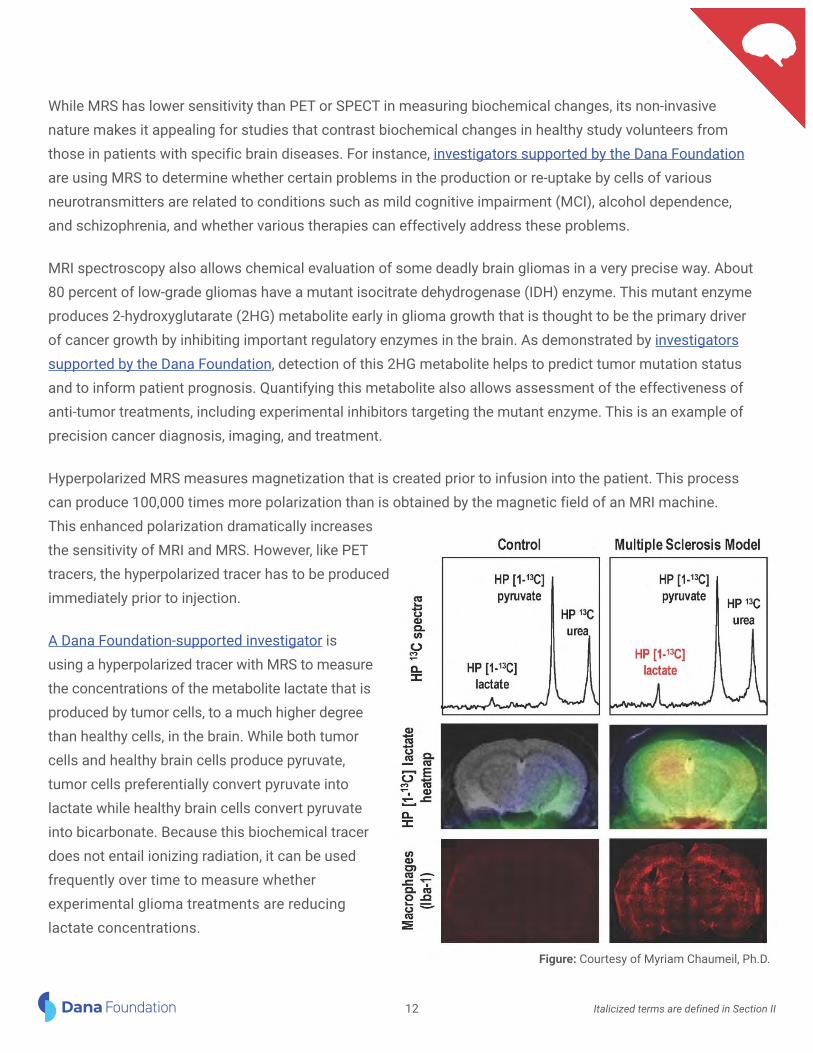
Hyperpolarized MRS measures magnetization that is created prior to infusion into the patient. This process can produce 100,000 times more polarization than is obtained by the magnetic field of an MRI machine. This enhanced polarization dramatically increases the sensitivity of MRI and MRS. However, like PET tracers, the hyperpolarized tracer has to be produced immediately prior to injection.
A Dana Foundation-supported investigator is using a hyperpolarized tracer with MRS to measure the concentrations of the metabolite lactate that is produced by tumor cells, to a much higher degree than healthy cells, in the brain. While both tumor cells and healthy brain cells produce pyruvate, tumor cells preferentially convert pyruvate into lactate while healthy brain cells convert pyruvate into bicarbonate. Because this biochemical tracer does not entail ionizing radiation, it can be used frequently over time to measure whether experimental glioma treatments are reducing lactate concentrations.
Figure: Courtesy of Myriam Chaumeil, Ph.D.
12 Italicized terms are defined in Section II

Hyperpolarized MRS is being used by another Dana Foundation-supported investigator to detect and monitor neuroinflammatory processes in neurological diseases and injuries that have an inflammatory component, such as multiple sclerosis and traumatic brain injury. In these instances, two HP probes—HP arginine (a newly designed probe) and HP [1-13C] pyruvate which is currently the most clinically translatable probe—are being developed to track the presence of immune cells (microglia) in the brain.
Diffusion Tensor Imaging and Optical Coherence Tomography
Optical coherence tomography (OCT) is a non-invasive technique that performs cross-sectional imaging at high resolution of optic fiber layers in the retina. Analogous to ultrasound (which uses sound waves), OCT uses light waves to provide micron-scale images of retinal fiber layers and has been used in diabetic retinopathy, age-related macular degeneration, and recently in children with optic pathway gliomas. By combining DTI (of the optic radiations) and OCT (of the retinal nerve fiber layer), an investigator supported by the Dana Foundation is studying both the pre-synaptic and post-synaptic visual pathways to explore the association between microstructural damage, neuronal loss, and visual acuity loss.
Transcranial Optical Imaging
Functional near-infrared spectroscopy (fNIRS)- provides non-invasive measures of hemodynamic responses to neuronal activation in real world settings. It uses optical signals to approximate blood-oxygenation levels in localized regions of the brain. Participants typically wear a cap through which near-infrared light is projected through the scalp and skull and into the brain, recording the intensity of light that is diffusely refracted. Neural activation elicited in response to a stimulus produces increased blood flow to the activated area. Blood flow changes result in increased blood volume that is assessed by concentrations of oxyhemoglobin, deoxyhemoglobin, or the summed total.
This technique is widely used in studies in infants and children. It has been used to investigate the processing of objects and of socially and biologically relevant information, and language development. These qualities make it especially useful in developmental studies in infants and children. fNRIS is portable and can be used at crib, bedside, or in natural settings. During the last decade and more, fNIRS has become an important technique for assessing development, including studies of social communication and face processing in healthy development and in developmental disorders such as autism and attention-deficit/hyperactivity disorder (ADHD). In adults, fNIRS is used to study complex interactions such as sensory integration involved in balance control.
While fNIRS’ spatial resolution is much lower than that of fMRI and it cannot penetrate deep into the brain, studies in adults that simultaneously measure hemodynamic responses with fMRI and fNIRS have demonstrated a high degree of correlation. Additionally, fNIRS has better spatial resolution, albeit inferior temporal resolution, compared to EEG (electroencephalogram), which had been the major technique used in infants.
13 Italicized terms are defined in Section II

Diffuse optical spectroscopy (DOS) and diffuse correlation spectroscopy (DCS) are forms of optical spectroscopy that use near-infrared light that passes through intact skin and skull to measure cerebral blood flow and concentrations of oxygenated and deoxygenated blood. Used in combination, DOS/DCS monitors variations in light absorption and changes in light-scattering properties of tissues to yield information on oxygen metabolism in the brain. Knowledge of changes in brain oxygen metabolism helps clinical research discover the timing and causes of brain injury in critically ill pediatric patients. In a study supported by the Dana Foundation, DOS/DCS has been used successfully to show that time-to-surgery (time between birth and surgery) is a critical risk factor for brain injury in survivors of newborn heart surgery.
.
Figure: Courtesy of Daniel Licht, M.D.
Electrical and Magnetic Field Recording and Mapping Techniques
Whereas fMRI, fNIRS, and PET are based on the coupling of neural and vascular (blood flow) activities, electrophysiological methods directly reflect brain cells’ electrical activity. These non-invasive methods include electroencephalography (EEG) and magnetoencephalography (MEG).
EEG measures the electrical activity that is produced by neurons as recorded from electrodes placed along the scalp. MEG maps brain activity by measuring magnetic fields that are generated by post-synaptic electrical activity in the brain. Because these measures are based on electrical activity, their temporal resolution is much higher than neuroimaging methods that detect slower hemodynamic responses. Both EEG and MEG provide information about global as well as regional neural activity, but MEG provides better spatial resolution. Often one or the other of these electrophysiological methods is used concurrently or sequentially with fMRI or PET to provide complementary information about normal and disturbed brain function.
14 Italicized terms are defined in Section II

EEG is used clinically to measure physiological manifestations of abnormal cortical excitability, primarily in the diagnosis and management of epilepsy and other seizure disorders. EEG is also used to study sleep disorders. EEG is routinely used along with many other measures in intensive care to monitor head-injured patients in coma, providing information that helps physicians assess patients’ prognosis. Research supported by the Dana Foundation in comatose patients who have suffered a subarachnoid hemorrhage have found that EEG, rather than multimodal metabolic monitoring, provides the best prognosis of whether a patient is likely to recover consciousness.
EEG recordings can also be conducted while a patient is inside an MR scanner, but special hardware and software filtering techniques are required to remove the large electrical signals generated by the MRI scanner. EEG and fMRI are used together, for instance, to localize where in the brain a seizure starts and where it spreads thereafter, or for studies of neural activity in sleep or anesthesia.
With its superior spatial resolution, MEG is sometimes used by itself or in addition to using fMRI, PET, or DTI (all of which provide different types of information) in patients prior to undergoing brain surgery as a means to identify essential areas that need to be surgically spared. MEG also has applications in basic research on brain function. However, MEG instrumentation is also much more costly than EEG and requires a shielded room.
Image-Guided Therapies
While imaging, electrical, and magnetic technologies have primarily been used diagnostically (the latter two are also used for surgical mapping), there are emerging applications of these technologies to image-guided therapies.
Transcranial magnetic stimulation (TMS) is a non-invasive brain stimulation technique that can be used to temporarily modulate regional brain activity. With TMS, a large current is discharged through a coil to create a changing magnetic field that induces a corresponding changing electrical field in the brain.
TMS can be applied as a single pulse or repetitively. Single pulses are used to briefly stimulate or interrupt regional neural activity while r-TMS can induce longer- lasting changes. The frequency of repetitive stimulation is thought to determine whether the modulation is excitatory or inhibitory. TMS is used clinically and experimentally as a form of image-guided therapy. The federal Food and Drug Administration (FDA) has approved TMS used repetitively (rTMS) to treat major depressive disorder (MDD), pain associated with migraine, and obsessive-compulsive disorder (OCD). In stroke patients with motor deficits, rTMS is being Figure: TMS being used in a study with a research participant.
Figure courtesy of Roy Hamilton, M.D., M.S.
15 Italicized terms are defined in Section II

used experimentally to try to restore the balance of excitation between motor cortices in each brain hemisphere. While TMS can be applied to general brain regions such as lateral frontal cortex, or moved systematically around the scalp to localize specific regions such as motor cortex based on the evoked motor responses, more advanced TMS studies used structural or functional imaging data to guide the placement of TMS coils. Anoth-er therapeutic form of TMS, called theta burst stimulation (cTBS) is being used experimentally to see if it can predict the capacity in patients with aphasia for brain plasticity and recovery of language and motor functions.
The ability to modulate regional brain function is complementary to functional neuroimaging in that imaging results may be used to guide the location of TMS neuromodulation, while changes in brain function due to TMS neuromodulation can be used to further test putative functional regions or networks. Researchers continue to gain a better understanding of mechanisms of actions and optimal doses (such as frequency and patterns of delivery). This technique is also being tested experimentally in other neurological conditions such Parkinson’s disease, dystonia, and schizophrenia. In sum, TMS serves both to provide information on brain functions and to treat some brain functions.
Diffusion tractography’s representation of white matter connections can be used to guide the location of deep brain stimulation (DBS) electrodes used to treat refractory depression and obsessive-compulsive disorder (OCD). To help optimize the placement of DBS electrodes in the nucleus accumbens, an investigator supported by the Dana Foundation is building a detailed map of the brain connections that pass through the nucleus accumbens using diffusion tractography and validating the map against a new optical imaging technique called CLARITY that provides much more detailed and complementary visualization of the connections compared to tractography in post-mortem tissue.
High- and Low-Intensity Focused Ultrasound in Surgical and Medical Situations
High-intensity focused ultrasound (HIFU) is another emerging approach for neuromodulation in combination with neuroimaging. Focused ultrasound uses acoustic lenses to focus ultrasound energy deep in the brain. This can have physiological consequences including changes in blood flow, increases in blood-brain-barrier permeability, and heating. Localized heating can be monitored in real time during treatment using MRI thermometry, which is based on diffusion imaging. HIFU is used by neurosurgeons to ablate tissue in localized brain areas in movement disorders such as tremor, and for chronic pain, without observable damage to intervening tissue or vasculature. HIFU ablations, however, are irreversible.
In contrast, the effects of low-intensity focused ultrasound pulsation (LIFUP) on neurons is reversible and non-destructive. LIFUP, therefore, can be used for both neuronal excitation and inhibition. An investigator supported by the Dana Foundation is testing the efficacy of thalamic stimulation with MRI image-guided LIFUP to try to increase arousal and/or awareness in patients with disorders of consciousness.
16 Italicized terms are defined in Section II

Fluorescence-Assisted Resection and Exploration
Optical fluorescence imaging uses near-infrared (NIR) fluorescent light that penetrates several millimeters into brain tissue and can identify normal brain structures and tissues involved in disease, including glioma and neurodegenerative disease. An investigator supported by the Dana Foundation used optical fluorescence imaging, combined with targeted contrast agents that differentiate disease tissues from healthy tissue, to provide neurosurgeons with real time image guidance to improve the completeness of resection and decrease surgical complications in patients with brain tumors.
Figure: Brain tumor imaging using MRI, NIR-1, and NIR-II imaging with targeted contrast agents. Figure courtesy of Hak Soo Choi, Ph.D.
Cellular and Molecular Imaging
Cellular and molecular imaging techniques answer questions about the biochemical activities of cells and their molecules, and how these are altered by disease, injury, and their treatments. They do so at a much higher resolution in space and time than PET, SPECT, and MRS. Cellular and molecular imaging techniques use several types of light microscopes, and various types of “optical probes,” which are molecules that have been specially labeled to emit light of various wavelengths to “contrast” the target cells of interest from other cells. While “intravital” light microscopy—used to visualize live organisms—was developed more than 170 years ago, the development in recent times of many types of highly specialized light-emitting probes, advances in specialized light microscopes, and computerization have transformed the science of optical imaging.
Optical Imaging in Live Laboratory Animals
Optical scanning techniques image the actions of molecules and cells that are illuminated with bioluminescent or fluorescent probes in live laboratory animals. This imaging requires surgical removal of a part of the skull to
17 Italicized terms are defined in Section II

expose the cortex. Thereafter, scientists visualize actions of cells or molecules. Voltage-sensitive dyes, (molecules that bind across a neuron’s membrane), enable scientists to detect spiking and synaptic activity of neurons in the exposed cortex.
Optical tomographic imaging is the major technique for visualizing cells and molecules in live laboratory animals. This technology uses near infrared (NIR) fluorescent probes; fluorescence greatly increases the sensitivity of NIR imaging. The brain area of interest is exposed by removing the skull covering that area and biochemical activity that occurs deeper within the animals’ tissues can be observed. While animal tissue absorbs and scatters visible light, the amount of NIR light absorbed by tissues is less, enabling this imaging to penetrate further. The amount of resolution provided by NIR alone, however, is insufficient for identifying which specific cells within any location are actually emitting the light. This problem is addressed with the application of genetic and adoptive transfer techniques for creating fluorescent probes to use with NIR optical imaging.
Optical Imaging Probes
Advances in molecular and cellular imaging are largely due to the development of novel types of light-emitting probes and ingenious ways of labeling them for use in living laboratory animals. These methods can be used to monitor cellular and molecular function, including studies in vivo in which microscopy using optical probes is carried out in the brain of a living animal. Currently, however, these methods are invasive in that they involve administering experimental chemicals or genetically modifying animals to produce optical probes through cellular protein synthesis, along with invasive surgery to allow microscope visualization of a brain region of interest. Thus, while optical imaging methods are transforming knowledge of cellular and molecular function by allowing these to be studied in situ and in three dimensions, these methods are generally not yet amenable for use in studying the human brain.
Bioluminescent probes and fluorescent probes are two major types of optical probes used in neuroscience research. Bioluminescent probes use “luciferase,” an enzyme, to generate and emit light by an organism, providing real time analyses of disease processes at the molecular level in living organisms, including laboratory animals. Luciferase is the enzyme in fireflies and glowworms that makes them light up. These are the best known examples of organisms that naturally produce bioluminescence, but deep-sea marine organisms and some bacteria and fungi also produce bioluminescence.
Fluorescent protein probes are green fluorescent protein and its yellow, blue, and cyan-colored mutants; and red fluorescent proteins. In addition, there are hundreds of other fluorescent probes that are not fluorescent proteins. Fluorescent probes are introduced into and visualized in the animal or its tissue cultures when excited by ultraviolet or visible light and viewed with optical imaging techniques. Fluorescence is the absorption and subsequent re-radiation of light by an organism. Fluorescence was known and used for microscopy, including intravital imaging for many years prior to the discovery of fluorescent proteins.
18 Italicized terms are defined in Section II

The discovery of fluorescent proteins occurred after scientists, isolating a blue bioluminescent protein from a specific type of jellyfish, observed another protein that produced green fluorescence when illuminated with ultraviolet light. The 2008 Nobel Prize in Chemistry (Osamu Shimomura, Martin Chalfie, and Roger Y. Tsien) was awarded for the discovery and development of the green fluorescent protein. The gene for green fluorescent protein was cloned in the early 1990s; but its utility as a molecular probe occurred later, after scientists used fusion products to track gene expression in bacteria and nematodes.
The colored proteins, plus fusion proteins and biosensors—all of which are referred to as fluorescent proteins—are used primarily to visualize molecules in living cells. Moreover, multiple (different colored) fluorescent probes can simultaneously identify several target molecules within a cell and show their actions.
The fluorescent proteins are fused to specific proteins and enzymes in the laboratory. They are primarily introduced into the animal through production of “transgenic” strains, whereby the fluorescent protein is introduced into the germline (sequence of germ cells containing genetic material), often under the control of tissue-specific or cell type-specific promoters.
In another approach, engineered genes that encode fluorescent protein fusions, rather than the proteins themselves, are introduced into the animals by attaching them to harmless viruses, which serve as vectors to carry the fluorescent protein fusions into the animal. The fluorescently labeled cells in tissues of interest are then imaged.
To introduce bioluminescent and fluorescent probes into the animal, a widely used technique is genetic transfer. The gene that produces bioluminescence or fluorescence is cloned in the laboratory and introduced into a laboratory animal in one of two ways. The gene may be inserted into a harmless virus (called a vector) and introduced into the animal. Or, the gene is inserted into a stem cell and introduced into the animal so that the differentiated cell that the stem cell develops into will express the luminescence or fluorescent protein. The scientists at the forefront of gene transfer technology (Mario Capecchi, Sir Martin J. Evans and Oliver Smithies) were awarded the Nobel Prize in Physiology of Medicine in 2007 for their discoveries of principles for introducing specific gene modifications in mice by the use of embryonic stem cells.
Adoptive transfer, another technique, involves tagging specific cells, such as in an animal model of a disease, and transferring those tagged cells into another laboratory animal to see how the cells work. Adoptive transfer is used to label cells that are “naturally occurring probes” in the body, in that they migrate to specific targets. As an example, lymphocytes (a family of white blood cells, either T or B cells) produce antibodies that are taught by immune dendritic cells to attack a specific foreign invader such as an infectious agent. Scientists label the antibody protein with a fluorescent tag and inject the material into another laboratory animal; or, they "stain" tissue sections or isolated cells within tissues and image the tissues or cells using a technique called flow cytometry.
19 Italicized terms are defined in Section II

Cellular and Molecular Labeling for Image-Guided Therapies
Use of “dendrimers” with fluorescent probes is another approach to labeling particles that travel to a target in the brain. Unlike antibodies, which are proteins produced by living cells, dendrimer nanoparticles are compounds that are synthesized in the laboratory. Dendrimers can serve as fluorescent imaging probes for improved neuro-imaging of tumors. They also have been explored as a vehicle for delivering targeted therapies in the brain.
Successfully delivering a drug across the blood-brain-barrier to enter the brain has been an issue that has limited the efficacy of many anti-cancer treatments for patients with brain tumors. Direct intra-tumoral delivery using convection-enhanced delivery (CED) has been one solution to overcome this limitation. Other systemic drug delivery techniques, however, will likely be critical to achieving long term disease control with malignancies of the brain. A number of novel drug delivery approaches have either recently been FDA-approved or are in clinical development as promising new anti-cancer treatments. These include liposomes, microspheres, polymer microcapsules, polymer conjugates, and nanoparticles.
Dendrimers, in particular, have emerged as especially versatile synthetic nanoparticles that can serve both as fluorescent imaging probes for improved neuroimaging of tumors, as well as provide a delivery vehicle for targeted therapy “payloads.” Nevertheless, as brain gliomas arise from the brain’s own glial cells, immune cells recognize glioma cells as “self” and do not attack. Gliomas, therefore, capitalize on immune “checkpoint” mechanisms that are designed to prevent “autoimmunity,” the process by which immune cells mistakenly identify the body’s own cells as foreign and attack them. Since checkpoint mechanisms block immune activation, they create a permissive tumor microenvironment that protects gliomas and enables them to grow. Research supported by the Dana Foundation is leveraging optical imaging methods to characterize the molecular genetics of glioblastoma cells and their interactions with the immune system to try to optimize therapeutic strategies to synergistically to target glioblastoma and its tumor microenvironment. Dendrimer therapies are being used for the specific purpose of clearing out the tumor microenvironment. Other current research is focusing on modulating the tumor microenvironment using checkpoint inhibitors. These two types of therapies clearly can be synergistic and have the potential to work together in the future.
Another technique for imaging and targeting drug delivery is the use of aptamers, which are made from nucleic acids. Aptamers can be produced to have exquisite binding specificity for defined molecular targets, similar to antibodies. They can be attached to the surface polymeric particles for imaging and to enable targeted drug delivery.
Optical Imaging Microscopes
The variations in the ways that different bioluminescent and fluorescent probes provide information are influenced by the type of microscope or other imaging technologies used. There are three major types of light
20 Italicized terms are defined in Section II

microscopes: fluorescence, two photon, and confocal. All three are fluorescent microscopy techniques used to study molecules in living cells. They provide insights into how the molecules and the cells they compose behave normally, and how they are altered by disease, injury, treatment, or other experiences. Each type of microscope has its strengths and limitations.
Fluorescence microscopes are used with fluorescent probes that emit light of short wavelength to reveal biochemical activities within a cell in human and in animal tissue cultures. Fluorescence microscopes have the highest resolution of all cellular imaging devices. This enables them to be used to identify a single fluorescently labeled molecule or differentiate activities of several differently colored fluorescent molecules in the same cell. This process is referred to as “subcellular” resolution of molecular activities within a cell.
Fluorescently labeled cells are excited by laser or incandescent light. The fluorescent label (probe), which is designed to go to a specific molecule or region of a cell, absorbs a “photon” of energy supplied by the laser or incandescent light. Then the photon is excited to emit light at a particular wavelength, depending upon the specific probe used. The emitted light is recorded as a photographic image, video, fluorescence decay trace, or as “photo multiplier tube” signals from serial points that are displayed and analyzed by computer to provide flexible images.
Most fluorescent light microscope probes emit light of short wavelengths, which is visible with the naked eye, and useful for imaging molecules or cells close to the surface. Visible light does not pass through tissue well, however, and these techniques are suitable only in cases where the distances traveled in tissue by the probe are small (micrometers in length). As a result, the tissue grown in laboratory cultures needs to be very thin. Tissue grown for visualization with the other two main types of light microscopes, confocal and multi-photon, can be much thicker.
Confocal laser scanning microscopy provides the ability to simultaneously collect multiple images in digital form from serial sections of thick tissue specimens, and flexibly display and analyze them via computer. Confocal imaging is undertaken in thick tissue cultures and in small laboratory animals. The blur-free images are taken point-by-point using “photo multiplier tubes” that provide sensitive and fast registration of the intensity of emitted light. The points are then reconstructed by computer, rather than projected through a microscope’s eyepiece.
Confocal laser scanning imaging relies extensively on fluorescent probes of longer wavelength to monitor dynamic processes such as: cellular integrity, membrane fluidity, transduction of cellular signals, enzyme activities, movements of proteins, and the migration of cells in the developing animal embryo. Additionally, confocal laser screening microscopes facilitate study of brain synapses (communication junctions between two nerve cells) and cell circuitry (neural networks), as do multi-photon techniques.
Multi-photon, including the commonly used two-photon, laser microscopy rely on the simultaneous absorption
21 Italicized terms are defined in Section II

Figure. Two-photon laser scanning microscopic image of the
of two or more photons by a molecule to image fluorescent probes with longer wavelengths that penetrate deeper into tissues. It is used in thick tissue cultures and small laboratory animals, often to study cellular actions over time in the brain. As an example, multi-photon imaging visualizes actions of immune microglia and of antibodies summoned to the brain to fight infections and cancers.
In research supported by the Dana Foundation, two-photon (2P) calcium imaging is being used in an experimental animal model of a severe rare neurodevelopmental disorder known as Dravet syndrome to understand the basic mechanisms of febrile seizures, which is the most common seizure type. Dravet syndrome is due to a mutation of the gene SCN1A that predominantly affects neurons that use the inhibitory neurotransmitter called GABA. This neurotransmitter normally inhibits other brain cells and therefore is thought to protect against seizures. The technique of 2P calcium imaging allows investigators to record an optical readout of hundreds or thousands of neurons simultaneously in awake, behaving mice, including during temperature-induced seizures. After using this imaging technology to define the cellular architecture of seizures and identify cellular and circuit mechanisms underlying seizure onset, investigators then will try to manipulate the circuitry to recover normal GABA network function to prevent seizure generation.
Fluorescence resonance energy transfer (FRET) is undertaken with microscopic imaging in tissue cultures to reveal the interaction between two or more fluorescent probes. This interaction of adjacent probes is used to monitor the assembly or fragmentation of molecules, as occurs in the binding of a molecule to its receptor. Fluorescent-and FRET- generated signals are relatively weak and therefore limited to tissue cultures. To undertake such studies in small laboratory animals requires the use of macroscopic optical imaging scanners.
Array Tomography
Array tomography uses arrays of serial ultrathin tissue sections of fixed tissue specimens. The technique has become important in exploring circuit and molecular architectures of the dynamic events that underlie nervous system development and function. Array tomography combines and goes beyond capabilities of optical fluorescence and electron
genetically-encoded calcium indicator GCaMP6s (green) with
labeling of neurons in layer 2/3 primary somatosensory cortex
of a Dravet syndrome (Scn1a+/-) mouse, with co-labeling of a
prominent subset of inhibitory GABAergic interneurons known
as parvalbumin-positive fast-spiking cells (red).
Figure courtesy of Ethan Goldberg, M.D., Ph.D.
22 Italicized terms are defined in Section II

microscopy. Ordered arrays of ultrathin serial sections that are constructed and repeatedly stained on glass microscope slides provide for quantitative imaging at high-resolution of large numbers of antigens and fluorescent proteins in tissue ultrastructure.
This technique, in combination with two-photon imaging and electron microscopy, has been integral to exploring the role of microglia in pruning synapses during development and later in life. Such pruning is thought to guide healthy brain development; excessive or premature pruning may lead to developmental problems and later in life may prematurely remove weak synapses.
Light Sheet Swept Confocally-Aligned Planar Excitation
A related approach to studying tau accumulation is with the newly developed light sheet swept confocally-aligned planar excitation (SCAPE) microscope. Light sheet microscopy is used for imaging sensitive biological samples or biological processes that occur rapidly in vivo. The sample’s illumination occurs in a single plane at a time, perpendicular to the sample, so this technique avoids any out-of-focus excitation and damage due to light. Optical sectioning is intrinsic, and 3-D images are recorded by moving the sample through the light sheet.
Scientists supported by the Dana Foundation are using the light sheet microscope SCAPE to study tau accumulation in vestibular circuits in a living zebrafish model for progressive supranuclear palsy (PSP) that has DNA from a PSP patient inserted into the genome. SCAPE is fast enough to measure changes in neuronal shapes and functions while simultaneously measuring progressive failures in synapses that connect cells responsible for balance and gaze. Investigators are tracking the time-course of degeneration that affects molecules, functions (neuronal loss), and are able to correlate dynamic changes in molecular and cellular activity with behaviors measures of eye movement responses to changes in position.
In other research supported by the Dana Foundation, light sheet microscopy is combined with CLARITY preparation of the mouse brain to enable a 3-D scan of the entire ex vivo brain with unprecedented speed and resolution. CLARITY preparation removes lipids so that the brain becomes a transparent “matrix” that is amenable to optical microscopy and light sheet microscopy is capable of resolving individual neurons and their connections. In this project, scientists are characterizing abnormalities in molecules and connections across multiple brain regions from a mouse model on insulin resistance to try to gain new insights into links between Type 2 diabetes and neurodegeneration.
Optogenetics: Stimulating Neurons in Awake Behaving Animals
One of the most impactful new optical technologies is optogenetics, first described in 2005. Introducing a microbial opsin gene makes neurons precisely responsive to light. So, rather than using electrical techniques to
23 Italicized terms are defined in Section II

stimulate neurons, optogenetics uses optically-activated proteins to activate them. Although optogenetics is not an imaging technique, the ability to precisely control the activity of specific populations of neurons in specific brain regions provides a powerful means of studying complex brain functions in animal models during imaging studies that were previously limited to very simple manipulations, such as electrical forepaw stimulation to study the motor network.
Optogenetics combines genetics and optics to exert this control. Genes that confer light responsiveness are inserted into neurons. Light then can be delivered into freely behaving mammals using fiberoptics. The optically activated proteins enable scientists to selectively and precisely activate and inhibit specific populations of neurons. Activation or inhibition is controlled by genetically encoded switches using bursts of light. Optogenetics is providing insight into how single neurons operate alone and within neural networks.
Optogenetics primarily utilizes channelrhodopsin-2 (ChR2). This protein from algae is a light-activated cation channel that can induce neuronal depolarization and action potentials with unparalleled spatial, temporal, and neurochemical precision. Discovery of the properties and utility of ChR2, an “all-in-one” light-activated protein followed discoveries in the 1970s of two other similar light-activated proteins from microbes, bacteriorhodopsins, and halorhodopsins. These (and now other similar) proteins are inserted into cultured cells or in the brains of live animals to investigate the specific structure and function of a neural network. Clinically, optogenetic science has the potential to modulate the activity of brain networks that are involved or implicated in neurological and psychiatric diseases.
Single-Cell RNA-Seq
Single-cell RNA-seq is an emerging confluence of technologies being used to reveal when genes in individual cells switch on through time during development. The journal Science selected it as the 2018 Breakthrough of the Year. Scientists anticipate that microscopy will soon play a role in this research.
The combination of techniques that is called single-cell RNA-seq involves isolating thousands of intact cells from living organisms, sequencing the proteins expressed by genes via their RNA in each cell and—through computerization or cell labeling—reconstructing their relationships. A research group has identified the relationship of more than 100 cell types in the brains of zebrafish. Researchers are looking to use these combined techniques with new microscopy techniques to visualize where this molecular activity occurs in a cell and how it is affected by nearby cells.
Combining Imaging Technologies to Advance Human Health
As these examples illustrate, new combination techniques are already advancing the threshold for applying imaging innovations to further understanding brain functions and the effects of experiences, diseases, and
24 Italicized terms are defined in Section II

therapies in altering these.
A decade ago, we would not have envisioned that PET imaging with probes that attach only to the protein amyloid could help in the probable diagnosis of Alzheimer’s disease and assess effects of therapies to reduce amyloid or prevent its further deposition in the brain. We would not have foreseen that PET (which has relatively poorer spatial resolution) combined with optical imaging in laboratory animals could visualize fluorescently marked neurons in the brain to reveal how one neuron wires with another to form neural circuits, and to monitor this process over time during development to see changes in response to disease or experience.
Since biochemical changes in cells precede changes that occur in response to disease, identifying these cellular changes could provide the means to diagnose diseases in their earliest stages, when they are most likely to be responsive to effective therapies. Imaging biochemical changes in molecules, rather than physical differences between normal and diseased tissues, has the potential not only to improve early identification and diagnosis of diseases, but also to quickly assess the efficacy of various treatments.
Combining molecular imaging with anatomical and physiological imaging technologies, as these examples illustrate, is fundamentally advancing scientific understanding of how the brain functions and the translation of that understanding to improve human health.
Section II Imaging Techniques at a Glance - Defined terms from Section I
Adoptive transfer is used in molecular imaging to tag specific cells, such as in an animal model of a disease, and transfer those tagged cells into another laboratory animal to see how they work. The technique is used to label cells that are “naturally occurring probes” in the body, such as immune T cells, which produce antibodies that migrate to an infection. (Please also see genetic transfer, a related technique.)
Advanced dynamic contrast-enhanced MRI is a molecular technique for imaged-guided therapy that is used experimentally to try to identify blood-brain-barrier dysfunction in people with major depressive disorder (MDD). Using the tracer gadolinium, clinical investigators may be able to detect subtle blood-brain-barrier leakage that enables inflammatory cytokines to enter the brain and disrupt brain circuitry. Gadolinium injected into the blood transfers into brain tissue at the sites of blood-brain-barrier dysfunction can be visualized with MRI. Used in combination with a functional MRI technique called multi-echo multi-band functional MRI, researchers then measure connectivity of the neural circuit involved in reward and anhedonia, to see if the circuit is disrupted in MDD patients with blood-brain-barrier dysfunction and high levels of inflammation. This approach may lead to novel anti-inflammatory approaches to treatment.
25

Angiography uses a radiopaque dye injected through a catheter into a blood vessel to detect a blockage or narrowing of downstream vessels, or other vascular lesions such as aneurysms or arteriovenous malformations of the vessel. The vessel is outlined on x-ray as white. Angiographic data now are digitized and analyzed using algorithms that subtract pre-and post-contrast images (termed “digital subtraction angiography”). Angiography provides the highest resolution images of vascular structures in the brain and is widely used clinically.
Array tomography uses arrays of serial ultrathin sections on fixed tissue specimens. The technique is used to explore circuit and molecular architectures of dynamic events underlying nervous system development and function. Array tomography combines and goes beyond the capabilities of optical fluorescence and electron microscopy. Ordered arrays of ultrathin serial sections that are constructed and repeatedly stained on glass microscope slides provide for quantitative imaging at high-resolution of large numbers of antigens and fluorescent proteins in tissue ultrastructure. This technique, in combination with two-photon imaging and electron microscopy, has been used to explore the role of microglia in the brain in advantageous or deleterious synaptic pruning.
Arterial spin labeling (ASL) is a perfusion contrast used with fMRI. ASL MRI uses magnetically labeled arterial blood water as a flow tracer, rather than an exogenously administered radiotracer, to quantify regional cerebral blood flow (CBF) noninvasively. It provides absolute quantification of CBF, which renders it sensitive to both static function and changes occurring over longer intervals. Since alterations in cerebral blood flow represent the proximate cause of ischemic cerebrovascular diseases, ASL MRI has been translated to clinical use for the diagnosis and management of disorders of cerebral blood flow. ASL MRI also is used to measure the effects of chronically administered drugs on regional brain function. These measures rely on a coupling between changes in regional neural activity and changes in cerebral blood flow.
Bioluminescent probes are used in molecular imaging. They utilize the enzyme luciferase to generate and emit light by an organism, providing real-time analyses of disease processes—particularly infections and cancer progression—at the molecular level in living organisms, including laboratory animals. The enzyme is found in fireflies, glowworms, deep sea marine organisms, and some bacteria and fungi.
BOLD (blood oxygenation level dependent) MRI is the contrast agent used in most fMRI imaging (please see functional MRI below). BOLD contrast reflects a complex interaction between the volume of blood, its flow, and its transport of oxygen by an iron-containing protein in red blood cells. Functional contrast is produced when the oxygen is released from the iron and taken up and used by brain cells (indicating that they are active). After the iron loses the oxygen, the iron becomes highly magnetized when exposed to the MRI magnetic field. BOLD MRI scans are sensitive to changes in regional brain function. Task-based BOLD fMRI has been used in research to study numerous brain systems involved in functions such as language, memory, and decision-making. It is used clinically in presurgical mapping of brain regions to identify areas that control functions and should not be surgically removed.
26

Computer assisted tomography (CT) uses special x-ray equipment to obtain and reconstruct three-dimensional anatomical images of bone, soft tissues, and air. An x-ray emitter rotated around a patient measures the rays’ intensities from different angles. For brain imaging, numerous x-ray beams are passed through the head at different angles. Special sensors measure the amount of radiation absorbed by different tissues and a computer uses the differences in x-ray absorption to form cross-sectional images or “slices” of brain called “tomograms.” CT can be used with or without contrast agents that typically contain radiodense iodine. Contrast agents facilitate easy visualization of blood vessels or structures lacking a blood brain barrier. CT can show bone, soft tissues, and blood vessels in the same images. CT is quick, has few contraindications, and is useful, therefore, in emergency situations to identify brain structure abnormalities such as brain swelling, bleeding arising from ruptured aneurysms, hemorrhagic stroke, and head injury.
Confocal laser scanning microscopy is a molecular imaging technique that monitors dynamic processes such as synaptic activity and cell death. The technique enables simultaneous collection in digital form of multiple images from serial sections of thick tissue specimens that are flexibly displayed and analyzed via computer. The blur-free images are taken point-by-point and, with sensitive and fast registration of the intensity of emitted light, are reconstructed via computer.
Cryo-electron microscopy (Cryo-EM) is a technique used to determine the high-resolution structure of proteins, nucleic acids, and other biomolecules—and to some extent how they move and change as they function—in tissue samples that have been cooled to cryogenic temperatures and embedded in rapidly cooled “glassy” (vitreous) water. Cryo-EM fires beams of electrons at proteins in frozen tissue samples, using more gentle electron beams than those used in traditional transmission electron microscopes and sophisticated methods of image processing. Cryo-EM is used, for instance, to deduce the structure of the specific protein in autopsied brain tissue of a person who had Alzheimer’s disease, which ultimately may provide insights into disease progression.
Deep brain stimulation (DBS) involves implanting electrodes in specific areas in the brain and externally stimulating the electrodes to measure electrical activities of neurons and their electrochemical pathways. DBS is used therapeutically to treat intractable Parkinson’s disease and essential tremor and is being studied for possible use in intractable depression and other brain conditions. It is also used in a few highly specialized centers to explore the neuronal underpinnings of cognition.
Dendrimer nanoparticles are used in molecular imaging with fluorescent probes to label particles that travel to a target. Dendrimer nanoparticles are compounds that are synthesized in the laboratory from materials (often polymers) and assembled into high molecular weight spherical particles. A fluorescent probe is added to the dendrimer nanoparticles which then target and light up specific cells. A therapeutic drug also can be added to dendrimer nanoparticles, which can cross the blood-brain-barrier and enter the brain. They have been studied as treatment delivery mechanisms in glioma, but the processes that guard against autoimmunity have thus far
27

impeded this approach. However, dendrimer therapies are being used for the specific purpose of clearing out the tumor microenvironment. Other current research is focusing on modulating the tumor microenvironment with checkpoint inhibitors. These two types of therapies can clearly be synergistic and have the potential to work together in the future.
Diffuse optical spectroscopy (DOS) and Diffuse correlation spectroscopy (DCS) are types of transcranial optical imaging. They are forms of optical spectroscopy, which uses near-infrared light that passes through intact skin and skull to measure cerebral blood flow and concentrations of oxygenated and deoxygenated blood. Used in combination, DOS/DCS monitors variations in light absorption and changes in light-scattering properties of tissues to yield information on oxygen metabolism in the brain. Knowledge of changes in brain oxygen metabolism helps in determining the timing and causes of brain injury in critically ill pediatric patients. DOS/DCS has been used successfully to show that time-to-surgery (time between birth and surgery) is a critical risk factor for brain injury in survivors of newborn heart surgery.
Diffusion-perfusion-weighted MRI is a combination technique used to estimate the “ischemic penumbra.” This is the brain tissue that has suffered from reduced blood flow following ischemic stroke but has not yet died and is the target of intensive therapy.
Diffusion spectrum imaging (DSI) is a high-resolution diffusion MRI sequence used to discern the complex crossing structure of white matter fibers more effectively than can be achieved with diffusion-weighted imaging (please see DW-MRI). DSI is used, for example, to explore whether white matter tracts from the brain’s small raphe nucleus can help determine which patients with depression are most likely to respond to treatment with selective serotonin reuptake inhibitors (SSRIs).
Diffusion tensor MRI (DTMRI) leverages the variation (anisotropy) in water diffusion in brain white matter to create detailed maps of white matter tracts. DTI has high spatial resolution and complex diffusion encoding compared to DWI (please see diffusion-weighted imaging) so that the directionality of water diffusion can be resolved in individual voxels. Computerized mathematical models estimate the most likely pathways of white matter tracts through voxels and provide color coded 3-dimensional maps of the pathways connecting between gray matter regions in the cortex or in subcortical nuclei. DTI measures microscopic water motion in tissues, and in the brain this motion is facilitated along white matter tracts that connect brain regions. Computerized mathematical models construct the images of the white matter tracts. It is used extensively pre-surgically to identify which tracts are functionally essential and should not be removed during surgical treatment for a brain tumor.
Diffusion tractography is a technique that is being used in the NIH Connectome project and also is used experimentally for image-guided therapies. It is an extension of diffusion tensor imaging (DTI) and traces brain pathways based on water diffusion patterns in brain tissue that are measured by magnetic resonance imaging
28

(MRI). Diffusion tractography utilizes data from diffusion imaging technologies to computationally reconstruct a pathway of a nerve fiber bundle (white matter) by following the orientation along which water diffusion is most rapid, because the orientation with the fastest diffusion rate corresponds to the long axis of nerve fiber bundles. Diffusion tractography is used as an image-guided therapy primarily to compare specific white matter pathways in different diseases and look for qualitatively different pathways involved in congenital disorders or injury recovery. This technology is beginning to develop clinically relevant information in several brain diseases such as multiple sclerosis, schizophrenia, depression, and ALS and other neurodegenerative diseases. In severe intractable depression, it is used experimentally to help guide optimal deep brain stimulation (DBS) electrode implantation. Diffusion-weighted MRI is sensitive to the microscopic motion of water, which differs in gray and white matter and varies according to brain health or pathology. In the healthy brain, gray matter has a higher diffusion rate than white matter. Additionally, gray matter diffusion is essentially equal in all directions, (isotropic) while white matter diffusion varies (it is “anistropic”). DW-MRI maps can provide information on the degree (“fractional anisotropy”) and magnitude of variation. DW-MRI is used clinically to confirm acute ischemic stroke (artery blockage or narrowing) where water diffusion decreases within minutes. Decreased diffusion also can be seen with DW-MRI in inflammatory brain disorders and tumors. A new class of genetically encoded “reporter genes” (which attach to and tag a gene of interest) has been used with DW-MRI to visualize gene expression in a mouse model of brain tumor by altering the diffusion of water across cell membranes.
Electroencephalography (EEG) is an electrophysiological method that directly reflects brain cells’ electrical activity. It measures the electrical activity that is produced by neurons as recorded from electrodes placed along the scalp. It provides information about global as well as regional neural activity, although it has lower spatial resolution compared to MEG (please see magnetoencephalography). EEG is used clinically to measure physiological manifestations of abnormal cortical excitability, primarily in diagnosing and managing epilepsy and other seizure disorders. It is also used with other measures in intensive care to monitor head-injured patients in coma. Recent studies in comatose patients who have suffered a subarachnoid hemorrhage have found that EEG, rather than multimodal metabolic monitoring, provides the best prognosis of the likelihood of recovering consciousness. EEG is also used to study sleep disorders. EEG is used with fMRI and PET to provide complementary information about normal and disturbed brain function; for instance, EEG and fMRI are used to localize seizure foci and spread.
Fluorescence resonance energy transfer (FRET) is a molecular imaging technique that reveals the interaction between two or more fluorescent probes in tissue cultures. It is used, for instance, to visualize a molecule binding to its receptor on a cell.
Functional near-infrared spectroscopy (fNIRS) is a form of transcranial optical imaging that non-invasively measures hemodynamic responses to neuronal activation in real world settings and is widely used in studies in infants and children. Participants wear a cap through which near-infrared light is projected through the scalp and skull into the brain. fNIRS uses optical signals to approximate blood-oxygenation levels in localized brain
29

regions. It records the intensity of light that is diffusely refracted. Neural activation elicited in response to a stimulus produces increased blood flow to the activated area. Blood flow changes result in increased blood volume. The spatial resolution of fNIRS is low compared to that of fMRI and it cannot penetrate deep into the brain, but studies in adults demonstrate that fNIRS and fMRI have a high degree of correlation. fNIRS has better spatial (but not temporal) resolution compared to EEG (please see electroencephalogram) which had been the major technique used in infants. fNIRS has become important in assessing development by investigating processing of objects, socially and biologically relevant information, and language. fNIRS in adults is used to study complex interactions like sensory integration in balance control.
Functional MRI (fMRI) is based on the principle that changes in regional cerebral blood flow and metabolism are coupled to changes in regional neural activity involved in brain functioning. Significant contrast in tissue can be attributed to changes either in blood flow alone, in metabolism alone, or in blood flow and metabolism. Using blood oxygenation level dependent (BOLD) contrast with MRI dramatically increases imaging speed. BOLD contrast reflects a complex interaction, not yet completely understood, between cerebral blood flow, cerebral blood volume, cerebral metabolic rate, and biophysical interactions between the brain and the local magnetic field. BOLD MRI scans are sensitive to changes in regional brain function. This ability has facilitated the direct testing in humans of hypotheses about the organization of regional brain function derived from behavioral testing and from studies in patients with focal lesions (please see magnetic resonance imaging.) fMRI methods have been translated clinically such as for presurgical mapping of function-controlling brain regions prior to neurosurgical procedures.
Fluorescence microscopes are used with fluorescent probes that emit light of short wavelength to reveal biochemical activities within a cell in human and animal tissue cultures. These microscopes have the highest resolution of all cellular imaging devices. They can be used to identify a single fluorescently labeled molecule or differentiate activities of several differently colored fluorescent molecules in the same cell.
Fluorescence optical imaging is used for image-guided therapy. Near-infrared (NIR) fluorescent light penetrates several millimeters into brain tissue and can identify normal brain structures and disease tissues, including glioma and neurodegenerative diseases. Optical fluorescence imaging, combined with targeted contrast agents that differentiate disease tissues from healthy tissue, helps provide neurosurgeons with real time image guidance to improve the completeness of resection and to decrease surgical complications. Fluorescence optical imaging is one of many optical imaging technologies that have been emerging since the early 2000s.
Fluorescent probes are used in molecular imaging to visualize molecules and their actions. The probes are green fluorescent protein, its yellow, blue, and cyan-colored mutants, and red fluorescent proteins. Fluorescent probes that emit light of short wavelengths are used with fluorescent light microscopes to image molecules that are close to the surface in laboratory cultures of thin human or animal tissues. Fluorescent probes that
30

emit light of longer wavelengths are introduced into small laboratory animals and used with other microscopes (please see confocal laser scanning microscopy and multi-photon laser microscopy) and excited by ultraviolet light to show molecules deeper in tissues, in the small laboratory animal or in thick animal tissues in laboratory cultures.
Genetic transfer is used in molecular imaging to introduce bioluminescent and fluorescent probes into the animal. The gene that produces bioluminescence or fluorescence is cloned in the laboratory and introduced into a laboratory animal. The gene is introduced into the animal either by inserting it into a harmless virus (called a vector) that gets into a specific type of cell, or by inserting it into a stem cell that differentiates into a cell that expresses the luminescence or fluorescent protein. (Please also see adoptive transfer, a related technique.)
High- and low-intensity ultrasound are emerging as types of image-guided therapy. High-intensity focused ultrasound (HIFU) is used neurosurgically to treat movement disorders by ablating tissue in localized brain areas. HIFU uses acoustic lenses to focus ultrasound energy deep in the brain, which can have physiological consequences including changes in blood flow, increases in blood brain barrier permeability, and heating. Localized heating can be monitored in real time during treatment using MRI thermometry, which is based on diffusion imaging. HIFU is used by neurosurgeons to ablate tissue in localized brain areas in movement disorders, including tremor, and for chronic pain. There are no reports of observable damage to intervening tissue or vasculature. HIFU ablations, however, are irreversible. Low-intensity focused ultrasound pulsation (LIFUP) can be used to excite or inhibit neurons. LIFUP is reversible and non-destructive. MRI image-guided LIFUP currently is being experimentally used to stimulate thalamic connections in patients with disorders of consciousness to try to increase their arousal and/or awareness.
Intravital light macroscopic imaging technologies use light-emitting probes as contrast to visualize specific molecules and the cells they compose in small laboratory animals and in a few larger laboratory animals. The molecules can be imaged everywhere they occur in the body as opposed to a single location (please see intravital light microscope technologies), and the molecules can be imaged as they move throughout the body, including the brain. (Please also see macroscopic optical scanning techniques.)
Intravital light microscopic technologies use light-emitting probes as contrast to visualize the activities of specific molecules and the cells they compose. Imaging is undertaken in tissues surgically biopsied from humans and laboratory animals. Imaging is also undertaken in small laboratory animals but is confined to the specific location under view. Laser doppler ultrasound employs laser technology to combine information from both light and sound. It is a non-invasive and highly sensitive method for measuring even tiny changes in the rate of blood flow velocity (speed) within arteries throughout the body, including the brain. Its primary use in the brain is for monitoring severely head-injured patients, especially those in coma in intensive care units.
31

Light sheet microscopy is used for imaging sensitive biological samples or biological processes that occur rapidly in vivo. The sample’s illumination occurs in a single plane at a time, perpendicular to the sample, so this technique avoids any out-of-focus excitation. Optical sectioning is intrinsic, and 3-D images are recorded by moving the sample through the light sheet. Scientists are using the light sheet microscope SCAPE, for instance, to measure tau accumulation in vestibular circuits in a transparent larval-stage transgenic zebrafish that has DNA from a patient with progressive supranuclear palsy (PSP) inserted into the genome. SCAPE is fast enough to measure changes in neuronal shapes and functions while simultaneously measuring progressive failures in synapses connecting cells responsible for balance and gaze to track the time-course of degeneration that affects molecules, functions (neuronal loss), and behaviors (balance and eye movement problems). In another example, using CLARITY with a light sheet microscope enables a 3-D scan of an animal’s entire brain with unprecedented speed and resolution. The brain becomes a transparent “matrix” to understand, for instance, insulin resistance in the brain; researchers are quantitively analyzing brain insulin resistance encompassing heterogeneous abnormalities in cells’ molecules and connections across multiple brain regions.
Low-intensity focused ultrasound pulsation (LIFUP) is a type of imaged-guided therapy. Because its effects on neurons are reversible and non-destructive, LIFUP is being experimentally used for both neuronal excitation and inhibition. MRI image-guided LIFUP currently is being experimentally used to stimulate thalamic connections in patients with disorders of consciousness to try to increase their arousal and/or awareness (please see high-and low-intensity ultrasound).
Macroscopic optical scanning techniques image the actions of molecules and cells that are illuminated with bioluminescent or fluorescent probes in live laboratory animals. These techniques enable scientists to visualize actions of cells or molecules anywhere they occur within living small laboratory animals, and in some cases in laboratory sheep and pigs. (Please also see intravital macroscopic imaging technologies.)
Magnetoencephalography (MEG) is an electrophysiological method that directly reflect brain cells’ electrical activity. MEG maps brain activity by measuring magnetic fields that are generated by post-synaptic electrical activity in the brain. Like EEG (please see electroencephalography), MEG provides information about global as well as regional neural activity, but MEG provides better spatial resolution. With its superior spatial resolution, MEG is used to investigate the localization of sensorimotor and cognitive functions in the brain. MEG is nearly as effective as EEG in accurately predicting the foci of refractory epileptic seizures. Using MEG is costly; it requires a shielded room.
Magnetic resonance imaging (MRI) is a non-invasive technology with high resolution that uses radiofrequency waves to probe tissue structure and function without requiring exposure to ionizing radiation. The radiofrequency signals readily penetrate the skull and spinal column so the tissue can be imaged with no interference. Radiofrequency energy is non-ionizing. MRI provides the best visualization of parenchymal (tissue) abnormalities in the brain and spinal cord, including tumors, demyelinating lesions, infections, vascular lesions such as stroke,
32

developmental abnormalities, and traumatic injuries (please also see structural MRI.) Flow-sensitive variations of MRI used clinically include magnetic resonance angiography and magnetic resonance venography, used to detect stenosis (narrowing) or clotting of arteries and veins, diffusion-sensitized MRI used to detect acute strokes just after onset, and perfusion-sensitized MRI used to demonstrate regional cerebral blood flow and blood volume.
Magnetic resonance spectroscopy (MRS) is a non-invasive technique that identifies biochemical changes in the brain associated with various diseases and conditions. While MRI-generated images are based on magnetic resonance signals from brain water (please see MRI), MRS measures weaker signals from brain metabolites identified according to their signal characteristics. MRS produces a “spectrum” reflecting the concentrations in a specific area of various molecules identified by their chemical composition. Each molecule type has a unique radio wave frequency (“radiofrequency’); the strength of the radiofrequency depends upon the molecule’s concentration in that area. MRS generally has poorer spatial resolution but greater chemical specificity compared to MRI. MRS is also less sensitive to biochemical changes compared to PET or SPECT, but because it is non-invasive it is used in studies contrasting biochemical changes in healthy volunteers versus patients with a specific brain disease. Additionally, with specialized tracers, MRS can be used to identify the size and stage of specific kinds of brain tumors that contain high levels of certain chemicals and can be used with specialized agents for diagnosis and treatment assessment.
For example, 2HG MRS can detect the 2HG metabolite produced early in low grade gliomas by a mutant enzyme, and MRS measurements help to predict tumor mutation status, assess treatment effectiveness, and inform patient prognosis. In another example, MRS used with a hyperpolarized tracer can measure concentrations of the metabolite lactate that is produced by tumor cells to a much higher degree than healthy cells in the brain. Because this biochemical tracer does not entail ionizing radiation, hyperpolarized MRS can be used frequently over time to assess experimental treatment efficacy. Hyperpolarized MRS is also being used experimentally to detect and monitor neuroinflammatory processes when present in neurological diseases and injuries.
Multi-photon laser microscopy is a molecular imaging technology that is used to study the actions of specific cells in the brain over time. The technology relies on the simultaneous absorption of two or more photons by a molecule to image fluorescent probes with long wavelengths that penetrate deep into tissues. It is used in thick tissue cultures and small laboratory animals. Two-photon microscopy is one of the most commonly used cellular imaging technologies in brain research.
Optical coherence tomography (OCT) is a non-invasive technique that performs cross-sectional imaging at high resolution of optic fiber layers in the retina. Analogous to ultrasound’s use of sound waves, OCT uses light waves to provide micron-scale images of retinal fiber layers. It has been used in diabetic retinopathy and age-related
33

macular degeneration. Recently it has been used in children with optic pathway gliomas. OCT of the retinal nerve fiber layer in combination with DTI (please see diffusion tensor imaging) of the optic radiations enables examination of both the pre-synaptic and post-synaptic visual pathway to explore the association between microstructural damage, neuronal loss, and visual acuity loss.
Optical probes are used in cellular and molecular imaging. They are molecules that have been specially labeled to emit light of various wavelengths, to “contrast” the target cells of interest from other cells.
Optical tomographic imaging is a molecular imaging technology used to study biochemical activity that occurs deep within the tissues of live laboratory animals. Near infrared (NIR) light is used in combination with fluorescent probes. Light of a specific wavelength is shined on the animal; in turn this light excites the target molecule to emit light at a different wavelength, which is monitored by tomographic detectors placed in a circle around the animal to collect light coming from various directions. Computers combine the multiple individual views into three-dimensional images.
Optogenetics is a technique for stimulating neurons in awake behaving animals. It is not specifically an imaging technique, but rather a method for precisely controlling the activity of specific populations of neurons in specific brain regions to study complex brain functions in animal models. Introducing a microbial opsin gene makes neurons precisely responsive to light. So, rather than using electrical techniques to stimulate neurons, optogenetics uses optically-activated proteins to activate them. Optogenetics combines genetics and optics to exert this control. Genes that confer light responsiveness are inserted into neurons. Light can then be delivered into freely behaving mammals using fiberoptics. Activation or inhibition of populations of neurons is controlled by genetically encoded switches using bursts of light. Optogenetics is providing insight into how single neurons operate alone and within neural networks. Clinically, optogenetic science has the future potential to modulate the activity of brain networks that are involved or implicated in neurological and psychiatric diseases.
Perfusion-weighted MRI shows areas of the brain in which blood flow has been altered. Most perfusion-weighted MRI uses dynamic imaging to monitor the passage of exogenous contrast through the brain. In clinical practice, it is usually quantified as the time to peak signal since that can be determined quickly at the scanner console. Its use has been to define regions of brain ischemia in acute stroke and, in combination with diffusion-weighted MRI, to estimate the “ischemic penumbra” (brain tissue with reduced blood flow but not yet infarcted and requiring rapid intervention). Clinicians, however, are shifting to using CT scanning because it is quicker and has fewer contraindications.
Photoacoustic tomography (PAT) is a technology that is being developed to help with the challenging effort to correlate functional imaging data from the human brain and cellular/molecular data from human tissues and laboratory animals. PAT is a hybrid technology attempting to bridge this gap between patterns of functional brain activity and cellular/molecular actions. PAT has been utilized in laboratory rats to provide non-invasive
34

brain imaging at high resolution with the skull intact. PAT retains intrinsic optical contrast characteristics while providing ultrasound’s limited-diffraction high spatial resolution. The PAT technique is based on contrasts in optical absorption, providing visualization of the structure in lesioned and healthy brains, blood flow responses to hypoxia, and cerebral cortical responses to neural activities.
Positron emission tomography (PET) measures physiological functioning in the brain. It was first used with the radiotracer FDG, which distributes according to regional glucose utilization (because it is trapped in the glycolysis cascade) to identify metabolic areas active during cognitive and motor tasks. With PET, the regional distribution of exogenously administered positron-emitting tracers is measured using tomographic imaging. PET is composed of sensitive detector arrays and image reconstruction algorithms. It detects gamma rays emitted from an exogenously administered radioisotope (low dose of ionizing radiation) injected intravenously. Using “coincidence detection,” PET identifies two gamma rays with opposing trajectories created from the positron-emitting tracer. This provides excellent spatial resolution through localization of the gamma ray origin. Various tracers are used clinically to: assess regional patterns of brain hypometabolism; measure amyloid accumulation to detect neuropathological changes associated with Alzheimer’s disease; measure cerebral blood flow and changes in regional brain function due to cognitive and sensorimotor tasks; and map neurotransmitter receptor distribution in the brain and explore how disease may alter this. PET radiochemistry research includes visualizing cellular and molecular processes in the human brain and how these are altered in neurological and psychiatric diseases; and, using PET analogs of novel therapeutic agents, verifying target engagement of novel compounds.
Resting-state functional connectivity MRI (rs-fcMRI) is used to study the network properties of the brain. It identifies spatially distributed networks in the brain, their dynamic properties, and alterations. It was developed based on the observation that while fMRI detects activation associated with administered tasks, spontaneous fluctuations in the BOLD signal occurred while participants were at rest and correlated within known neural networks. “Resting state” fMRI is yielding new insights into global brain organization. It is often referred to as rs-fMRI.
Structural MRI measures the nuclear magnetic resonance of the body’s own molecules, water protons, to create a computerized three-dimensional image of tissues. Protons in the nuclei of hydrogen atoms in water move (oscillate) between two points and vibrate when they are exposed to a strong magnetic field. They absorb energy in the frequency of radio waves; and then they remit this energy in the same radiofrequency (a process called resonance) when they return to their original state. Small differences in the protons’ oscillations are mathematically analyzed by computer to build a three-dimensional image of tissues. Variations in the molecular environment of water located in different brain structures and compartments provide contrast and the ability to see the spatial orientation of various brain structures. The contrast differentiates, for instance, the brain’s gray matter (primarily nerve cell bodies) from white matter (primarily axons and their myelin sheaths)
35

which are the nerve cell communication cables that connect brain regions. Many disease processes result in water content changes. These are reflected in the image produced to provide diagnostic information.
Single photon emission computed tomography (SPECT) measures physiological functioning in the brain. SPECT is conceptually similar to PET (please see positron emission tomography) but provides lower spatial resolution. SPECT, like PET, detects gamma rays that are emitted from an exogenously administered gam-ma-emitting radioisotope and requires exposure to a low dose of ionizing radiation that is injected intravenous-ly. In contrast to PET, SPECT detects single rays directly, and uses commercially available stable low-level radioisotopes so it is less expensive and more convenient for clinical use. For instance, SPECT is used clinically in the diagnosis of Parkinson’s disease.
Transcranial magnetic stimulation (TMS) is a non-invasive brain stimulation technique that is used to map cortical functions in the brain. With TMS, a large electromagnetic coil is placed on the scalp near the forehead. An electromagnet placed above the coil is used to create a rapidly changing magnetic field, inducing weak electric currents. Neurosurgical teams are beginning to use TMS pre-surgically to actively stimulate and map motor activity regions in the brain that need to be surgically spared. TMS has the opposite problem from MEG (please see magnetoencephalography) in that its direct activation identifies just the regions that are stimulated rather than all motor regions. Unlike the mapping function, a repetitive form of TMS, called rTMS, is used therapeutically to treat depression.
Ultrasound uses sound waves to determine the locations of surfaces within tissues and differentiates surfaces from fluids. It does so by measuring the time that occurs between the production of an ultrasonic pulse to the production of the echo created when the surface reflects the pulse. The skull significantly distorts the signals, but ultrasound is a valuable brain imaging technique in neonates where signals can be reliably obtained through the fontanelle. Transcranial ultrasound is still used clinically to monitor flow in the major intracranial arteries through thinner areas in the skull (so-called “bone windows”) that are variably available. This method, though, generally does not produce images. (please see high- and low-intensity ultrasound for use in image-guided therapies).
X-rays measure the density of tissues. They use photons, a quantum of visible light that possesses energy. The photons are passed through the body, deflected and absorbed to different degrees by tissues, and recorded as they pass out of the body onto a silver halide film. Dense structures such as bone, which block most of the photons, appear white; structures containing air appear black; and muscle, fat, and fluids appear in various shades of gray.
36