by competing with mir-145 expression to promote bladder
TRANSCRIPT

Page 1/20
Pseudogene OCT4-pg5 Upregulates OCT4BExpression To Promote Bladder Cancer ProgressionBy Competing With miR-145Wuer Zhou
General Hospital of Southern Theatre CommandYue Yang
General Hospital of Southern Theatre Command https://orcid.org/0000-0002-2478-1601Wei Wang ( [email protected] )
General Hospital of Southern Theatre Command https://orcid.org/0000-0001-7237-1159Chenglin Yang
General Hospital of Southern Theatre commandZhi Cao
General Hospital of Southern Theatre commandXiaoyu Lin
General Hospital of Southern Theatre CommandBangqi Wang
General Hospital of Southern Theatre CommandYuansong Xiao
General Hospital of Southern Theatre CommandXiaoming Zhang
General Hospital of Southern Theatre Command
Primary research
Keywords: OCT4-pg5, OCT4B, miR-145, competing endogenous RNA, bladder cancer
Posted Date: September 9th, 2021
DOI: https://doi.org/10.21203/rs.3.rs-832388/v1
License: This work is licensed under a Creative Commons Attribution 4.0 International License. Read Full License
Page 2/20
Abstract
BackgroundOctamer-binding transcription factor 4 pseudogene 5 (OCT4-pg5) contributes to tumor progression inmany cancer types, but contributions to bladder cancer (BC) have not been investigated.
MethodsReal-time quantity PCR (RT-qPCR) was performed to measure OCT4-pg5 and OCT4B expressions indifferent bladder cell lines and different grades of cancer. The effects of OCT4-pg5, OCT4B and miR-145on proliferation and metastasis were determined by in vitro and in vivo experiments. Luciferase reporterassay was carried out to reveal the interaction among OCT4-pg5, OCT4B and miR-145. Flow cytometrywas performed to explore the effects of OCT4-pg5 and OCT4B expression on the cell cycle stagedistribution of T24 cells.
ResultsOCT4-pg5 expression was signi�cantly increased in BC cell lines, which was correlated with OCT4Bexpression and advanced tumor grade. Overexpression of OCT4-pg5 and OCT4B promoted theproliferation and invasion of BC cells, while miR-145 suppressed these activities. Mechanically, OCT4-pg53’ untranslated region (3’UTR) competed for miR-145, thereby increasing OCT4B expression. In addition,OCT4-pg5 promoted EMT by activating the Wnt/β-catenin pathway and upregulating the expressionlevels of matrix metalloproteinases (MMPs) 2 and 9 as well as transcription factors zinc �nger E-boxbinding homeobox (ZEB) 1 and 2. Furthermore, elevated expression of OCT4-pg5 and OCT4B reduced thesensitivity of BC cells to cisplatin by reducing apoptosis and increasing the proportion of cells in G1.
ConclusionsThese �ndings indicate that OCT4-pg5/miR-145/OCT4B axis promotes the progression of BC by inducingEMT via Wnt/β-catenin pathway and enhances the cisplatin resistance. It could be prospect for thetherapeutic approaches for BC.
BackgroundBladder cancer (BC) is one of the most common urological malignant neoplasms, with approximately570,000 new cases diagnosed in 2020 [1]. Incidence and mortality of BC are positively correlated withsmoking and GDP per capita [2], so incidence is expected to continue rising in developing countries.Moreover, BC is characterized by high rates of recurrence, metastasis, and insensitivity to chemotherapy
Page 3/20
[1]. Consequently, it is critical to elucidate the molecular pathogenesis of BC to identify more e�cacioustreatment targets.
Octamer-binding transcription factor 4 (OCT4), one of the POU domain-containing family of transcriptionfactors, is implicated in the pathogenesis of multiple cancer types. The OCT4 gene can generate at leastthree distinct mRNA transcripts and proteins by alternative splicing and alternative translation initiation[3, 4]. Hypoxia stimulates a short OCT4 isoform, OCT4B, via a hypoxia inducible factor (HIF)2α-dependentpathway to induce epithelial–mesenchymal transition (EMT) and facilitate cancer dissemination [5–7].Further, OCT4B is highly expressed in bladder cancer (BC) and glioblastoma cells, and expression iscorrelated with poorer histopathological grade and clinical prognosis [8, 9], but its pathogenic function inBC is still unknown.
MicroRNA-145 (miR-145, chromosome 5q) functions as a tumor-suppressor in multiple cancers, includingnon-small-cell lung, bladder, and colorectal cancer, by downregulating its oncogenes [10]. In addition, miR-145 can bind directly to the 3’untranslated region (3’UTR) of OCT4 mRNA, thereby inhibiting theproliferation, in�ltration, and migration of breast cancer [10], while it is still lack of reports in BC.
Long non-coding RNAs (lncRNAs), de�ned as untranslated RNA transcripts longer than 200 nucleotides,are now recognized as important regulators of tumorigenesis and progression [11], and are consideredpotential biomarkers for the early detection, diagnosis, and prognosis of BC [12]. Pseudogenes are non-functional copies of genes that also produce lncRNAs [13], and it is now recognized that pseudogenelncRNAs can regulate tumor progression, mainly by competing with miRNAs for binding to parent genes[14] and thereby interfering with miRNA-mediated gene suppression. For instance, OCT4-pg5 is an OCT4pseudogene typically transcribed in cancer tissues that may upregulate OCT4 expression [15] by actingas an ‘RNA sponge’ to prevent inhibition by miR-145 [16]. However, the functions of OCT4-pg5, OCT4B,and miR-145 in BC have not been established.
In this study, we report that OCT4-pg5 expression is elevated in BC cells and tissues concomitantly withOCT4B, and that higher expression of both gene is strongly associated with more advanced pathologicalgrade and stage. Subsequent functional studies including cell viability, cell migration, and Luciferase-based gene expression assays further indicated that OCT4-pg5 acts as a competing endogenous RNA(ceRNA) promoting OCT4B expression by sponging miR-145, thereby interfering with miR-145-mediatedOCT4B downregulation and increasing BC cell metastatic capacity and drug resistance.
Materials And MethodsCell culture and clinical samples
Five human bladder cancer cell lines (T24, EJ, BIU-87, 5637, and TCC-SUP) and an immortalized humanbladder epithelial cell line (SV-HUC-1) were obtained from the American Type Culture Collection (ATCC,Manassas, VA, USA). The human bladder cancer cell line BIU-87 was preserved by our department(Department of Urology, General Hospital of Southern Theater Command, Guangzhou, China). Cells were

Page 4/20
maintained in RPMI 1640 (Gibco, Grand Island, NY) or DMEM (Gibco) supplemented with 10% fetalbovine serum (FBS; Gibco) in an atmosphere of 5% CO2 at 37 °C.
A total of 140 human bladder cancer tissue samples and 34 adjacent bladder epithelial tissue sampleswere obtained from the General Hospital of Southern Theater Command (China) from February 2016 toOctober 2019. Inclusion criteria were con�rmed non-muscle-invasive bladder cancer (NMIBC, n=70) ormuscle-invasive bladder cancer (MIBC, n=70), while the exclusion criterion was metastasis before surgery.All patients provided informed written consent, and the study was approved by the Institute ResearchEthics Committee, General Hospital of Southern Theater Command, China.
Plasmid construction and transfection
For OCT4-pg5 and OCT4B overexpression, the portion of the OCT4-pg5 3’UTR and OCT4B mRNAcontaining the miR-145 binding site were introduced into the pcDNA3.1(+) vector (Invitrogen, Carlsbad,CA). Target gene PCR products were digested with BamHI//NotI and cloned into the vectors, followed byDNA sequence veri�cation. Human OCT4-pg5 siRNAs (si-OCT4-pg5), OCT4B siRNAs (si-OCT4B), miR-145inhibitors, miR-145 mimics, and appropriate negative controls were acquired from Vipotion Biotechnology(Guangzhou, China). For in vitro assays, si-OCT4-pg5, si-OCT4B, miR-145 mimics, and control [emptypcDNA3.1(+)] were transfected into T24 cells, while pcDNA3.1(+)/OCT4-pg5, miR-145 mimics,pcDNA3.1(+)/OCT4-pg5 plus miR-145 mimics, pcDNA3.1(+)/OCT4B, or control vectors were transfectedinto 5637 cells as indicated using Lipofectamine 2000 reagent (Invitrogen) according to themanufacturer’s protocol.
RNA extraction and real-time PCR analysis
Total RNA was extracted from tissues or cells using TRIZOL reagent (Invitrogen Life Technologies)according to the manufacturer's instructions. Real-time PCR was performed using a Stratagene Mx3000PReal-time PCR System (Applied Biosystems, Agilent Stratagene, America) and Bestar qPCR RT Kit (DBIBioscience, Shanghai, China). The ampli�cation procedure was as follows: 94 °C for 2 min, followed by40 cycles of 94 °C for 20 s, 58 °C for 20 s, and 72 °C for 20 s. A dissociation step was performed togenerate a melting curve for con�rmation of ampli�cation speci�city. For miR-145, U6 was used as theinternal reference gene, while b-actin was used as the reference for other genes. Relative gene expressionlevels were calculated using the 2-ΔΔCt method. All primers used were designed and produced by VipotionBiotechnology, and are listed in Table S6.
Luciferase reporter assay
The wild-type 3’UTR sequence of OCT4 containing the putative miR-145 binding site was cloned intopsiCHECK2 (Promega, Madison, WI, USA) to construct a 3’UTR luciferase reporter. For miRNA targetanalysis, cell lines (HEK293 and HepG2) were transfected with the luciferase reporter and co-transfectedwith empty vector (control), miR-145, wild-type OCT4-pg5 (wt-OCT4-pg5), wild-type OCT4-pg5 plus miR-145 or empty (control), miR-145, mut-type OCT4-pg5 (mut-OCT4-pg5), or mutant-type OCT4-pg5 plus miR-
Page 5/20
145 plasmid as indicated. The luciferase activities were measured 48 h after transfection using a Dual-Luciferase Reporter Assay Kit (Promega) according to the manufacturer’s instructions.
Immuno�uorescence staining
After transfection, T24 or 5637 cells were grown on glass chamber slides to 90% con�uence, �xed with4% paraformaldehyde for 20 min, permeabilized with 0.1% Triton X-100 in phosphate‐buffered saline(PBS) for 30 min, and blocked with 3% bovine serum albumin (BSA) in PBS for 1h at room temperature.Cells were then incubated with anti-OCT4B, anti-β-catenin, anti-E-cadherin, anti-vimentin, and (or) anti-Snail antibodies overnight at 4 °C. Immunolabeled cells were incubated with FITC-conjugated secondaryantibody (Bioworld, Atlanta, GA, USA) for 1 h and �nally counterstained with DAPI for 15 min. Stainingpatterns were examined and captured using a laser scanning confocal microscope.
Western blotting
Total cellular protein was extracted in lysis buffer (Beyotime; Shanghai, China) and quanti�ed using theBradford method. Proteins were separated by 10% sodium dodecyl sulfate-polyacrylamide gelelectrophoresis (SDS-PAGE) and transferred electrophoretically onto polyvinylidene di�uoride membranes(Millipore, USA). The membranes were incubated overnight at 4 °C with the following primary antibodies:anti-OCT4B, anti-N-cadherin, anti-β-catenin (all from Santa Cruz Biotechnology; Dallas, TX, USA), anti-vimentin, anti-Snail, anti-E-cadherin, anti-MMP2, anti-MMP9, anti-ZEB1, anti-ZEB2, and anti-GAPDH (allfrom Affbiotech; Shanghai, China). Protein levels were quanti�ed by densitometry using Image-Pro Plus6.0.
Cell viability assay
Cell proliferation was measured using the Cell Counting Kit-8 (Dojindo, Cat. No.CK04) according to themanufacturer’s instructions. T24 or 5637 cells were seeded in 96-well culture plates at 1×10⁴ cells/well,cultured overnight, transfected with the indicated plasmids for 48 h, washed, and cultivated in completemedium for the indicated growth period (0, 24, 48, and 72 h). Cells were then treated with 10 ml /well CellCounting Kit-8 solution for 4 h, and total viable cell number estimated by the absorbance at 450 nm usinga microplate reader (Thermo Fisher Scienti�c, Multiskan MK3).
Migration and invasion assays
In vitro migration and invasion assays were conducted using uncoated and Matrigel-coated 24-welltranswell chambers (pore size of 8 µM; Costar, Corning, NY, USA), respectively, according to themanufacturer’s instructions. T24 or 5637 cells were plated at 5×10⁵/well in the upper chambers of 24-welltranswell plates with FBS-free medium, while the bottom chambers were �lled with culture mediumcontaining 20% FBS. After 48 h of incubation at 37 °C in a humidi�ed 5% CO2 atmosphere, cells in theupper chambers were removed, and migratory or invasive cells were stained with 0.1% crystal violet
Page 6/20
solution for 15 min. Total cell numbers from 6 randomly chosen �elds per membrane were quanti�ed at200× magni�cation. Mean cell numbers from triplicate assays were calculated for each condition.
Wound-healing assay
After transfection, T24 cells were seeded into 6-well plates at 2×10⁵ cells/well and allowed to grow to90% con�uence in complete medium. Cell monolayers were then scratched (wounded) using a sterileplastic pipette tip (200 ml), washed three times with PBS to remove cell debris, and incubated in serum-free medium for 24 h. Cells migrating into the wound area were photographed under an invertedmicroscopy at designated times, and the average distance of migration was calculated.
Colony formation assays
Transfected T24 or 5637 cells were seeded in 6-well plates at 500 cells/well and cultured for 10 days.Colonies were �xed in paraformaldehyde, stained with crystal violet, photographed, and counted.
Cell cycle and apoptosis analyses
Cell cycle and apoptosis analyses were conducted by �ow cytometry (FCM) using a FACS Calibur �owcytometer (BD Biosciences, San Jose, CA, USA). For cell cycle analysis, transfected cells were plated at5×105/mL in 6-well plates, incubated for 4–6 h in complete medium, washed with PBS, and thenincubated in fresh complete medium for another 48 h. Cells were harvested, centrifuged, and �xed in 70%cold ethanol for 2 h. DNA staining was conducted using 300 ml /well cell cycle staining kit solution(Vazyme Biotech, Nanjing, China) for at least 15 min under darkness. For cell apoptosis analysis, cellstransfected as indicated were stained using an AnnexinV-FITC Apoptosis Detection Kit (Vazyme Biotech).The proportion of apoptotic cells was analyzed by FCM using Cell Quest software.
Xenograft tumor model
Six-week-old BALB/c nude mice were acquired from the Model Animal Research Center of SouthernMedical University. All animal procedures were performed according to protocols approved by theInstitutional Animal Care and Use Committee of the General Hospital of Southern Theater Command. Toestablish the xenograft tumor model, 5×10⁶ T24 cells stably transfected with si-OCT4-pg5 or NC-si-RNA,and 5×10⁶ 5637 cells stably transfected with pcDNA3.1(+)/OCT4-pg5 or NC-pcDNA3.1(+) were injectedsubcutaneously in the left �ank of separate BALB/c nude mouse groups (5 mice per group). Tumorvolumes were evaluated every three days and calculated according to the equation
where A is the largest diameter and B is the perpendicular diameter. After 27 days, mice were sacri�cedand tumors were isolated and weighted.
Page 7/20
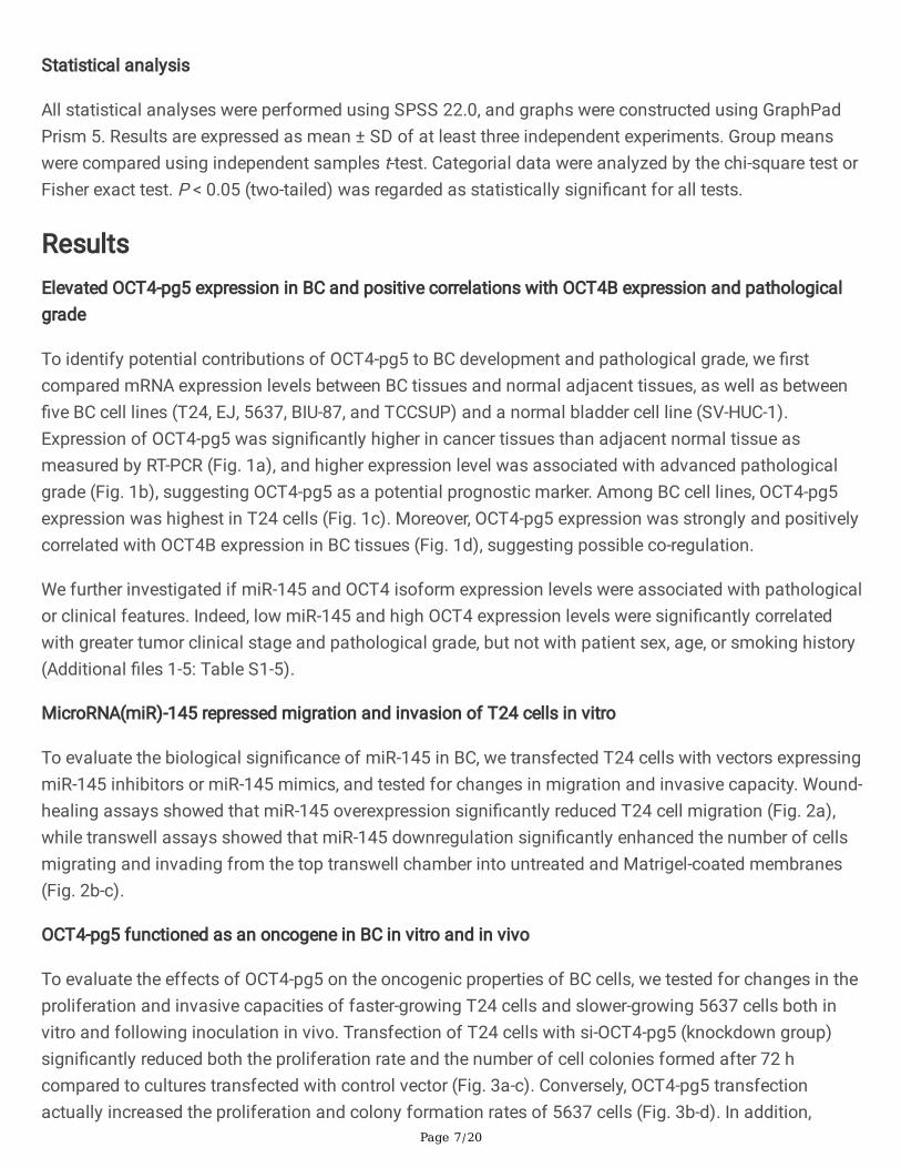
Statistical analysis
All statistical analyses were performed using SPSS 22.0, and graphs were constructed using GraphPadPrism 5. Results are expressed as mean ± SD of at least three independent experiments. Group meanswere compared using independent samples t-test. Categorial data were analyzed by the chi-square test orFisher exact test. P < 0.05 (two-tailed) was regarded as statistically signi�cant for all tests.
ResultsElevated OCT4-pg5 expression in BC and positive correlations with OCT4B expression and pathologicalgrade
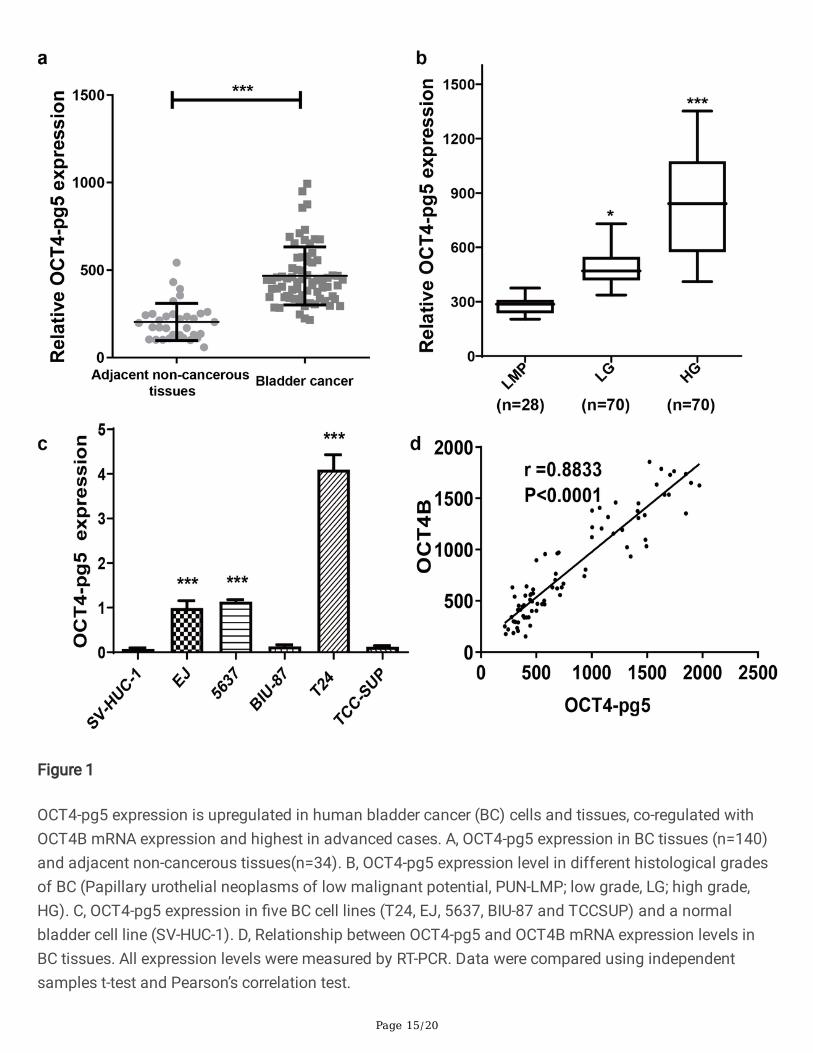
To identify potential contributions of OCT4-pg5 to BC development and pathological grade, we �rstcompared mRNA expression levels between BC tissues and normal adjacent tissues, as well as between�ve BC cell lines (T24, EJ, 5637, BIU-87, and TCCSUP) and a normal bladder cell line (SV-HUC-1).Expression of OCT4-pg5 was signi�cantly higher in cancer tissues than adjacent normal tissue asmeasured by RT-PCR (Fig. 1a), and higher expression level was associated with advanced pathologicalgrade (Fig. 1b), suggesting OCT4-pg5 as a potential prognostic marker. Among BC cell lines, OCT4-pg5expression was highest in T24 cells (Fig. 1c). Moreover, OCT4-pg5 expression was strongly and positivelycorrelated with OCT4B expression in BC tissues (Fig. 1d), suggesting possible co-regulation.
We further investigated if miR-145 and OCT4 isoform expression levels were associated with pathologicalor clinical features. Indeed, low miR-145 and high OCT4 expression levels were signi�cantly correlatedwith greater tumor clinical stage and pathological grade, but not with patient sex, age, or smoking history(Additional �les 1-5: Table S1-5).
MicroRNA(miR)-145 repressed migration and invasion of T24 cells in vitro
To evaluate the biological signi�cance of miR-145 in BC, we transfected T24 cells with vectors expressingmiR-145 inhibitors or miR-145 mimics, and tested for changes in migration and invasive capacity. Wound-healing assays showed that miR-145 overexpression signi�cantly reduced T24 cell migration (Fig. 2a),while transwell assays showed that miR-145 downregulation signi�cantly enhanced the number of cellsmigrating and invading from the top transwell chamber into untreated and Matrigel-coated membranes(Fig. 2b-c).
OCT4-pg5 functioned as an oncogene in BC in vitro and in vivo
To evaluate the effects of OCT4-pg5 on the oncogenic properties of BC cells, we tested for changes in theproliferation and invasive capacities of faster-growing T24 cells and slower-growing 5637 cells both invitro and following inoculation in vivo. Transfection of T24 cells with si-OCT4-pg5 (knockdown group)signi�cantly reduced both the proliferation rate and the number of cell colonies formed after 72 hcompared to cultures transfected with control vector (Fig. 3a-c). Conversely, OCT4-pg5 transfectionactually increased the proliferation and colony formation rates of 5637 cells (Fig. 3b-d). In addition,
Page 8/20
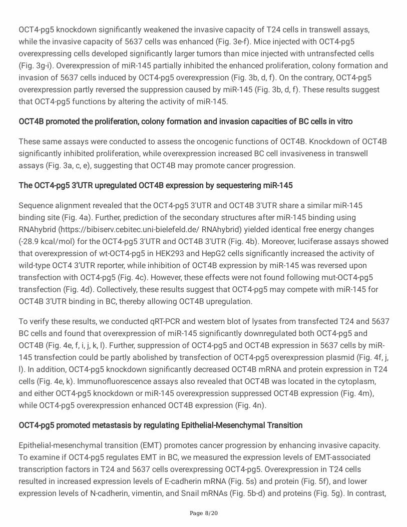
OCT4-pg5 knockdown signi�cantly weakened the invasive capacity of T24 cells in transwell assays,while the invasive capacity of 5637 cells was enhanced (Fig. 3e-f). Mice injected with OCT4-pg5overexpressing cells developed signi�cantly larger tumors than mice injected with untransfected cells(Fig. 3g-i). Overexpression of miR-145 partially inhibited the enhanced proliferation, colony formation andinvasion of 5637 cells induced by OCT4-pg5 overexpression (Fig. 3b, d, f). On the contrary, OCT4-pg5overexpression partly reversed the suppression caused by miR-145 (Fig. 3b, d, f). These results suggestthat OCT4-pg5 functions by altering the activity of miR-145.
OCT4B promoted the proliferation, colony formation and invasion capacities of BC cells in vitro
These same assays were conducted to assess the oncogenic functions of OCT4B. Knockdown of OCT4Bsigni�cantly inhibited proliferation, while overexpression increased BC cell invasiveness in transwellassays (Fig. 3a, c, e), suggesting that OCT4B may promote cancer progression.
The OCT4-pg5 3’UTR upregulated OCT4B expression by sequestering miR-145
Sequence alignment revealed that the OCT4-pg5 3'UTR and OCT4B 3'UTR share a similar miR-145binding site (Fig. 4a). Further, prediction of the secondary structures after miR-145 binding usingRNAhybrid (https://bibiserv.cebitec.uni-bielefeld.de/ RNAhybrid) yielded identical free energy changes(-28.9 kcal/mol) for the OCT4-pg5 3'UTR and OCT4B 3'UTR (Fig. 4b). Moreover, luciferase assays showedthat overexpression of wt-OCT4-pg5 in HEK293 and HepG2 cells signi�cantly increased the activity ofwild-type OCT4 3’UTR reporter, while inhibition of OCT4B expression by miR-145 was reversed upontransfection with OCT4-pg5 (Fig. 4c). However, these effects were not found following mut-OCT4-pg5transfection (Fig. 4d). Collectively, these results suggest that OCT4-pg5 may compete with miR-145 forOCT4B 3’UTR binding in BC, thereby allowing OCT4B upregulation.
To verify these results, we conducted qRT-PCR and western blot of lysates from transfected T24 and 5637BC cells and found that overexpression of miR-145 signi�cantly downregulated both OCT4-pg5 andOCT4B (Fig. 4e, f, i, j, k, l). Further, suppression of OCT4-pg5 and OCT4B expression in 5637 cells by miR-145 transfection could be partly abolished by transfection of OCT4-pg5 overexpression plasmid (Fig. 4f, j,l). In addition, OCT4-pg5 knockdown signi�cantly decreased OCT4B mRNA and protein expression in T24cells (Fig. 4e, k). Immuno�uorescence assays also revealed that OCT4B was located in the cytoplasm,and either OCT4-pg5 knockdown or miR-145 overexpression suppressed OCT4B expression (Fig. 4m),while OCT4-pg5 overexpression enhanced OCT4B expression (Fig. 4n).
OCT4-pg5 promoted metastasis by regulating Epithelial-Mesenchymal Transition
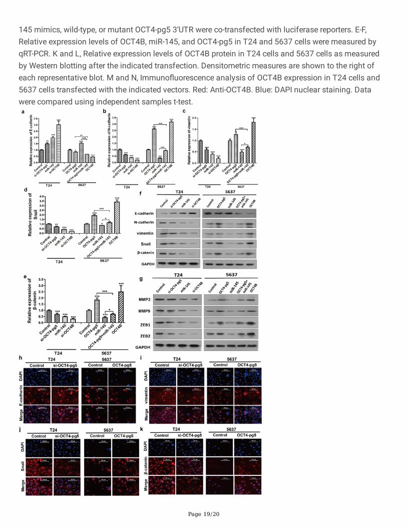
Epithelial-mesenchymal transition (EMT) promotes cancer progression by enhancing invasive capacity.To examine if OCT4-pg5 regulates EMT in BC, we measured the expression levels of EMT-associatedtranscription factors in T24 and 5637 cells overexpressing OCT4-pg5. Overexpression in T24 cellsresulted in increased expression levels of E-cadherin mRNA (Fig. 5s) and protein (Fig. 5f), and lowerexpression levels of N-cadherin, vimentin, and Snail mRNAs (Fig. 5b-d) and proteins (Fig. 5g). In contrast,
Page 9/20
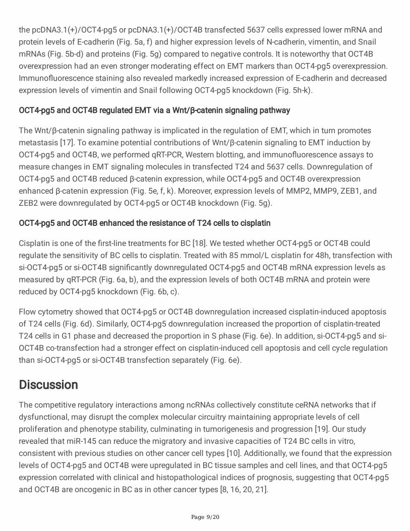
the pcDNA3.1(+)/OCT4-pg5 or pcDNA3.1(+)/OCT4B transfected 5637 cells expressed lower mRNA andprotein levels of E-cadherin (Fig. 5a, f) and higher expression levels of N-cadherin, vimentin, and SnailmRNAs (Fig. 5b-d) and proteins (Fig. 5g) compared to negative controls. It is noteworthy that OCT4Boverexpression had an even stronger moderating effect on EMT markers than OCT4-pg5 overexpression.Immuno�uorescence staining also revealed markedly increased expression of E-cadherin and decreasedexpression levels of vimentin and Snail following OCT4-pg5 knockdown (Fig. 5h-k).
OCT4-pg5 and OCT4B regulated EMT via a Wnt/β-catenin signaling pathway
The Wnt/β-catenin signaling pathway is implicated in the regulation of EMT, which in turn promotesmetastasis [17]. To examine potential contributions of Wnt/β-catenin signaling to EMT induction byOCT4-pg5 and OCT4B, we performed qRT-PCR, Western blotting, and immuno�uorescence assays tomeasure changes in EMT signaling molecules in transfected T24 and 5637 cells. Downregulation ofOCT4-pg5 and OCT4B reduced β-catenin expression, while OCT4-pg5 and OCT4B overexpressionenhanced β-catenin expression (Fig. 5e, f, k). Moreover, expression levels of MMP2, MMP9, ZEB1, andZEB2 were downregulated by OCT4-pg5 or OCT4B knockdown (Fig. 5g).
OCT4-pg5 and OCT4B enhanced the resistance of T24 cells to cisplatin
Cisplatin is one of the �rst-line treatments for BC [18]. We tested whether OCT4-pg5 or OCT4B couldregulate the sensitivity of BC cells to cisplatin. Treated with 85 mmol/L cisplatin for 48h, transfection withsi-OCT4-pg5 or si-OCT4B signi�cantly downregulated OCT4-pg5 and OCT4B mRNA expression levels asmeasured by qRT-PCR (Fig. 6a, b), and the expression levels of both OCT4B mRNA and protein werereduced by OCT4-pg5 knockdown (Fig. 6b, c).
Flow cytometry showed that OCT4-pg5 or OCT4B downregulation increased cisplatin-induced apoptosisof T24 cells (Fig. 6d). Similarly, OCT4-pg5 downregulation increased the proportion of cisplatin-treatedT24 cells in G1 phase and decreased the proportion in S phase (Fig. 6e). In addition, si-OCT4-pg5 and si-OCT4B co-transfection had a stronger effect on cisplatin-induced cell apoptosis and cell cycle regulationthan si-OCT4-pg5 or si-OCT4B transfection separately (Fig. 6e).
DiscussionThe competitive regulatory interactions among ncRNAs collectively constitute ceRNA networks that ifdysfunctional, may disrupt the complex molecular circuitry maintaining appropriate levels of cellproliferation and phenotype stability, culminating in tumorigenesis and progression [19]. Our studyrevealed that miR-145 can reduce the migratory and invasive capacities of T24 BC cells in vitro,consistent with previous studies on other cancer cell types [10]. Additionally, we found that the expressionlevels of OCT4-pg5 and OCT4B were upregulated in BC tissue samples and cell lines, and that OCT4-pg5expression correlated with clinical and histopathological indices of prognosis, suggesting that OCT4-pg5and OCT4B are oncogenic in BC as in other cancer types [8, 16, 20, 21].
Page 10/20
Pseudogenes are implicated in the initiation and progression of cancers, at least in part by acting asmicroRNA decoys that disrupt normal miRNA-mediated regulation of oncogene [22]. miR-145 can bind toOCT4 mRNA as well as to the OCT4 pseudogenes OCT4-pg1, 3, 4 and 5 [3], and recent reports havedemonstrated that OCT4-pg4 can regulate OCT4 expression and compete with miR-145 in hepatocellularcarcinoma [23]. Additionally, OCT4-pg5 acts as a miR-145 sponge in endometrial cancer, resulting inelevated OCT4 expression [16]. Similarly, PTENP1 has been shown to function as a ceRNA that competesfor miRNAs targeting the PTENP1 3′UTR [24], while miR-145 can regulate OCT4 expression by targetingthe OCT4 3′UTR in various cancer types [25, 26]. Given that the OCT4-pg5 3′UTR and OCT4B 3′UTR sharethe same miR-145 binding site and show the same free energy change upon binding, it appears thatOCT4-pg5 competes equally with miR-145 for the OCT4B 3’UTR but does not trigger mRNA degradation.Indeed, inhibition of OCT4B expression by miR-145 could be reversed by OCT4-pg5 overexpression.Collectively, these results strongly suggest that OCT4-pg5 acts as an oncogene by functioning as aceRNA, thereby elevating parental OCT4B expression.
In several cancer types, overexpression of OCT4B was reported to prevent apoptosis [7] and increasemigration, invasion, and extracellular matrix degradation capacities by inducing various EMT-relatedgenes [27]. Further, several studies found that OCT4 upregulated the transcription factors ZEB1, ZEB2,and Snail by activating β-catenin, while miR-145 inhibited EMT by blocking the expression of OCT4,thereby downregulating the expression of Snail, ZEB1, and ZEB2 [28–30]. We also found that expressionsof ZEB1, ZEB2, Snail, and two additional metastasis-related genes, MMP2 and MMP9, were upregulatedin BC cells by OCT4B overexpression as well as by OCT4-pg5 overexpression. However, OCT4Bdemonstrated stronger EMT induction potential than OCT4-pg5, suggesting that OCT4-pg5 may induceEMT indirectly by regulating OCT4B expression. The Wnt/β-catenin signaling pathway is known to pro-mote metastasis by inducing EMT [17], and the LEF1/β-catenin-dependent WNT signaling pathway canbe activated by OCT4 [31]. Indeed, silencing β-catenin blocked Oct4/Nanog-mediated EMT [32]. In thecurrent study, β-catenin was activated by OCT4-pg5 and OCT4B, suggesting that OCT4-pg5 and OCT4Bmay induce EMT via Wnt/β-catenin signaling.
It was reported that OCT4 knockdown protected NSCLC cells from apoptosis and enhanced sensitivity tocisplatin treatment [33], while overexpression of OCT4 pro-moted the differentiation of lung cancer cellsinto SLCCs and increased cisplatin resistance [34]. Similarly in the current study, OCT4-pg5 and OCT4Bexpression protected T24 BC cells from cisplatin damage, while OCT4-pg5 knockdown increasedcisplatin-induced apoptosis and reduced the proportion of G1 cells, possibly by disinhibiting OCT4expression. Further studies are needed to identify the biological mechanism linking MMP2 and MMP9upregulation to OCT4-pg5/miR-145/OCT4B axis activity and to con�rm that this axis can regulatecisplatin sensitivity in BC cases, thereby altering disease progression and clinical outcome.
ConclusionsOur study demonstrates that OCT4-pg5 can promote oncogenesis by competing with miR-145 for bindingto OCT4B mRNA, thereby disrupting miR-145-mediated OCT4B downregulation and leading to OCT4B
Page 11/20
overexpression, and ultimately greater BC cell proliferative and invasive capacity. These �ndings identifynew potential therapeutic targets for BC.
AbbreviationsOCT4, Octamer-binding transcription factor 4;
OCT4-pg5, Octamer-binding transcription factor 4 pseudogene 5;
BC, bladder cancer;
OCT4B, Octamer-binding transcription factor 4B;
MiR-145, MiRNA-145;
EMT, Epithelial-to-mesenchymal transition;
3’UTR, 3’ untranslated regions;
CeRNAs, Competing endogenous RNAs;
LncRNAs, Long non-coding RNAs;
ATCC, American Type Culture Collection;
NMIBC, Non-muscle-invasive bladder tumors;
MIBC, Muscle-invasive bladder tumors;
PBS, Phosphate‐buffered saline;
SDS-PAGE, Sodium dodecyl sulfate-polyacrylamide gel;
FCM, Flow cytometry.
DeclarationsAcknowledgements
Not applicable.
Authors’ contributions
WZE, YY and CLY prepared and wrote the original draft. WZE conducted the experiments and analyzeddata. WW conceived the study. ZC, XYL, BQW, YSX, and XMZ validated the paper. WZE, YY and CLY
Page 12/20
contributed equally to this paper and should be considered as co-�rst author. All the authors have readand agreed to the �nal version of the manuscript.
Funding
This study was supported by the Guangzhou Science and Technology Plan Project Basic and AppliedBasic Research Project (202002030030), the Guangdong Province Basic and Applied Basic ResearchFund Project (2020A1515010044), and the National Science Fund subsidized project 81372744 .
Availability of data and materials
The data presented in this study are available on request from the corresponding author.
Ethics approval and consent to participate
All the patients were informed of consent forms, and the study was approved by the Institute ResearchEthics Committee, General Hospital of Southern Theater Command, China. All animal procedures wereperformed according to the protocols approved by the Institutional Animal Care and Use Committee atGeneral Hospital of Southern Theater Command.
Consent for publication
All human tissue samples were obtained with informed consent from all subjects.
Competing interests
The authors declare no con�ict of interest.
References1. Sung H, Ferlay J, Siegel RL, Laversanne M, et al. Global Cancer Statistics 2020: GLOBOCAN
Estimates of Incidence and Mortality Worldwide for 36 Cancers In 185 Countries. CA Cancer J Clin.2021;0:1–41.
2. Teoh JY, Huang J, Ko WY, et al. Global trends of bladder cancer incidence and mortality, and theirassociations with tobacco use and gross domestic product per capita. Eur Urol. 2020;78:893–906.
3. Patra SK. Roles of OCT4 in pathways of embryonic development and cancer progression.Mechanisms of Ageing Development. 2020;189:111286.
4. Wang X, Zhao Y, Xiao Z, et al. Alternative translation of OCT4 by an internal ribosome entry site andits novel function in stress response. Stem Cells. 2009;27:1265–75.
5. Poursani EM, Mehravar M, Shahryari A, Mowla SJ, Mohammad Soltani B. Alternative splicinggenerates different 5' UTRs in OCT4B variants. Avicenna J Med Biotechnol. 2017;9:201–4.
�. Lin SC, Chung CH, Chung CH, et al. OCT4B mediates hypoxia-induced cancer dissemination.Oncogene. 2019;38:1093–105.
Page 13/20
7. Meng L, Hu H, Zhi H, et al. OCT4B regulates p53 and p16 pathway genes to prevent apoptosis ofbreast cancer cells. Oncol Lett. 2018;16:522–8.
�. Poursani EM, Mehravar M, Mohammad Soltani B, Mowla SJ, Trosko JE. A Novel variant of OCT4entitled OCT4B3 is expressed in human bladder cancer and astrocytoma cell lines. Avicenna J MedBiotechnol. 2017;9:142–5.
9. Choi SH, Kim JK, Jeon HY, Eun K, Kim H. OCT4B Isoform promotes anchorage-independent growth ofglioblastoma cells. Mol Cells. 2019;42:135–42.
10. Xu WX, Liu Z, Deng F, et al. MiR-145: a potential biomarker of cancer migration and invasion. Am JTransl Res. 2019;11:6739–53.
11. Chi Y, Wang D, Wang J, Yu W, Yang J. Long non-coding RNA in the pathogenesis of cancers. Cells.2019;8:1015.
12. Taheri M, Omrani MD, Ghafouri-Fardsun S. Long non-coding RNA expression in bladder cancer.Biophys Rev. 2018;10:1205–13.
13. Hirotsune S, Yoshida N, Chen A, et al. An expressed pseudogene regulates the messenger-RNAstability of its homologous coding gene. Nature. 2003;423:91–6.
14. Hu X, Yang L, Mo Y. Role of pseudogenes in tumorigenesis. Cancers (Basel). 2018;10:256.
15. Poursani EM, Mohammad SB, Mowla SJ. Differential expression of OCT4 pseudogenes inpluripotent and tumor cell lines. Cell J. 2016;18:28–36.
1�. Bai M, Yuan M, Liao H, et al. OCT4 pseudogene 5 upregulates OCT4 expression to promoteproliferation by competing with miR-145 in endometrial carcinoma. Oncol Rep. 2015;33:1745–52.
17. Khan AQ, Ahmed EI, Elareer NR, Junejo K, Steinhoff M, Uddin S. Role of miRNA-regulated cancer stemcells in the pathogenesis of human malignancies. Cells. 2019;8:840.
1�. Yoshida T, Kates M, Fujita K, Bivalacqua TJ, McConkey DJ. Predictive biomarkers for drug responsein bladder cancer. Int J Urol. 2019;26:1044–53.
19. Chan JJ, Tay Y. Noncoding RNA. RNA Regulatory networks in cancer. Int J Mol Sci. 2018;19:1310.
20. Choi SH, Kim JK, Jeon HY, Eun K, Kim H. OCT4B Isoform promotes anchorage-independent growth ofglioblastoma cells. Mol Cells. 2019;42:135–42.
21. Soheili S, Asadi MH, Farsinejad A. Distinctive expression pattern of OCT4 variants in different typesof breast cancer. Cancer Biomark. 2017;18:69–76.
22. Lou W, Ding B, Fu P. Pseudogene-derived lncRNAs and their miRNA sponging mechanism in humancancer. Front Cell Dev Biol. 2020;8:85.
23. Wang L, Guo ZY, Zhang R, et al. Pseudogene OCT4-pg4 functions as a natural microRNA sponge toregulate OCT4 expression by competing for miR-145 in hepatocellular carcinoma. Carcinogenesis.2013;34:1773–81.
24. Poliseno L, Salmena L, Zhang J, Carver B, Haveman WJ, Pandol� PP. A coding-independent functionof gene and pseudogene mRNAs regulates tumour biology. Nature. 2010;465:1033–8.

Page 14/20
25. Matsushita R, Yoshino H, Enokida H, Goto Y, Miyamoto K, Yonemori M, et al. Regulation of UHRF1 bydual-strand tumor-suppressor microRNA-145 (miR-145-5p and miR-145-3p): inhibition of bladdercancer cell aggressiveness. Oncotarget. 2016;7:28460–87.
2�. Wu Y, Liu S, Xin H, et al. Up-regulation of microRNA-145 promotes differentiation by repressing OCT4in human endometrial adenocarcinoma cells. Cancer. 2011;117:3989–98.
27. Zhou JM, Hu SQ, Jiang H, et al. OCT4B1 promoted EMT and regulated the self-renewal of CSCs inCRC: effects associated with the balance of miR-8064/PLK1. Mol Ther Oncolytics. 2019;15:7–20.
2�. Zhao H, Kang X, Xia X, et al. MiR-145 suppresses breast cancer cell migration by targeting FSCN-1and inhibiting epithelial-mesenchymal transition. Am J Transl Res. 2016;8:106–3114.
29. Gao Y, Zhang Z, Li K, et al. Linc-DYNC2H1-4 promotes EMT and CSC phenotypes by acting as asponge of miR-145 in pancreatic cancer cells. Cell Death Dis. 2017;8:e2924.
30. Li C, Lu L, Feng B, et al. The lincRNA-ROR/miR-145 axis promotes invasion and metastasis inhepatocellular carcinoma via induction of epithelial-mesenchymal transition by targeting ZEB2. SciRep. 2017;7:4637.
31. Sun L, Liu T, Zhang S, Guo K, Liu Y. Oct4 induces EMT through LEF1/β-catenin dependent WNTsignaling pathway in hepatocellular carcinoma. Oncol Lett. 2017;13:2599–606.
32. Liu L, Zhu H, Liao Y, et al. Inhibition of Wnt/β-catenin pathway reverses multi-drug resistance andEMT in Oct4+/Nanog + NSCLC cells. Biomed Pharmacother. 2020;127:110225.
33. Liu X, Ma M, Duan X, Zhang H, Yang M. Knockdown of OCT4 may sensitize NSCLC cells to cisplatin.Clin Transl Oncol. 2017;19:587–92.
34. Chiou SH, Wang ML, Chou YT, et al. Co-expression of Oct4 and Nanog enhances malignancy in lungadenocarcinoma by inducing cancer stem cell-like properties and epithelial-mesenchymal trans-differentiation. Cancer Res. 2010;70:10433–44.
Figures
Page 15/20
Figure 1
OCT4-pg5 expression is upregulated in human bladder cancer (BC) cells and tissues, co-regulated withOCT4B mRNA expression and highest in advanced cases. A, OCT4-pg5 expression in BC tissues (n=140)and adjacent non-cancerous tissues(n=34). B, OCT4-pg5 expression level in different histological gradesof BC (Papillary urothelial neoplasms of low malignant potential, PUN-LMP; low grade, LG; high grade,HG). C, OCT4-pg5 expression in �ve BC cell lines (T24, EJ, 5637, BIU-87 and TCCSUP) and a normalbladder cell line (SV-HUC-1). D, Relationship between OCT4-pg5 and OCT4B mRNA expression levels inBC tissues. All expression levels were measured by RT-PCR. Data were compared using independentsamples t-test and Pearson’s correlation test.

Page 16/20
Figure 2
Overexpression of miR-145 reduces the migration and invasion potential of T24 BC cells in vitro. A,Effects of miR-145 on T24 cell migration as examined by wound healing assay (magni�cation, ×50). Band C, transwell assays used to evaluate the effects of miR-145 on T24 cell invasion B and migration C.Photographs of cell invasion and migration were acquired from polycarbonate membranes stained withcrystal violet (magni�cation, ×200). Data were compared using independent samples t-test.

Page 17/20
Figure 3
OCT4-pg5 and OCT4B function as oncogenes in BC cells in vitro and in vivo. A and B, CCK-8 assaysshowing viable T24 and 5637 cell numbers at different times post-transfection with the indicatedtranscripts. C and D, Colony formation assays to estimate the proliferation rates of T24 and 5637 BCcells. E and F, Transwell assays to determine the invasion capacities of T24 and 5637 cells. G, Effect ofOCT4-pg5 overexpression on tumor growth in vivo. H, Tumor growth curves measured every three days

Page 18/20
after inoculation of T24 or 5637 cells. I, Tumor weight was measured on the 27th day post-inoculation.Data were compared using independent samples t-test.
Figure 4
OCT4-pg5 3’UTR regulates the expression of OCT4B by sequestering miR-145. A, The sequence alignmentof miR-145 with OCT4B and OCT4-pg5 3'UTRs. B, The binding force and possible secondary structureanalyzed by RNAhybrid. C and D, Luciferase activity of OCT4B reporters in T24 and 5637 BC cells. MiR-
Page 19/20
145 mimics, wild-type, or mutant OCT4-pg5 3’UTR were co-transfected with luciferase reporters. E-F,Relative expression levels of OCT4B, miR-145, and OCT4-pg5 in T24 and 5637 cells were measured byqRT-PCR. K and L, Relative expression levels of OCT4B protein in T24 cells and 5637 cells as measuredby Western blotting after the indicated transfection. Densitometric measures are shown to the right ofeach representative blot. M and N, Immuno�uorescence analysis of OCT4B expression in T24 cells and5637 cells transfected with the indicated vectors. Red: Anti-OCT4B. Blue: DAPI nuclear staining. Datawere compared using independent samples t-test.
Page 20/20
Figure 5
OCT4-pg5 promotes metastasis by inducing EMT through the Wnt/β-catenin pathway. A-E, RelativemRNA expression levels of E-cadherin, vimentin, Snail and β-catenin in T24 cells and 5637 cells aftertransfection as measured by RT-PCR (normalized to β-actin). F and G, Relative protein expression levels ofE-cadherin, vimentin, Snail, β-catenin, MMP2, MMP9, ZEB1 and ZEB2 in T24 cells and 5637 cells aftertransfection as measured by Western blot (normalized to GAPDH). H-K, Immuno�uorescence staining forE-cadherin, vimentin, Snail and β-catenin in T24 cells and 5637 cells. Red: anti-E-cadherin, anti-vimentin,anti-Snail, and anti-β-catenin. Blue: DAPI nuclear staining. Data were compared using independentsamples t-test.
Figure 6
Crosstalk between OCT4-pg5 and OCT4B enhances the resistance of T24 cells to cisplatin. A and B,OCT4-pg5 and OCT4B expression levels in T24 cells as measured by qRT-PCR, C, Western blot analysis ofOCT4B protein level in T24 cells. GAPDH was used as the control. D, Effects of OCT4-pg5 3’UTR andOCT4B on the apoptotic rate of T24 cells as measured by �ow cytometry. UL: necrotic cells, UR: terminalapoptotic cells, LR: early apoptotic cells, LL: normal cells. Cells in UR and LR were counted and analyzed.E, Effects of OCT4-pg5 and OCT4B expression on the cell cycle stage distribution of T24 cells asmeasured by �ow cytometry. Data were compared using independent samples t-test.
Supplementary Files
This is a list of supplementary �les associated with this preprint. Click to download.
SupplementalTables.xls