by j. c. newhook and d. a. titchen* from the department of
TRANSCRIPT
J. Physiol. (1974), 237, pp. 415-430 415With 3 plateau and 1 text-figurePrinted in Great Britain
EFFECTS OF VAGOTOMY,ATROPINE, HEXAMETHONIUM AND ADRENALINE ON THEDESTINATION IN THE STOMACH OF LIQUIDS SUCKED BY
MILK-FED LAMBS AND CALVES
BY J. C. NEWHOOK AND D. A. TITCHEN*From the Department of Physiology and Anatomy, Massey University,Palmerston North, New Zealand and Department of Veterinary
Preclinical Sciences, University of Melbourne, Parkville,Victoria 3052, Australia
(Received 13 August 1973)
SUMMARY
1. The normal passage to the abomasum of liquid sucked by lambsand calves was confirmed in radiographic studies to be changed to passageof liquid to the reticulum and rumen after cervical or abdominal vagotomy.
2. The effects of hexamethonium (8-10 mg kg-1 i.v.) were similar tothose of vagotomy.
3. Atropine (200-800,g kg-' i.v.) had either no detectable effect onthe destination in the stomach of liquid sucked by lambs or caused apartial failure of the reticular groove mechanism.
4. Fluid sucked by lambs passed wholly to the abomasum afteradrenaline (5-40 #ug kg-' i.v.).
5. Atropine and adrenaline caused a greater dysfunction of the reticulargroove mechanisms in calves than in lambs.
6. Contraction of the reticular groove was observed via rumen fistulaein three lambs. Eversion of the caudal oesophagus into the reticulumwhich occurred when saliva was swallowed into the reticulum and rumenwas not observed when liquids were sucked from a bottle.
7. The continued passage to the abomasum of sucked liquid inatropinized lambs has been taken as an indication of the importance ofcaudal oesophageal reactions, tonic activity of the reticular groove andthe atropine-resistant vagally induced opening of the reticulo-omasalorifice in the reticular groove mechanism.
* Address: Department of Veterinary Preclinical Sciences, University ofMelbourne, Parkville, Victoria 3052, Australia.

J. C. NEWHOOK AND D. A. TITCHEN
INTRODUCTION
Liquids sucked by young ruminants pass to the most caudal part oftheir stomach, the abomasum, without entering the reticulum and rumen.This is attributed to the reticular groove which forms a conduit fromthe oesophagus through the reticulum to the reticulo-omasal orifice.Contraction of the groove has been palpated when calves with rumenfistulae suck (Wester, 1926; Schalk & Amadon, 1928; XWise, 1939). Thegroove contracted in the foetal lamb (Duncan & Phillipson, 1951) asit did in decerebrate calves and lambs in response to stimulation of thevagus nerves (Comline & Titchen, 1951).The effectiveness of contraction of the reticular groove in directing
sucked liquid to the abomasum depends on the reticulo-omasal orificebeing open so that liquid passing from the oesophagus into the reticulargroove may enter the omasum and thence the abomasum. Whereascontraction of the reticular groove is blocked by atropine (Wester,1926; Comline & Titchen, 1951) opening of the reticulo-omasal orificein the sheep evoked by efferent vagus nerve stimulation is resistant toatropine (Newhook & Titchen, 1972). In the present work the effect ofatropine on the destination of liquid sucked by lambs and calves wasstudied and a comparison made of its effects and those of cervical andabdominal vagotomy and hexamethonium. Brief preliminary reports ofsome of the observations have been presented (Newhook, 1970; Newhook& Titchen, 1969).
METHODS
Animals and their management. These were lambs and calves of both sexes. InNew Zealand the lambs studied were of the Romney or Cheviot breeds or theircrosses, and in Australia Romney or Leicester crosses, obtained within 72 hr ofbirth and after they had received colostrum from their dams. In the laboratorythey were kept in individual cages on wire floors without bedding. They were fedfour times each day with a proprietary lamb rearing mixture ('Fostermilk', Glaxo,New Zealand) derived from dried cow's milk, or with reconstituted evaporatedwhole milk. The lambs sucked these from a bottle and teat. They were allowed asmuch as they would drink at each meal. From 3 weeks of age dried buttermilkpowder was added to the lamb rearing mixture in increasing quantities until itformed three quarters of the ration. The lambs and calves always had access towater but never to solid food. The lambs were kept closely clipped about theirforelegs and hindquarters to reduce wool eating. All were allowed each day toroam for a period in a pen on a concrete floor. The animals were accustomed tobeing fed from a bottle when restrained in headstocks.
Radiographic apparatus and procedures. The X-ray generator used was either oftwo mobile machines with stationary anode tubes (a 'Watvic D3', Watson Victor,Australia, with a mechanical timer and a 'Konrad 60', Watson Victor, Australia,with an electronic timer), or a G.E. (U.S.A.) 'Patrician' apparatus with rotating
416

ROUTE OF LIQUID SUCKED BY LAMBS AND CALVES 417
anode tube. The two mobile machines were used without the aid of grids, butsometimes a 6: 1 Liebel Flarsheim grid was used with the larger apparatus.The X-ray films (Kodak 'Blue Brand', 3M type R, Ilford 'Red Seal') were used
in cassettes fitted with intensifying screens (Du Pont 'High Speed' and 'Par Speed').Development was with 'Phenisol' (Ilford) and fixation with 'Hypam' (Ilford).The contrast media were an oesophageal paste ('Microtrast', Damancy) or
powders ('Raybar', Bell Craig: 'Micropaque', Damancy). The paste (70% w/wbarium sulphate in an aqueous vehicle) was added to milk up to 30% v/v. Bothpowders are readily suspended in liquid and were mixed with milk to make up to30% v/v of the liquid. All the mixtures used flowed readily through the teats andwere sucked avidly by lambs and calves.
Observations on the course of swallowed liquid were made with all three machineswhilst liquid was being sucked. Each mobile apparatus was used in the animals'usual quarters. The larger G.E. machine is a fixed installation to which animalshad to be taken. In an effort to accustom them to this procedure the lambs weregiven at least one feed daily for 3 or 4 days of each week in the X-ray room. Oneanimal consistently refused to suck in this room. It was excluded from the experi-mental observations.
Radiographic observations were made before, during and after sucking on atleast one occasion before any experimental procedures or administration of drugs.The animals were fed from the same bottle and teat during normal feeding andexperimental sessions. The rate at which they sucked was 2-12 ml. sec-L. Theamounts of contrast material received were usually limited by giving a maximumof 120 ml. milk contrast medium mixtures at each radiographic session except aftervagotomy and during observations per fistulam when over 200 ml. have been given.The standard procedure adopted was to take a preliminary radiograph of eachanimal whilst standing and held by an acrylic sheet (Perspex, I.C.I.) against thefilm cassette. Three or more radiographs were taken, at the start of, during, andafter sucking 100-120 ml. milk-contrast media mixtures. Some radiographs werealso taken in a dorsoventral plane to confirm the distribution of contrast mediumin the stomach.The drugs used were all injected i.v. into a jugular vein. Atropine sulphate
(B.D.H.) was given in doses of the base of 200-800 ,ug kg-', hexamethonium (KochLight) 8-10 mg kg-', adrenaline hydrochloride 5-40 /ug kg-'.
Surgical procedures were undertaken with precautions to maintain asepsis andunder anaesthesia induced and maintained with halothane (B.P.). Induction wasundertaken with the aid of a Hall's dog mask (B.O.C.) in conjunction with a Fluotec(B.O.C.) vaporizer in a closed circuit anaesthetic apparatus. Maintenance was viaa cuffed endotracheal tube.
Cervical or abdominal vagotomy was through mid-line cervical or epigastricincisions. Rumen fistulae used for direct observation of the reticular groove wereprepared in the left side of the animal in the mid-dorsal sac with the two stagetechnique described by Jarrett (1948).
Markers placed on the lips of the reticular groove were Weck 'hemoclips'(Edward Weck & Co. Long Island City, N.Y.) the use of which with special forcepshas been described by Samuels, Roedling, Katz & Cincotti (1966). In the presentwork medium size clips were used after being re-shaped so that when closed theyhad a quadrilateral outline, with a circumference of about 11 mm.A speculum with its own illumination was used for viewing structures in the
stomach. It had an external diameter of 2-5 cm and an internal diameter of 2 cm.The rumen and reticulum were emptied of fluid by suction and the intermittentlydeposited saliva was also removed by suction. The end of the suction tube wascovered with gauze to prevent it holding to the mucosa.
J. C. NEWHOOK AND D. A. TITCHENAcute experiments. The completeness of vagus nerve section was confirmed at
acute experiments under chloralose anaesthesia or after decerebration underhalothane anaesthesia, when the effects were examined of electrical stimulation ofthe vagus nerve trunks cranial and caudal to the point of transaction. The procedureswere as described by Newhook & Titchen (1972). The observations on the effectsof vagotomy reported all refer to animals in which stimulation of the cervicalvagus nerve trunks failed to elicit movements of the stomach.
Studies in calves. Six Jersey or Jersey-cross calves (four males, two females)were used in the studies in New Zealand. Observations were made on calves3-22 days of age. They were maintained in the same manner as lambs and subjectedto similar experimental procedures. All radiography was undertaken on them inthe room in which they were normally housed, using one or other of the mobilemachines.
RESULTS
The appearance of contrast material in the oesophagus and the stomachwas followed radiographically in 47 milk-fed lambs 2-140 days old,during and after sucking.The abomasal destination of sucked liquid was the invariable normal
result. This was indicated by the accumulation of contrast material ina ventral position in association with outlines of the abomasal folds, theabomasal gas cap in the mid-epigastric region (Figs. 1 and 2, P1. 1) andsometimes clear impressions of the pyloric antrum of the abomasum.Movement of contrast material from the abomasum into the duodenumhas been detected within 200 see of the commencement of its ingestion.Benzie & Phillipson (1957) and Kay, Orskov & Wenham (1972) reportedrapid movement of material caudally from the abomasum.Two liquid-gas interfaces were present in the stomach. Liquid in the
abomasum extended ventrally towards the floor of the abdomen andhad overlying it the mid-epigastric abomasal gas cap which increasedduring sucking. The other liquid-gas interface was more dorsal and theliquid did not extend as far ventrally towards the floor of the abdomen;dorsal to it was a gas cap extending to the level of the vertebral column.This liquid and gas was in the rumen. Direct observation has shown thatsaliva swallowed at times when lambs are not sucking enters the reticulumand rumen. These different fluid levels were detected as early as ina lamb 4 days old.
Radiographic observations of the oesophagus. The usual appearance ofsucked material in the oesophagus was of distinct boluses with broadcaudal faces and tapering cranially. 116 boluses measured in twenty-fourlambs 3-140 days old were 25-100 mm long, another twenty-five were110-180 mm long. The total length of the oesophagus was 240-370 mm.The entire oesophagus contained contrast material (Fig. 3, P1. 2) inseven radiographs (six animals) of a total of forty-three in which all of
418
ROUTE OF LIQUID SUCKED BY LAMBS AND CALVES 419
the oesophagus was included in the field. Discrete boluses occupying lessthan half the length of the oesophagus were also detected in one ormore radiographs taken at the same session as that in which all of theoesophagus contained contrast material.A dilatation of the caudal thoracic oesophagus has been detected
(Figs. 1 and 2, P1. 1). This corresponds to the description of the phrenicampulla provided by Ruckebusch & Bost (1962). It was not invariablypresent (Fig. 3, P1. 2).The passage into the reticular groove of sucked material was marked by its
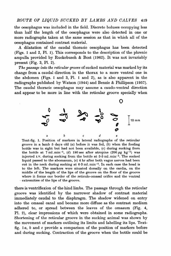
change from a caudal direction in the thorax to a more ventral one inthe abdomen (Figs. 1 and 3, P1. 1 and 2), as is also apparent in theradiographs published by Watson (1944) and Benzie & Phillipson (1957).The caudal thoracic oesophagus may assume a caudo-ventral directionand appear to be more in line with the reticular groove specially when
0
0ty 15 '4 t>° IiOi10mm
a b c d eText-fig. 1. Position of markers in lateral radiographs of the reticulargroove in a lamb 5 days old (a) before it was fed, (b) when the feedingbottle was in sight but had not been available, (c) during sucking fromthe bottle at 7 ml. min-', (d) 180 sec after atropine (200 jug kg-') wasinjected i.v. during sucking from the bottle at 5*5 ml.min-'. The suckedliquid passed to the abomasum, (e) 4 hr after both vagus nerves had beencut in the neck during sucking at 6-3 ml. min-'. In each case the head isto the left. The markers were situated dorsally on the cardia, on themiddle of the length of the lips of the groove on the floor of the groovewhere it forms one border of the reticulo-omasal orifice and the ventral
- extremities of the lips of the groove.
there is ventriflexion of the hind limbs. The passage through the reticulargroove was identified by the narrower shadow of contrast materialimmediately caudal to the diaphragm. The shadow widened on entryinto the omasal canal and became more diffuse as the contrast mediumadhered to, or spread between the leaves of the omasum (Fig. 4,P1. 2), clear impressions of which were obtained in some radiographs.Shortening of the reticular groove in the sucking animal was shown bythe movement of markers outlining its limits and labelling its lips. Text-fig. la, b and c provide a comparison of the position of markers beforeand during sucking. Contraction of the groove when the bottle could be

J. C. NEWHOOK AND D. A. TITCHEN
seen but before sucking started was detected with these markers (Text-fig. lb). Wise (1939) and Orskov, Benzie & Kay (1970) reported theactivation of the reticular groove mechanism as a conditioned responsein the calf and lambs in anticipation of being allowed to suck.
Effects of cervical and abdominal vagotomyAfter both vagus nerves were cut in the neck or in the abdomen
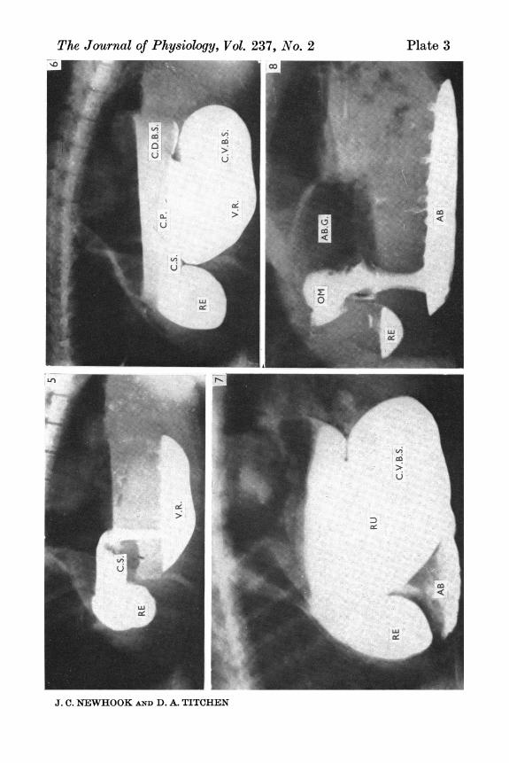
immediately caudal to the diaphragm, sucked liquid passed to thereticulum and cranial sac of the rumen whence it flowed over the cranialpillar into the ventral rumen (Fig. 5, P1. 3). Nine of the eleven animalssubjected to complete vagotomy continued to suck avidly. They displayedthe usual excitement and interest in anticipation of receiving the teatand drank vigorously taking all of the liquid made available. One lambwhich had bilateral cervical vagotomy was reluctant to suck and whenit did made only a few sucking movements at a time. It had a markedruminal tympanities and sucked only 15 ml. during the radiographicobservations. Another animal which had sucked to satiation 1 hr beforecervical vagotomy did not suck at any time in the next 24 hr. The passageto the reticulum and rumen of the 120 ml. or more of liquid sucked ledto clear outlines being obtained of the reticulum, cranial sac of the rumenbetween the rumino-reticular fold and cranial pillar, the ventral sac ofthe rumen and, as in Fig. 6 of P1. 3, of the ventral margins of the caudaldorsal blind sac of the rumen. The ruminal gas cap which increased onsucking after vagotomy provided a less clear demarcation of the dorsallimits of the rumen than did the contrast medium ventrally - part of thiswas due to overlying intestinal gas shadows. These features providedclear evidence of the reticular and ruminal destination of liquid suckedafter vagotomy. The passage of traces of sucked material into theabomasum was detected in seven of the lambs with vagotomy after theysucked; some of the liquid sucked was seen to have passed into theabomasum over the course of up to 6 hr after sucking (Fig. 7, P1. 3).
Cervical vagotomy was followed by a failure of discrete bolus formationin the oesophagus caudal to the level of transaction. The caudal cervicaland thoracic regions of the oesophagus were distended throughout theirlength after cervical vagotomy, with narrowing at the level of the firstrib and between this site and the heart. The caudal thoracic oesophagealdilatation identified as the phrenic ampulla was never detected afterbilateral cervical vagotomy.
After the abdominal vagus nerve trunks were cut there was a tendencyfor greater lengths of the oesophagus to be filled with sucked liquid.Of forty-one radiographs made in lambs after abdominal vagus nervesection contrast medium was present in the whole length of the oesophagus
420
ROUTE OF LIQUID SUCKED BY LAMBS AND CALVES 421
in twelve and in only four radiographs were there boluses of less thanhalf the length of the oesophagus. This contrasts with the situationdescribed in the normal lambs (p. 418-419).
Contrast medium was detected in the larynx and trachea of threeanimals with both abdominal vagus nerve trunks cut. None of theseanimals had any obvious difficulty or distress on sucking. Contrastmaterial was detected in the trachea in one lamb in which both vagusnerves had been cut in the neck.
Effect of atropine on the course of sucked liquidLambs continued to suck avidly after atropine was injected into a
jugular vein in doses of 200-800,ug kg-'. The greatest effect producedwas a failure of some swallowed liquid to pass to the abomasum, whichoccurred on twenty occasions in eighteen lambs 7-48 days old. All suckedliquid passed to the abomasum on eighteen occasions in twelve lambs(7-29 days old) after atropine. The radiographic appearance when theadministration of atropine i.v. was followed by some material enteringthe reticulum is presented in Fig. 8 of P1. 3. Contrast medium was notedin the trachea in five animals after they had received atropine (500/tg kg-').None had displayed reluctance, difficulty or clumsiness on sucking afteratropine was given.The omasum appeared dilated after the i.v. injection of atropine. In
the absence of quantitative measures this must be clearly identified asan impression. The passage caudally from the abomasum into theduodenum of contrast medium was detected within 10 min of the startof sucking in one of these animals after atropine (500 /ug kg-') was given.A comparison of the position of markers on the reticular groove underresting conditions, during sucking in the untreated lamb, after it receivedatropine and, on another occasion, after the vagus nerves were cut isprovided in Text-fig. 1 made by tracing the outlines of markers fromradiographs.
Effect of hexamethoniumIn contrast to the range of responses after atropine the administration
of hexamethonium (8-10 jug kg-' i.V.) was followed by the entry of liquidswallowed during sucking, into the reticulum and rumen. Observationswere made in five lambs on a total of six occasions, on each of whichthe 100-120 ml. they were allowed to suck was taken avidly and withoutdifficulty.
Effect of adrenalineThe i.V. injection of adrenaline in doses of 5-40 ,ug kg-' to three lambs
on a total of eight occasions was without effect on the eagerness with

J. C. NEWHOOK AND D. A. TITCHENwhich the animals sucked. They did so eagerly and apparently effectivelysince all the contrast medium sucked passed to the abomasum withoutevidence of entering the reticulum or rumen.
Direct observations of the reticular grooveThese were made in three lambs 44-93 days old fed only by sucking
from a bottle, and which had fistulae in the mid-dorsal rumen. Whenthe animals were not sucking the lips of the reticular groove were incontact with each other along their whole length and showed localizedcontractions of the length of the lips and movements in which the rightlip moved to cover the left. When saliva was swallowed the lips of thegroove were separated in their dorsal parts by the oesophagus protrudingbetween them and delivering saliva into the reticulum and cranial sacof the rumen.
During sucking the reticular groove shortened and rotated along itslength. The reticulum adjacent to the groove also contracted, and thusoverlay and partially obscured the groove from view. When the lambssucked after the administration of atropine (500 jug kg-' i.v.) the lips ofthe groove simply remained in contact. They were not seen to shortenor twist although they showed localized contractions similar to thoseseen before atropine was given. In these three lambs after the admini-stration of atropine, sucked material passed largely to the abomasum.They received up to 240 ml. milk at one time, an estimated 10 ml. ofwhich escaped into the reticulum passing in a sheet between the lipsalong nearly their entire length. Eversion of the oesophagus still occurredwhen saliva was swallowed by the atropinized lamb.
Direct observations were made of the reticular groove during and for10 min after the administration of 5-40 fig/kg of adrenaline in one lamb.On one occasion a separation of its lips in the dorsal part of the reticulargroove was observed which persisted for 15 sec. It was thought that thespontaneous contractions of the groove diminished after adrenaline wasinjected on two occasions. Apart from this, the form of the contractionsof the groove during sucking remained substantially as they were onoccasions when adrenaline had not been given. Fluid was not observedescaping between the lips of the groove when the lamb sucked.
Direct observation was made of the reticular groove in one animal4-5 hr after both vagus nerves were cut in the neck. The lips of thegroove, which were in contact with one another over their full length,did not exhibit any spontaneous activity. Saliva dribbled from theoesophagus, the dilated lumen of which could occasionally be seen duringrespiratory movements. When the animal sucked the fluid flooded intothe reticulum along the whole length of the reticular groove.
422

ROUTE OF LIQUID SUCKED BY LAMBS AND CALVES 423
Observations on stimulation of the vagus nervesThese were made in the course of testing the completeness of section
of the vagus nerves. The reticulum, reticular groove and reticulo-omasalorifice were observed during stimulation of the vagus nerves of lambsafter decerebration or under chloralose anaesthesia. The absence ofreticular and reticular groove contractions on stimulation of each of thevagus nerves in the neck (cranial to the site of transaction with cervicalvagotomy) was the criterion of complete section of the vagus nerves.In the course of confirming the reactivity of the preparations to stimu-lation of the vagus nerves responses of the reticular groove to cervicalvagus nerve trunk stimulation (caudal to the point of previous section)were examined.
TABLE 1. Destination within the stomach of suckled fluid in six calvessubjected to the procedures listed before being suckled
Destination of suckled fluid
Abomasum and Reticulo-Treatment of animals Abomasum reticulo-rumen rumen
Controls including six observations 12 -in three animals after saline i.v.
Atropine 200 /,tg/kg three calves 2 1Atropine 500 fig/kg six calves 1 5Atropine 600 /zg/kg three calves - 3Atropine 800 jug/kg six calves 1 7 1(3 twice)
Adrenaline 5 fig/kg three calves 3Adrenaline 10 fig/kg three calves 2 1Adrenaline 20 fg/kg six calves - 3 3Bilateral cervical vagotomy (three - - 3calves)
Before atropine, stimulation of the vagus nerves caudal to the pointof their section in the neck caused bradycardia, a marked blanching ofthe mucosa of the lips of the reticular groove and surrounding regionsof the reticulum, a shortening of the groove and thickening and a rollingmovement of the lips, specially of the right lip. After the stimulus therewas commonly a separation of the lips of the groove at their omasal endwhich persisted for 2-5 sec after stimulation of the vagus at 5, 10 or20 sec-' for 10 sec. After atropine, a reduced but definite thickening ofthe right lip of the groove and a slight rolling movement of it have beenobserved on stimulation of the vagus nerves although the other effectswere abolished. When the lips of the groove which normally lay lightly
J. C. NEWHOOK AND D. A. TITCHENapposed were separated, the reactions of the reticulo-omasal orificepreviously reported with vagal stimulation (Newhook & Titchen, 1972)were obtained.
Observations in calvesFour sets of observation of the destinations of suckled fluid were
undertaken in calves, namely, (1) control observations including sixmade after the i.v. injection of saline, (2) observations made in all of thecalves after atropine had been injected I.v., (3) observations made afterthe injection i.v. of adrenaline (5-20 ,tg/kg) and (4) observations made inthree calves after both vagus nerves were cut in the neck. The resultsare summarized in Table 1. All of the observations recorded were madewhen the animals sucked vigorously. None of the procedures seemed toaffect the ability or desire of the animals to suck at the time the obser-vations were made.
DISCUSSION
Experimental conditionsSince animals continued to suck avidly following all of the procedures
adopted, the results are presented as effects of the experimental proceduresadopted and not due to loss of a desire to suck, or the forced ingestionof liquid. The importance of this is clear from the studies reported byWatson (1944), Orskov et al. (1970) and Lawlor, Hopkins & Kealy (1971)in which the reticular groove mechanism was shown to be active whenliquid is taken in the course of sucking but not when lambs drink toslake their thirst, nor when they bite at a teat rather than suck fromit. When they swallow liquids with which they are dosed these pass tothe rumino-reticulum unless they have some particular taste characteristic(Watson & Jarrett, 1944). The importance of behavioural factors wasalso shown in studies in conscious animals in which it was observedthat the groove may contract when animals anticipate being fed (Wise1939). Orskov et al. (1970) showed the effectiveness of the reticulargroove mechanism as a psychic response. They demonstrated the passageto the abomasum of a suspension of contrast material injected into theoesophagus at times when lambs were teased with a bottle; in theabsence of teasing contrast material injected into the oesophagus passedto the rumino-reticulum. Markers placed on the reticular groove clearlypermitted identification of its contraction in lambs in anticipation ofreceiving milk from a bottle (Text-fig. 1).Duncan (1953) reported that vagotomized lambs continued to suck
but made no comment on their behaviour when they did so. In thesame report it was noted that adult sheep also continued to eat after
424

ROUTE OF LIQUID SUCKED BY LAMBS AND CALVES 425
vagotomy and that this led to distension of the rumino-reticulum withfood. In the present work, nine of the eleven lambs subjected to completevagotomy sucked vigorously post-operatively taking all of the liquidoffered them. Lambs may have been more readily satiated than beforevagotomy: this possibility was not examined. Sucking was limited toone episode post operatively at which the lambs were given up to about200 ml. Similarly observations on the effects of atropine, hexamethoniumand adrenaline were made without apparent effects on the avidity withwhich animals sucked. A limited amount of the liquid was usually givenwhilst these agents had their effects, and thus large loads of barium inthe gut, and possible untoward effects of milk in the rumino-reticulumwere avoided.
Observations in normal animalsThe features used here to identify the passage in the stomach of sucked
liquid have been described by Czepa & Stigler (1926), Watson (1944) andin detail by Benzie & Phillipson (1957). These and the present studiesincluded observations both whilst animals were sucking and on thedestination of liquid after sucking was complete. Our identifications havebeen aided by the use of markers placed on the reticular groove and haveled to a conservative indication of the extent of the reticular groove. Instudies made whilst animals were sucking there are indications of theintermittent passage of liquid in the oesophagus and the reticular groove.Benzie & Phillipson (1957) identified, in cineradiographic studies, itspassage in a pulsatile manner from the oesophagus through the groove.An intermittent flow of liquid through the groove was palpated bySchalk & Amadon (1928). This pulsed delivery is most probably referableto oesophageal activity and in particular that of the phrenic ampulla.The rapid passage of some sucked liquid into the duodenum confirms
observations made by Benzie & Phillipson (1957) and Kay et al. (1972).This may be basically a reflex response. Andersson, Landgren, Neil &Zottermann (1950) found that intestinal motility was reflexly stimulatedwhen the central end of the superior laryngeal nerve was stimulated inthe cat. This explains the well known augmentation of intestinal motilityon swallowing. Similarly an increase in intestinal motility was noted inreflex studies on the reticular groove in decerebrate preparations whenswallowing was stimulated (Comline & Titchen, 1951). Orskov et al. (1970)provided clear evidence in lambs of contractions of the caudal part ofthe abomasum and the presence of liquid in the duodenum 20 min afterthey sucked liquid or it was injected into the oesophagus during teasing.It seems that many, if not all, of the characteristic reactions duringsucking have been evoked as a conditioned response.

J. C. NEWHOOK AND D. A. TITCHEN
Effects of vagotomyDuncan (1953) showed that the reticular groove mechanism failed
following vagotomy after which liquid sucked by lambs was found inthe rumen. This observation has been confirmed in the present work.Radiographic evidence presented here suggests that after vagotomysucked liquid enters the reticulum and antrum of the rumen and thenflows into the ventral rumen. There are confusing reports in the literatureon the effects of vagotomy on the reticulo-omasal orifice (see Duncan,1953). Our observations indicate that it is not completely closed aftervagotomy after which some liquid has been detected passing from thereticulum into the abomasum (Fig. 7, P1. 3). Recurring rhythmic openingand closing movements of the reticulo-omasal orifice were recorded inspinal and anaesthetized preparations of lambs and sheep which hadboth vagus nerves cut (Newhook & Titchen, 1972). If these movementsare present in the conscious lamb and calf after vagotomy they wouldprovide some opportunity for the passage of liquid towards the abomasum.Recurrent e.m.g. discharges have been recorded from circular muscle ofthe reticulo-omasal orifice in adult sheep after hexamethonium andvagotomy (R. Derrick, B. Patten & D. A. Titchen, unpublished). Move-ment through the reticulo-omasal orifice would be expected to be slowhowever, in the absence, after vagotomy, of sustained opening mediatedby the vagus (Newhook & Titchen, 1972) and without propulsive move-ments to contribute to the passage of material through the orifice (Stevens,Sellers & Spurrell, 1960).
Effects of atropine, hexamethonium and adrenalineAtropine blocks contraction of the reticular groove (Wester, 1926;
Comline & Titchen, 1951) but in the present study did not invariablyblock the activity of the reticular groove mechanism. Radiographicevidence suggests some shortening of the groove may still be presentin lambs after atropine (Text-fig. 1). This shortening after atropine maybe due to contraction of the band of skeletal muscle which passes fromthe oesophagus through the floor of the reticular groove towards thereticulo-omasal orifice (Trautmann & Fiebiger, 1957) and even as far asthe reticulo-omasal orifice (Watson, 1944). Contraction of this band ofskeletal muscle might account for the movements of the reticular grooveseen after atropine in the present experiments with stimulation of thevagus nerves. Direct observations reported now in the lamb and previouslyin the calf (Wester, 1926) indicate the loss, after atropine, of the fullshortening of the groove and the inversion movements of its lips (themovement recorded in acute experiments by Comline & Titchen, 1951).
426

ROUTE OF LIQUID SUCKED BY LAMBS AND CALVES 427
Watson (1944) discussed the relation between the morphology andfunction of the reticular groove indicating that the groove might beable to contribute to the passage of liquid towards the reticulo-omasalorifice in the absence of contraction. Our observations suggest that themechanism continues to operate in lambs in the absence of completegroove contraction. The importance of caudal oesophageal and reticulo-omasal orifice activity in this continued operation of the reticular groovemechanism must be considered. The eversion of the oesophagus betweenthe lips of the groove when saliva was swallowed into the rumen suggeststhere are different caudal oesophageal reactions which contribute to thedestination of liquid in the stomach. The oesophageal eversion on swal-lowing saliva persisted after atropine was given. Lambs 10-12 weeks old,although entirely milk fed, have been found to have some atropineresistant spontaneous parotid salivary secretion (J. Patterson & D. A.Titchen, unpublished). It could be suggested that there was a similarreaction of the caudal oesophagus with the delivery of saliva and ofsucked liquid but that contraction of the groove prevented eversion ofthe oseophagus on sucking. This is not supported by the observationsthat after atropine blocked full groove contraction sucked liquid continuedto pass to the abomasum and that it did so without any sign of eversionof the oesophagus. Caudal oesophageal motility was not obviously modifiedby atropine. The sensitivity to atropine and the function of the welldeveloped circular layer of smooth muscle in this region of the oesophagus(Abe, 1959) has not been studied. The continued passage of sucked liquidto the abomasum after atropine would depend on the reticulo-omasalorifice being open. The vagal opening of this sphincter persists afteratropine but is blocked by hexamethonium (Newhook & Titchen, 1972).This loss of vagal control of the reticulo-orifice could account for themarkedly different effects of atropine and of hexamethonium.
Adrenaline and adrenal medullary secretion were shown in decerebratepreparations to reduce reflexly stimulated contractions and tonic activityof the reticular groove (Comline & Titchen, 1951). We have no explanationof the failure of adrenaline to do so in the present experiments in lambs,even with very large doses. The site of the effect on the reticular grooveof adrenaline and of circulating catecholamines released in response tostimulation of the splanchnic nerves has not been demonstrated. Complexeffects of adrenaline have been noted in conscious and decerebratepreparations of adult sheep. Kay (1959) showed that rumination (aprocedure commonly taken as an indication of animals being at ease)regularly followed the intravenous injection of adrenaline. This wasrelated to facilitation of receptors concerned in the initiation of regurgi-tation. The complexity of reactions to adrenaline is indicated by the

428 J. C. NEWHOOK AND D. A. TITCHEN
observations that inhibition of reticular contractions, stimulation of asingle slow reticular contraction or a series of reticular contractions mayfollow i.v. injections of adrenaline in decerebrate preparations (Titchen,1958).The observations of complete failure of the reticular groove mechanism
after vagotomy and hexamethonium but not atropine yield evidence tosupport the views advanced by Watson (1944) and Phillipson (1970)that the reticular groove contraction is one part of a mechanism contri-buting to the passage of sucked fluid to the abomasum. The reactionsof the caudal oesophagus, the morphology, tonic contraction and reflexcontractions of the reticular groove and opening of the reticulo-omasalorifice are important parts of this mechanism.
The skilled assistance received from Mrs M. Nandrup S.R.N. and Mr J. PattersonB.Sc. is acknowledged with gratitude. One of us (D.A.T.) is pleased to record theaid received from the Australian Research Grants Committee and from the MelbourneUniversity Veterinary Research Fund. J. C. N. was able to continue these studieswhilst on leave spent in part in the University of Melbourne.
REFERENCES
ABE, S. (1959). On the histology and the innervation of the oesophagus and thefirst forestomach of goat. Archos histol. jap. 16, 109-129.
ANDERSSON, B., LANDGREN, S., NEIL, E. & ZOTTERMANN, Y. (1950). Reflexaugmentation of intestinal motility caused by stimulation of the superiorlaryngeal nerve. Acta physiol. scand. 20, 253-257.
BENZIE, D. & PHIrPSON, A. T. (1957). The Alimentary Tract of the Ruminant.Edinburgh: Oliver and Boyd.
ComImNE, R. S. & TITCHEN, D. A. (1951). Reflex contractions of the oesophagealgroove in young ruminants. J. Physiol. 115, 210-226.
CZEPA, A. & STIGLER, R. (1926). Der wiederkauermagen im Rbntgenbild. PfluigersArch. ges. Physiol. 212, 300-356.
DUNCAN, D. L. (1953). The effects of vagotomy and splanchnotomy on gastricmotility in the sheep. J. Physiol. 119, 157-169.
DUNCAN, D. L. & PHILLIPSON, A. T. (1951). The development of the motor responsesin the stomach of the foetal sheep. J. exp. Biol. 28, 32-40.
JARRETT, I. G. (1948). Production of rumen and abomasal fistulae in sheep.J. Coun. scient. ind. Res. Aust. 21, 311-315.
KAY, R. N. B. (1959). Rumination in sheep caused by injection of adrenaline.Nature, Lond. 183, 552-553.
KAY, R. N. B., ORsxov, E. R. & WENHAM, G. (1972). Radiographic studies of theoesophagus and stomach of the suckling lamb and kid. J. Physiol. 227, 2-3P.
LAWLOR, M. J., HOPKINS, S. P. & KEALY, J. K. (1971). The functioning of theoesophageal groove reflex and comparison of the performance of lambs fedindividually and in groups. Br. J. Nutr. 26, 439-448.
NEWHOOK, J. C. (1970). Visual studies of the oesophageal groove in lambs and itsresponses to intravenous atropine. J. Anat. 106, 199.
NEWHOOK, J. C. & TITCHEN, D. A. (1969). Radiographic study of the oesophagealgroove in lambs. J. Anat. 104, 405.

ROUTE OF LIQUID SUCKED BY LAMBS AND CALVES 429
NEWHOOx, J. C. & TITCHEN, D. A. (1972). Effects of stimulation of efferent fibresof the vagus on the reticulo-omasal orifice of the sheep. J. Physiol. 222, 407-418.
ORSKOV, E. R., BENZIE, D. & KAY, R. N. B. (1970). The effects of feeding procedureon closure of the oesophageal groove in young sheep. Br. J. Nutr. 24, 785-795.
PHmILIPSON, A. T. (1970). Ruminant digestion. In Dukes' Physiology of DomesticAnimals, 8th edn., chapt. 22, ed. SWANSON, M. J. Ithaca: Comstock.
RuCKEBUSCH, Y. & BOST, J. (1962). Aspects fonctionnels du cardia chez lesruminants. Rev. Med. vet. 113, 211-227.
SAMUELS, P. B., ROEDLING, H., KATZ, R. & CINCOTTI, J. J. (1966). A new hemo-static clip: 2-year review of 1007 cases. Ann. Surg. 163, 427-431.
SCHALK, A. F. & AMADON, R. S. (1928). Physiology of the ruminant stomach(bovine). Bull. N. Dak. agric. Exp. Stn No. 216.
STEVENS, C. E., SELLERS, A. F. & SPURRELL, F. A. (1960). Function of the bovineomasum in ingesta transfer. Am. J. Physiol. 198, 449-455.
TITCHEN, D. A. (1958). Reflex stimulation and inhibition of reticulum contractionsin the ruminal stomach. J. Physiol. 141, 1-21.
TRAUTMANN, A. & FIEBIGER, J. (1957). Fundamental of the Histology of DomesticAnimals. Translated and revised by HABEL, R. E. & BIBERSTEIN, E. L. Ithaca:Comstock.
WATSON, R. H. (1944). Studies on Deglutition in Sheep. I. Observations on thecourse taken by liquids through the stomach of the sheep at various ages frombirth to maturity. Bull. Coun. scient. ind. Res. Melb. 180, 1-94.
WATSON, R. H. & JARRETT, I. G. (1944). Studies on Deglutition in Sheep. II.Observations on the influence of copper salts on the course taken by liquids intothe stomach of the sheep. Bull. Coun. scient. ind. Res. Melb. 180, 95-126.
WESTER, J. (1926). Die Physiologie und Pathologie der Vormagen beim Rinde.Berlin: Richard Schoetz. Cited by PHILLIPSON, A. T. (1970) Ruminal Digestion.In Dukes' Physiology of Domestic Animals, 8th edn., chapt. 22, ed. SWANSON, M. J.Ithaca: Comstock.
WISE, G. H. (1939). Factors affecting the reactions of the esophageal groove ofdairy calves. J. Dairy Sci. 22, 465.
EXPLANATION OF PLATES
All of the radiographs were made of lambs whilst standing and except in Figs. 6and 7, whiltt sucking. In each, cranial is to the left, dorsal uppermost. The figurenumbers are in the top right hand corner.
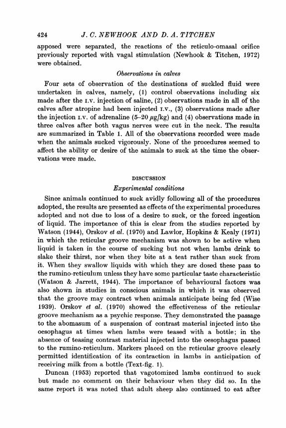
PLATE 1
Fig. 1. Radiograph made during sucking of milk-contrast mixture by a lamb4 days old. Contrast medium is present in three sites in the thoracic oesophagus,the most cranial bolus dorsal to the shoulder joint (S) is separate from morecaudally situated material in the mid-thoracic oesophagus which is partly separatedfrom the caudal phrenic ampulla (P.A.) by a distinct constriction. Contrast mediumpresents a continuous shadow from the phrenic ampulla, through the reticulargroove (R.G.), the omasum (OM) into the abomasum (AB). Liquid-gas interfacespresent are in the epigastric position between the abomasal gas (AB.G.) and liquid(AB), and in the dorsal position between the ruminal gas (RU.G.) and liquid.Fig. 2. The 4-day-old lamb in which this radiograph was taken was sucking at thetime. Contrast medium in the caudal thoracic oesophagus is partially separated by
J. C. NEWHOOK AND D. A. TITCHENa constriction cranial to the phrenic ampulla (P.A.). The reticular groove (R.G.)is free of contrast material which is present in the omasum (OM) and abomasum(AB) and has passed into the pyloric antrum (PYL).
PLATSE 2
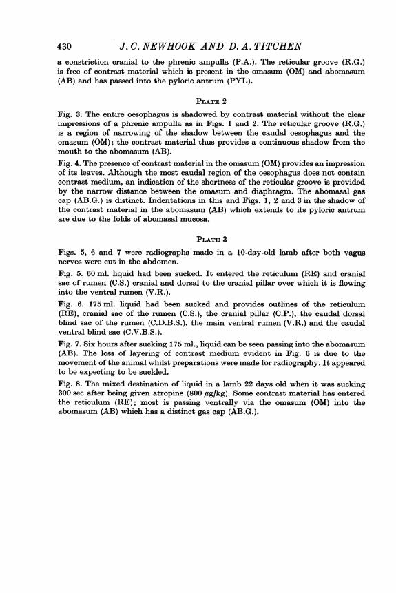
Fig. 3. The entire oesophagus is shadowed by contrast material without the clearimpressions of a phrenic ampulla as in Figs. 1 and 2. The reticular groove (R.G.)is a region of narrowing of the shadow between the caudal oesophagus and theomasum (OM); the contrast material thus provides a continuous shadow from themouth to the abomasum (AB).Fig. 4. The presence of contrast material in the omasum (OM) provides an impressionof its leaves. Although the most caudal region of the oesophagus does not containcontrast medium, an indication of the shortness of the reticular groove is providedby the narrow distance between the omasum and diaphragm. The abomasal gascap (AB.G.) is distinct. Indentations in this and Figs. 1, 2 and 3 in the shadow ofthe contrast material in the abomasum (AB) which extends to its pyloric antrumare due to the folds of abomasal mucosa.
PLATE 3
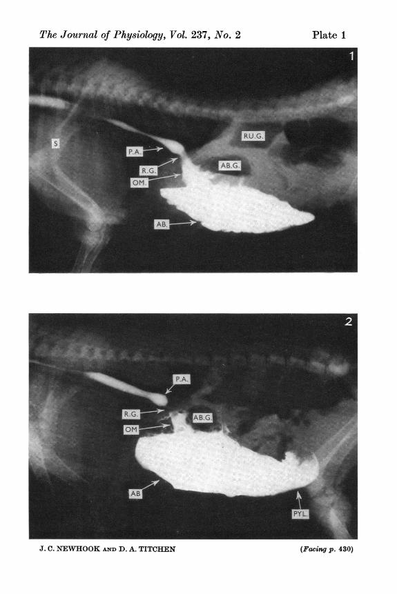
Figs. 5, 6 and 7 were radiographs made in a 10-day-old lamb after both vagusnerves were cut in the abdomen.Fig. 5. 60 ml. liquid had been sucked. It entered the reticulum (RE) and cranialsac of rumen (C.S.) cranial and dorsal to the cranial pillar over which it is flowinginto the ventral rumen (V.R.).Fig. 6. 175 ml. liquid had been sucked and provides outlines of the reticulum(RE), cranial sac of the rumen (C.S.), the cranial pillar (C.P.), the caudal dorsalblind sac of the rumen (C.D.B.S.), the main ventral rumen (V.R.) and the caudalventral blind sac (C.V.B.S.).Fig. 7. Six hours after sucking 175 ml., liquid can be seen passing into the abomasum(AB). The loss of layering of contrast medium evident in Fig. 6 is due to themovement of the animal whilst preparations were made for radiography. It appearedto be expecting to be suckled.Fig. 8. The mixed destination of liquid in a lamb 22 days old when it was sucking300 sec after being given atropine (800 fig/kg). Some contrast material has enteredthe reticulum (RE); most is passing ventrally via the omasum (OM) into theabomasum (AB) which has a distinct gas cap (AB.G.).
430
The Journal of Physiology, Vol. 237, No. 2
J. C. NEWHOOK AND D. A. TITCHEN
Plate I
(Facing p. 430)
The Journal of Physiology, Vol. 237, No. 2
ikt :
J. C. NEWHOOK AND D. A. TITCHEN
Plate 2
The Journal of Physiology, Vol. 237, No. 2w , ,I IV
A n'~~~~~~~~~~~M
J. C. MEWHOOK AND D. A. TITCHEN
Plate 3