by - the journal of biological chemistry · · 2003-02-07is hydrolyzed to glucose by intestinal...
TRANSCRIPT
GASTROINTESTINAL DIGESTION OF STARCH
I. THE ACTION OF OLIGO-1,6-GLUCOSIDASE ON BRANCHED SACCHARIDES
BY JOSEPH LARNER AND C. M. McNICKLE*
(FTOWZ the Division of Biochemistry, Noyes Laboratory of Chemistry, University of Illinois: Urbana, I~.!in.??.nis)
(Received for publication, December 3, 1954)
Digestion of starch in mammals occurs chiefly in the mouth and small intestine through the catalytic activity of ar-amylase and maltase (a-glu- cosidase). Extensive cw-amylolysis of amylose and amylopectin results in the formation of maltose, glucose, and a mixture of branched oligosaccha- rides (cr-amylase dextrins) (1). The latter compounds, whose structure is currently being investigated by several groups (2, 3), contain the a-l,6 linkages of the amylopectin fraction intact, since these linkages are not subject to hydrolysis by a-amylase (4). Meyer and Gonon (4) have re- cently suggested that, ‘if hydrolysis of amylopectin by ar-amylase is carried out to completion, isomaltose is the sole branched product.
Glucose is subsequently absorbed through the intestinal wall. Maltose is hydrolyzed to glucose by intestinal maltase and absorbed as glucose (5). Any maltose which escapes hydrolysis in the intestine is subsequentlyhydro- lyzed by maltase present in the blood (5). We have recently shown (6) that intestinal mucosa contains a new enzyme termed oligo-1,6-glucosi- dase, which specifically hydrolyzes the a-l,6 linkages of isomaltose, “pa- nose, ” and cr-amylase dextrins.’ The three enzymes, a-amylase, oligo-1 ,6- glucosidase, and maltase, catalyze the essentially complete digestion of starch in the gastrointestinal tract.
Oligo-1 ,6-glucosidase differs in its action from amylo-l ,6-glucosidase and R enzyme. It is the purpose of the present report to describe the activity and properties of this new enzyme.
* This work was supported in part by a grant from the Graduate College of the University of Illinois, Urbana.
1 Since the previous work was published (6)) a report of similar enzymatic activity has come to our attention (7). An enzyme from bovine intestinal juice and intestinal mucosa, tentatively called or-limitdextrinase, was shown by increase in reducing power alone to hydrolyze the a-amylase dextrins. No separation from maltase ac- tivity was achieved. For further details, see “Discussion.”
723
by guest on June 4, 2018http://w
ww
.jbc.org/D
ownloaded from

724 OLIGO-1,6-GLUCOSIDASE
Methods and Materials
Enzymes and Coenxymes-Extracts were prepared from hog small in- testine obtained as soon as possible from the slaughter-house.2 All steps were carried out in the cold room at 3”. The intestines were opened, washed with cold distilled H20, and the mucosa removed by scraping with a knife. The scrapings (usually 50 gm.) were thoroughly ground in a mortar with an equal weight of washed sand. The mass was extracted by continued grinding with 5 volumes of cold distilled water and clarified by centrifugation at the highest speed of the Servall centrifuge, model SS-1, for 30 minutes. The opalescent reddish fluid was filtered through Whatman No. 1 paper and dialyzed for 4 to 6 hours against 0.85 per cent NaCl. It was stored at 3” with a small test-tube containing toluene suspended over the surface. When prepared and stored in this manner, the enzyme usually retained full activity for 3 weeks.
Hexokinase from brewers’ yeast was prepared by an unpublished method supplied by C. R. Park of Vanderbilt University. Concentrated stock solutions were diluted with 1 per cent amorphous insulin, frozen in small aliquots, and used as needed.3 Zuischenferment was purified from yeast by the method of Kornberg (8). Amylo-1,6-glucosidase was prepared from pooled supernatant fluids remaining after the crystallization of muscle phosphorylase, as previously described (9). Adenosine triphosphate was purchased from the Pabst Laboratories, and triphosphopyridine nucleotide (TPN) from the Sigma Chemical Company or prepared by the method of Kornberg and Horecker (10).
Substrate--Amylase dextrins were prepared by extensive action of salivary amylase on waxy maize amylopectin.3 Two digestions were carried out with undiluted saliva. 1 ml. of filtered saliva was incubated with 186 mg. of amylopectin at 30’ for 25.5 hours, at which time digestion had proceeded to the extent of 44.0 per cent in terms of glucose equivalents. It was treated a second time with 0.5 ml. of filtered saliva for 16 hours at 30”, during which time digestion proceeded an additional 5.7 per cent. Enzymatic activity was stopped by heating at 100”. Glucose and maltose were removed by four fermentations with bakers’ yeast. The absence of these two sugars was checked by paper chromatography. The average
2 Older hogs, weighing 100 pounds or more, seem to be a better source of enzyme than younger animals.
a We gratefully acknowledge the gift of the following materials: amorphous in- sulin from Dr. 0. K. Behrens, Eli Lilly and Company; waxy maize amylopectin from Dr. T. J. Schoch, Corn Products Refining Company; isomaltose from Dr. Allene Jeanes, Northern Utilization Research Branch; panose from Dr. S. C. Pan, The Squibb Institute for Medical Research; and clinical dextran from Dr. E. B. Hodge, Commercial Solvents Corporation.
by guest on June 4, 2018http://w
ww
.jbc.org/D
ownloaded from
J. LARNER AND C. M. MCNICKLE 725
degree4 of polymerization of the branched dextrins was 6.5. The mixture of “branched” oligosaccharides which contained isomaltose and at least three higher saccharides was stored in the frozen state and used as needed. Muscle phosphorylase limit dextrin was prepared as previously described
(9). Analytical-Paper chromatography was carried out by the multiple as-
cent method described by French and coworkers (11) and Jeanes and co- workers (12). The chromatograms were sprayed with alkaline 3,5-dinitro- salicylate according to Jeanes and coworkers (12), or with ammoniacal AgN03 according to the method of Hough (13). Reducing sugars were determined by the Nelson method (14) with the alkaline Reagent 60 of Shaffer and Somogyi (15) with the longer boiling time of 30 minutes. De- proteinization was accomplished by the Ba(OH)2-ZnSOr method of So- mogyi (16) with the modifications suggested by Hers and Kusaka (17).
Protein was determined by the biuret method of Robinson and Hogden (18). In some cases protein was determined nephelometrically after pre- cipitation by trichloroacetic acid essentially by the method suggested by Burton and Stadtman (19). Crystalline bovine serum albumin was used as the standard. Phosphate was determined by the method of Fiske and Subbarow (20).
Hydrolysis of Isomaltose, Panose, and a-Amylase Dextrins
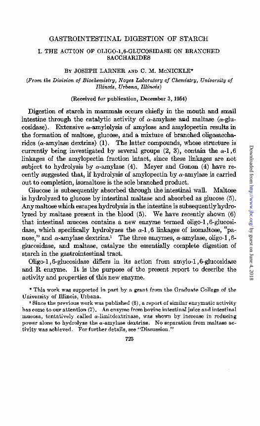
Incubation of intestinal extracts with isomaltose, panose, or the a-amy- lase dextrins leads to a rapid increase in reducing power. Typical experi- ments are shown in Fig. 1. Substrates were incubated with enzyme in the absence of buffer at 30”. Aliquots were removed at zero time and at time intervals, deproteinized by the Ba(OH)2-ZnS04 method, and the re- ducing power was determined. Essentially complete hydrolysis of iso- maltose (Curve 1) (98.5 per cent) and the a-amylase dextrins (Curve 3) (103 per cent) had occurred in 210 and 30 minutes, respectively. Panose (Curve 2) was hydrolyzed 87.5 per cent in 180 minutes. All hydrolyses are recorded in terms of glucose equivalents. Since these experiments were designed to produce essentially complete hydrolysis, the amounts of enzyme and substrate added vary, and the rates of hydrolysis are therefore not com- parable. Seiji (7), using a-amylase dextrins as substrate, records hydroly- ses in the range of 12.4 to 58.5 per cent in terms of glucose equivalents.
In order to rule out phosphorolytic reactions, intestinal extracts were dialyzed to remove inorganic phosphate. Before dialysis, analysis for in-
4 Determined by increase in reducing-power after acid hydrolysis. Meyer and Gonon (4) report that 50 per cent hydrolysis of amylopectin by crystalline pan- creatic a-amylase liberates branched dextrins of a degree of polymerization of 6.0.
by guest on June 4, 2018http://w
ww
.jbc.org/D
ownloaded from
726 OLIGO-1,6-GLUCOSIDASE
organic phosphate revealed 125 to 150 y per ml. After 4 to 6 hours of di- alysis against 0.85 per cent NaCl, these levels had decreased to 4.7 and 9.8 y per ml. Enzymatic activity was not decreased by dialysis. In addition, deproteinization was routinely carried out by the Ba(OH)&nSOa method of Somogyi, which removes phosphorylated sugars.
Bacterial Contamination-That intact bacteria are not directly responsi- ble for enzymatic activity is shown by the fact that enzymatic activity was retained after filtration through a bacteriological filter (sintered glass). For example, incubation of 1 .O ml. of intestinal extract (1.97 mg. of protein)
FIG. 1. Hydrolysis of branched saccharides by oligo-1,6-glucosidase. Curve 1, isomaltose. The isomaltose reaction mixture contained 2 mg. of isomaltose, 58 units of oligo-1,6-glucosidase; volume 2.2 ml. Curve 2, panose. The panose reaction mixture contained 15 mg. of panose, 177 units of oligo-1,6-glucosidase; volume 6.2 ml. Curve 3, cw-amylase dextrins. The cu-amylase dextrin reaction mixture con- tained 0.199 mg. of wamylase dextrins, 216 units of oligo-1,6-glucosidase; volume 6.2 ml. For the definition of a unit, see the text.
with 4.3 mg. of panose at 30” for 210 minutes resulted in 37.5 per cent hy- drolysis before filtration, and 16.2 per cent after filtration through a bac- teriological filter. The loss in activity is explained by the fact that the extract had been frozen and thawed between the two experiments, a pro- cedure known to reduce oligo-1,6-glucosidase activity in crude extracts. Also, after centrifugation at the highest speed of Servall model SS-1 for 30 minutes, extracts had between lo6 and lo6 organisms per ml., which decreased to lo3 organisms per ml. after storage for 18 hours in the pres- ence of toluene.6 Assuming 1012 organisms per gm., lo6 organisms per ml. would amount to less than 0.01 per cent of the protein present in the extracts (usually about 10 mg. of protein per ml.). However, the possi-
5 We wish to thank J. R. Stamer and I. C. Gunsalus of the Department of Bac- teriology for carrying out these counts.
by guest on June 4, 2018http://w
ww
.jbc.org/D
ownloaded from
J. LARNER AND C. M. MCNICKLE 727
bility that enzymatic activity is of bacterial origin is still open to question. This possibility seems less likely in view of the fact that extracts made from hogs which had received antibiotics in the ration sufficient to elicit a growth response had enzyme activities similar to controls which had re- ceived no antibiotics.6 There was also no significant difference in the ac- tivity of extracts prepared by grinding with sand in the presence of toluene. However, to arrive at a more definite conclusion, this problem is currently being investigated with germ-free animals.
I 2 3 4 I 2 3 4
FIG. 2 FIG. 3
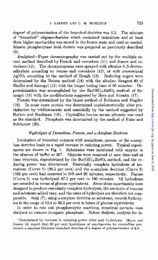
FIG. 2. Hydrolysis of isomaltose by oligo-1,6-glucosidase. Amount of stippling roughly proportional to intensity of spot on original chromatogram. G, glucose; M, maltose; I, isomaltose; P, panose; Column 1, controls; Column 2, 34.2 per cent hydrolysis; Column 3, 64.5 per cent hydrolysis; Column 4, 95.0 per cent hydrolysis.
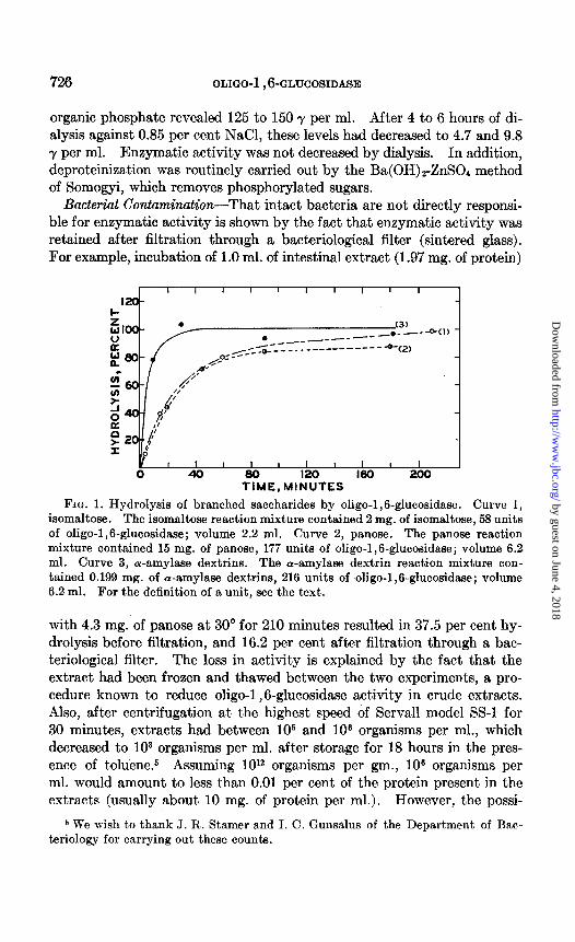
FIG. 3. Hydrolysis of panose by oligo-1,6-glucosidase. Letter designations are the same as in Fig. 2. Column 1, controls; Column 2,49.5 per cent hydrolysis; Col- umn 3,73.8 per cent hydrolysis; Column 4,97.5 per cent hydrolysis.
Paper Chromatography-Product formation was investigated by three methods; fermentability by bakers’ yeast, paper chromatography, and coupling with a specific microenzymatic test system.
The product formed after the hydrolysis of isomaltose was completely fermentable by bakers’ yeast. Paper chromatography (Fig. 2) revealed that the only product which appeared either at intermediate stages or after essentially complete hydrolysis of isomaltose was free glucose.
At intermediate stages of the hydrolysis of panose (Fig. 3), glucose and small amounts of maltose were present, demonstrating the hydrolysis of the a-l,6 linkages. At complete hydrolysis, maltose disappeared and glucose
6 The antibiotic mixture fed (36 mg. per pound of ration) was made up of equal parts of Terramycin, procaine penicillin, and streptomycin. We wish to thank Dr. A. H. Jensen of the Animal Science Department for making these animals available to us.
by guest on June 4, 2018http://w
ww
.jbc.org/D
ownloaded from
728 OLIGO-1,6-GLUCOSIDASE
appeared as the only product. Maltase present in the extracts as a con- taminant is responsible for the formation of glucose from maltose. In addition, faint spots appeared at late stages of the hydrolysis of panose (not visible in Fig. 3) which migrated at slower rates than isomaltose. These probably represent the result of transferring action taking place dur- ing the over-all hydrolysis, most likely oligosaccharides arising from maltase action. In maltose controls paper chromatographic analysis revealed, in addition to maltose and glucose, the presence of oligosaccharide spots at
. DEXTRAN
PANOSE l
ISOMALTOSE
r
AD
TIME. MINUTES
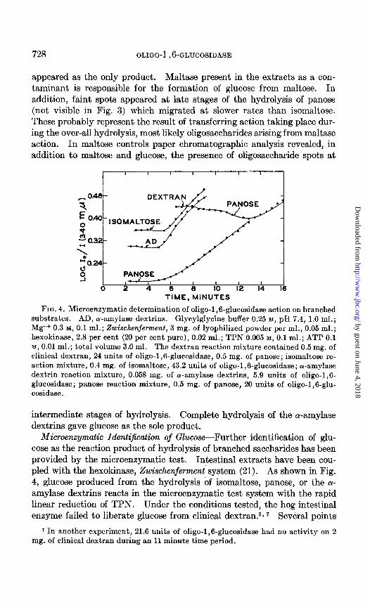
FIG. 4. Microenzymatic determination of oligo-1,6-glucosidase action on branched substrates. AD, cu-amylase dextrins. Glycylglycine buffer 0.25 M, pH 7.4, 1.0 ml.; Mg++ 0.3 M, 0.1 ml.; Zwischenferment, 3 mg. of lyophilized powder per ml., 0.05 ml.; hexokinase, 2.8 per cent (20 per cent pure), 0.02 ml.; TPN 0.005 M, 0.1 ml.; ATP 0.1 M, 0.01 ml.; total volume 3.0 ml. The dextran reaction mixture contained 0.5 mg. of clinical dextran, 24 units of oligo-1,6-glucosidase, 0.5 mg. of panose; isomaltose re- action mixture, 0.4 mg. of isomaltose, 43.2 units of oligo-1,6-glucosidase; or-amylase dextrin reaction mixture, 0.053 mg. of Lu-amylase dextrins, 5.9 units of oligo-1,6- glucosidase; panose reaction mixture, 0.5 mg. of panose, 20 units of oligo-1,6-glu- cosidase.
intermediate stages of hydrolysis. Complete hydrolysis of the a-amylase dextrins gave glucose as the sole product.
Microenzymatic Identijication of Glucose-Further identification of glu- cose as the reaction product of hydrolysis of branched saccharides has been provided by the microenzymatic test. Intestinal extracts have been cou- pled with the hexokinase, Zwischenjerment’ system (21). As shown in Fig. 4, glucose produced from the hydrolysis of isomaltose, panose, or the (Y- amylase dextrins reacts in the microenzymatic test system with the rapid linear reduction of TPN. Under the conditions tested, the hog intestinal enzyme failed to liberate glucose from clinical dextran.3* 7 Several points
r In another experiment, 21.6 units of oligo-1,6-glucosidase had no activity on 2 mg. of clinical dextran during an 11 minute time period.
by guest on June 4, 2018http://w
ww
.jbc.org/D
ownloaded from
J. LARNER AND C. M. MCNICKLE 729
merit further discussion. With weak oligo-1 ,6-glucosidase activity, there usually is an initial lag period of about 1 minute after substrate addition before TPN reduction begins. This is probably a function of the time re- quired for saturation of hexokinase and Zwischenferment by the glucose and glucose-6-phosphate produced subsequent to the hydrolysis of the oligo- saccharides. Intestinal extracts as freshly prepared contain large quan- tities of glucose which interfere in the microenzymatic determination. Dialysis against 0.85 per cent NaCl for 4 to 6 hours served to lower the glucose content to levels low enough so that there was no serious inter- ference in the test. More highly purified glucosidase preparations con- tained no detectable glucose.
0 ox)4 0.08 0.12 0.10 420 0.24 EXTRACT ADDED, ML.
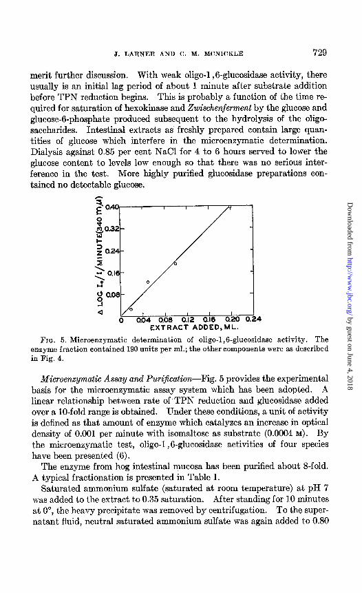
FIG. 5. Microenzymatic determination of oligo-1,6-glucosidase activity. The enzyme fraction contained 190 units per ml.; the other components were as described in Fig. 4.
Microenzymatic Assay and Purijication-Fig. 5 provides the experimental basis for the microenzymatic assay system which has been adopted. A linear relationship between rate of TPN reduction and glucosidase added over a lo-fold range is obtained. Under these conditions, a unit of activity is defined as that amount of enzyme which catalyzes an increase in optical density of 0.001 per minute with isomaltose as substrate (0.0004 M). By the microenzymatic test, oligo-1,6-glucosidase activities of four species have been presented (6).
The enzyme from hog intestinal mucosa has been purified about &fold. A typical fractionation is presented in Table I.
Saturated ammonium sulfate (saturated at room temperature) at pH 7 was added to the extract to 0.35 saturation. After standing for 10 minutes at O”, the heavy precipitate was removed by centrifugation. To the super- natant fluid, neutral saturated ammonium sulfate was again added to 0.80
by guest on June 4, 2018http://w
ww
.jbc.org/D
ownloaded from
730 OLIGO-1,6-GLUCOSIDASE
saturation. After standing for 10 minutes at 0”, the precipitate was col- lected by centrifugation, dissolved in a minimal volume of 0.85 per cent NaCl, and dialyzed for 6 hours against a large volume of 0.85 per cent NaCl. This step usually results in about a Z-fold increase in specific activity with about a 60 to 80 per cent recovery. The protein concentration was de- termined (usually 2.5 per cent) and the reddish opalescent solution diluted to a protein concentration of 1 per cent. 0.2 ml. of alumina Oy (22) (dry weight 14.8 mg. per ml.) was added per ml. of extract, and the mixture stirred for 5 minutes at 0”. After centrifugation, the slightly opalescent
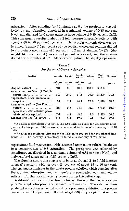
TABLE I Purification of Oligo-i ,6-glucosidase
Fraction Activity
units per ml.
Original extract. . . . . . . . . . . 104 Ammonium sulfate (0.35-0.80
saturation)................... 440 Supernatant after alumina ad-
sorption...................... 94 Ammonium sulfate (O-O.80 satu-
ration) . . . . . ..(............... 320 Supernatant after calcium phos-
phate gel adsorption*. . 232 Ethanol fraction (18440J,)t. . 384
Protein
mg;,fier
9.8
25.0
2.1
9.4
4.8 4.8
-
.- Specific activity
uds jer m&T.
10.6
17.6
44.7
34.0
48.3 80.0
T-
.-
-
VdUlXle
ml.
170.0
30.0
72.5
12.5
5.5 1.8
Total units
17,680
13,200
6,810
4,000
1,275 692
- 1
.-
-
R.%OW~
)er celZt
74.6
38.5
22.6
16.9 10.1
* An aliquot containing 1700 out of the 4000 units was used for the calcium phos- phate gel adsorption. The recovery is calculated in terms of a recovery of 3000 units.
t An aliquot containing 1160 out of the 3000 units was used for the ethanol frac- tionation. The recovery is calculated in terms of a recovery of 1790 units.
supernatant fluid was treated with saturated ammonium sulfate (as above) to a concentration of 0.8 saturation. The precipitate was collected by centrifugation, dissolved in a minimal volume of 0.85 per cent NaCl, and dialyzed for 6 hours against 0.85 per cent NaCl.
The alumina adsorption step results in an additional 2- to 3-fold increase in specific activity with an over-all recovery of about 35 to 40 per cent. The enzyme is unstable in the dilute protein solution which remains after the alumina adsorption and is therefore concentrated with ammonium sulfate. Further loss in activity occurs during this latter step.
Additional purification has been achieved through the use of calcium phosphate gel adsorption and ethanol fractionation. The calcium phos- phate gel adsorption is carried out after a preliminary dilution to a protein concentration of 1 per cent. 0.2 ml. of gel (23) (dry weight 10.4 mg. per
by guest on June 4, 2018http://w
ww
.jbc.org/D
ownloaded from
J. LARNER AND C. M. MCNICKLE 731
ml.) was added per ml. of extract, stirred for 5 minutes at O’, and centri- fuged. The supernatant fluid was then treated with 95 per cent ethanol at a temperature of from 0” to -5” and the fraction between 18 and 44 per cent ethanol collected by centrifugation, dissolved in a small volume of 0.85 per cent NaCl, and dialyzed for 2 hours against 0.85 per cent NaCl.
I t I 2 IO 14 18 22
L,MIN”TES
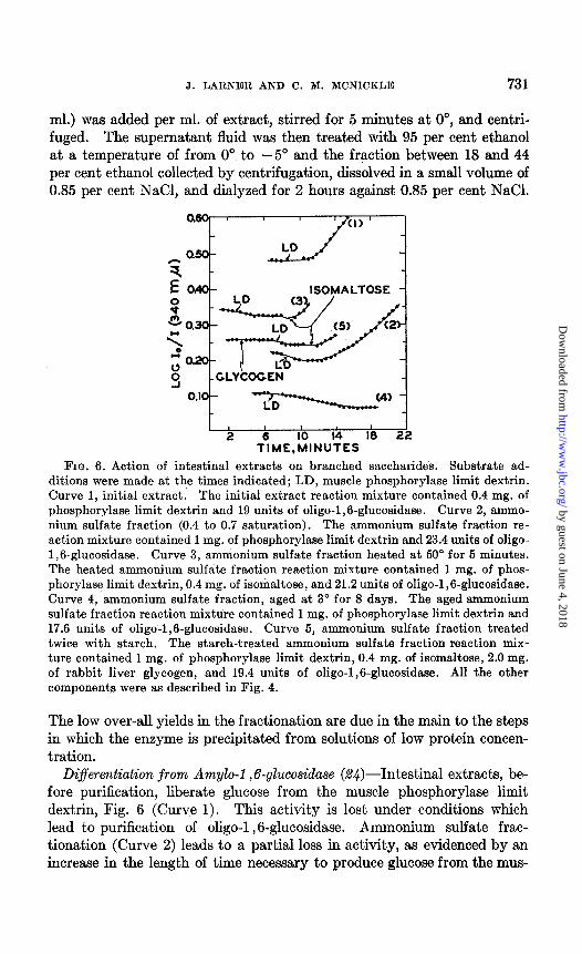
FIG. 6. Action of intestinal extracts on branched saccharides. Substrate ad- ditions were made at the times indicated; LD, muscle phosphorylase limit dextrin. Curve 1, initial extract. The initial extract reaction mixture contained 0.4 mg. of phosphorylase limit dextrin and 19 units of oligo-1,6-glucosidase. Curve 2, ammo- nium sulfate fraction (0.4 to 0.7 saturation). The ammonium sulfate fraction re- action mixture contained 1 mg. of phosphorylase limit dextrin and 23.4 units of oligo- 1,6-glucosidase. Curve 3, ammonium sulfate fraction heated at 50” for 5 minutes. The heated ammonium sulfate fraction reaction mixture contained 1 mg. of phos- phorylase limit dextrin, 0.4 mg. of isomaltose, and 21.2 units of oligo-1,6-glucosidase. Curve 4, ammonium sulfate fraction, aged at 3” for 8 days. The aged ammonium sulfate fraction reaction mixture contained 1 mg. of phosphorylase limit dextrin and 17.6 units of oligo-1,6-glucosidase. Curve 5, ammonium sulfate fraction treated twice with starch. The starch-treated ammonium sulfate fraction reaction mix- ture contained 1 mg. of phosphorylase limit dextrin, 0.4 mg. of isomaltose, 2.0 mg. of rabbit liver glycogen, and 19.4 units of oligo-1,6-glucosidase. All the other components were as described in Fig. 4.
The low over-all yields in the fractionation are due in the main to the steps in which the enzyme is precipitated from solutions of low protein concen- tration.
Differentiation from Amylo-1,6-glucosidase (%$)-Intestinal extracts, be- fore purification, liberate glucose from the muscle phosphorylase limit dextrin, Fig. 6 (Curve 1). This activity is lost under conditions which lead to purification of oligo-1 ,6-glucosidase. Ammonium sulfate frac- tionation (Curve 2) leads to a partial loss in activity, as evidenced by an increase in the length of time necessary to produce glucose from the mus-
by guest on June 4, 2018http://w
ww
.jbc.org/D
ownloaded from
732 OLIGO-1,6-GLUCOSIDASE
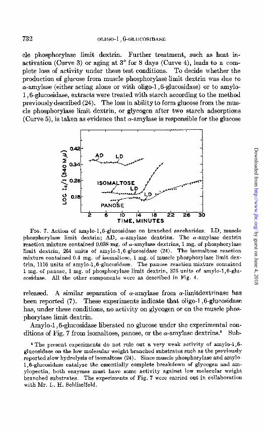
cle phosphorylase limit dextrin. Further treatment, such as heat in- activation (Curve 3) or aging at 3” for 8 days (Curve 4), leads to a com- plete loss of activity under these test conditions. To decide whether the production of glucose from muscle phosphorylaselimit dextrin was due to a-amylase (either acting alone or with oligo-1,6-glucosidase) or to amylo- 1,6-glucosidase, extracts were treated with starch according to the method previously described (24). The loss in ability to form glucose from the mus- cle phosphorylase limit dextrin, or glycogen after two starch adsorptions (Curve 5)) is taken as evidence that ol-amylase is responsible for the glucose
r ’ I I
I I I I I 2 6 IO 14 18 22 26 30
TIME. MINUTES
FIG. 7. Action of amylo-1,6-glucosidase on branched saccharides. LD, muscle phosphorylase limit dextrin; AD, or-amylase dextrins. The oc-amylase dextrin reaction mixture contained 0.038 mg. of ol-amylase dextrins, 1 mg. of phosphorylase limit dextrin, 264 units of amylo-1,6-glucosidase (24). The isomaltose reaction mixture, contained 0.4 mg. of isomaltose, 1 mg. of muscle phosphorylase limit dex- trin, 1116 units of amylo-1,6-glucosidase. The panose reaction mixture contained 1 mg. of panose, 1 mg. of phosphorylase limit dextrin, 375 units of amylo-1,6-glu- cosidase. All the other components were as described in Fig. 4.
released. A similar separation of ar-amylase from a-limitdextrinase has been reported (7). These experiments indicate that oligo-1 ,6-glucosidase has, under these conditions, no activity on glycogen or on the muscle phos- phorylase limit dextrin.
Amylo-l ,6-glucosidase liberated no glucose under the experimental con- ditions of Fig. 7 from isomaltose, panose, or the oc-amylase dextrins.8 Sub-
* The present experiments do not rule out a very weak activity of amylo-1,6- glucosidase on the low molecular weight branched substrates such as the previously reported slow hydrolysis of isomaltose (24). Since muscle phosphorylase and amylo- 1,6-glucosidase catalyze the essentially complete breakdown of glycogen and am- ylopectin, both enzymes must have some activity against low molecular weight branched substrates. The experiments of Fig. 7 were carried out in collaboration with Mr. L. H. Schliselfeld.
by guest on June 4, 2018http://w
ww
.jbc.org/D
ownloaded from
J. LARNER AND C. M. MCNICKLE 733
sequent addition of muscle phosphorylase limit dextrin led to rapid glucose production, indicating the activity of the test system.
TABLE II
Ratio of Maltase to Oliqo-1, B-glucosidase Activity _ ._
Oligo-1,6-glucosidase fraction
Initial extract, 10 days at 3”. . . . . Ammonium sulfate fraction (0.p0.7 saturation). . . .
“ I‘ “ incubated 5 min. at 50”. , Supernatant after 1 alumina adsorptiont. . . . .
I‘ ‘I 2 ‘I adsorptions . . . . . . . ‘I “ 1 calcium phosphate gel adsorp-
tiont . . . . . . . . . . . . . . . . . . . . . . Supernatant after 2 calcium phosphate gel adsorp-
tions..............................................
-
_-
-
Maltase activity
w*
units per ml.
1920 4200 2210 1040 828
880
480
-
--
-
Oligo-1 ,6- glucosidase activity (2)
units per ml
296 500 288 170 58
57
26
-
_ -
-
Ratio (0: (2)
6.5 8.4 7.7 6.1
14.3
15.4
18.5
* The reaction mixture is the same as that for determination of oligo-1,6-gluco- sidase activity (Fig. 4), except that maltose was used as substrate.
t Alumina and calcium phosphate gel adsorptions were carried out as described in the text.
TABLE III
Stability of Oligo-f ,6-glucosidase
I Activity
I R‘XOVe~
units )lw ml. per cent
Control, pH 7.0. . . . . . . . . . . . . . . . . . . . . . . 276 pH 5.58, 60 min. at room temperature.. . . . . 110 39.8
I‘ 9.77, 60 “ “ “ “ . . . . 148 53.6
Enzyme solutions were brought to the indicated pH’s by the addition of 0.05 N HCl or 0.75 N NaOH. After standing for the indicated time, the solutions were neutralized to pH 7 with either solid NaHCOs or 2 N HCl, and tested microenzymati- tally. Two types of controls were run, with and without NaCl added in amounts equal to that present in the experimental vessels. There was no significant differ- ence in these controls.
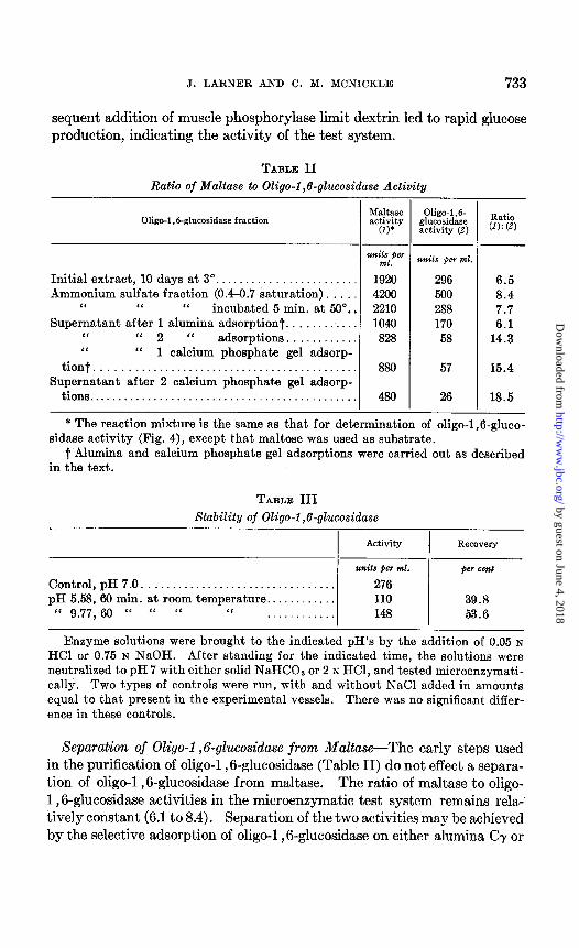
Separation of Oligo-1,6-glucosidase from Maltase-The early steps used in the purification of oligo-1,6-glucosidase (Table II) do not effect a separa- tion of oligo-1,6-glucosidase from maltase. The ratio of maltase to oligo- 1,8glucosidase activities in the microenzymatic test system remains rela- tively constant (6.1 to 8.4). Separation of the two activities may be achieved by the selective adsorption of oligo-1 ,6-glucosidase on either alumina CT or
by guest on June 4, 2018http://w
ww
.jbc.org/D
ownloaded from
734 OLIGO-I ,6-GLUCOSIDASE
calcium phosphate gel. Under these conditions, the ratio of the two ac- tivities was increased9 to 18.5. Seiji (7), fractionating with acetone and ammonium sulfate, was unable to separate these two activities.
Stability and pH Optimum-The crude enzyme is stablelo for about 3 weeks at 3”. After this time, activity is decreased. A decrease in glu- cosidase activity of initial extracts is also brought about by storing in the frozen state. More highly purified preparations are stable in the frozen state. Lyophilization from either 0.85 per cent NaCl or glycylglycine buffer (0.025 M, pH 7.5) resulted in a loss of about 50 per cent of activity.
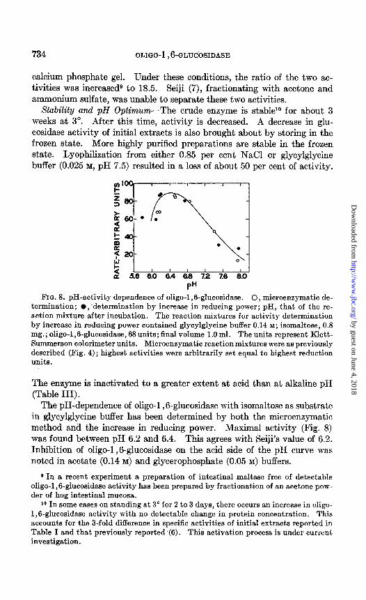
FIG. 8. pH-activity dependence of oligo-1,6-glucosidase. 0, microenzymatic de- termination; l ;determination by increase in reducing power; pH, that of the re- action mixture after incubation. The reaction mixtures for activity determination by increase in reducing power contained glycylglycine buffer 0.14 M; isomaltose, 0.8 mg.; oligo-1,6-glucosidase, 68 units; final volume 1.0 ml. The units represent Klett- Summerson calorimeter units. Microenzymatic reaction mixtures were as previously described (Fig. 4) ; highest activities were arbitrarily set equal to highest reduction units.
The enzyme is inactivated to a greater extent at acid than at alkaline pH (Table III).
The pH-dependence of oligo-1 ,6-glucosidase with isomaltose as substrate in glycylglycine buffer has been determined by both the microenzymatic method and the increase in reducing power. Maximal activity (Fig. 8) was found between pH 6.2 and 6.4. This agrees with Seiji’s value of 6.2. Inhibition of oligo-1,8glucosidase on the acid side of the pH curve was noted in acetate (0.14 M) and glycerophosphate (0.05 M) buffers.
9 In a recent experiment a preparation of intestinal maltase free of detectable oligo-1,6-glucosidase activity has been prepared by fractionation of an acetone pow- der of hog intestinal mucosa.
10 In some cases on standing at 3“ for 2 to 3 days, there occurs an increase in oligo- 1,6-glucosidase activity with no detectable change in protein concentration. This accounts for the a-fold difference in specific activities of initial extracts reported in Table I and that previously reported .(6). This activation process is under current investigation.
by guest on June 4, 2018http://w
ww
.jbc.org/D
ownloaded from
J. LARNER AND C. M. MCNICKLE 735
DISCUSSION
The evidence presented in the preceding sections establishes the presence of a new enzyme oligo-1,6-glucosidase in extracts of small intestinal mucosa. Oligo-1,6-glucosidase is involved in the terminal stages of starch digestion in the small intestine. Acting together with cY-amylase and maltase, oligo- 1,6-glucosidase catalyzes the essentially complete breakdown of starch to glucose in digestion. That this latter enzyme is of physiological signifi- cance in the whole animal is shown by the fact that isomaltose in the rat gives rise to as large an increase in liver glycogen as does maltose.”
Purified oligo-1,6-glucosidase from .the hog does not liberate glucose from clinical dextran, glycogen, or the muscle phosphorylase limit dextrin under the conditions tested. It is therefore differentiated from amylo-1,6- glucosidase, which in turn has essentially no activity on the low molecular weight substrates. A partial separation from maltase has been achieved by differential adsorption on alumina CT, and calcium phosphate gel.
Seiji (7) has presented evidence that the enzymatic hydrolysis of ar- amylase dextrins is catalyzed by an enzyme tentatively called ar-limit- dextrinase, obtained from bovine intestinal mucosa and intestinal juice. The a-amylase dextrins which he prepared may have contained maltose (RF of maltose 0.26, RF of one spot in dextrin mixture 0.23) and are there- fore subject to hydrolysis by the maltase present in the extracts.
Other plant, fungal, and bacterial a-l ,gsplitting enzymes may now be mentioned. Oligo-1 ,6-glucosidase may be differentiated from R enzyme by the inability of the latter to hydrolyze isomaltose and panose (1). Limitdextrinase, an enzyme with similar activity to oligo-1 ,6-glucosidase from Aspergillus oryzae culture filtrates, has been reported (25). Similar hydrolysis of isomaltose and panose by mold culture filtrates has been re- ported by Pan and coworkers (26) and by Tsuchiya and coworkers (27). It is not known at present whether amyloglucosidase (y-amylase) (28-30) or the maltase of Clostridium acetobutylicum (31) hydrolyzes a-1 ,6 linkages in addition to a-1,4.
SUMMARY
The hydrolysis of the a-l ,6 linkages of isomaltose, panose, and a-amylase dextrins by an enzyme from intestinal mucosa called oligo-1 ,6-glucosidase has been demonstrated. Hydrolysis has been followed by increase in re- ducing power, paper chromatography, and by coupling to a specific micro- enzymatic test system.
Oligo-1 ,6-glucosidase has essentially no activity against the muscle phos- phorylase limit dextrin. It is thus differentiated from amylo-1,6-glucos-
11 Personal communication from Dr. R. C. Corley, Department of Chemistry, Purdue University, Lafayette, Indiana.
by guest on June 4, 2018http://w
ww
.jbc.org/D
ownloaded from
736 OLIGO-1,6-GLUCOSIDASE
idase, which in turn has essentially no activity against isomaltose, panose, or the cu-amylase dextrins. Partial purification and separation of oligo-1 ,6- glucosidase from a-amylase and maltase have been achieved.
Acting together with maltase, oligo-1 ,6-glucosidase catalyzes the com- plete hydrolysis of ar-amylase dextrins and thus allows essentially complete digestion of starch to proceed in the gastrointestinal tract.
BIBLIOGRAPHY
1. Whelan, W. J., Biochem. Sot. Symposia, 11, 17 (1953). 2. Nordin, P., Wild, G., and French, D., Abstracts, American Chemical Society,
124th meeting, Chicago, 53C, Sept. (1953). 3. Whelan, W. J., and Roberts, P. J. P., Nature, 170,748 (1952). 4. Meyer, K. H., and Gonon, W. F., Helv. chim. acta, 34, 308 (1951). 5. Peters, J. P., and Van Slyke, D. D., Quantitative clinical chemistry; Interpreta-
tions, Baltimore, 2nd edition, 1 (1946). 6. Larner, J., and McNickle, C. M., J. Am. Chem. Sot., 76,4747 (1954). 7. Seiji, M., J. Biochem., Japan, 40, 519 (1953). 8. Kornberg, A., J. Biol. Chem., 163, 805 (1950). 9. Larner, J., J. Biol. Chem., 212,9 (1955).
10. Kornberg, A., and Horecker, B. L., Biochem. Preparations, 3, 24 (1953). 11. French, D., Knapp, D. W., and Pasur, J. H., J. Am. Chem. Sot., 73, 5150 (1950). 12. Jeanes, A., Wise, C. S., and Dimler, R. J., Anal. Chem., 23,415 (1951). 13. Hough, L., Nature, 166, 400 (1950). 14. Nelson, N., J:BioZ. Chem., 163, 375 (1944). 15. Shaffer, P. A., and Somogyi, M., J. BioZ. Chem., 100, 695 (1933). 16. Somogyi, M., J. BioZ. Chem., 160,69 (1945). 17. Hers, H. G., and Kusaka, T., Biochim. et biophys. actu, 11, 427 (1953). 18. Robinson, H. W., and Hogden, C. G., J. BioZ. Chem., 136, 727 (1940). 19. Burton, R. M., and Stadtman, E. R., J. BioZ. Chem., 202, 873 (1953). 20. Fiske, C. H., and Subbarow, Y., J. BioZ. Chem., 66,375 (1925). 21. Slein, M. W., J. BioZ. Chem., 166,753 (1950). 22. Wilstatter, R., Kraut, H., and Erbacher, O., Ber. them. Ges., 66,244s (1925). 23. Keilin, F. R. S., and Hartree, E. F., Proc. Roy. Sot. London, Series B, 124, 397
(1938). 24. Cori, G. T., and Larner, J., J. BioZ. Chem., 168,17 (1951). 25. Underkofler, L. A., and Roy, D. K., Cereal Chem., 28,18 (1961). 26. Pan, S. C., Nicholson, L. W., and Kolachov, P., Arch. Biochem. and Biophys., 42,
406 (1953). 27. Tsuchiya, H. M., Montgomery, E. M., and Corman, J., J. Am. Chem. Sot., 71,
3265 (1949). 28. Corman, J., and Langlykke, A. F., Cereal Chem., 26,190 (1948). 29. Kerr, R. W., Cleveland, F. C., and Katebeck, W. J., J. Am. Chem. Sot., 73, 3916
(1951). 30. Phillips, L. L., and Caldwell, M. L., J. Am. Chem. Sot., 73,3559 (1961). 31. French, D., and Knapp, D. W., J. BioZ. Chem., 187,463 (1950).
by guest on June 4, 2018http://w
ww
.jbc.org/D
ownloaded from
Joseph Larner and C. M. McNickleBRANCHED SACCHARIDES
OLIGO-1,6-GLUCOSIDASE ONSTARCH: I. THE ACTION OF
GASTROINTESTINAL DIGESTION OF
1955, 215:723-736.J. Biol. Chem.
http://www.jbc.org/content/215/2/723.citation
Access the most updated version of this article at
Alerts:
When a correction for this article is posted•
When this article is cited•
alerts to choose from all of JBC's e-mailClick here
tml#ref-list-1
http://www.jbc.org/content/215/2/723.citation.full.haccessed free atThis article cites 0 references, 0 of which can be by guest on June 4, 2018
http://ww
w.jbc.org/
Dow
nloaded from