by wan-jung lin
TRANSCRIPT

THE NUCLEAR ACTIONS OF IGFBP-3
By
WAN-JUNG LIN
A thesis submitted in partial fulfillment ofthe requirements for the degree of
MASTER OF SCIENCES
WASHINGTON STATE UNIVERSITYDepartment of Animal Sciences
AUGUST 2006

ii
To the Faculty of Washington State University:
The members of the Committee appointed to examine the thesis of
WAN-JUNG LIN find it satisfactory and recommend that it be accepted.
___________________________________Chair
___________________________________
___________________________________

iii
ACKNOWLEDGEMENT
In the two years work in Animal Sciences WSU, I have been supported and
accompanied by so many great people. First, I would like to thank my advisor and big
brother Dan (Dr. Rodgers) who is not only a great scientist, but also a most kind person.
Under his scientific supervision and enthusiasm for helping students, I learned a great
deal more than I expected. In addition, I was grateful to have had worked with Momo
(Dr. Oufattole), for his experienced help in the laboratory, his willingness to engage in
discussions, and his encouragement for both research and life. Furthermore, I would like
to thank all the members and friends of the Rodgers’ lab who sweetened up my lab work
every day and were always available for any questions and help. I am also grateful to
have those friends who always give me mental support either in the US (Arriba) or
Taiwan.
Moreover, I am thankful for the assistance and support from my committee
members Dr. Rodgers, Dr. Johnson and Dr. McLean, throughout my masters thesis
program.
Knowing these people is a most precious experiences, even though they couldn’t
realize how much I learned from them and how important they are.
Last, but not least, I would like to thank my mother, father, brother Kenny and
sister Phoebe who always offered support and love.

iv
NUCLEAR ACTIONS OF IGFBP-3
Abstract
by Wan-Jung Lin, M.S.Washington State University
August 2006
Chair: Buel Dan Rodgers
IGF-binding protein (IGFBP)-3 has intrinsic antiproliferative and proapoptotic
functions that are independent of IGF binding and may involve nuclear localization. We
determined that exogenous IGFBP-3 rapidly translocates to myoblast nuclei and that a
22-residue peptide containing the metal binding domain (MBD) and nuclear localization
sequence (NLS) can similarly direct chimeric GFP into myoblast nuclei. Furthermore, a
non-IGF-binding IGFBP-3 mutant inhibited myoblast proliferation without stimulating
apoptosis. These results suggest that IGFBP-3 inhibits muscle cell growth in an IGF-
independent manner that may be influenced by its rapid nuclear localization. We
therefore identified IGFBP-3 interacting proteins by screening a rat L6 myoblast cDNA
library using the yeast two-hybrid assay and two N-terminal deletion mutants as bait:
BP3/231 (231 residues, L61 to K291) and BP3/ 111 (K181 to K291). Proteins previously
known to interact with IGFBP-3 as well as several novel proteins were identified,
including RNA polymerase II binding subunit 3 (Rpb3). The domain necessary for Rpb3
binding was subsequently identified using different IGFBP-3 deletion mutants and was
localized to the MBD/NLS epitope. Rpb3/IGFBP-3 binding was confirmed by

v
coimmunoprecipitation assays with specific antisera, whereas a NLS mutant IGFBP-3 did
not associate with Rpb3, suggesting that a functional NLS is required. Rpb3 facilitates
recruitment of the polymerase complex to specific transcription factors and is necessary
for the transactivation of many genes. Its association of Rpb3 with IGFBP-3 provides a
functional role for IGFBP-3 in the direct modulation of gene transcription.
vi
TABLE OF CONTENTS
Page
ACKNOWLEDGEMENTS............................................................................................. iii
ABSTRACT ................................................................................................................... iv
LIST OF TABLES.......................................................................................................... vi
LISTOF FIGURES ........................................................................................................ vii
CHAPTERS
1. IGF-INDEPENDENT ROLES OF INSULIN-LIKE GROWTH FACTOR BINDING PROTEIN (IGFBP)-3..............................................................……..1
2. RNA POLYMERASE II SUBUNIT 3 (RPB3), A POTENTIAL NUCLEARTARGET OF IGFBP-3………………………………………………………...10
Introduction………………………………………………………………...10
Materials and Methods……………………………………………………..10
Results……………………………………………………………………...18
Discussion………………………………………………………………….32
3. COMMERCIAL AND APPLIED ASPECTS OF IGFBP-3 THERAPEUTICS
………………………………………………………………………………….37
Summary…………………………………………………………………...37
Clinical Implications and Animal Applications……………………………39
4. REFERENCES………………………………………………………………...47
vii
FIGURES
1. Anatomy of an IGFBP………………………………………………………………….4
2. IGF-independent inhibition of myoblast proliferation by IGFBP-3…………………..19
3. IGFBP-3 nuclear localization in L6 myoblasts………………………………………..21
4. IGFBP-3 and MBD peptide time course for nuclear localization in L6 myoblasts…...23
5. Identification of IGFBP-3-interacting proteins in L6 myoblasts……………………...25
6. Binding interactions between IGFBP-3 & a RNA polymerase II subunit, Rpb3……..27
7. IGFBP-3/Rpb3 co-immunoprecipitation requires an intact NLS domain…………….29
8. IGFBP-3/Rpb3 co-immunoprecipitation from L6 myoblast nuclear lysates………….30
9. IGFBP-3 inhibition of myoblast proliferation requires an intact NLS domain……….31
10.Model of IGFBP-3 regulated myoblast growth inhibition and subsequent
differentiation…………………………………………………………………………….38
TABLES
1. Subcloning primers for IGFBP-3 and Rpb3 deletion mutants……………………..….16

1
CHAPTER ONE
INTRODUCTION TO THE IGF-INDEPENDENT ROLES OF INSULIN-LIKE
GROWTH FACTOR BINDING PREOTEIN (IGFBP)-3
The insulin-like growth factor (IGF) system plays a critical role in modulating cell
growth and the development of the central nervous system, skeletal muscle, reproductive
organs and other tissues. It includes both IGF-I and II, a single trans-membrane tyrosine-
kinase receptor and six high affinity IGF binding proteins (IGFBP-1 to -6) (LeRoith &
Roberts 2003, Duan & Xu 2005). Of the two factors, IGF-I is better characterized and is
mainly synthesized in the liver under the primary control of growth hormone (GH).
Indeed, most of GH’s actions are mediated by stimulating IGF-I synthesis in hepatocytes
(Grimberg & Cohen 2000) or its secretion locally, which thereby controls the growth of
somatic tissues, including skeletal muscle, by paracrine and autocrine mechanisms
(Adams & Gregory 2002).
The biological actions of both IGFs are transduced intracellularly by the type I
IGF receptor (IGF-IR). Ligand binding to the two α subunits induces conformational
changes in the intracellular β subunits, which in turn autophosphorylates (LeRoith &
Roberts 2003). Activation of the β subunit tyrosine kinase domains stimulates two
principle down stream signaling cascades; the mitogen-activated protein (MAP) kinase
pathway (LeRoith & Roberts 2003) and the phosphoinositol-3 kinase (PI3-K) pathway
(Yamamoto et al. 1992). Phosphorylation of Shc (SH2 containing proto-oncogene)
initiates the MAP Kinase pathway and recruits other factors including the growth factor

2
receptor-bound 2(Grb2)/Sos complex, which subsequently activates Ras (a GTP-binding
protein kinase) and the different MAPK pathways (LeRoith et al. 1995). This signaling
cascade induces cell proliferation and differentiation by controlling the duration of
MAPK activities and is largely responsible for the IGFs mitogenic actions in both normal
and transformed cells (Marshall 1995). Activation of the PI3-K pathways begins with the
phosphorylation of the insulin receptor substrates (IRS-1 to -4) which then recruit SH2
domain containing proteins including the p85 subunits of PI3-K (Backer et al. 1992).
This in turn leads to the activation of Akt and stimulates cell survival by inhibiting
apoptotic signals. IRS-1 also interacts with other proteins such as Syp (phosphotyrosine
phosphatase), which has been shown to positively regulate the mitogenic actions of IGF-
I, Nck (adaptor protein) and also Grb2/Sos (Backer et al.1992, Xiao et al. 1994).
The physiological effects of IGF-I in many tissues, but specially in muscle, have
been identified in both IGF-I null mice (Liu et al. 1993), which leads to skeletal muscle
hypoplasia and death immediately after birth, and in IGF-I over-expressing transgenic
mice, which have approximately 30% greater muscle mass than their non-transgenic
littermates (Mathews et al. 1988). In addition to regulating myoblast proliferation
through MAPK dependent pathways (LeRoith & Roberts 2003), IGF-I signaling also
stimulates myoblast differentiation through the survival pathway mediated by PI3-K and
eventually the induction of p21, a cyclin-dependent kinase (CDK) inhibitor. Like other
CDK inhibitors, p21controls the CDKs by forming inhibitory complexes during the G1
and S phases of the cell cycle which stimulates cell cycle withdrawal (Lawlor & Rotwein
2000).

3
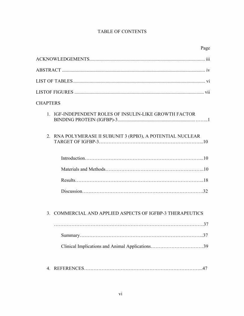
The activity and bioavailability of IGF-I and -II are extensively modulated by six
highly homologous IGFBPs that have equal or slightly higher affinities for the IGFs than
the type-I receptor (Yan et al. 2004). Systemically, the IGFBPs bind IGFs and prevent
their clearance by increasing the circulating half-lives. They also regulate IGF-I and -II
distribution and can either block or facilitate access to the receptor (Firth & Baxter 2002).
All IGFBPs share highly conserved carboxyl and amino terminal domains and each
contain a variable central linker domain (Clemmons 2001). The amino terminal domain is
responsible for IGF binding while the carboxyl terminal cystein-rich domain is
responsible for interacting with other proteins. The linker domain is often modified post-
translationally in a manner specific to each IGFBP (Duan & Xu 2005). That either
includes glycosylation (IGFBP-3, -4 & -5) and/ or phosphorylation (IGFBP-1, -3 & -5)
(see figure 1). The former does not affect IGF binding affinity (Bach et al. 1992, Carr et
al. 1994) but it increases IGFBP stability in part by decreasing susceptibility to
proteolysis (Firth & Baxter 1999, Marinaro et al.2000). It is unclear how phosphorylation
affects most IGFBPs. However, the IGF-I binding ability of IGFBP-1 increases six fold
after phosphorylation (Jones et al.1991). By contrast, the affinity of phosphorylated
IGFBP-3 for IGF-I is unchanged (Hoeck & Mukku 1994), although the growth inhibitory
actions of IGFBP-3 are enhanced by phosphorylation (Hollowood et al.2002). This
suggests that some of IGFBP-3’s actions are independent of IGF binding and that
phosphorylation of IGFBP-3 is involved.
In serum, about 75% of IGF-I is transported in a 150 kDa ternary complex with
IGFBP-3 and the acid-labile subunit. The complex also forms with IGFBP-5, which is
structurally very similar to IGFBP-3, although this occurs infrequently. The formations of

4
Figure 1. Anatomy of an IGFBP. There is 50% homology between different IGFBPsand 40-80% between different species. These differences are mainly come from twomajor post-translational modifications, glycosylation and phosphorylation, in the linkerdomain which give each binding protein possess specific function.
IGF-binding domainH2N- -CO2H
RGD, integrin receptor binding- IGFBP-1 confirmed- IGFBP-2 putative
Val49, Tyr50, Pro62 & Lys68-Leu74
phosphorylated- IGFBP-1 by casein kinase I & II- IGFBP-3 by PKA, PKC & CK II
NLS = KGRKR
metal binding domain (BP-3)
c c c c c c c c c c c c serine rich c c c c

5
these complexes is primarily responsible for maintaining the circulating IGF-reservoir
and in prolonging IGF half-life as only 5% of IGF-I circulates in a free and unbound state
(Firth & Baxter 2002). The remaining 20% forms 40~50 kDa complexes with the
remaining IGFBPs. These smaller complexes readily cross the vascular endothelium
where they regulate IGF availability at the tissue and cellular level (Payet et al. 2004).
The modulation of IGF actions by “stimulatory” or “inhibitory” IGFBPs varies
depending upon the cell type, the presence of other interacting proteins and experimental
conditions. For example, IGFBP-4 and -6 have been consistently identified to inhibit IGF
actions locally, although IGFBP-1, -2, -3 and -5 have been demonstrated to either
stimulate or inhibit the effects of IGF-I (Duan & Xu 2005). When IGF-I and IGFBP-3 are
co-incubated with human skin fibroblasts, IGFBP-3 inhibits the proliferative effects of
IGF-I (Conover et al. 1996). Another study using primary human chondrocytes indicates
that an IGF-I analogue des(1-3) IGF-I incapable of binding to IGFBPs is more potent in
stimulating cell proliferation than normal IGF-I, which also suggests that endogeneously
produced IGFBPs inhibit IGF-I actions locally (Matsumoto et al. 2000). By contrast, but
using the same human skin fibroblast system, preincubating IGFBP-3 with IGF-I
facilitates the binding interaction between IGF-I and its receptor and helps to stimulate
the proliferation (De Mellow & Baxter 1988). The IGFBP-3 has the potential to decrease
the IGF-I proliferation ability in human osteosarcoma cells which is IGF-I dependent by
cloned QAYL-IGF-I, a mutated IGF-I with reduced affinity to IGFBP3 (Schmid et al.
2001). Another interesting study indicates the IGFBP-3 has differential physiological
effects at high and low concentrations as it inhibits cell growth at low concentrations, but
promotes growth at high concentrations (McCaig et al. 2002).
6
These regulatory actions are generally dependent upon ligand binding, however,
recent research has shown that some IGFBPs have functions completely independent
from their interactions with the IGFs. This is particularly true for IGFBP-3 which was
first observed to possess anti-proliferation functions in chicken embryo fibroblasts,
although this was demonstrated using an IGFBP-3 fragment that lacked the IGF-I binding
domain (Lalou et al. 1996). Similar IGF-independent and inhibitory actions have since
been described in many different cell types including prostate cancer cells (Cohen et al.
1994, Angelloz-Nicoud & Binoux 1995), fibroblasts (Cohen et al. 1993, Lalou et al.
1995), breast cancer cells (Oh et al. 1995), mesenchymal chondroprogenitor cells
(Longobardi et al. 2003), renal carcinoma cells (Cheung et al. 2004) and skeletal muscle
myoblasts (Foulstone et al. 2003, Pampusch et al. 2003, Xi et al. 2004). These functions
have been identified for the most part by using different IGFBP-3 mutants and deletion
constructs, such as the GGG (Buckway et al. 2001) and LLGQ (Yan et al. 2004) IGFBP-
3 mutants and the 22-/25- kDa proteolyzed fragment (Lalou et al. 1996) that do not bind
the IGFs, but somehow still inhibit their actions. Studies with Long-R3-IGF-I, a mutant
IGF-I analogue incapable of binding to IGFBPs but fully capable of normal interactions
with its receptors (Tomas et al. 1992), and with cells from IGF-IR knock out mouse
embryos (Rajah et al. 1997) have also demonstrated IGF-independent growth inhibitory
actions of IGFBP-3.
Although the GH/ IGF-I axis is the principle regulator of IGFBP-3 production in
the liver and consequentially circulating levels of IGFBP-3, the gene expression of
IGFBP-3 can be induced by several anti-proliferative and pro-apoptotic factors in many
different cell types. This coincides with the well documented IGF independent growth

7
inhibitory and apoptotic functions of IGFBP-3. There are several apoptotic factors
identified that exert their effect through regulating the mRNA and protein levels of
IGFBP-3 including transforming growth factor (TGF)-β I in PEMC (Kamanga-Sollo et
al. 2005), breast cancer cells (Fanayan et al. 2000), and prostate cancer cells (Cohen et al.
2000); myostatin in PEMC (Kamanga-Sollo et al.2005); retinoid acid in breast cancer
cell (Gucev et al. 1996); tumor necrosis factor (TNF)-α in breast cancer cells (Rozen et
al. 1998); tumor suppressor p53 in breast cancer cells (Schedlich & Graham 2002) and
prostate cancer cells (Rajah et al. 1997); and 1,25 dihydroxyvitamin D3 in prostate
cancer cells (Boyle et al. 2001). These studies suggest that IGFBP-3 is not only a typical
binding protein, but that it serves as an anti-proliferation factor as well. Indeed, cells with
high levels of IGF-I expression more readily transform into malignant cells (LeRoith &
Roberts 2003) and IGFBP-3 is an anti-cancer IGF-I antagonist in many cancerous cells
(Lee & Cohen 2002) including those from lung (Yu et al.1999), breast (Schedlich &
Graham 2002), prostate (Hong et al. 2002) and colon (Ma et al. 1999). Although the
specific inhibitory mechanisms have not been elucidated, recent studies have identified a
novel apoptotic signaling pathway in F9 embryonal carcinoma cells. Nuclear IGFBP-3
binds the Nur77/ RXRalpha heterodimer and facilitates its translocation from the nucleus
to the mitochondria. This results in the release of cytoplasmic cytochrome c, the
activation of the pro-apoptotic factor caspase (Lee et al. 2005) and eventually apoptosis.
These anti-proliferative effects do not always result in apoptosis and have been
associated with myoblast differentiation. In cultures of primary myosatellite cells from
adult human skeletal muscle, the IGFBP-3 has higher secretion positively in respond to
cell differentiation caused by both IGF-I and non-IGFBP binding Long-R3-IGF-I which

8
indicates IGFBP-3 may be involved in myoblast differentiation in both IGF-dependent/-
independent manners (Foulstone et al. 2003). Besides, the mRNA and protein level of
IGFBP-3 changes with differentiation processes in porcine embryonic myogenic cells
(Johnson et al. 1999). Furthermore, IGFBP-3 antisense oligonucleotides similarly
reduced IGFBP-3 production and myoblast differentiation (Foulstone et al. 2003).
Combined, these studies suggest that IGFBP-3 is a multifunctional regulator of
proliferation and differentiation in muscle cells.
Quite remarkably, IGFBP3 has been demonstrated to translocate from
extracellular compartments into the nuclei of different cell types including opossum
kidney cells (Li et al. 1997), human lung cancer cells (Jaques et al. 1997), keratinocytes
(Wraight et al. 1998) and human breast cancer cells (Schedlich et al. 1998). This nuclear
transportation is depend on the C- terminal nuclear localization sequence (NLS) and is
not mediated by classical receptor endocytosis (Schedlich et al. 1998), but may involve
caveolae-mediated endocytosis. (Lee et al. 2004). The precise mechanism of IGFBP-3
translocation is not known, however, it directly associates with Transferrin and its
receptor (Lee et al. 2004). This suggests that one mechanism may include the “piggy
back” of IGFBP-3 with Transferrin into the cell. Receptors for Transferrin are widely, but
not ubiquitously, expressed in many tissues, which may explain IGFBP-3’s unique ability
to nuclear locate in many, but not all cell types (Lee et al. 2004). The ability to nuclear
locate is shared by IGFBP-5 which is structurally homologous to IGFBP-3 and contains
many conserved elements including a heparin binding motif (HBM) and a NLS within its
carboxyl terminal domain. The anti-proliferative functions of IGFBP-3 are required for
the TGF-β stimulated growth inhibition of some human breast cancer cells and these

9
effects are independent of IGF-I binding (Oh et al.1995, Kim et al. 2004). Besides breast
cancer cells, IGFBP-3 also forms a complex with nuclear RXR-alpha inside the nucleus
of LAPC-4 prostate cancer cells under the treatment of retinoid acid (Liu et al. 2000) and
thereby stimulates apoptosis in an IGF-I-independent manner (Hong et al. 2002). By
studying the apoptotic resistant mechanism of senescent fibroblasts, Hampel et al. (2005)
reported that IGFBP-3 nuclear localization may be required for apoptosis in some cells.
Although the functional role of nuclear IGFBP-3 is still unclear at present, these studies
suggest that IGFBP-3 might exert its IGF-I independent anti-proliferative functions by
controlling gene expression inside the nucleus. The nuclear localization of IGFBP-3 and -
5 is a dynamic paradigm shift in fundamental peptide hormone signaling and suggests
that some large hydrophilic peptides may be able to influence cellular processes without
second messenger mediation. However, very few intracellular binding partners for either
IGFBP-3 or -5 have been identified and clearly defined nuclear functions are also
unknown. This illustrates the need for more functional studies into the unique and
intracellular actions of these related proteins.

10
CHAPTER TWO
RNA POLYMERASE II (RPB3), A POTENTIAL NUCLEAR TARGET OF
IGFBP-3
Introduction
IGF-I has been identified as a multifunctional cytokine in regulating both aspects
of muscle development, proliferation and differentiation, which is dependent upon cell
cycle progression and by contrast, growth arrest (Mathews et al. 1988, Liu et al. 1993).
Recent studies suggest that IGFBP-3 may inhibit IGF-I stimulated myoblast proliferation
by both IGF-I-dependent and -independent means (Pampusch et al. 2003), which in turn
may influence differentiation. These include direct anti-proliferation effects in L6
myogenic cells and a mediating role in the TGF-β and myostatin-induced growth
inhibition of porcine embryonic myogenic cells (Kamanga-Sollo et al. 2003). These
effects complement the well known anti-proliferative effects of IGFBP-3 in several
cancer cell lines which are independent of direct IGF binding, but dependent upon
IGFBP-3 nuclear translocalization (Firth & Baxter 2002). The functional role, however,
of nuclear IGFBP-3 in modulating skeletal muscle growth remains unclear. We therefore
propose that IGFBP-3 could interact with nuclear targets to inhibit myoblast proliferation.
Materials and Methods
Stable transformation of Chinese hamster ovary (CHO) cells
The cDNA for a previously described non-IGF-binding human IGFBP-3 with
glycine substitutions for residues critical to IGF-binding (I56, L80, and L81) was kindly

11
provided by Ron Rosenfeld. The mutant cDNA was subcloned from pCMV6-(GGG)BP-
3 into the pcDNA3.1 expression vector creating pcDNA3.1-(GGG)BP-3. CHO cells were
transfected with 5 µg pcDNA3.1 or pcDNA3.1-(GGG)BP-3 and LipofectAmine2000
(Invitrogen, Carlsbad, CA) according to the manufacturer’s protocol. Stably transfected
cells were derived in 600 µg/ml G418, and six monoclonal CHO/empty vector (EV) and
CHO/(GGG)- IGFBP-3 lines were eventually expanded. The presence of mutant protein
in the serum-free conditioned medium from three CHO/(GGG)IGFBP-3 clonal cell lines
was verified by Western blotting with αhIGFBP-3 (Diagnostic Systems Laboratory,
Webster, TX). In addition, serum free conditioned medium was collected from confluent
CHO/(GGG)IGFBP-3 clone 1 cells after 48 h and the concentration of mutant protein
was measured using a hIGFBP-3 enzyme-linked immunosorbant assay (Diagnostic
Systems Laboratory).
Proliferation and apoptosis assays
Equal numbers of CHO/EV and CHO/(GGG)IGFBP-3 cells were grown to
confluency in F-12K medium, which was then replaced with myoblast growth medium
(DMEM/10% fetal bovine serum) for 24 h. This conditioned media was then used to
replace the nonconditioned growth medium on myoblasts that were previously plated in
96-well plates and grown to approximately 50% confluency. Proliferation (Cell- Titer 96;
Promega, Madison, WI) and apoptosis (caspase-3 and -7, ApoONE; Promega) assays
were performed on these cells after 24 h according to the manufacturers’ protocols.
Proliferation assays were also performed on L6 myoblasts (50% confluent) cultured in
serum-free DMEM supplemented with 1 nm Long-R3-(LR3) IGF-I (Diagnostic Systems

12
Laboratory) and 1.0 or 0.5 nm recombinant IGFBP-3 or the MDGEA IGFBP-3 that has a
mutated NLS; 228KGRKR to 228MDGEA (Schedlich et al. 2000). Experiments were
repeated twice (Fig. 1, n = 12/experiment; see Fig. 8, n = 7/experiment) and differences
between groups were determined by a Student’s t test or by an ANOVA coupled to
Fisher’s least significant difference test for multiple mean comparisons. Statistical
analyses were performed on individual assays and on pooled data by expressing the
arbitrary values as a percentage of controls (Fig. 1, n = 24; see Fig. 8, n = 14) and
differences were detected using both approaches.
Fluorescent microscopy
Rat L6 myoblasts were plated in DMEM supplemented with 10% fetal bovine
serum and grown to 50% confluency on coverslips. Cells were then cultured in the
absence or presence of 500 ng/ml recombinant human IGFBP-3 (Protigen, Inc.,
Mountainview, CA) for 60 min. After washing three times in PBS, cells were then fixed
in 1% paraformaldehyde on ice for 15 min, washed three additional times, and
permeabilized on ice with 0.2% Triton X-100 for 15 min. Nonspecific binding sites were
blocked by incubating cells in 3% normal goat serum diluted in PBS for 1 h at room
temperature. Cells were subsequently probed with αhIGFBP-3 (1:200; Diagnostic
Systems Laboratory) for 1 h at room temperature, washed three times and then similarly
incubated with a fluorescein isothiocyanate (FITC)-conjugated rabbit antigoat secondary
antibody (RAGFITC, 1:300; Vector Laboratories, Burlingame, CA) in the dark. Cell nuclei
were labeled by staining DNA with 5 µg/ml 4’,6-diamidino-2- phenylindole (DAPI) for 2
min, also in the dark. Coverslips were then mounted with Fluoromount (Diagnostic

13
BioSystems, Pleasanton, CA) antifading reagent and sealed. Positive IGFBP-3
immunoreactivity and DNA staining of cells treated with hIGFBP-3 and of controls-
untreated cells stained with both primary and secondary antisera or treated cells stained
with secondary alone- was detected using fluorescent microscopy. In separate
experiments, immunolocalization of exogenous IGFBP-3 was also determined as
described using cells that were treated for 5, 20, and 60 min. In these experiments,
IGFBP-3 immunoreactivity was visualized using a two-photon Leica (Bannockburn, IL)
TCS SP confocal microscope.
To determine whether the MBD region of IGFBP-3, which also contains the NLS
and caveolin binding box, possibly facilitates plasma as well as nuclear membrane
translocation in these cells, myoblasts were incubated with 500 ng/ml GFP-chimeric
peptides (Protigen, Inc.) that corresponded to either the carboxy-terminal MBD/NLS
region (242KKGFYKKKQCRPSKGRKRGFCW263) or to residues 176–194 of IGFBP-3
(KKGHAKDSQRYKVDESQS, control peptide). Cells were terminated after 5, 20, and
60 min, fixed and DNA was stained as described. Cellular localization of the chimeras
was then visualized by confocal microscopy. These were among many recently
characterized IGFBP-3 peptide-GFP chimeras, GFP32 and GFP31, respectively (Singh et
al. 2004).
Cellular fractionization and Western blotting
Replicate cultures of L6 myoblasts were grown to approximately 70% confluency
and treated with or without 500 ng/ml IGFBP-3 for 60 min. Myoblasts were also grown
to 50% confluency and stimulated to differentiate by serum withdrawal; replacing growth
14
medium with DMEM/ 2% horse serum. Myoblasts and fully differentiated myotubes
were washed five times in PBS at room temperature and cytosolic and nuclear protein
was isolated with the CelLytic NuCLEAR fractionation kit (Sigma, St. Louis, MO)
according to the manufacturer’s protocol. Protein concentration was determined by the
Lowry assay (Bio-Rad, Hercules, CA) and 30 µg from each replicated fraction were
separated by denaturing PAGE under reducing conditions (10% β-mercaptoethanol).
Protein was transferred onto a 0.2 µm polyvinylidene difluoride membrane (Bio-Rad),
which was subsequently blocked in 5% nonfat milk (Bio-Rad) prepared in 20 mm
Tris.HCl (pH 7.5), 137 mm NaCl, and 0.1% Tween 20 (TBST). The membrane was then
probed with αhIGFBP-3 (1:2000; Diagnostic Systems Laboratory) for 60 min in 5%
milk, subjected to 3- to 10-min washes in TBST and reprobed with a RAG secondary for
30 min, all at room temperature. After repeating the washes, positive immunogenic
reactions were visualized with enhanced chemiluminescence reagents in combination
with Hyperfilm-ECL (both from Amersham Life Science, Arlington Heights, IL).
Yeast two-hybrid assays
Total RNA was isolated from L6 myoblasts with Trizol (Invitrogen) and mRNA
was further isolated using oligo(dT) cellulose following the MicroPoly(A) Purist
(Ambion, Austin, TX) protocol. Double-stranded cDNA was generated using Moloney
murine leukemia virus reverse transcriptase and oligo(dT) primers with the BD SMART
kit (BD Biosciences, Palo Alto, CA) and a cDNA library was constructed from PCR-
amplified cDNA using the BD Matchmaker library construction and screening kit (BD
Biosciences). Two separate bait vectors were constructed from the wild-type hIGFBP-3

15
cDNA, BP3/231 (231 amino acids, L61-K291) and BP3/111 (K181-K291), which were
ultimately used in the screening assay. Flanking EcoRI and BamHI sites were introduced
by PCR (25 cycles of 94 C for 30 sec, 60 C for 30 sec, and 68 C for 1 min) using primers
listed in Table 1. The resulting amplicons were then subcloned into pGBKT7 in-frame
with the GAL4 DNA binding domain producing pGBKT7-BP3/231 and -BP3/111.
Before screening, yeast (AH109) were transformed with both plasmids separately to
ensure that neither induced reporter gene transactivation alone. Yeast were then
cotransformed with the GAL4 activation domain pGADT7-Rec vector, double stranded
cDNA and pGBKT7-BP3/231 or BP3/111 vectors and allowed to grow at 30C on high-
stringency synthetic dropout (SD) plates lacking tryptophan, leucine, adenine, and
histidine (Trp-/ Leu-/ Ade-/ His-). Colonies were collected and cultured in 3 ml of liquid
SD/ -Trp/ -Leu/ -Ade/ -His for 24 h. Cells were harvested, resuspended in 100 µl of SD/ -
Trp/ -Leu/ -Ade/ -His medium supplemented with 100 U of Lyticase (Sigma) and
incubated at 37 C for 1 h under vigorous shaking. Twenty microliters of 20% sodium
dodecyl sulfate were added to each sample and tubes vortexed vigorously. Samples were
put through a freeze/thaw cycle at -20 C and vortexed to ensure complete cell lysis. Both
pGADT7 and pGBKT7 recombinant plasmids were then isolated using the QIAprep
miniprep kit (QIAGEN, Valencia, CA) and transformed into DH5α Escherichia coli
strain. pGADT7 library plasmids containing cDNA for IGFBP-3-interacting proteins
were amplified and separated from pGBKT7 vectors by growing transformed DH5α in
the presence of ampicillin alone. Different IGFBP-3 and Rpb3 deletion mutants were
similarly constructed using PCR as described above (see Table 1) and used to confirm the

16
Table1. Subcloning primers for IGFBP-3 and Rpb3 deletion mutants
Name Forward primer (5'–3') Reverse primer (5'–3')Rpb3/120 CATGCCATGGTGATGCCGTACGCCAACCAG GCGGATCCGAGATCTCGGGAGGTAACATGRpb3/158 GCGAATTCCGAGATCTCATCTCCAACAGC GCGGATCCAACCTAGTCTCCGCAGCAGGChBP3/231 CGGAATTCCTGGTGCGCGAGCCGGGCTGC CGGGATCCCTTGCTCTGCATGCTGTAGChBP3/111 CGGAATTCAAAGACAGCCAGCGCTACAAAG CGGGATCCCTTGCTCTGCATGCTGTAGChBP3/39 GCGAATTCCCCAGGGGTGTACACATTCC GCGGATCCCTGCCCATACTTATCCACACACRpb3/120, Amino acids M1-L120; Rpb3/158, R118-N275; hBP3/231, L61-K291; hBP3/111, K181-K291;and hBP3/39, P232-P270.
17
library screening results as well as to identify the interacting domains. These include
BP3/39 (P232-P270) subcloned into pGBKT7 and Rpb3/120 (M1-L120) and Rpb3/157
(R118- N275) into pGADT7. Such forced interactions were performed by first growing
transformed yeast on Trp-/ Leu- plates and by restreaking colonies on Trp-/ Leu-/ Ade-/
His- plates.
Coimmunoprecipitation assays
Rpb3 (purchased under the commercial name of POLR2C; USBiological,
Swampscott, MA) was incubated for 3 h on a rotary shaker at room temperature with
either the recombinant IGFBP-3 or the MDGEA-IGFBP-3 mutant in 500µl of the binding
buffer (150 mm NaCl, 1 mm MgCl2, 1 mm CaCl2 and 0.5% CHAPS in 1X PBS) and at a
concentration of 1µg/ml each. Antibodies against either IGFBP-3 (rabbit anti-IGFBP-3
IgG; Santa Cruz Biotechnology, Santa Cruz, CA) or Rbp3 (chicken anti- POLR2C IgY;
ProSci Inc., Poway, CA) were added at titers of 1:50 and 1:200, respectively, and the
reactions incubated for an additional 1 h. When chicken anti-POLR2C antibodies were
used, rabbit antichicken IgY antibodies (Immunology Consultants Laboratory, Newberg,
OR) were added (1:200) for an additional 1 h incubation (IgY type antibodies are not
recognized by proteins A or G). Protein/antibody complexes were precipitated using 50µl
of Protein G Plus/Protein A-Agarose beads (Calbiochem, San Diego, CA) prewashed
with the binding buffer. Precipitated complexes were washed three times with fresh
binding buffer, solubilized in Laemmli Loading buffer and boiled for 10 min. Fractions
were loaded on a SDS-PAGE gel and immunoblotted for the presence of IGFBP-3, the
MDGEA-IGFBP-3 mutant, or of Rpb3. Binding interactions between exogenous IGFBP-

18
3 and endogenous Rpb3 were also investigated using nuclear lysates. Proliferating L6
myoblasts (~80% confluent) were incubated with or without 1µg/ml IGFBP-3 for 60 min.
Cells were washed thoroughly, and nuclear protein was extracted using the Nuclear
Complex Co-IP Kit according to the manufacturer’s protocol (Active Motif, Carlsbad,
CA). To preserve potentially unstable protein/protein interactions, enzymatic shearing
was performed at 4 C for 90 min instead of the standard 10 min at 37 C. Nuclear protein
was immunoprecipitated as described above using the manufacturer’s buffer and protocol
with the exception of the following modifications: each reaction included 500 µg of
nuclear protein mixed with or without 5µg of polyclonal αIGFBP-3 (Santa Cruz
Biotechnology) and both the incubation and the washing buffers were supplemented with
150 mm NaCl and 0.5% CHAPS, but did not contain dithiothreitol. Immunoprecipitated
proteins were then detected by immunoblotting as described with either αIGFBP-3 or
αRpb3.
Results
IGF-independent inhibition of myoblast proliferation
The presence of mutant protein in the conditioned medium of three
CHO/(GGG)IGFBP-3 cell lines was confirmed by Western blotting (Fig. 2A) and was
additionally measured in the serum-free conditioned medium of the first clonal line by an
ELISA (338.5 ng/ml). Conditioned myoblast growth media (DMEM/10% fetal bovine
serum) from control CHO cells stably transfected with an empty vector (CHO/EV) or
from CHO/(GGG)IGFBP-3 clone 1 were then used to determine the IGF-independent

19
Figure 2. IGF-independent inhibition of myoblast proliferation by IGFBP-3. (A)
Western immunoblotting of conditioned media from three stably transfected CHO cell
lines overexpressing a non- IGF-binding IGFBP-3 termed (GGG)BP-3. (B) Detection of
IGFBP-4 in conditioned media of CHO cells transfected with an empty vector (EV) or
with (GGG)IGFBP-3. Media from confluent cells were concentrated 10-fold by spin
filtration and the end volumes were normalized. An equal volume from each medium was
then analyzing by western blotting with αIGFBP-4. (C) Conditioned medium from CHO
cells transfected with an empty vector or from CHO/(GGG)BP-3 cells was added to
proliferating myoblasts for 24 hr. Cell number and apoptosis assays were performed as
described in Materials and Methods. The arbitrary values from both proliferation and
apoptosis assays are expressed as a % of controls, CHO cell conditioned medium values
(n=24). (p<0.05).

20
effects of IGFBP-3 on myoblast proliferation. The number of proliferating myoblasts
cultured for 24 h with medium containing (GGG)IGFBP-3 was approximately 35% less
than cells cultured with CHO/EV conditioned medium (Fig. 2C). Thus, IGFBP-3 directly
inhibited myoblast proliferation in vitro without necessarily sequestering locally
produced IGF-I or that contained within the serum. This effect was not due to plating
efficiency or to apoptosis because caspase-3/7 activity was identical in both groups,
suggesting that the (GGG)IGFBP-3-induced reduction in myoblast cell number was due
to either a reduced proliferation rate or to cell cycle growth arrest, but not to cell death.
CHO cells express predominantly IGFBP-4, which were determined to be equal in
conditioned medium from both CHO/EV and CHO/(GGG)IGFBP-3 cells (Fig. 2B).
Levels of IGFBP-4 were in fact identical in media from all CHO/ (GGG)IGFBP-3 clonal
cell lines despite sometimes very different levels of mutant IGFBP-3 (data not shown).
Therefore, the over expression of (GGG)IGFBP-3 does not result in a compensatory
increase in IGFBP-4 secretion. These results additionally suggest that the growth
inhibitory effects of the CHO/(GGG)IGFBP-3 medium was due to the mutant IGFBP-3
and not to changes in IGFBP-4 production.
Nuclear localization of recombinant IGFBP-3 in rat L6 myoblasts
IGFBP-3 immunoreactivity was absent in control cells incubated without
recombinant protein and stained with both primary and secondary antisera (Fig. 3, middle
panel) and was similarly lacking in control cells incubated with 500 ng/ml IGFBP-3, but
stained with secondary antibody alone (Fig. 3, left panel). Thus, these primary and
secondary antisera do not cross-react with nonspecific proteins under these conditions. In

21
Figure 3. IGFBP-3 nuclear localization in L6 myoblasts. (A) Cells were incubated -/+
500 ng/ml IGFBP-3 for 1 h, fixed and stained -/+ anti-human IGFBP-3 (αBP-3) and -/+
FITC-conjugated rabbit anti-goat secondary (RAGFITC). Blue DAPI staining of nuclei
(D) and green RAGFITC staining of IGFBP-3 (F) images are inset of overlay images.
Co-localization/staining of DNA and IGFBP-3 is indicated by turquoise. Left and middle
panels indicate no non-specific staining of secondary and primary antisera, respectively.
(B) Western blotting of cellular fractions from L6 myoblasts & myotubes incubated with
IGFBP-3. Myoblasts and fully differentiated myotubes were incubated -/+ 500 ng/ml
IGFBP-3 for 60 min. Cytosolic (c) and nuclear (n) fractions were then blotted with
hIGFBP-3. (BP-3 = short & long exposures of 20 ng IGFBP-3 peptide)

22
cells treated with recombinant protein and stained with both antisera, IGFBP-3
immunoreactivity was located within myoblast nuclei as indicated by overlapping
staining of DNA (blue) and IGFBP-3 (green) (Fig. 3, right panel). These results suggest
that exogenous IGFBP-3 translocates across the plasma and nuclear membranes of
proliferating myoblasts in vitro and ultimately nuclear locates within the 60-min culture
period. Cells were fixed in paraformaldehyde and washed extensively (six times total)
before permeabilization. Therefore, nuclear localization of IGFBP-3 is not due to
contamination of nuclear and other intracellular compartments during the
immunolabeling procedure. Plasma membrane and/or cytosolic staining was also detected
in these cells (Fig.3, right panel) but could not be distinguished from one another using
normal fluorescent microscopy. Subsequent studies were, therefore, performed using a
confocal microscope. Western blotting of equal amounts of cytosolic and nuclear protein
from cells incubated with IGFBP-3 for 60 min independently verified the presence of
IGFBP-3 in the nucleus (Fig. 3B). The protein was intact in both cellular fractions and
was not proteolyzed. Thus, the in situ immunofluorescence of IGFBP-3 in the former
assays is not due to immunoreactive byproducts of IGFBP-3 degradation. Absolute levels
of IGFBP-3 were greater in nuclear protein fractions than in cytosolic, which is consistent
with the transitional movement of IGFBP-3 from the cytosol to the nucleus. Intact
IGFBP-3 was additionally detected in nuclear protein of fully differentiated myotubes as
well as in proliferating myoblasts, which suggests that a nuclear role for IGFBP-3 is
preserved even after differentiation.

23
Figure 4. IGFBP-3 and MBD peptide time course for nuclear localization in L6
myoblasts. (A column) Confocal microscopy of myoblasts incubated with 500 ng/ml
IGFBP-3 for 5 and 20 min. Control cells incubated in the absence of IGFBP-3 were
negative (data not shown). (B column) Myoblasts incubated with 500 ng/ml of a GFP-
chimeric peptide that corresponds to the MBD/NLS domain of IGFBP-3 (MBD/NLS-
GFP, residues 242-263) or to (C column) a control peptide (residues 176-194 of IGFBP-
3). Images of DAPI-stained nuclei (D) and RAGFITC-stained IGFBP-3/GFP fluorescent
(F) are inset of overlay images.

24
IGFBP-3 nuclear localization time-course
Immunolocalization of IGFBP-3 was determined in cells treated for 5 and 20 min
(Fig. 4A). A minimal amount of exogenous IGFBP-3 was located within the nuclei after
only 5 min as the majority of immunoreactivity was cytosolic and perinuclear. By
contrast, both cytosolic and nuclear compartments were entirely engulfed with IGFBP-3
after just 20 min. This rapid pattern of translocation was mimicked by the GFP chimera
containing the MBD/NLS peptide (Fig. 4B), but not by the control chimera (Fig. 4C).
These results suggest that the MBD/NLS region of IGFBP-3 may facilitate the crossing
of both plasma and nuclear membranes. They further suggest that a NLS/MBD peptide
could similarly direct other proteins or large hydrophilic molecules across these
membranes and into the cytosol and/or nuclei of myoblasts and possibly other cell types.
The identification of nuclear IGFBP-3 using fluorescent microscopy, confocal
microscopy, and Western blotting all suggest, moreover independently verify, that
IGFBP-3 rapidly translocates to myoblast nuclei.
Identification of IGFBP-3-interacting proteins
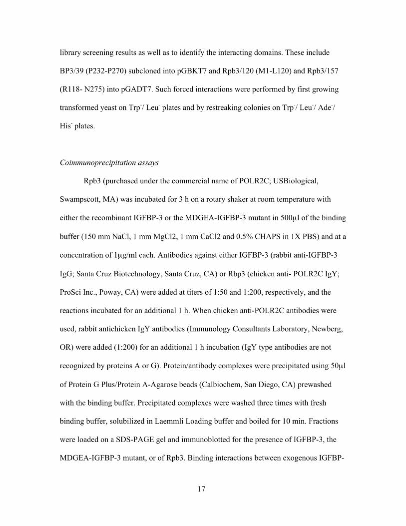
A total of 57 clones (12 duplicates) were identified as putative IGFBP-3-
interacting proteins after screening a custom L6 myoblast cDNA expression library using
a high stringency yeast two-hybrid assay (Fig. 5) and two different IGFBP-3 deletion
mutants as bait: BP3/231 and BP3/111 (Fig. 6). Fifteen clones were isolated with
BP3/231 and 42 with BP3/111, possibly indicating steric hindrance by the N-terminal
region. Thirteen proteins have been previously shown to directly interact with IGFBP-3,
and three of these were also identified in our screens, including fibronectin, type Iα-
25
Figure 5. Identification of IGFBP-3-interacting proteins in L6 myoblasts. A custom
cDNA expression library was screened using a high stringency yeast 2-hybrid assay (BD
Bioscience) and two different IGFBP-3 deletion mutants as “bait”: BP3/231 and BP3/111
(see Figure 5). A total of 57 clones were identified, 15 with BP3/231 and 42 with
BP3/111. These include 12 duplicates as well as 3 previously identified IGFBP-3-
interacting proteins (table). The functional distribution of each protein isolated with the
indicated bait construct is shown in the histogram (cyto, cytosolic; endo, endocrine;
ECM, extracellular matrix; met, metabolic; sig, signaling; trans, transcription). Previously
identified IGFBP-3-interacting proteins are shown in the table, including those also
identified in our assays (bold). *method of detection = yeast 2-hybrid screening (Y2H),
ligand affinity/chromatography (affi); #functional class = extracellular matrix (ECM),
endocrine or related (endo)

26
collagen and IGFBP-3 itself, which independently validates our assay (Fig. 5). Several
diverse functional classes were represented among the clones, although extracellular
matrix proteins were best represented with 16 total clones (11 different proteins and five
duplicates). Most interesting, however, was the identification of a nuclear pore protein as
well as six different proteins involved in transcription including three clones of the rat
homolog for RNA polymerase II (RNAPII) binding subunit 3 (Rpb3, a.k.a. subunit C,
GenBank accession no. NP_001012491) and two clones of a structurally similar subunit
of RNA polymerase I (NP_001008331).
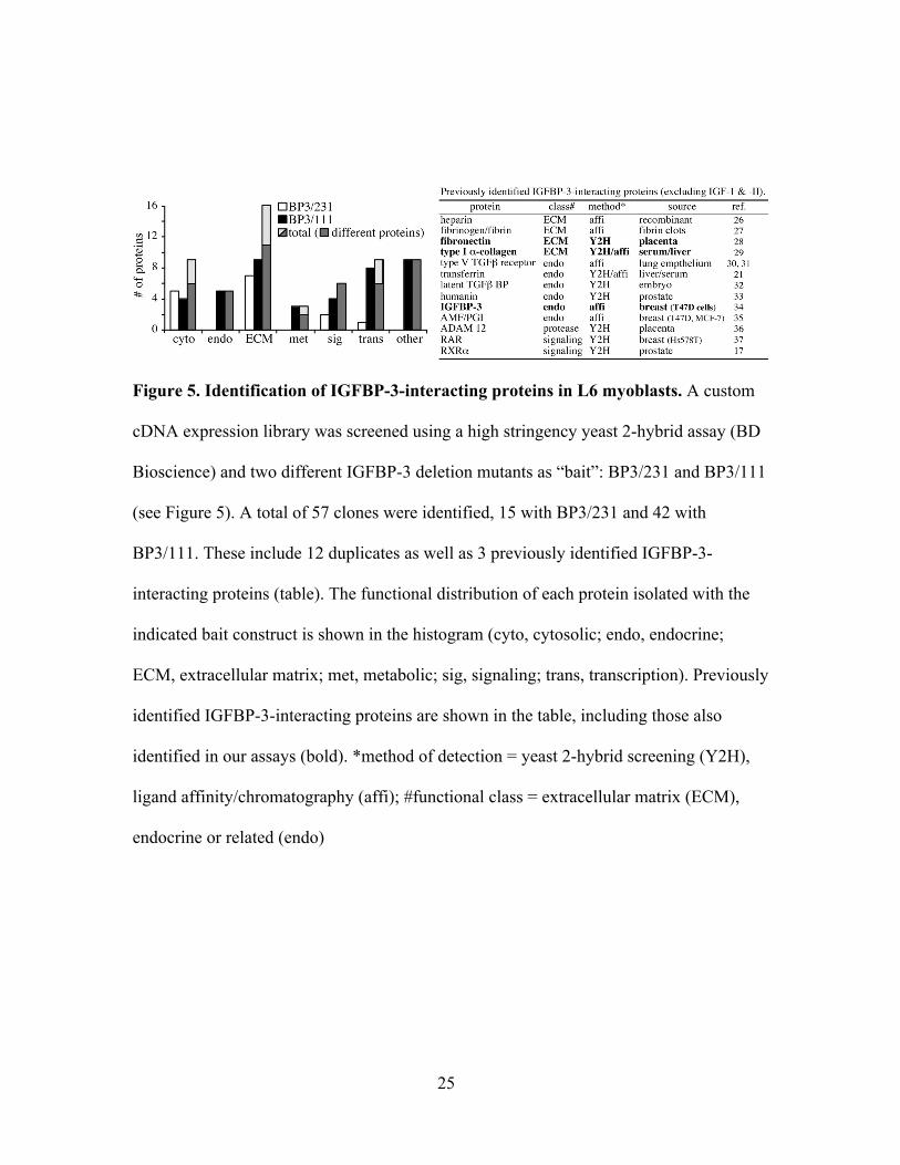
To confirm the binding interactions of BP3/ 231 and BP3/ 111 with Rpb3, yeast
two-hybrid assays were performed using yeast cotransfected with either the full-length
Rpb3 or the negative control activation domain vector, simian virus 40 (SV40), in
combination with BP3/231, BP3/111 or with a negative control DNA-binding domain
vector, Lamin C. With each transfection, the presence of both DNA-binding and
activation domain vectors was verified by conditional growth on Leu-/ Trp- plates (Fig.
6). Cells overexpressing either BP3/231 or BP3/111 and Rpb3 grew after replating on
Leu- /Trp- /His- /Ade- plates, which confirmed Rpb3 binding to these regions of IGFBP-3.
However, neither of the negative controls grew under these conditions. Similar results
were also obtained using RNA polymerase 1–1 (Pol-I; Fig. 6), which is structurally
similar to Rpb3. This suggests that the Rpb3/IGFBP-3 binding interaction is specific
within the limitations of this assay. To help identify the interacting domains, assays were
also performed on yeast cotransfected with a smaller construct that codes for the 39
amino acid epitope containing the MBD/NLS (BP3/39) in combination with constructs
for either the first 120 amino acids of Rpb3 (Rpb3/120) or the last 157 (Rpb3/157).
27
Figure 6. Binding interactions between IGFBP-3 & a RNA polymerase II subunit,
Rpb3. RNA Polymerase II binding subunit 3 (Rpb3) and the homologous protein from
the RNA Polymerase I complex (Pol-I) were identified in the Y2H screen, both with
BP3/111. These interactions were confirmed by co-expressing the indicated proteins in
Y2H assays. The putative Rpb3-binding domain within IGFBP- 3 was also identified
using these assays. (left) Cartoon of IGFBP-3 and Rpb3 deletion mutants. Functional
domains of IGFBP-3 are shaded and annotated and each IGFBP-3 and Rpb3 mutant is
named based on the total number of residues within each mutant (WT, wild-type). (right)
Yeast were co-transfected with plasmids containing the cDNA for the indicated mutants
(IGFBP-3 in pGBKT7 DNA-binding vector; polymerases in pGADT7-Rec[2] activation
domain vector) and spread upon leucine/tryptophan deficient plates (Leu-/Trp-). Only
cells containing both plasmids will grow under these conditions. Individual colonies were
also cultured under restrictive conditions (Leu-/Trp-/His-/Ade- = histidine & adenine
deficient as well) where growth requires the interaction between both proteins and the
consequential expression of reporter genes. (SV40+p53 = positive control; Lamin C and
SV40 = negative controls)
28
Conditional growth only occurred in cells overexpressing BP3/39 and Rpb3/120,
indicating that binding occurs between the N-terminal half of Rpb3 and the region of
IGFBP-3 that contains the MBD/ NLS epitope.
NLS requirement for IGFBP-3/Rpb3 binding and IGFBP-3-stimulated growth inhibition
The direct association of IGFBP-3 with Rpb3 was also confirmed using co-
immunoprecipitation assays and antisera specific for each recombinant protein. When co-
incubated, western blotting revealed the presence of both proteins in the αIGFBP-3 and
αRpb3 immunoprecipitates (Fig. 7) and there was no indication of antibody cross-
reactivity (left & middle panels, last lanes). By contrast, only a minimal amount of Rpb3
was detected within the αIGFBP-3 immunoprecipitates when Rpb3 was incubated with
the MDGEA mutant instead of wild-type IGFBP-3 (right panel, lane 2 vs. 3). These data
suggest that an intact NLS is required for significant IGFBP-3/Rpb3 binding. This
interaction was also confirmed by coimmunoprecipitating nuclear lysates from cells
treated exogenously with wild-type IGFBP-3 for 60 min (Fig. 8). Both IGFBP-3 and
endogenous Rpb3 were detected in the precipitates indicating coassociation of these two
proteins within nuclear lysates. The presence of Rpb3 was not due to non-specific
interactions with the antiserum or with the protein A/G agarose as precipitates from cells
treated without IGFBP-3 or lysates incubated with agarose alone did not contain Rpb3
(Fig. 8, last two lanes in both panels).
Proliferation assays were also conducted on myoblasts cultured for 48 h with LR3
IGF-I and either IGFBP-3 or the MDGEA mutant. As expected, LR3 IGF-I alone
increased cell growth by almost 50% compared to controls (Fig. 9). Both doses of

29
Figure 7. IGFBP-3/Rpb3 co-immunoprecipitation requires an intact NLS domain.
Recombinant IGFBP-3 or the NLS-mutant that does not nuclear locate (MDGEA, see
Firth & Baxter (5)) were incubated with recombinant Rpb3 and immunoprecipitated as
described in the Materials and Methods with either IGFBP-3 or Rpb3 antisera as
indicated. Precipitated complexes were washed, solubilized in SDS-PAGE loading buffer
and immunoblotted with the indicated antisera. Antibody specificity was controlled by
immunoprecipitating IGFBP-3 with αRpb3 or vice versa (left & middle panels, last
lanes).

30
Figure 8. IGFBP-3/Rpb3 co-immunoprecipitation from L6 myoblast nuclear lysates.
Proliferating cells were treated with or without recombinant IGFBP-3 for 60 min, washed
thoroughly and nuclear lysates were isolated as described in the Materials and Methods.
Equal amounts of nuclear protein from each treatment group were immunoprecipitated
with αIGFBP-3 as indicated and samples were blotted as in Fig. 7. Non-specific binding
of nuclear protein to the protein-A/G agarose or to IGFBP-3 was controlled by the
inclusion of samples lacking either IGFBP-3 antibodies or peptide.

31
Figure 9. IGFBP-3 inhibition of myoblast proliferation requires an intact NLS
domain. Proliferating L6 myoblasts were incubated for 48 h in serum-free DMEM with
or without 1.0 nM LR3 IGF-I (does not recognize IGFBPs) in the presence of 1.0 and 0.5
nM wild-type IGFBP-3 (B) or the MDGEA mutant IGFBP-3 (M), which is incapable of
nuclear localization (5). Cell number was determined using the CellTiter 96 colorimetric
assay (Promega) according to the manufacturers’ protocol (mean values + SEM are
shown; n=14/treatment).

32
IGFBP-3, however, attenuated these effects, which confirm previous studies indicating a
growth inhibitory role IGFBP-3 in these cells as well as the data presented in Fig. 2. This
inhibitory effect was lost when cells were treated with the MDGEA mutant IGFBP-3.
Thus, IGF-independent growth inhibition of myoblast proliferation requires IGFBP-3
nuclear localization and possibly Rpb3 binding.
Discussion
Incubating myoblasts with IGF-I has a dual effect on the cell cycle as the growth
factor simultaneously stimulates proliferation and differentiation (Adams 2002). It also
stimulates the synthesis and secretion of IGFBP-3 as cells begin to differentiate, a process
that appears to be significantly influenced by IGFBP-3 (Foulstone et al. 2003, Johnson et
al. 2003, Pampusch et al. 2003). We have shown that an IGFBP-3 analogue with no
appreciable affinity for either IGF-I or -II can inhibit myoblast proliferation under growth
conditions, which is the first step in the initiation of differentiation. Similar roles have
been defined for IGFBP-3 in negatively regulating myoblast growth using primary
human (Foulstone et al. 2003) and porcine (Johnson et al. 2003, Pampusch et al. 2003)
myosatellite cells as well as rat L6 myoblasts (Xi et al. 2004) that incorporate both IGF-
dependent and -independent mechanisms. Thus, IGF stimulated myoblast differentiation
appears to be mediated, at least in part, by the local production of IGFBP-3, which serves
a similar role for TGFβ1 and myostatin (Kamanga-Sollo et al. 2003).
Myoblast nuclear localization of IGFBP-3 was independently demonstrated in
separate experiments using fluorescent and confocal microscopy and by western blotting
of nuclear protein. The translocation of intact IGFBP-3 across both plasma and nuclear

33
membranes was rapid and occurred within 5 min, which is consistent with a functional
nuclear role for IGFBP-3 in these cells and suggests that nuclear immunoreactivity was
not due to non-specific endocytosis and subsequent proteolysis of IGFBP-3. The presence
of IGFBP-3 within the nuclei of many different cell types in addition to rat myoblasts and
myotubes (Jaques et al. 1997, Li et al. 1997, Schedlich et al. 1998, Wraight et al. 1998,
Liu et al. 2000) suggests that IGFBP-3 helps in regulating fundamental cellular
processes. However, IGFBP-3 stimulates apoptosis in many of these cells, but had no
effect on the myoblasts described herein. This suggests that although the mechanisms
required for IGFBP-3 nuclear entry may be relatively common, its function is likely cell
type-specific and dependent upon the unique expression of intracellular and nuclear
proteins. Some IGF-independent effects of IGFBP-3 are dependent upon its nuclear
localization, which may include myoblast growth inhibition. Indeed, the MDGEA mutant
IGFBP-3, which does not nuclear locate, is incapable of inhibiting LR3 IGF-stimulated
proliferation (Fig. 9) and does not associate with Rpb3 in coimmunoprecipitation assays
(Fig. 7). We have additionally demonstrated exogenous IGFBP-3 binding to endogenous
Rpb3 in nuclear lysates (Fig. 8). These data suggest that nuclear localization and possibly
Rpb3 binding, two related yet possibly exclusive events, are both necessary prerequisites
for the IGF-independent inhibition of myoblast growth. The NLS domain is necessary for
a number or binding interactions including the association of IGFBP-3 with importin-β
(Firth & Baxter 2002, Schelich et al. 2000). However, the NLS is also a highly and
negatively charged domain that when mutated, may alter the three dimensional structure
of the C-terminal region, which would explain its dependence for so many binding
interactions.

34
The first intracellular protein demonstrated to directly bind IGFBP-3 was RXRα
(Liu et al. 2000), although Schedlich et al. 2004 recently demonstrated retinoic acid
receptor (RAR) binding as well. Binding to RXRα enhanced transcriptional activity and
apoptosis in the former study, whereas RAR binding specifically inhibited ligand
activation and the formation of RXR:RAR heterodimers in the latter. Thus, a minimum
role for IGFBP-3 in the nucleus appears to include the modulation of transactivation for
some RXR- and RAR-responsive genes, which in turn would have different effects
depending upon the cell type and the presence of different retinoid receptors. Gene
expression requires recruitment of the RNAPII complex whose core enzyme is composed
of 12 different subunits. Rpb3 is the third largest subunit and is located externally away
from the core DNA-binding domain (Cramer 2004). Assembly of the RNAPII complex is
dependent upon Rpb3, although its functional role, as is currently known, is to facilitate
recruitment of RNAPII to specific transcription factors, which in turn initiates gene
transcription (Tan et al. 2000, Corbi et al. 2002, De Angelis et al. 2003). Thus, nuclear
IGFBP-3 may not need to bind DNA directly to influence transcription as it could assist
in RNAPII recruitment to transcription factor complexes, including those involving RXR
and RAR. Rpb3 is ubiquitously expressed in all tissues, however, its levels are
considerably higher in cardiac and smooth muscle than in any other tissue (Fanciulli et al.
1996, 1998), which is suggestive of an alternative and muscle-specific role. Indeed, Rpb3
binds myogenin and activating transcription factor (ATF) 4 and directly facilitates their
transactivational activity while simultaneously stimulating myoblast differentiation
(Corbi et al. 2002, De Angelis et al. 2003). Corbi et al. 2005 recently demonstrated Rpb3
shuttling between cytoplasmic and nuclear compartments. Rpb3 association with HCR

35
(α-helix coiled-coil rod homologue) in the cytoplasm prevents nuclear entry and
myoblast differentiation. Thus, a functional role for IGFBP-3 in regulating myoblast
differentiation could include the “delivery” of Rpb3 to nuclear targets.
Lee et al. recently determined that cellular internalization of secreted IGFBP-3 is
mediated by its binding to transferrin and to caveolin. Although internalization, nuclear
entry and the apoptotic effects of IGFBP-3 were all completely blocked by inhibitors of
caveolae formation, a minimal amount of a non-transferrin-binding mutant IGBFP-3
(K228E/R230G) still localized to the nucleus. In a similar study, Singh et al.
characterized several peptides based on an IGFBP-3 epitope within its C-terminal domain
and defined critical motifs within these GFP-chimeric peptides necessary for cellular
internalization. Chimeras containing the transferrin- and partial caveolin-binding motifs
all localized to the nucleus whereas those that lacked both motifs (GFP34) or the partial
caveolin-binding motif alone (GFP36) remained extracellular. Co-immunoprecipitation,
cross-linking and/or ligand blotting assays were used to demonstrate direct binding
between IGFBP-3 or the MBD/NLS peptide with caveolin and either transferrin or its
receptor (Lee et al. 2004, Singh et al. 2004). Together, these studies suggest that IGFBP-
3 internalization is mediated by transferrin and caveolin, both of which are expressed in
skeletal muscle cells including L6 myoblasts (Shimo-Oka et al. 1986, Scherer et al.
1997), and that caveolin-binding in particular is critical.
Previous studies with transformed skeletal muscle cell lines suggested that
IGFBP-5 was the principle IGFBP secreted by muscle cells (Florini et al. 1996). By
contrast, recent studies with primary skeletal muscle stem cells from human and porcine
sources suggest that IGFBP-3 is specifically involved in the growth regulation of PEMC,

36
human skeletal muscle cells, and L6 myogenic cells. We hypothesize that IGFBP-3
partially mediates IGF-stimulated myoblast differentiation by inhibiting cell growth and
that nuclear localization of IGFBP-3 is critical to this process (Fig. 9). Future studies will
further explore the ability of IGFBP-3 to initiate differentiation and to regulate gene
expression via its association with Rpb3. They will also determine if other proteins
identified in our screening assay contribute to or enhance these effects. Nevertheless,
these and other recent studies strongly suggest that skeletal muscle development is
influenced by the IGF-independent actions of IGFBP-3 and that nuclear localization is
required.

37
CHAPTER THREE
COMMERCIAL AND APPLIED ASPECTS OF IGFBP-3 THERAPEUTICS
Summary
Previous studies, including those discussed herein, indicate that IGFBP-3 serves as a
multifunctional growth regulator in different cell types. Using non-IGF binding analogs
of IGFBP-3, we confirmed that IGFBP-3 directly inhibits myoblast proliferation in an
IGF-independent manner and that the NLS sequence is important for this function. We
observed the rapid translocation of IGFBP-3 into L6 myoblast nuclei using various
techniques. A GFP-NLS chimera also possesses the same rapid nuclear translocation
ability and suggests that the NLS peptide could help deliver hydrophobic therapeutics to
muscle cell nuclei. We additionally screened a custom cDNA library derived from L6
myoblasts using a Yeast 2-Hybrid assay and IGFBP-3 as bait. Herein, we identified
several interacting proteins, most notably Rpb3, which was confirmed with
coimmunoprecipitation assays. We also determined that the NLS sequence is important
for IGF-independent myoblast growth inhibition. Several additional IGFBP-3 analogues
have been generated by site-directed mutagenesis and will help in future studies seeking
to better define the IGFBP-3/ Rpb3 binding interaction. We therefore present a model
(Fig. 10) where the local production of IGFBP-3 inhibits myoblast proliferation in an
IGF-independent manner. This requires an intact NLS and its rapid translocation of
IGFBP-3 into the nucleus where it interacts with RNAPII via Rpb3. We hypothesize that

38
Figure 10. Model of IGFBP-3 regulated myoblast growth inhibition and subsequent
differentiation. (1) IGF-I binds and activates its receptor, which (2) signals to the
nucleus and stimulates IGFBP-3 gene expression and subsequently protein secretion (3).
IGFBP-3 then binds IGF-I (4) thereby removing the mitogenic signal. It also translocates
to the nucleus (5) where it binds Rpb3 (6) and influences the expression of myogenin-
regulated genes (7). This in turn suppresses proliferation and initiates differentiation.

39
this interaction enhances the transactivational activity of two myoblast differentiation
factors- myogenin and ATF-4- and in turn inhibits proliferations.
Medical and Veterinary implications
Several valuable studies of IGFBP-3’s potential actions in the clinic have been
recently performed. These include the use of IGFBP-3 alone or in combination with IGF-
I (trade name IPLEX, formerly SomatoKine) to treat patients with primary IGF-I
deficiency (IGFD), GH insensitivity syndrome (GHIS, Laron syndrome), severe burning
injuries, catabolic stress or even cancer. IGFD and GHIS are often caused by mutated GH
receptors and lead to growth retardation, increased circulating GH levels, reduced
circulating IGF-I, -II and IGFBP-3’s levels, and hyperglycemia (Woods et al. 1997).
Many of these symptoms can be ameliorated by IGF-I treatment, which normalizes the
growth and metabolic functions (LeRoith et al. 2001). As mentioned earlier, IGFBP-3
circulates with IGF-I as a 150 kDa complex that include the ALS protein (Firth & Baxter
2002). The purpose of the complex is to prolong the half-life of IGF-I in serum from
minutes to hours. IPLEX is more effective than IGF-I alone in increasing and stabilizing
serum IGF-I levels in IGFD and GHIS patients and is especially useful for treating
children with severe short stature as it stimulates muscle protein synthesis and connects
the metabolic conditions without causing adverse side-effect such as IGF-I-induced
hypoglycemia (Kemp & Thrailkill 2005). The ability of IPLEX to prevent muscle
wasting during fasting conditions has also been studied in semi-starved rat. The result
shows that plasma IGF-I level is 20% higher in rats treated with complex than IGF-I
alone and almost recover to the same amino acid concentrations in the plasma as freely-

40
fed control rats which were significantly reduced in saline and IGF-I alone treatments
(Svanberg et al. 2000). Furthermore, only IPLEX stimulated amino acid uptake, as
assessed by radio-labeled phenylalanine incorporation, in muscle during long term
starvation experiments (Svanberg et al. 2000), indicating that IGF-I alone is once again
insufficient.
Another potential therapeutic use of IPLEX is for treating severely burned patients.
Burn injuries induce cachexia, hypermetabolism, inflammation and immune system
dysfunction (Herndon et al. 1999). Treating burn patients with GH and IGF-I can
effectively enhance recovery of muscle wasting and increase resistance to infections
(Edmondson et al. 2003). However, GH and IGF-I treatments normally lead to
hyperglycemia (Singh et al. 1998) and hypoglycemia (Clark 2004), respectively.
Administering IPLEX to severe burned adults (Debroy et al. 1999) and children
(Herndon et al. 1999) successfully improved the net protein synthesis and maintained
high serum IGF-I concentrations. In the adult patients, who had 24% total body surface
area (TBSA) burns, serum IGF-I levels were raised from 118±11 to 489±90 ng/ml with
1.0 mg/kg/day IPLEX for five days (Debroy et al. 1999). In the children, who had >40 %
TBSA burns and had received three times surgical procedures, serum IGF-I levels were
raised from 100±10 to 275±52 µg/ml using an IPLEX dose of 0.5mg/kg/day over five
to seven days (Spies et al. 2002). None of these participants developed hypoglycemia or
electrolyte imbalance which can be caused by IGF-I therapy. IPLEX also attenuates the
induction of acute phase proteins and proinflammatory cytokines after tissue injuries
associated with infections, inflammations or burn injury (Moshage 1997).
Proinflammatory cytokines aid in tissue recovery and help maintain systematic

41
homeostasis after trauma, however, overproduction can be detrimental and produce tissue
damage itself, hypermetabolism, multiple organ failure or even death (Moshage 1997,
Pruitt et al. 1995). Clinical studies indicate that IPLEX decreases the type I acute phase
proteins and proinflammatory cytokines such as tumor necrosis factor (TNF) -α and
interleukin-1β, both of which inhibit the GH/IGH-I growth axis and are associated with
increased mortality in severely burned children (Jeschke et al. 2000, Pruitt et al. 1995,
Spies et al. 2002). These studies strongly indicate that IPLEX is an effective therapeutic
for treating burn injuries as it stimulates muscle growth and prevents trauma-induced
muscle wasting, stimulates tissue recovery and blocks immune responses that interfere
with these processes.
Diabetes is the most common metabolic disease and affects over 14 million people
in the United States alone (National Center for Chronic Disease Prevention and Health
Promotion, www.cdc.gov). Type I diabetes, or insulin-dependent diabetes mellitus
(IDDM), is caused by the autoimmune attack of the insulin-secreting β-cells in the
pancreas and usually presents during childhood or early adulthood. This type of diabetes
accounts for only 10% of diabetic, who require insulin supplements to regulate glucose
metabolism. On the other hand, patients with type II diabetes or non-insulin-dependent
diabetes mellitus (NIDDM), have normal or elevated circulating insulin levels. This
disease usually presents in middle-age and is associated with obesity, although it is also
the primary endocrine disorder of children due to the prevalence of obesity in our youth
(Berry et al. 2006). Livers of type II diabetics are unable to respond to insulin, which
produces hyperglycemia and in turn, hyperinsulinaemia. The exact causes of type II
diabetes are unknown but are believed to be due to an increased susceptibility to

42
environmental influences (Hattersley & Pearson 2006). IGF-I possesses about 6% of
insulin’s ability to produce hypoglycemia (Guler et al. 1987) and can ameliorate diabetic
symptoms at high concentrations (Zenobi et al. 1992). It has also been suggested as a
potential therapy for some diabetics as it can inhibit β cell apoptosis as well (Thrailkill
2000). IGF-I is rapidly cleared, however, suggesting that IPLEX could enhance the
therapeutic potential of IGF-I in treating diabetes. For patients with type I diabetes,
administrating IPLEX at a dose of 2mg/kg/day for two weeks reduced the insulin dose
requirement by 49% (Clemmons et al. 2000). When given to type II diabetics, IPLEX
reduced the insulin dose requirement by 54% (Clemmons et al. 2005). None of the side
effects associated with IGF-I therapy occur in both type I and type II diabetics, which
include edema, weight gain, tachycardia, dyspnea, myalgia and fatigue (Jabri et al. 1994,
Clemmons et al. 2000) developed. These studies suggest that IPLEX can significantly
regulate carbohydrate metabolism and increase the efficiency of insulin supplementation
without producing adverse side effects. These studies are also the basis for Phase II
clinical trials that will assess the long-term safety of treating diabetics with IPLEX
(www.insmed.com).
Myotonic muscular dystrophy (MD), the most prevalent form of muscular
dystrophy, is a dominantly inherited disease caused by the unstable repeated expansion of
CTG nucleotides (Type I MD) in the 3’ UTR region of the myotonic dystrophy protein
kinase (DMPK) gene or CCTG (Type II MD) nucleotides in the intron of zinc finger
protein 9 (ZNF9) gene. This disease mainly affects the function of smooth and skeletal
muscle cells and commonly leads to myotonia (Machuca-Tzili et al. 2005). Although
there is no direct evidence that IPLEX can decrease MD symptoms, it reduces muscle

43
protein degradation and preserves the number of muscle fibers during atrophy
(Zdanowicz & Teichberg 2003). Administration of IGF-I (1mg/kg) for eight weeks to
mdx mice- a murine model for Duchenne muscular dystrophy- increases the force per
cross-sectional area of diaphragm by 49% and enhances fatigue resistance as well
(Gregorevic et al. 2002). The growth promoting effects of IPLEX are greater and softer
than IGF-I therapy and thus, IPLEX is a likely candidate therapeutic for treating some
muscular dystrophies as well.
IGFBP-3 is a multifunctional molecule with potent anti-tumor properties. It
regulates the tumor-promoting actions of IGF-I directly (Grimberg & Cohen 2000), but
also inhibits tumor growth via IGF-independent means in several cancer cell types (Firth
& Baxter 2002, Silha et al. 2006). Insmed is also conducting clinical trials (Phase I) on
the growth inhibitory effects of IGFBP-3 in breast and colorectal cancer cells, a study
that is based on several previous studies performed independently of Insmed (Butt et al.
2002, Gucev et al. 1996, LeRoith & Roberts 2003). Blouin et al. 2003 demonstrated that
IGFBP-3 reduced the proliferation rate of MCF-7 breast cancer cells by 55% and HT-29
colorectal cancer cells by 49%. The co-administration of IGFBP-3 with 2 Gy radiation
therapy has an additive effect on inhibiting the growth of MCF-7 and HT-29 carcinoma
cells by 3.5 and 1.6 fold. The same study also investigated the in vivo effect of IGFBP-3.
These receiving MCF-7 xenografts were injected twice daily with 20mg/kg IGFBP-3,
only 3of 12 mice formed tumors with a mean volume 37.7mm3 in the IGFBP-3 treated
group while 8 of 12 control mice formed tumors with a mean volume 113.9mm3 in
control group. Thus, IGFBP-3 reduced both the incidence and size of MCF-7 tumors.
IGFBP-3’s anti-tumor functions are also enhanced when given in combination with
44
chemotherapeutic drugs (Yu et al. 2003). When using the same MCF-7 breast cancer
tumor xenografts model, co-treatment of 10 mg/kg IGFBP-3 and 13 or 20 mg/kg
Paclitaxel was 33% and 61%, respectively, more effective than Paclitaxel treatment
alone. Similar results were also obtained with LoVo colorectal carcinoma xenografts, as
treating with 30 mg/kg IGFBP-3 and 10 mg/kg CPT-11 was 30% to 69% more effective
than CPT-11 alone. These studies suggest IGFBP-3 can be used as a not only single anti-
tumor treatment, but that it also significantly enhances the efficiency of other
chemotherapeutic therapies. However, the co-treatment of IGFBP-3 with either radiation
or chemotherapeutic drugs may inhibit proliferation effects differently depending on the
agents and on whether the cells are transformed or not (Perks et al. 2003). Most
importantly, the apoptotic functions of IGFBP-3 in cancer cells is largely IGF-
independent (Lee & Cohen 2002) and often requires nuclear translocation (Firth et
al. 2002). These growth inhibitory functions need to be further characterized, however, as
our observation that IGFBP-3 binds Rpb3 in the nucleus which is expressed ubiquitously
in all cell types- suggests that IGFBP-3 may influence gene expression by directly
modulating RNAPII activity in a tissue specific manner that depends on other interacting
proteins. Nevertheless, its apoptotic actions in different cancer cells and its role in
regulating IGF-I levels in circulation indicate that the potential clinical benefit of IGFBP-
3 is not limited to fighting tumors, but is multifunctional.
Animal production implications
The IGF-I system has been implicated in many aspects of bovine mammary gland
development and function (Akers 2006). As mentioned earlier, IGFBP-3 mediates the

45
inhibitory functions of TGF-β and retinoid acid on transformed mammary epithelial cells,
which represses IGF-I stimulated proliferation (Gucev et al. 1996). However, IGFBP-3
possesses inverse functions in normal breast epithelial cells that are concentration
dependent. It decreases cell number by 27.8% at lower concentrations (25 ng/ml) but
increases cell number by 37 % at higher concentrations (100ng/ml) (Perks et al. 2002).
The production of IGFBP-3 is primarily regulated by the GH/IGF-I axis. Therefore, these
findings suggest that IGFBP-3 is an important regulator of breast epithelial cell
proliferation and that it may help mediate GH-stimulated lactation efficiency.
The dual natural of IGFBP-3’s actions also extend to the regulation of skeletal
muscle growth and development. Although it supports growth by maintaining a
circulating reservoir of IGF-I, it also mediates the suppressive effects of transforming
growth factor (TGF)-β and myostatin on skeletal muscle cell lines and on porcine
embryonic myogenic cells (Kamaga-Sollo et al. 2003). Myostatin is an extremely potent
inhibitor of skeletal muscle growth as it directly inhibits myoblast proliferation.
Mutations in the myostatin gene can create double-muscling in domestic livestock that
similar to the myostatin null mouse (McPherron & Lee 1997). Thus, manipulating the
production and/ or bioactivity of IGFBP-3 could potentially perturb myostatin’s anti-
proliferative actions in muscle cells and augment the muscle mass of domesticated
species.

46
CHAPTER FOUR
REFERENCES
1. Adams GR 2002 Exercise Effects on Muscle Insulin Signaling and Action:Invited Review: Autocrine/paracrine IGF-I and skeletal muscle adaptation. J ApplPhysiol 93:1159-1167
2. Akers RM 2006 Major Advances Associated with Hormone and Growth FactorRegulation of Mammary Growth and Lactation in Dairy Cows. J. Dairy Sci.89:1222-1234
3. Angelloz-Nicoud P& Binoux M 1995 Autocrine regulation of cell proliferation bythe insulin-like growth factor (IGF) and IGF binding protein-3 protease system ina human prostate carcinoma cell line (PC-3)136.12.5485. Endocrinology136:5485-5492
4. Bach LA, Thotakura NR& Rechler MM 1992 Human Insulin-Like Growth-FactorBinding Protein-6 Is O-Glycosylated. Biochemical and Biophysical ResearchCommunications 186:301-307
5. Backer JM, Myers MG, Jr., Shoelson SE, Chin DJ, Sun XJ, Miralpeix M, Hu P,Margolis B, Skolnik EY, Schlessinger J& et al. 1992 Phosphatidylinositol 3'-kinase is activated by association with IRS-1 during insulin stimulation. Embo J11:3469-3479
6. Berry D, Urban A& Grey M 2006 Understanding the Development andPrevention of Type 2 Diabetes in Youth (Part 1). Journal of Pediatric Health Care20:3-10
7. Boyle BJ, Zhao XY, Cohen P& Feldman D 2001 Insulin-like growth factorbinding protein-3 mediates 1 alpha,25-dihydroxyvitamin d(3) growth inhibition inthe LNCaP prostate cancer cell line through p21/WAF1. J Urol 165:1319-1324
8. Buckway CK, Wilson EM, Ahlsen M, Bang P, Oh Y& Rosenfeld RG 2001Mutation of three critical amino acids of the N-terminal domain of IGF-bindingprotein-3 essential for high affinity IGF binding. J Clin Endocrinol Metab86:4943-4950
9. Butt AJ, Fraley KA, Firth SM& Baxter RC 2002 IGF-Binding Protein-3-InducedGrowth Inhibition and Apoptosis Do Not Require Cell Surface Binding andNuclear Translocation in Human Breast Cancer Cells. Endocrinology 143:2693-2699
10. Carr J, Grant P, Francis G, Owens J, Wallace J& Walton P 1994 Isolation andcharacterization of ovine IGFBP-4: protein purification and cDNA sequence. JMol Endocrinol 13:219-236
11. Cheung CW, Vesey DA, Nicol DL& Johnson DW 2004 The roles of IGF-I andIGFBP-3 in the regulation of proximal tubule, and renal cell carcinoma cellproliferation. Kidney Int 65:1272-1279
12. Clark RG 2004 Recombinant human insulin-like growth factor I (IGF-I): risks andbenefits of normalizing blood IGF-I concentrations. Horm Res 62 Suppl 1:93-100
13. Clemmons DR, Moses AC, McKay MJ, Sommer A, Rosen DM& Ruckle J 2000The Combination of Insulin-Like Growth Factor I and Insulin-Like GrowthFactor-Binding Protein-3 Reduces Insulin Requirements in Insulin-Dependent

47
Type 1 Diabetes: Evidence for in VivoBiological Activity. J Clin EndocrinolMetab 85:1518-1524
14. Clemmons DR 2001 Use of Mutagenesis to Probe IGF-Binding ProteinStructure/Function Relationships. Endocr Rev 22:800-817
15. Clemmons DR, Moses AC, Sommer A, Jacobson W, Rogol AD, Sleevi MR&Allan G 2005 Rh/IGF-I/rhIGFBP-3 administration to patients with type 2 diabetesmellitus reduces insulin requirements while also lowering fasting glucose. GrowthHormone & IGF Research 15:265-274
16. Cohen P, Lamson G, Okajima T& Rosenfeld RG 1993 Transfection of the humaninsulin-like growth factor binding protein-3 gene into Balb/c fibroblasts inhibitscellular growth. Mol Endocrinol 7:380-386
17. Cohen P, Peehl DM, Graves HC& Rosenfeld RG 1994 Biological effects ofprostate specific antigen as an insulin-like growth factor binding protein-3protease. J Endocrinol 142:407-415
18. Cohen P, Nunn SE& Peehl DM 2000 Transforming growth factor-beta inducesgrowth inhibition and IGF-binding protein-3 production in prostatic stromal cells:abnormalities in cells cultured from benign prostatic hyperplasia tissues. JEndocrinol 164:215-223
19. Conover CA, Clarkson JT& Bale LK 1996 Factors regulating insulin-like growthfactor-binding protein-3 binding, processing, and potentiation of insulin-likegrowth factor action. Endocrinology 137:2286-2292
20. Corbi N, Di Padova M, De Angelis R, Bruno T, Libri V, Iezzi S, Floridi A,Fanciulli M& Passananti C 2002 The a-like RNA polymerase II core subunit 3(RPB3) is involved in tissue-specific transcription and muscle differentiation viainteraction with the myogenic factor myogenin. FASEB J.:02-0123fje
21. Cramer P 2004 RNA polymerase II structure: from core to functional complexes.Current Opinion in Genetics & Development 14:218-226
22. De Angelis R, Iezzi S, Bruno T, Corbi N, Di Padova M, Floridi A, Fanciulli M&Passananti C 2003 Functional interaction of the subunit 3 of RNA polymerase II(RPB3) with transcription factor-4 (ATF4). FEBS Letters 547:15-19
23. De Mellow JSM& Baxter RC 1988 Growth hormone-dependent insulin-likegrowth factor (IGF) binding protein both inhibits and potentiates IGF-I-stimulatedDNA synthesis in human skin fibroblasts. Biochemical and Biophysical ResearchCommunications 156:199-204
24. Debroy MA, Wolf SE, Zhang XJ, Chinkes DL, Ferrando AA, Wolfe RR&Herndon DN 1999 Anabolic effects of insulin-like growth factor in combinationwith insulin-like growth factor binding protein-3 in severely burned adults. JTrauma 47:904-910; discussion 910-901
25. Duan C& Xu Q 2005 Roles of insulin-like growth factor (IGF) binding proteins inregulating IGF actions. General and Comparative Endocrinology 142:44-52
26. Edmondson SR, Thumiger SP, Werther GA& Wraight CJ 2003 Epidermalhomeostasis: the role of the growth hormone and insulin-like growth factorsystems. Endocr Rev 24:737-764
27. Fanayan S, Firth SM, Butt AJ& Baxter RC 2000 Growth inhibition by insulin-likegrowth factor-binding protein-3 in T47D breast cancer cells requires transforming

48
growth factor-beta (TGF-beta ) and the type II TGF-beta receptor. J Biol Chem275:39146-39151
28. Fanciulli M, Bruno T, Cerboni C, Bonetto F, Iacobini C, Frati L, Piccoli M,Floridi A, Santoni A& Punturieri A 1996 Cloning of a novel human RNApolymerase II subunit downregulated by doxorubicin: new potential mechanismsof drug related toxicity. FEBS Letters 384:48-52
29. Fanciulli M, Bruno T, Di Padova M, De Angelis R, Lovari S, Floridi A&Passananti C 1998 The interacting RNA polymerase II subunits, hRPB11 andhRPB3, are coordinately expressed in adult human tissues and down-regulated bydoxorubicin. FEBS Letters 427:236-240
30. Firth S& Baxter R 1999 Characterisation of recombinant glycosylation variants ofinsulin-like growth factor binding protein-3. J Endocrinol 160:379-387
31. Firth SM& Baxter RC 2002 Cellular Actions of the Insulin-Like Growth FactorBinding Proteins. Endocr Rev 23:824-854
32. Florini J, Ewton D& Coolican S 1996 Growth hormone and the insulin-likegrowth factor system in myogenesis. Endocr Rev 17:481-517
33. Foulstone EJ, Savage PB, Crown AL, Holly JM& Stewart CE 2003 Role ofinsulin-like growth factor binding protein-3 (IGFBP-3) in the differentiation ofprimary human adult skeletal myoblasts. J Cell Physiol 195:70-79
34. Gregorevic P, Plant DR, Leeding KS, Bach LA& Lynch GS 2002 Improvedcontractile function of the mdx dystrophic mouse diaphragm muscle after insulin-like growth factor-I administration. Am J Pathol 161:2263-2272
35. Grimberg A& Cohen P 2000 Role of insulin-like growth factors and their bindingproteins in growth control and carcinogenesis. J Cell Physiol 183:1-9
36. Gucev ZS, Oh Y, Kelley KM& Rosenfeld RG 1996 Insulin-like growth factorbinding protein 3 mediates retinoic acid- and transforming growth factor beta2-induced growth inhibition in human breast cancer cells. Cancer Res 56:1545-1550
37. Guler H, Zapf J& Froesch E 1987 Short-term metabolic effects of recombinanthuman insulin-like growth factor I in healthy adults. N Engl J Med 317:137-140
38. Hampel B, Wagner M, Teis D, Zwerschke W, Huber LA& Jansen-Durr P 2005Apoptosis resistance of senescent human fibroblasts is correlated with the absenceof nuclear IGFBP-3. Aging Cell 4:325-330
39. Hattersley AT& Pearson ER 2006 Minireview: Pharmacogenetics and Beyond:The Interaction of Therapeutic Response, {beta}-Cell Physiology, and Genetics inDiabetes. Endocrinology 147:2657-2663
40. Herndon DN, Ramzy PI, DebRoy MA, Zheng M, Ferrando AA, Chinkes DL,Barret JP, Wolfe RR& Wolf SE 1999 Muscle protein catabolism after severeburn: effects of IGF-1/IGFBP-3 treatment. Ann Surg 229:713-720; discussion720-712
41. Hoeck WG& Mukku VR 1994 Identification of the Major Sites ofPhosphorylation in Igf Binding Protein-3. Journal of Cellular Biochemistry56:262-273
42. Hollowood AD, Stewart CEH, Perks CM, Pell JM, Lai T, Alderson D& HollyJMP 2002 Evidence implicating a mid-region sequence of IGFBP-3 in its specificIGF-independent actions. Journal of Cellular Biochemistry 86:583-589

49
43. Hong J, Zhang G, Dong F& Rechler MM 2002 Insulin-like growth factor (IGF)-binding protein-3 mutants that do not bind IGF-I or IGF-II stimulate apoptosis inhuman prostate cancer cells. J Biol Chem 277:10489-10497
44. Jabri N, Schalch DS, Schwartz SL, Fischer JS, Kipnes MS, Radnik BJ, TurmanNJ, Marcsisin VS& Guler HP 1994 Adverse effects of recombinant humaninsulin-like growth factor I in obese insulin-resistant type II diabetic patients.Diabetes 43:369-374
45. Jaques G, Noll K, Wegmann B, Witten S, Kogan E, Radulescu RT& Havemann K1997 Nuclear Localization of Insulin-Like Growth Factor Binding Protein 3 in aLung Cancer Cell Line. Endocrinology 138:1767-
46. Jeschke MG, Barrow RE& Herndon DN 2000 Insulinlike growth factor I plusinsulinlike growth factor binding protein 3 attenuates the proinflammatory acutephase response in severely burned children. Ann Surg 231:246-252
47. Johnson BJ, White ME, Hathaway MR& Dayton WR 1999 Decreased steady-state insulin-like growth factor binding protein-3 (IGFBP-3) mRNA level isassociated with differentiation of cultured porcine myogenic cells. J Cell Physiol179:237-243
48. Johnson BJ, White ME, Hathaway MR& Dayton WR 2003 Effect ofdifferentiation on levels of insulin-like growth factor binding protein mRNAs incultured porcine embryonic myogenic cells. Domestic Animal Endocrinology24:81-93
49. Jones JI, D'Ercole AJ, Camacho-Hubner C& Clemmons DR 1991Phosphorylation of insulin-like growth factor (IGF)-binding protein 1 in cellculture and in vivo: effects on affinity for IGF-I. Proceedings Of The NationalAcademy Of Sciences Of The United States Of America 88:7481-7485
50. Kamanga-Sollo E, Pampusch MS, White ME& Dayton WR 2003 Role of insulin-like growth factor binding protein (IGFBP)-3 in TGF-beta- and GDF-8(myostatin)-induced suppression of proliferation in porcine embryonic myogeniccell cultures. J Cell Physiol 197:225-231
51. Kamanga-Sollo E, Pampusch MS, White ME, Hathaway MR& Dayton WR 2005Insulin-like growth factor binding protein (IGFBP)-3 and IGFBP-5 mediate TGF-beta- and myostatin-induced suppression of proliferation in porcine embryonicmyogenic cell cultures. Exp Cell Res 311:167-176
52. Kemp SF& Thrailkill KM 2005 SomatoKine: is there a use in treating growthdisorders? Curr Opin Investig Drugs 6:373-377
53. Kim HS, Ingermann AR, Tsubaki J, Twigg SM, Walker GE& Oh Y 2004 Insulin-like growth factor-binding protein 3 induces caspase-dependent apoptosis througha death receptor-mediated pathway in MCF-7 human breast cancer cells. CancerRes 64:2229-2237
54. Lalou C, Lassarre C& Binoux M 1995 A proteolytic fragment of insulin-likegrowth factor (IGF) binding protein-3 that fails to bind IGF is a cell growthinhibitor. Prog Growth Factor Res 6:311-316
55. Lalou C, Lassarre C& Binoux M 1996 A proteolytic fragment of insulin-likegrowth factor (IGF) binding protein-3 that fails to bind IGFs inhibits themitogenic effects of IGF-I and insulin. Endocrinology 137:3206-3212
50
56. Lawlor MA& Rotwein P 2000 Insulin-Like Growth Factor-Mediated Muscle CellSurvival: Central Roles for Akt and Cyclin-Dependent Kinase Inhibitor p21. Mol.Cell. Biol. 20:8983-8995
57. Le Roith D, Scavo L& Butler A 2001 What is the role of circulating IGF-I?Trends Endocrinol Metab 12:48-52
58. Lee KW& Cohen P 2002 Nuclear effects: unexpected intracellular actions ofinsulin-like growth factor binding protein-3. J Endocrinol 175:33-40
59. Lee KW, Liu B, Ma L, Li H, Bang P, Koeffler HP& Cohen P 2004 Cellularinternalization of insulin-like growth factor binding protein-3: distinct endocyticpathways facilitate re-uptake and nuclear localization. J Biol Chem 279:469-476
60. Lee KW, Ma L, Yan X, Liu B, Zhang XK& Cohen P 2005 Rapid apoptosisinduction by IGFBP-3 involves an insulin-like growth factor-independentnucleomitochondrial translocation of RXRalpha/Nur77. J Biol Chem 280:16942-16948
61. LeRoith D, Werner H, Beitner-Johnson D& Roberts C, Jr 1995 Molecular andcellular aspects of the insulin-like growth factor I receptor. Endocr Rev 16:143-163
62. LeRoith D& Roberts CT, Jr. 2003 The insulin-like growth factor system andcancer. Cancer Lett 195:127-137
63. Li W, Fawcett J, Widmer HR, Fielder PJ, Rabkin R& Keller GA 1997 Nucleartransport of insulin-like growth factor-I and insulin-like growth factor bindingprotein-3 in opossum kidney cells. Endocrinology 138:1763-1766
64. Liu JP, Baker J, Perkins AS, Robertson EJ& Efstratiadis A 1993 Mice carryingnull mutations of the genes encoding insulin-like growth factor I (Igf-1) and type1 IGF receptor (Igf1r). Cell 75:59-72
65. Liu B, Lee HY, Weinzimer SA, Powell DR, Clifford JL, Kurie JM& Cohen P2000 Direct functional interactions between insulin-like growth factor-bindingprotein-3 and retinoid X receptor-alpha regulate transcriptional signaling andapoptosis. J Biol Chem 275:33607-33613
66. Longobardi L, Torello M, Buckway C, O'Rear L, Horton WA, Hwa V, RobertsCT, Jr., Chiarelli F, Rosenfeld RG& Spagnoli A 2003 A novel insulin-like growthfactor (IGF)-independent role for IGF binding protein-3 in mesenchymalchondroprogenitor cell apoptosis. Endocrinology 144:1695-1702
67. Ma J, Pollak MN, Giovannucci E, Chan JM, Tao Y, Hennekens CH& StampferMJ 1999 Prospective Study of Colorectal Cancer Risk in Men and Plasma Levelsof Insulin-Like Growth Factor (IGF)-I and IGF-Binding Protein-3. J Natl CancerInst 91:620-625
68. Machuca-Tzili L, Brook D& Hilton-Jones D 2005 Clinical and molecular aspectsof the myotonic dystrophies: a review. Muscle Nerve 32:1-18
69. Marinaro JA, Neumann GM, Russo VC, Leeding KS& Bach LA 2000 O-glycosylation of insulin-like growth factor (IGF) binding protein-6 maintains highIGF-II binding affinity by decreasing binding to glycosaminoglycans andsusceptibility to proteolysis. European Journal Of Biochemistry / FEBS 267:5378-5386
70. Marshall CJ 1995 Specificity of receptor tyrosine kinase signaling: Transientversus sustained extracellular signal-regulated kinase activation. Cell 80:179-185

51
71. Mathews LS, Hammer RE, Behringer RR, D'Ercole AJ, Bell GI, Brinster RL&Palmiter RD 1988 Growth enhancement of transgenic mice expressing humaninsulin-like growth factor I. Endocrinology 123:2827-2833
72. Matsumoto T, Tsurumoto T, Goldring M& Shindo H 2000 Differential effects ofIGF-binding proteins, IGFBP-3 and IGFBP-5, on IGF-I action and binding to cellmembranes of immortalized human chondrocytes. J Endocrinol 166:29-37
73. McCaig C, Fowler CA, Laurence NJ, Lai T, Savage PB, Holly JM& Perks CM2002 Differential interactions between IGFBP-3 and transforming growth factor-beta (TGF-beta) in normal vs cancerous breast epithelial cells. Br J Cancer86:1963-1969
74. McPherron AC& Lee SJ 1997 Double muscling in cattle due to mutations in themyostatin gene. Proc Natl Acad Sci U S A 94:12457-12461
75. Moshage H 1997 Cytokines and the hepatic acute phase response. J Pathol181:257-266
76. Oh Y, Muller HL, Ng L& Rosenfeld RG 1995 Transforming growth factor-beta-induced cell growth inhibition in human breast cancer cells is mediated throughinsulin-like growth factor-binding protein-3 action. J Biol Chem 270:13589-13592
77. Pampusch M, Kamanga-Sollo E, White M, Hathaway M& Dayton W 2003 Effectof recombinant porcine IGF-binding protein-3 on proliferation of embryonicporcine myogenic cell cultures in the presence and absence of IGF-I. J Endocrinol176:227-235
78. Payet LD, Firth SM& Baxter RC 2004 The role of the acid-labile subunit inregulating insulin-like growth factor transport across human umbilical veinendothelial cell monolayers. J Clin Endocrinol Metab 89:2382-2389
79. Perks CM, McCaig C, Clarke JB, Clemmons DR& Holly JMP 2002 A non-IGFbinding mutant of IGFBP-3 modulates cell function in breast epithelial cells.Biochemical and Biophysical Research Communications 294:988-994
80. Pruitt JH, Copeland EM, 3rd& Moldawer LL 1995 Interleukin-1 and interleukin-1antagonism in sepsis, systemic inflammatory response syndrome, and septicshock. Shock 3:235-251
81. Rajah R, Valentinis B& Cohen P 1997 Insulin-like growth factor (IGF)-bindingprotein-3 induces apoptosis and mediates the effects of transforming growthfactor-beta1 on programmed cell death through a p53- and IGF-independentmechanism. J Biol Chem 272:12181-12188
82. Rozen F, Zhang J& Pollak M 1998 Antiproliferative action of tumor necrosisfactor-alpha on MCF-7 breastcancer cells is associated with increased insulin-likegrowth factor binding protein-3 accumulation. Int J Oncol 13:865-869
83. Schedlich LJ, Young TF, Firth SM& Baxter RC 1998 Insulin-like Growth Factor-binding Protein (IGFBP)-3 and IGFBP-5 Share a Common Nuclear TransportPathway in T47D Human Breast Carcinoma Cells. J. Biol. Chem. 273:18347-18352
84. Schedlich LJ, Le Page SL, Firth SM, Briggs LJ, Jans DA& Baxter RC 2000Nuclear import of insulin-like growth factor-binding protein-3 and -5 is mediatedby the importin beta subunit. J Biol Chem 275:23462-23470

52
85. Schedlich LJ& Graham LD 2002 Role of insulin-like growth factor bindingprotein-3 in breast cancer cell growth. Microsc Res Tech 59:12-22
86. Scherer PE, Lewis RY, Volonte D, Engelman JA, Galbiati F, Couet J, Kohtz DS,van Donselaar E, Peters P& Lisanti MP 1997 Cell-type and Tissue-specificExpression of Caveolin-2. CAVEOLINS 1 AND 2 CO-LOCALIZE AND FORMA STABLE HETERO-OLIGOMERIC COMPLEX IN VIVO. J. Biol. Chem.272:29337-29346
87. Schmid C, Ghirlanda-Keller C& Zapf J 2001 Effects of IGF-I and -II, IGFbinding protein-3 (IGFBP-3), and transforming growth factor-beta (TGF-beta) ongrowth and apoptosis of human osteosarcoma Saos-2/B-10 cells: lack of IGF-independent IGFBP-3 effects. Eur J Endocrinol 145:213-221
88. Shimo-Oka T, Hagiwara Y& Ozawa E 1986 Class specificity of transferrin as amuscle trophic factor. J Cell Physiol 126:341-351
89. Silha JV, Sheppard PC, Mishra S, Gui Y, Schwartz J, Dodd JG& Murphy LJ 2006Insulin-like growth factor (IGF) binding protein-3 attenuates prostate tumorgrowth by IGF-dependent and IGF-independent mechanisms. Endocrinology147:2112-2121
90. Singh KP, Prasad R, Chari PS& Dash RJ 1998 Effect of growth hormone therapyin burn patients on conservative treatment. Burns 24:733-738
91. Singh B, Charkowicz D& Mascarenhas D 2004 Insulin-like growth factor-independent effects mediated by a C-terminal metal-binding domain of insulin-like growth factor binding protein-3. J Biol Chem 279:477-487
92. Spies M, Wolf SE, Barrow RE, Jeschke MG& Herndon DN 2002 Modulation oftypes I and II acute phase reactants with insulin-like growth factor-1/bindingprotein-3 complex in severely burned children. Crit Care Med 30:83-88
93. Svanberg E, Ohlsson C, Kimball SR& Lundholm K 2000 rhIGF-I/IGFBP-3complex, but not free rhIGF-I, supports muscle protein biosynthesis in rats duringsemistarvation. Eur J Clin Invest 30:438-446
94. Tan Q, Linask KL, Ebright RH& Woychik NA 2000 Activation mutants in yeastRNA polymerase II subunit RPB3 provide evidence for a structurally conservedsurface required for activation in eukaryotes and bacteria. Genes Dev. 14:339-348
95. Thrailkill KM 2000 Insulin-like growth factor-I in diabetes mellitus: itsphysiology, metabolic effects, and potential clinical utility. Diabetes Technol Ther2:69-80
96. Tomas FM, Knowles SE, Owens PC, Chandler CS, Francis GL, Read LC&Ballard FJ 1992 Insulin-like growth factor-I (IGF-I) and especially IGF-I variantsare anabolic in dexamethasone-treated rats. Biochem J 282 ( Pt 1):91-97
97. Woods KA, Dastot F, Preece MA, Clark AJL, Postel-Vinay M-C, Chatelain PG,Ranke MB, Rosenfeld RG, Amselem S& Savage MO 1997 Phenotype: GenotypeRelationships in Growth Hormone Insensitivity Syndrome. J Clin EndocrinolMetab 82:3529-3535
98. Wraight CJ, Liepe IJ, White PJ, Hibbs AR& Werther GA 1998 Intranuclearlocalization of insulin-like growth factor binding protein-3 (IGFBP-3) during celldivision in human keratinocytes. J Invest Dermatol 111:239-242
99. Xi G, Kamanga-Sollo E, Pampusch MS, White ME, Hathaway MR& Dayton WR2004 Effect of recombinant porcine IGFBP-3 on IGF-I and long-R3-IGF-I-

53
stimulated proliferation and differentiation of L6 myogenic cells. J Cell Physiol200:387-394
100. Xiao S, Rose D, Sasaoka T, Maegawa H, Burke T, Jr, Roller P, Shoelson S&Olefsky J 1994 Syp (SH-PTP2) is a positive mediator of growth factor-stimulatedmitogenic signal transduction. J. Biol. Chem. 269:21244-21248
101. Yamamoto K, Altschuler D, Wood E, Horlick K, Jacobs S& Lapetina E 1992Association of phosphorylated insulin-like growth factor-I receptor with the SH2domains of phosphatidylinositol 3-kinase p85. J. Biol. Chem. 267:11337-11343
102. Yan X, Forbes BE, McNeil KA, Baxter RC& Firth SM 2004 Role of N- and C-terminal residues of insulin-like growth factor (IGF)-binding protein-3 inregulating IGF complex formation and receptor activation. J Biol Chem279:53232-53240
103. Yu H, Spitz MR, Mistry J, Gu J, Hong WK& Wu X 1999 Plasma levels ofinsulin-like growth factor-I and lung cancer risk: a case-control analysis. J NatlCancer Inst 91:151-156
104. Zdanowicz MM& Teichberg S 2003 Effects of Insulin-Like Growth Factor-1/Binding Protein-3 Complex on Muscle Atrophy in Rats. Experimental Biologyand Medicine 228:891-897
105. Zenobi PD, Jaeggi-Groisman SE, Riesen WF, Roder ME& Froesch ER 1992Insulin-like growth factor-I improves glucose and lipid metabolism in type 2diabetes mellitus. J Clin Invest 90:2234-2241