by wencke m. wagner - repository.up.ac.za
TRANSCRIPT
I
DIAGNOSTIC IMAGING
OF THE NORMAL
COMMON MARMOSET (Callithrix jacchus)
by
Wencke M. Wagner
Submitted in partial fulfillment of the requirements
for the degree of M.Med.Vet (Diagnostic Imaging)
in the Faculty of Veterinary Science, University of Pretoria
Pretoria
June 2004
UUnniivveerrssiittyy ooff PPrreettoorriiaa eettdd –– WWaaggnneerr,, WW MM ((22000055))

II
DIAGNOSTIC IMAGING
OF THE NORMAL
COMMON MARMOSET (Callithrix jacchus)
by
Wencke M. Wagner
Research conducted in the Diagnostic Imaging Section,
Department of Companion Animal Clinical Studies,
Faculty of Veterinary Science,
University of Pretoria and approved by the Faculty Ethics Committee.
Promoter:
ROBERT M. KIRBERGER
BVSc MMedVet(Rad) DipECVDI
Diagnostic Imaging Section,
Department of Companion Animal Clinical Studies,
Faculty of Veterinary Science
UUnniivveerrssiittyy ooff PPrreettoorriiaa eettdd –– WWaaggnneerr,, WW MM ((22000055))

III
To my family
and Albert
UUnniivveerrssiittyy ooff PPrreettoorriiaa eettdd –– WWaaggnneerr,, WW MM ((22000055))
IV
ACKNOWLEDGEMENTS
My special thanks to Prof. R.M. Kirberger for the supervision of this dissertation.
I also would like to thank specifically the radiology sisters from the Onderstepoort
Veterinary Academic Hospital for their support, the members of the Exotic Animal Clinic
for helping with the anaesthesia, the World Primate Centre for supplying the marmosets,
Wendy McLeod for her personal involvement and Kenneth Joubert for helping with the
statistical analysis.
Thank you.
UUnniivveerrssiittyy ooff PPrreettoorriiaa eettdd –– WWaaggnneerr,, WW MM ((22000055))
V
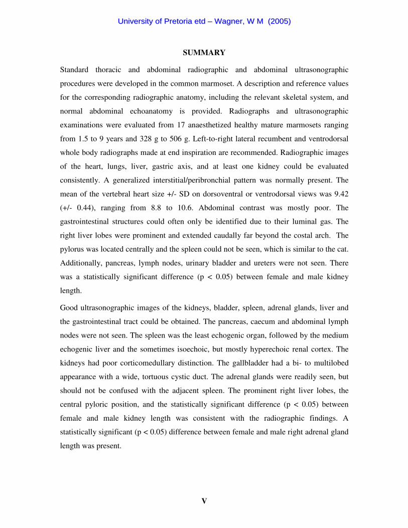
SUMMARY
Standard thoracic and abdominal radiographic and abdominal ultrasonographic
procedures were developed in the common marmoset. A description and reference values
for the corresponding radiographic anatomy, including the relevant skeletal system, and
normal abdominal echoanatomy is provided. Radiographs and ultrasonographic
examinations were evaluated from 17 anaesthetized healthy mature marmosets ranging
from 1.5 to 9 years and 328 g to 506 g. Left-to-right lateral recumbent and ventrodorsal
whole body radiographs made at end inspiration are recommended. Radiographic images
of the heart, lungs, liver, gastric axis, and at least one kidney could be evaluated
consistently. A generalized interstitial/peribronchial pattern was normally present. The
mean of the vertebral heart size +/- SD on dorsoventral or ventrodorsal views was 9.42
(+/- 0.44), ranging from 8.8 to 10.6. Abdominal contrast was mostly poor. The
gastrointestinal structures could often only be identified due to their luminal gas. The
right liver lobes were prominent and extended caudally far beyond the costal arch. The
pylorus was located centrally and the spleen could not be seen, which is similar to the cat.
Additionally, pancreas, lymph nodes, urinary bladder and ureters were not seen. There
was a statistically significant difference (p < 0.05) between female and male kidney
length.
Good ultrasonographic images of the kidneys, bladder, spleen, adrenal glands, liver and
the gastrointestinal tract could be obtained. The pancreas, caecum and abdominal lymph
nodes were not seen. The spleen was the least echogenic organ, followed by the medium
echogenic liver and the sometimes isoechoic, but mostly hyperechoic renal cortex. The
kidneys had poor corticomedullary distinction. The gallbladder had a bi- to multilobed
appearance with a wide, tortuous cystic duct. The adrenal glands were readily seen, but
should not be confused with the adjacent spleen. The prominent right liver lobes, the
central pyloric position, and the statistically significant difference (p < 0.05) between
female and male kidney length was consistent with the radiographic findings. A
statistically significant (p < 0.05) difference between female and male right adrenal gland
length was present.
UUnniivveerrssiittyy ooff PPrreettoorriiaa eettdd –– WWaaggnneerr,, WW MM ((22000055))

VI
This study emphasizes that significant species specific differences exist between dogs and
cats and the common marmoset. Simply applying canine or feline radiographic or
ultrasonographic interpretation principles may result in misdiagnosis.
UUnniivveerrssiittyy ooff PPrreettoorriiaa eettdd –– WWaaggnneerr,, WW MM ((22000055))

VII
TABLE OF CONTENTS
TITLE PAGE............................................................................................... I
DEDICATION ............................................................................................. III
ACKNOWLEDGEMENTS........................................................................ IV
SUMMARY.................................................................................................. V
LIST OF FIGURES .................................................................................... X
LIST OF TABLES ...................................................................................... XII
LIST OF APPENDICES............................................................................. XIII
GLOSSARY ................................................................................................. XIV
1. INTRODUCTION ................................................................................... 1
1.1. Hypothesis .............................................................................................. 1
1.2. Objectives............................................................................................... 1
1.3. Benefits of the study............................................................................... 2
2. LITERATURE REVIEW....................................................................... 3
2.1. Marmosets .............................................................................................. 3
2.1.1. Classification .................................................................................. 3
2.1.2. Anatomy ......................................................................................... 3
2.2. Radiography of the thorax and abdomen ............................................... 3
2.3. Radiographic anatomy of the thorax and abdomen................................ 4
2.4. Transcutaneous ultrasonography of the abdomen................................. 4
3. MATERIALS AND METHODS............................................................ 5
3.1. Animals and general preparation............................................................ 5
3.2. Radiography of the thorax and abdomen ............................................... 5
3.2.1. Animal preparation ......................................................................... 5
3.2.2. Radiographic examination.............................................................. 5
UUnniivveerrssiittyy ooff PPrreettoorriiaa eettdd –– WWaaggnneerr,, WW MM ((22000055))

VIII
3.2.3. Evaluation....................................................................................... 6
3.3. Radiographic anatomy of the thorax and abdomen................................ 6
3.3.1. Animals and preparation................................................................. 6
3.3.2. Radiographic examination.............................................................. 6
3.3.3. Evaluation....................................................................................... 6
3.3.4. Data analysis................................................................................... 8
3.4. Transcutaneous ultrasonography of the abdomen.................................. 8
3.4.1. Animal preparation ......................................................................... 8
3.4.2. Ultrasonographic examination........................................................ 8
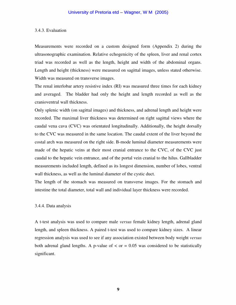
3.4.3. Evaluation....................................................................................... 9
3.4.4. Data analysis................................................................................... 9
4. RESULTS................................................................................................. 10
4.1. Animals .................................................................................................. 10
4.2. Radiography of the thorax and abdomen ............................................... 10
4.3. Radiographic anatomy of the thorax and abdomen ............................... 10
4.3.1. General............................................................................................ 10
4.3.2. Relevant skeletal system................................................................. 11
4.3.3. Thorax............................................................................................. 11
4.3.4. Abdomen ........................................................................................ 12
4.4. Transcutaneous ultrasonography of the abdomen.................................. 13
4.4.1. Technique ....................................................................................... 13
4.4.2. Ultrasonographic findings .............................................................. 14
4.5. Figures .................................................................................................... 17
4.6. Tables ..................................................................................................... 26
5. DISCUSSION .......................................................................................... 31
5.1. Animals .................................................................................................. 31
5.2. Radiography of the thorax and abdomen ............................................... 31
5.3. Radiographic anatomy of the thorax and abdomen................................ 32
5.3.1. Relevant skeletal system................................................................. 32
UUnniivveerrssiittyy ooff PPrreettoorriiaa eettdd –– WWaaggnneerr,, WW MM ((22000055))
IX
5.3.2. Thorax............................................................................................. 32
5.3.3. Abdomen ........................................................................................ 34
5.4. Transcutaneous ultrasonography of the abdomen ............................... 35
5.4.1. Technique ...................................................................................... 35
5.4.2. Ultrasonographic findings ............................................................. 35
5.5. Comparison between radiographic and ultrasonographic results........... 39
5.6. Limitations of the study.......................................................................... 40
6. CONCLUSION........................................................................................ 41
REFERENCES ............................................................................................ 42
APPENDICES ............................................................................................. 47
UUnniivveerrssiittyy ooff PPrreettoorriiaa eettdd –– WWaaggnneerr,, WW MM ((22000055))
X
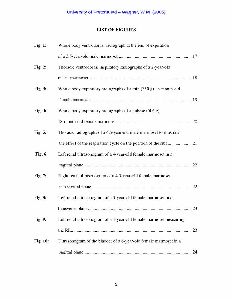
LIST OF FIGURES
Fig. 1: Whole body ventrodorsal radiograph at the end of expiration
of a 3.5-year-old male marmoset................................................................. 17
Fig. 2: Thoracic ventrodorsal inspiratory radiographs of a 2-year-old
male marmoset. ......................................................................................... 18
Fig. 3: Whole body expiratory radiographs of a thin (350 g) 18-month-old
female marmoset ........................................................................................ 19
Fig. 4: Whole body expiratory radiographs of an obese (506 g)
18-month-old female marmoset .................................................................. 20
Fig. 5: Thoracic radiographs of a 4.5-year-old male marmoset to illustrate
the effect of the respiration cycle on the position of the ribs ..................... 21
Fig. 6: Left renal ultrasonogram of a 4-year-old female marmoset in a
sagittal plane............................................................................................... 22
Fig. 7: Right renal ultrasonogram of a 4.5-year-old female marmoset
in a sagittal plane........................................................................................ 22
Fig. 8: Left renal ultrasonogram of a 3-year-old female marmoset in a
transverse plane ........................................................................................... 23
Fig. 9: Left renal ultrasonogram of a 4-year-old female marmoset measuring
the RI........................................................................................................... 23
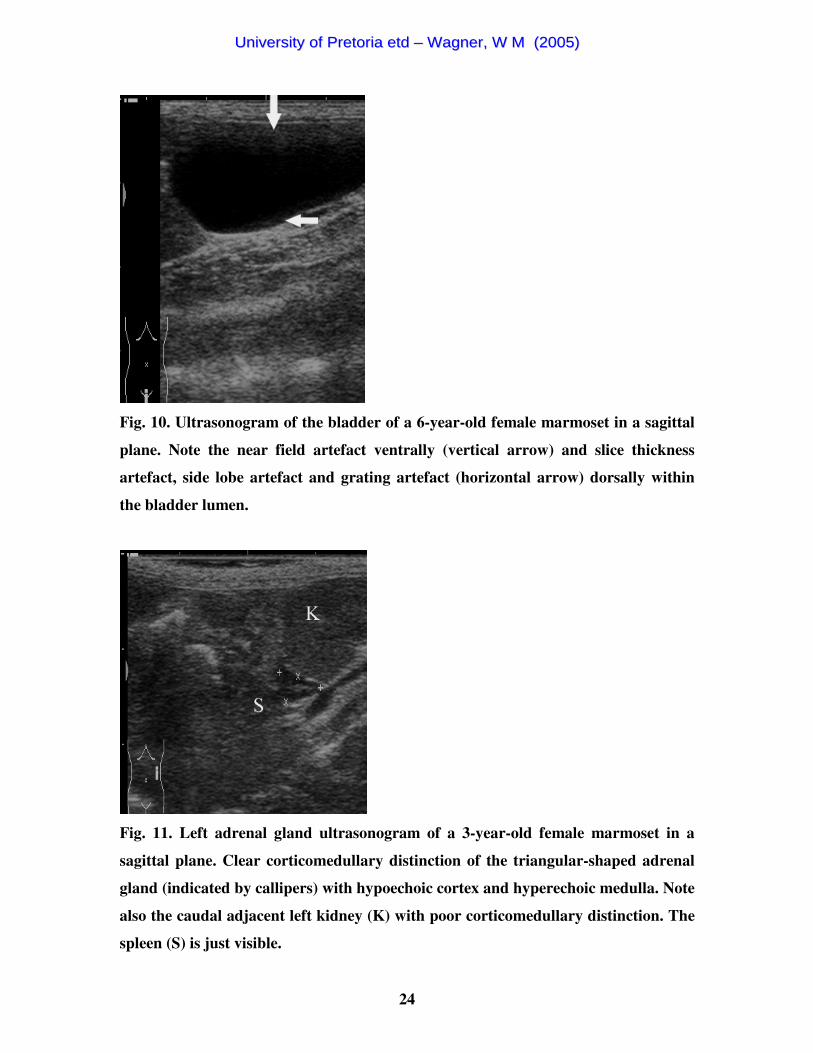
Fig. 10: Ultrasonogram of the bladder of a 6-year-old female marmoset in a
sagittal plane............................................................................................... 24
UUnniivveerrssiittyy ooff PPrreettoorriiaa eettdd –– WWaaggnneerr,, WW MM ((22000055))
XI
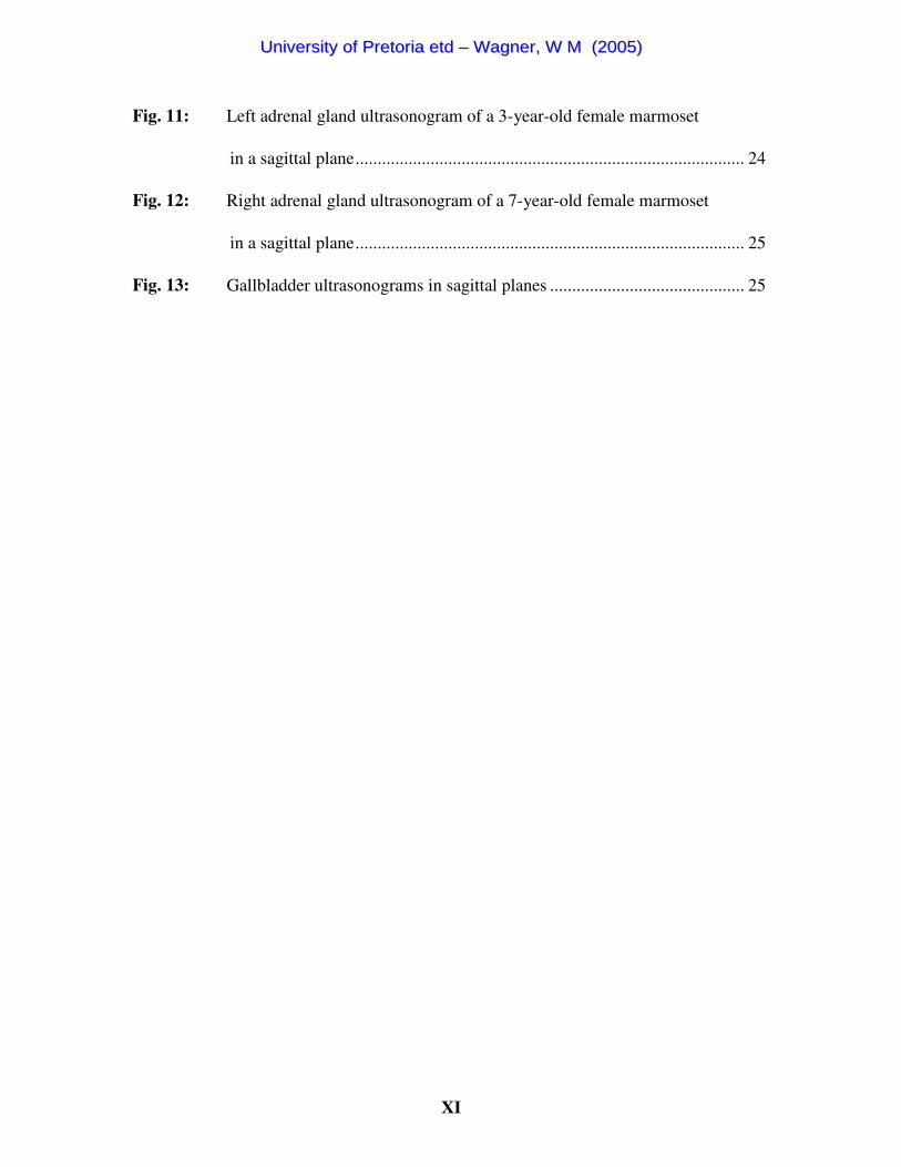
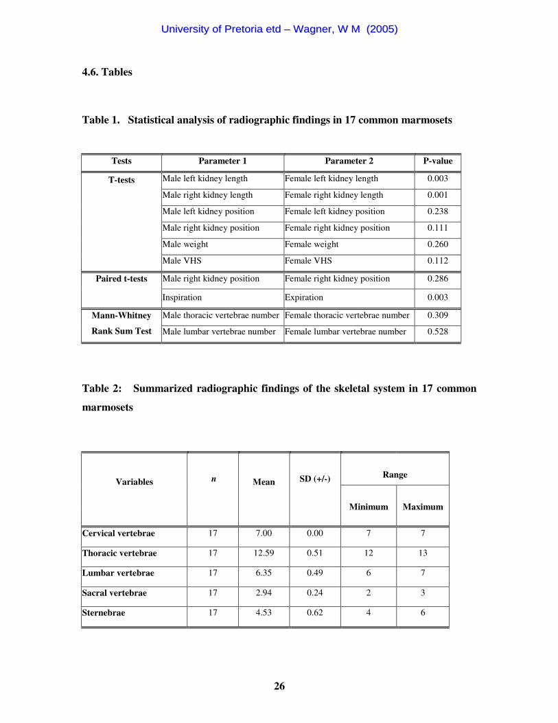
Fig. 11: Left adrenal gland ultrasonogram of a 3-year-old female marmoset
in a sagittal plane........................................................................................ 24
Fig. 12: Right adrenal gland ultrasonogram of a 7-year-old female marmoset
in a sagittal plane........................................................................................ 25
Fig. 13: Gallbladder ultrasonograms in sagittal planes ............................................ 25
UUnniivveerrssiittyy ooff PPrreettoorriiaa eettdd –– WWaaggnneerr,, WW MM ((22000055))
XII
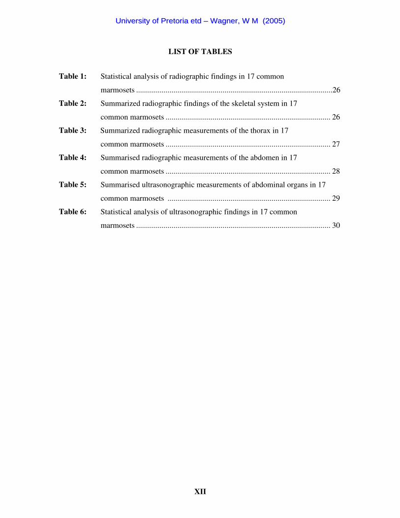
LIST OF TABLES
Table 1: Statistical analysis of radiographic findings in 17 common
marmosets ....................................................................................................26
Table 2: Summarized radiographic findings of the skeletal system in 17
common marmosets .................................................................................... 26
Table 3: Summarized radiographic measurements of the thorax in 17
common marmosets .................................................................................... 27
Table 4: Summarised radiographic measurements of the abdomen in 17
common marmosets .................................................................................... 28
Table 5: Summarised ultrasonographic measurements of abdominal organs in 17
common marmosets ................................................................................... 29
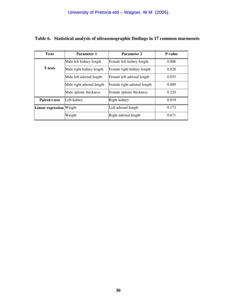
Table 6: Statistical analysis of ultrasonographic findings in 17 common
marmosets ................................................................................................... 30
UUnniivveerrssiittyy ooff PPrreettoorriiaa eettdd –– WWaaggnneerr,, WW MM ((22000055))
XIII
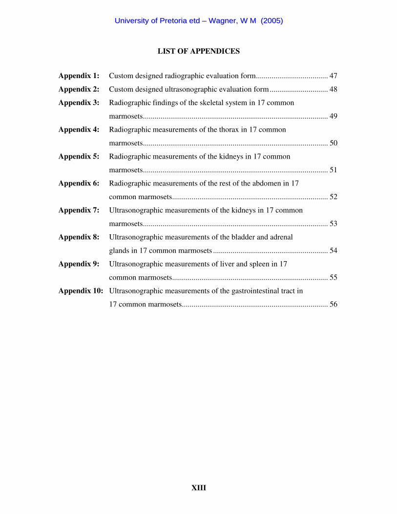
LIST OF APPENDICES
Appendix 1: Custom designed radiographic evaluation form..................................... 47
Appendix 2: Custom designed ultrasonographic evaluation form.............................. 48
Appendix 3: Radiographic findings of the skeletal system in 17 common
marmosets............................................................................................... 49
Appendix 4: Radiographic measurements of the thorax in 17 common
marmosets............................................................................................... 50
Appendix 5: Radiographic measurements of the kidneys in 17 common
marmosets............................................................................................... 51
Appendix 6: Radiographic measurements of the rest of the abdomen in 17
common marmosets................................................................................ 52
Appendix 7: Ultrasonographic measurements of the kidneys in 17 common
marmosets............................................................................................... 53
Appendix 8: Ultrasonographic measurements of the bladder and adrenal
glands in 17 common marmosets ........................................................... 54
Appendix 9: Ultrasonographic measurements of liver and spleen in 17
common marmosets................................................................................ 55
Appendix 10: Ultrasonographic measurements of the gastrointestinal tract in
17 common marmosets........................................................................... 56
UUnniivveerrssiittyy ooff PPrreettoorriiaa eettdd –– WWaaggnneerr,, WW MM ((22000055))
XIV
GLOSSARY
ABBREVATIONS USED IN TEXT: cm cm CVC caudal vena cava DV dorsoventral radiograph F female Fig. Figure g gram GIT gastrointestinal tract H height kg kilogram kVp kilovoltage peak L length L2 2nd lumbar vertebra (analogous for others) LI large intestine LLR right-to-left lateral recumbent radiograph M male mAs milliampere-seconds MHz megahertz mm millimeter
n number N patient number OVAH Onderstepoort Veterinary Academic Hospital RI resistive index RLR left-to-right lateral recumbent radiograph SD standard deviation T thickness T12 12th thoracic vertebra (analogous for others) VD ventrodorsal radiograph VHS vertebral heart size W width
UUnniivveerrssiittyy ooff PPrreettoorriiaa eettdd –– WWaaggnneerr,, WW MM ((22000055))

1
CHAPTER 1
1. INTRODUCTION
The common marmoset (Callithrix jacchus) is a popular pet in South Africa and is
commonly presented to the Exotic Animal Clinic of the Onderstepoort Veterinary
Academic Hospital (OVAH).
Diagnostic ultrasound and radiology are complementary and valuable diagnostic imaging
techniques in small and large animals. Both represent non-invasive, minimally stressful
and rapid diagnostic imaging tools. They are standard diagnostic procedures in small and
large animal practice, and radiology has become a standard diagnostic procedure in exotic
animal medicine. It is a commonly used technique for marmosets in the OVAH
Diagnostic Imaging Section, but there is minimal information available in the literature.
Ultrasound is the imaging tool of choice for soft tissue evaluation and is commonly used
in the diagnostic work-up and pregnancy diagnosis of marmosets at the OVAH.
The study was approved by the Animal Use and Care Committee of the Faculty of
Veterinary Science, University of Pretoria, and formed part of a multi-project study in
liaison with the OVAH Dental and Exotic Animal Clinics.
1.1. Hypothesis
It is believed that a standard thoracic and abdominal radiographic and abdominal
ultrasonographic procedure can be developed and that significant species specific
differences exist compared to the dog and cat. With increasing knowledge of normal
abdominal echoanatomy and normal radiographic anatomy it is anticipated that
diagnostic proficiency will improve.
1.2. Objectives
1) Develop a standard radiographic procedure for the thorax and abdomen in the
common marmoset
UUnniivveerrssiittyy ooff PPrreettoorriiaa eettdd –– WWaaggnneerr,, WW MM ((22000055))
2
2) Provide a description and reference values for the associated radiological normal
anatomy, including the relevant skeletal system
3) Describe the abdominal echoanatomy of the normal common marmoset using
transcutaneous ultrasound.
1.3. Benefits of the study
The research was undertaken because of the need for normal ultrasonographic and
radiographic descriptions of the common marmoset in order to enhance the efficiency of
these diagnostic imaging modalities for clinical application.
1) Optimalisation of the radiographic technique for the common marmoset
2) Knowledge of the radiographic anatomy of the common marmoset
3) Knowledge of the ultrasonographic anatomy of the common marmoset
UUnniivveerrssiittyy ooff PPrreettoorriiaa eettdd –– WWaaggnneerr,, WW MM ((22000055))
3
CHAPTER 2
2. LITERATURE REVIEW
2.1. Marmosets
2.1.1. Classification
Marmosets (Callithrix), tamarins (Saguinus and Leontopithecus) and Goeldi’s monkeys
(Callimico goeldii) are small neotropical primates indigenous to Central and South
America. These three groups are classified as Callitricchidae1.
The common marmoset (Callithrix jacchus) in particular is a popular pet in South Africa
and is commonly presented to the Exotic Animal Clinic of the OVAH.
2.1.2. Anatomy
Literature concerning the anatomy in the common marmoset relevant to this study is
limited to the skeleton, stating that Callitricchidae have on average 7 cervical, 13
thoracic, 7 lumbar and 3 sacral vertebrae2.
2.2. Radiography of the thorax and abdomen
Hatt3 describes an unusual case of metabolic bone disease in a common marmoset and its
radiographic findings.
Bush et al.4 describe the radiographic evaluation of diaphragmatic defects in golden lion
tamarins (Leontopithecus rosalia rosalia) which appear to be predisposed to this disease.
Articles have also been published on the common marmoset’s physeal closure times5-7.
To the best of the author’s knowledge there has been no work published on standard
radiographic technique in the common marmoset or associated species.
UUnniivveerrssiittyy ooff PPrreettoorriiaa eettdd –– WWaaggnneerr,, WW MM ((22000055))
4
2.3. Radiographic anatomy of the thorax and abdomen
To the best of the author’s knowledge there has been no work published on normal
radiographic anatomy of the common marmoset or associated species.
2.4. Transcutaneous ultrasonography of the abdomen
Two ultrasonographic reports could be found on Callitrichidae. One describes
ultrasonographic monitoring of prenatal growth and development in the common
marmoset8. The other briefly describes the normal ultrasonographic anatomy of some
abdominal organs in five marmosets9. Additionally, ultrasonographic descriptions exist of
the rhesus monkey’s ( Macaca mulatta) abdomen10, renal allograft vasculopathy in a non-
human primate model11 and of the normal kidney in the cynomolgus monkey (Macaca
fascicularis)12.
To the best of the author’s know ledge there has been no detailed work published on
normal echoanatomy of the common marmoset or associated species.
UUnniivveerrssiittyy ooff PPrreettoorriiaa eettdd –– WWaaggnneerr,, WW MM ((22000055))

5
CHAPTER 3
3. MATERIALS AND METHODS
3.1. Animals and general preparation
Twenty mature male (n=10) and non-pregnant female (n=10) marmosets (1.5 to 9 years)
were examined, all originating from the same facility. The animals were not related to
each other. Clinical examination, complete blood count and liver, kidney, and pancreas
specific biochemistry parameters were performed as part of a linked study by the Exotic
Animal Clinic of the OVAH to evaluate health status.
The marmosets were starved for 12 hours prior to scheduled morning procedures, but had
access to water till shortly before the examination. Marmosets were anaesthetised with
isoflurane inhalation using a mask to ensure patient compliance, and safety of handlers
and to minimise stress.
3.2. Radiography of the thorax and abdomen
3.2.1. Animal preparation
No additional preparation was needed.
3.2.2. Radiographic examination
A 100-speed mammography system was used (which is slower than a 100-speed
conventional system)13. The source-to-image distance was 105 cm using a table top
technique. Left-to-right lateral recumbent (RLR) and ventrodorsal (VD) whole body
radiographs were made using 44 kVp and 10 mAs for all marmosets. Centre point for
these radiographs was the middle of the last rib. Arms were initially positioned next to
the body, then cranially adjacent to the skull. For the first 5 marmosets additional right-
to-left lateral recumbent (LLR) and dorsoventral (DV) whole body radiographs were
UUnniivveerrssiittyy ooff PPrreettoorriiaa eettdd –– WWaaggnneerr,, WW MM ((22000055))

6
made for comparison. The best views were applied for the remaining marmosets. The
next 5 marmosets had additional radiographs centred and collimated to the thorax, made
at the end of inspiration and expiration. The following 5 marmosets had additional
radiographs, centred and collimated to the abdomen, made at end of inspiration and
expiration. These were all critically evaluated and the best technique applied to the
remaining marmosets.
3.2.3. Evaluation
Radiographs were subjectively compared. Differences in image quality and organ
position were compared between RLR versus LLR or VD versus DV radiographs.
Thoracic and abdominal contrast was compared at the end of inspiration and expiration.
The effect of arm position on the lung field was evaluated. The influence of collimating
to the thorax, abdomen or whole body was compared concerning image detail and
positioning.
3.3. Radiographic anatomy of the thorax and abdomen
3.3.1. Animal preparation
See 3.2.1.
3.3.2. Radiographic examination
See 3.2.2.
3.3.3. Evaluation
Relevant skeletal structures
Radiographs were evaluated and findings (including abnormalities) were recorded on a
custom designed form (Appendix 1). Vertebral and sternal numbers, transitional vertebra
UUnniivveerrssiittyy ooff PPrreettoorriiaa eettdd –– WWaaggnneerr,, WW MM ((22000055))

7
and alignment of the last lumbar vertebra to the other lumbar or sacral vertebrae were
recorded.
Thorax
Subcutaneous thoracic fat thickness, as a potential indicator for abdominal contrast, was
measured ventrally to the sternum on lateral views at the level of the 3rd rib and laterally
to the thoracic wall on VD/DV views at the level of the 7th rib. The tracheal angle to the
spine was measured (between the dorsal wall of the trachea at the 1st rib and the ventral
border of the cranial thoracic vertebrae). The position of the carina and the caudal vena
cava (CVC) diameter (midway between diaphragm and caudal cardiac border) were
determined on lateral views, and the vertebral heart size (VHS)14 was obtained on lateral
and DV/VD images. Lung patterns were noted as well as the most caudal position of the
diaphragmatic crura during inspiration and expiration on lateral views. To prevent the
influence of varying thoracic vertebral numbers, the cranial aspect of T12 was considered
as 12 and counted caudally (13, 14), independently of the thoracic vertebral number on
lateral views.
Abdomen
Kidney length was compared to L2 length, but also measured in cm, and its position was
determined in relationship to the vertebrae on lateral and VD/DV views. The angle of the
gastric axis was measured between a line drawn along the ventral aspect of the first 3-4
lumbar vertebrae and the gastric axis. The caudal extent of the liver beyond the most
caudoventral aspect of the costal arch was recorded in cm on lateral views. Visibility of
organs, large intestinal diameter in cm and in relation to L2 body length and abdominal
contrast were noted. Abdominal contrast (using peritoneal and retroperitoneal contrast
parameters) was considered excellent if both kidneys on VD and lateral views and the
caudal liver edge on lateral views could be seen, good if only both kidneys or one kidney
on VD and the caudal liver edge on lateral views was seen, and poor in all other cases.
Incidental findings were noted.
UUnniivveerrssiittyy ooff PPrreettoorriiaa eettdd –– WWaaggnneerr,, WW MM ((22000055))

8
3.3.4. Data analysis
A t-test analysis was used to compare male versus female left and right kidney length and
position, body weight, and VHS on DV/VD. A Mann-Whitney Rank Sum Test was used
to compare male and female number of thoracic and lumbar vertebrae. A paired t-test was
used to determine whether the position of the left and right kidney differed significantly
between males and females, and also whether inspiration and expiration differed
significantly within one individual. A p-value of < or = 0.05 was considered to be
statistically significant.
3.4. Transcutaneous ultrasonography of the abdomen
3.4.1. Animal preparation
Ultrasonographic preparation and examination followed the completed radiographic
examination. Each marmoset was placed on a heating pad in dorsal recumbency, had its
abdomen clipped and a generous amount of warmed ultrasound coupling gel (MMS,
Medmac Services, Johannesburg, Republic of South Africa) was applied to the skin. A
complete abdominal ultrasonographic examination was performed with a linear-array
multi-frequency transducer (Sonoline Omnia, Siemens, Berlin, Germany) operated at 9
MHz.
3.4.2. Ultrasonographic examination
Examinations were recorded on videotape and magnetic optic disc. A systematic
abdominal examination starting with the spleen, followed by the left kidney, bladder,
right kidney, liver and the gastrointestinal tract (GIT) was performed and then adrenal
glands and pancreas imaging was attempted. Due to the multi-project nature of this study,
ultrasonographic examinations were limited to 30-40 min, and the genital tract was not
included.
UUnniivveerrssiittyy ooff PPrreettoorriiaa eettdd –– WWaaggnneerr,, WW MM ((22000055))
9
3.4.3. Evaluation
Measurements were recorded on a custom designed form (Appendix 2) during the
ultrasonographic examination. Relative echogenicity of the spleen, liver and renal cortex
triad was recorded as well as the length, height and width of the abdominal organs.
Length and height (thickness) were measured on sagittal images, unless stated otherwise.
Width was measured on transverse images.
The renal interlobar artery resistive index (RI) was measured three times for each kidney
and averaged. The bladder had only the height and length recorded as well as the
cranioventral wall thickness.
Only splenic width (on sagittal images) and thickness, and adrenal length and height were
recorded. The maximal liver thickness was determined on right sagittal views where the
caudal vena cava (CVC) was orientated longitudinally. Additionally, the height dorsally
to the CVC was measured in the same location. The caudal extent of the liver beyond the
costal arch was measured on the right side. B-mode luminal diameter measurements were
made of the hepatic veins at their most cranial entrance to the CVC, of the CVC just
caudal to the hepatic vein entrance, and of the portal vein cranial to the hilus. Gallbladder
measurements included length, defined as its longest dimension, number of lobes, ventral
wall thickness, as well as the luminal diameter of the cystic duct.
The length of the stomach was measured on transverse images. For the stomach and
intestine the total diameter, total wall and individual layer thickness were recorded.
3.4.4. Data analysis
A t-test analysis was used to compare male versus female kidney length, adrenal gland
length, and spleen thickness. A paired t-test was used to compare kidney sizes. A linear
regression analysis was used to see if any association existed between body weight versus
both adrenal gland lengths. A p-value of < or = 0.05 was considered to be statistically
significant.
UUnniivveerrssiittyy ooff PPrreettoorriiaa eettdd –– WWaaggnneerr,, WW MM ((22000055))

10
4. RESULTS
4.1. Animals
Two animals (M3 and M9) were excluded from this study, since their white cell counts
were elevated compared to the others for unknown reasons. The remaining 18 marmosets
had a normal clinical examination, complete blood count and liver, kidney, and pancreas
specific biochemistry parameters. A third marmoset (M1) was excluded from this study
because mineralization of the aorta was noted. All results thus pertain to the remaining 17
healthy animals.
Marmosets weighed between 328 g to 506 g. Male and female body weight did not differ
significantly (Table 1). Both females and males could have prominent thoracic mammary
glands (Fig. 1).
4.2. Radiography of the thorax and abdomen
No difference in image quality was noted on RLR versus LLR or VD versus DV
radiographs. Thoracic contrast was slightly better at the end of inspiration, without a
concomitant decrease in abdominal contrast (Fig. 1). The cranial aspect of the lungs could
be better seen with cranial positioning of the arms, which also resulted in better visibility
of the shoulder joint on VD views (Fig. 2). Collimating to the thorax improved thoracic
contrast compared to whole body radiographs, however optimal positioning was often
difficult to maintain. Collimating to the abdomen did not improve abdominal contrast
compared to whole body radiographs.
4.3. Radiographic anatomy of the thorax and abdomen
4.3.1. General
Independently of the radiographic technique used, abdominal contrast was often poor
UUnniivveerrssiittyy ooff PPrreettoorriiaa eettdd –– WWaaggnneerr,, WW MM ((22000055))
11
(7/17 of which 6 were males) (Fig. 1) and only 3 animals had excellent abdominal
contrast (Figs. 3 & 4). Body weight or amount of thoracic wall fat did not correlate to
abdominal contrast.
4.3.2. Relevant skeletal system
All marmosets had 7 cervical vertebrae (Table 2). Males most often had 13 (6/8) thoracic
vertebrae and females almost equally had 12 (5/9) or 13 (4/9). Animals with 12 thoracic
vertebrae had 7 lumbar vertebrae, those with 13 had 6, almost always resulting in a
thoracolumbar vertebral number of 19 (16/17). However, no statistical significance
difference was present between female and male thoracic and lumbar vertebrae number.
The last lumbar vertebra was always markedly shorter than the other lumbar vertebrae.
Prominent accessory processes were present in these vertebrae. Transitional last lumbar
vertebra (uni- or bilateral) occurred commonly (10/17) (Appendix 3). The sacrum
consisted of 3 fused segments, except in one female, which had only 2 segments. In 3/17
marmosets the last sacral vertebra was not fused and in one marmoset the ventral half of
S2-3 was not fused. The last rib was always floating. The sternum consisted of a
manubrium sterni, 4-5 sternebrae and a xiphoid process. The sternebrae were fused in 3
animals (last 2, 3 or 5 sternebrae).
Incidental findings were thoracic, lumbar and lumbosacral spondylosis (Fig. 3B), old
healed fractures and one animal with symmetrical rib anomalies resembling avian
uncinate processes.
4.3.3. Thorax (Table 3)
Cardiovascular system
Heart. On DV/VD views the apex of the rectangular (with rounded edges) cardiac
silhouette was positioned to the left with extensive diaphragmatic contact (Figs. 1, 2, 3A
& 4A). No marked difference between RLR and LLR or DV and VD views was seen.
Lateral and DV/VD VHS measurements correlated very well. The cranial cardiac border
and/or the carina were often difficult to see on lateral views (Figs. 3B, 4B & 5) and
UUnniivveerrssiittyy ooff PPrreettoorriiaa eettdd –– WWaaggnneerr,, WW MM ((22000055))

12
measurement of VHS was easier on the DV/VD views. Vertebral heart size on DV/VD
views was 9.42 (+/- 0.44) with a range from 8.8 to 10.6. There was no statistical
difference between female and male VHS.
Blood vessels. On lateral views, the CVC had a mean diameter +/- SD of 4.3 +/- 0.6 mm,
ranging from 3.0-5.0. On the DV/VD it was not clearly visible. The aorta was not clearly
visible on any view. The pulmonary vasculature was better visible on lateral views.
Respiratory system
The carina was not always clearly visible with a mean position +/- SD of T5.74 +/-0.47,
ranging from T5 to 6.5 and mostly positioned at T6 (10/17) (Appendix 4). The mean +/-
SD of the tracheal angle was 7.62 +/-2.76 degrees, ranging from 2 to 12 degrees. A
generalized interstitial/peribronchial pattern was always present. The difference in crura
positioning during the respiratory cycle on the lateral views was significant. The mean
position of the cranial diaphragmatic crus during inspiration +/- SD was 13.51 +/- 0.52,
ranging from 12.5 to 14.2 and for expiration 13.16 +/- 0.47. Only one set of radiographs
was made at the incorrect respiratory cycle. The diaphragmatic cupula and crura shape
did not differ on VD or DV and on LLR and RLR.
On the lateral views the ribs were better superimposed on each other during inspiration
(12/16) facilitating thoracic evaluation (Fig. 5). This was only the case in 7/21 expiratory
views. No fissure lines or caudal mediastinum were seen. The medial edge of the scapula
should not be misinterpreted as a margin of the cranial mediastinum on DV/VD.
4.3.4. Abdomen (Table 4)
Lymphatic system
The spleen, thoracic and abdominal lymph nodes were not seen on any view.
Digestive system
Stomach. The angle of the gastric axis did not differ between inspiration and expiration
and had a mean +/- SD of 100.29 +/ 9.9 degrees, ranging from 85 to 117 degrees. The
UUnniivveerrssiittyy ooff PPrreettoorriiaa eettdd –– WWaaggnneerr,, WW MM ((22000055))

13
gastric axis could not be readily determined on DV/VD views due to poor visibility. The
pylorus extended only to the midline. Positional effects on gastric gas were not seen.
Intestines. Intestinal outline depended on abdominal contrast and luminal gas. The small
and large intestine could not be distinguished, however the largest distended bowel was
considered to be large intestine (Fig. 1) and measured up to 1.4 cm or 1.4 times L2
(Appendix 6).
Glandular system
The liver was positioned mainly on the right were it extended up to 3.6 cm caudally to the
last rib on lateral views (2.54 +/-0.91 cm with range of 1.0 to 3.6 cm) taking up most of
the ventral right cranial abdominal cavity (Figs. 3B & 5B). On VD/DV views the exact
caudal extent was never clearly visible, but could be estimated due to adjacent luminal
intestinal gas in 7/17 animals. On the lateral view the caudal tip of the liver could be seen
in 9/17 cases and could be estimated in 3 additional ones.
The pancreas and adrenals were not seen.
Urinary system
Both kidneys were only seen in 9/17 on VD/DV views and 3/17 on lateral views (Figs. 3
& 4). The right kidney was mostly positioned caudally to the left on VD/DV. On the
VD/DV the mean left kidney length +/- SD was 1.91 +/-0.16 cm, ranging from 1.6 to 2.1
cm versus 1.96 +/-0.11 cm, ranging from 1.8 to 2.1 cm for the right kidney. On DV/VD
the mean left and right kidney length compared to L2 was significantly different between
the males and females (1.78 versus 2.03). There was no statistical difference in kidney
position between females and males.
The urinary bladder and ureters were not seen.
4.4. Transcutaneous ultrasonography of the abdomen
4.4.1. Technique
Due to time limitations not all measurements were obtained for all marmosets.
UUnniivveerrssiittyy ooff PPrreettoorriiaa eettdd –– WWaaggnneerr,, WW MM ((22000055))
14
Since the spleen was often difficult to find and the adrenal glands were easily identified,
the order of the ultrasonographic examination was adapted to left kidney, left adrenal
gland, spleen, bladder, right kidney, right adrenal gland, liver, stomach, duodenum and
rest of intestines. Mean measurements and standard deviations for each organ are
summarized in Table 5. Findings were grouped into organ systems.
4.4.2. Ultrasonographic findings
Urinary System
Kidneys. The left oval-shaped kidney was located caudally to the spleen (Fig. 6) and left
adrenal gland. The right kidney was in close contact with the liver (Fig. 7). It was also
oval-shaped with an occasional more teardrop shaped caudal pole. The renal cortex was
hyperechoic to the spleen (Fig. 6), mainly hyperechoic, and only occasionally isoechoic
(5/17) to the liver. Corticomedullary distinction was generally poor. A corticomedullary
rim sign could occasionally be seen, but no medullary rim sign was observed. The
medulla was particularly thin in comparison to the cortex, which was accentuated on
transverse images (Fig. 8). A moderate amount of pelvic fat was present. The mean left
kidney length +/- SD was 19.04 +/- 1.95 mm, and there was a significant difference
between males 17.76 +/- 2.19 mm and females 20.17 +/- 0.57 mm (Appendix 7). The
mean right kidney length +/- SD was 18.21 +/- 2.09 mm, also with a significant
difference between males 17.06 +/- 2.47 mm and females 19.22 +/- 0.98. The right
kidney length was significantly shorter than the left (Table 6). There was no significant
relationship between kidney length and body weight (Table 6). The width was larger than
the height (Appendix 7). The interlobar artery flow profile displayed a steep systolic
peak, a clear spectral window and a smooth down slope to diastole (Fig. 9). The mean RI
+/- SD of the left kidney was 0.75 +/-0.04 versus right kidney 0.72 +/-0.05.
Bladder. The bladder was only seen in 12/17 marmosets (8 females) and was located
intra-abdominally and had a round shape with anechoic content. It was thin-walled (Fig.
10) with a mean thickness +/- SD of 0.49 +/-0.25 mm. Near field artefacts could be seen
ventrally and slice thickness, side lobe and grating artefacts resulted in the impression of
sludge dorsally within the bladder lumen similar to dogs and cats (Fig. 10).
UUnniivveerrssiittyy ooff PPrreettoorriiaa eettdd –– WWaaggnneerr,, WW MM ((22000055))
15
Lymphatic System
Spleen. Large shape variation existed (flat to thick sausage shaped, triangular and curved
to folded on itself). It was located cranial to the left kidney and more medially than in
dogs or cats. It had a homogeneous fine granular echotexture (Fig. 6) and was the most
hypoechoic of the spleen, liver and renal cortex triad. The mean width +/- SD was 13.2
+/- 3.11 mm. The mean thickness +/- SD was 6.65 +/-1.56 mm and males did not differ
significantly from females (Table 6).
Lymph nodes. None were detected.
Glandular System
Adrenal glands. The adrenal glands were easily found (Figs. 11 & 12) craniomedially to
the kidneys without using a landmark approach as in the dog. Both adrenal glands had a
triangular, blunted arrow shape. A clear corticomedullary distinction (Fig. 12) was
usually present with a hyperechoic medulla and hypoechoic cortex. The right adrenal
bordered the liver cranially and the CVC laterally. The mean length +/- SD of the left
adrenal gland was 4.8 +/-0.95 mm and its height 3.82 +/-0.8 mm. The mean length of the
right adrenal gland +/- SD was 4.79 +/-0.68 mm and its height 3.94 +/-1.02 mm. In
females, the right adrenal gland length was significantly larger than in males, but not the
left (Table 6). The right adrenal gland length versus body weight was not significantly
related (Table 6).
Liver. The liver had a coarse echotexture (Fig. 13), and was mainly hypoechoic, but
occasionally isoechoic to the renal cortices and hyperechoic to the spleen. The right side
of the liver was the most prominent and extended with a mean +/- SD of 13.81 +/-6.39
mm beyond the costal arch. The mean liver height +/- SD was 21.14 +/- 2.29 mm, and
dorsally to the CVC 6.24 +/- 0.61 mm. The walls of the hepatic veins were isoechoic to
the surrounding liver tissue with a mean luminal diameter +/- SD of 1.25 +/- 0.34 mm.
The mean CVC luminal diameter +/- SD was 2.89 +/-0.64 mm. Portal veins had
hyperechoic walls with a mean luminal diameter +/- SD of 2.00 +/-0.41 mm.
Spontaneous contrast could be seen in the entire hepatic vasculature, but most
prominently in the CVC.
UUnniivveerrssiittyy ooff PPrreettoorriiaa eettdd –– WWaaggnneerr,, WW MM ((22000055))

16
The right-sided gall bladder could easily be seen (Fig. 13) and was up to 16 mm long
(Appendix 9). It had a bi- to multilobed appearance with a thin hyperechoic wall (up to
0.6 mm) differentiating it from the surrounding liver (Appendix 9). No sludge was seen.
The cystic duct was tortuous and could be differentiated from the surrounding vessels by
the absence of a colour Doppler signal. Its mean diameter +/- SD was 2.55 mm +/- 0.91
mm.
Pancreas. It was not detected.
Digestive System
The classic gastrointestinal 5-layered appearance could be seen clearly with the
hyperechoic mucosa-lumen interface, anechoic mucosa, hyperechoic submucosa,
anechoic muscularis and hyperechoic serosa.
Stomach. The stomach was easily seen and was often collapsed. The pylorus was located
towards the midline. Mean stomach length and ventral wall thickness +/- SD were 15.24
+/- 5.2 mm and 0.87 +/- 0.22 mm respectively.
Duodenum. The duodenum was mostly empty and collapsed. Its mean total and wall
diameter +/- SD was 3.35 +/- 0.94 mm and 0.86 mm +/- 0.19 respectively with the
anechoic mucosa being the most prominent layer (Appendix 10).
Rest of gastrointestinal tract. Due to time limitations only 8 marmosets were evaluated.
The mean total diameter for the rest of the small intestine and colon +/- SD were 2.9 +/-
1.67 mm and 8.6 +/- 2.4 mm respectively. The caecum was never specifically looked for
and hence was not identified.
UUnniivveerrssiittyy ooff PPrreettoorriiaa eettdd –– WWaaggnneerr,, WW MM ((22000055))
17
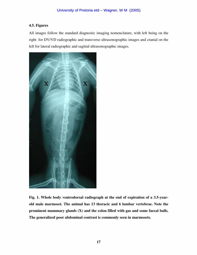
4.5. Figures
All images follow the standard diagnostic imaging nomenclature, with left being on the
right for DV/VD radiographic and transverse ultrasonographic images and cranial on the
left for lateral radiographic and sagittal ultrasonographic images.
Fig. 1. Whole body ventrodorsal radiograph at the end of expiration of a 3.5-year-
old male marmoset. The animal has 13 thoracic and 6 lumbar vertebrae. Note the
prominent mammary glands (X) and the colon filled with gas and some faecal balls.
The generalized poor abdominal contrast is commonly seen in marmosets.
UUnniivveerrssiittyy ooff PPrreettoorriiaa eettdd –– WWaaggnneerr,, WW MM ((22000055))

18
A
B
Fig. 2. Thoracic ventrodorsal inspiratory radiographs of a 2-year-old male
marmoset. Note the generalized interstitial/peribronchial infiltration. (A) Arms
positioned cranially adjacent to the skull. The cranial lung field and shoulder joint
can be seen better than in B. (B) Arms positioned next to the body, hampering
evaluation of the cranial lung field.
UUnniivveerrssiittyy ooff PPrreettoorriiaa eettdd –– WWaaggnneerr,, WW MM ((22000055))
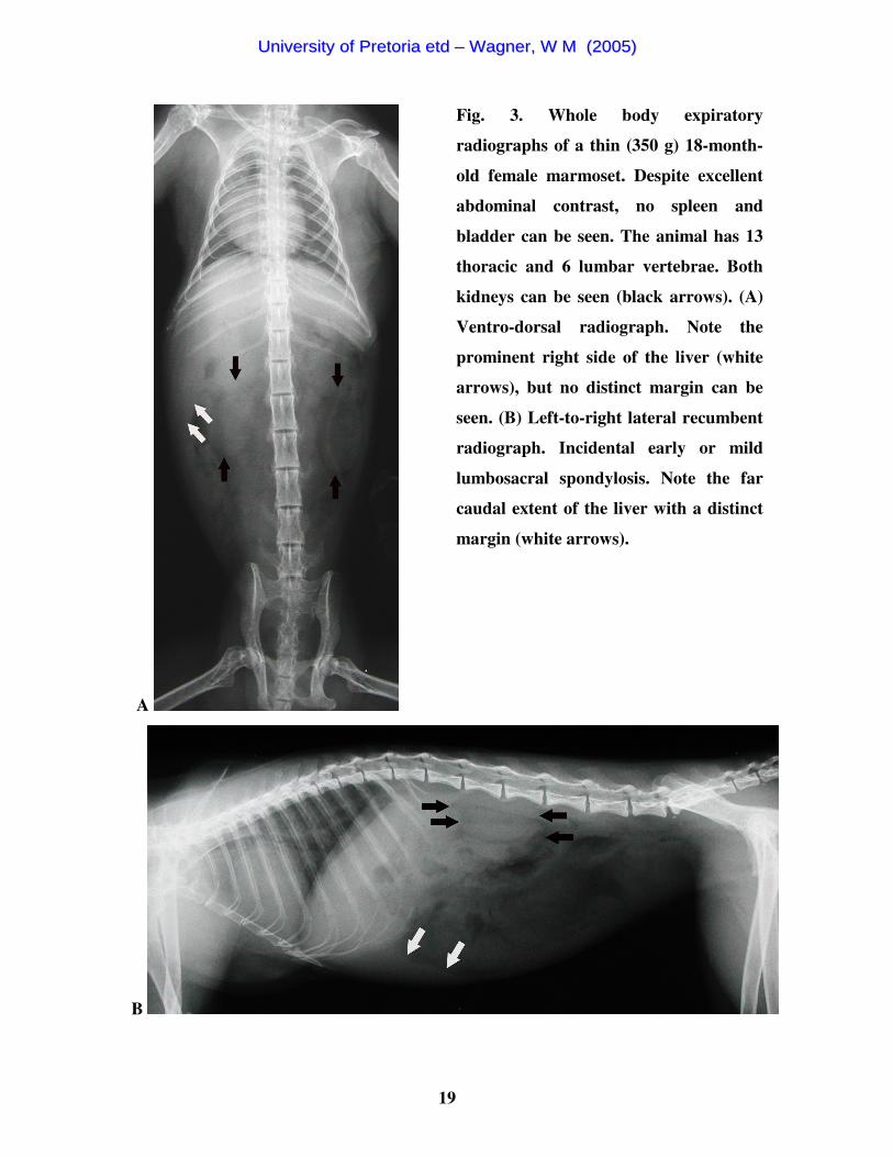
19
A
Fig. 3. Whole body expiratory
radiographs of a thin (350 g) 18-month-
old female marmoset. Despite excellent
abdominal contrast, no spleen and
bladder can be seen. The animal has 13
thoracic and 6 lumbar vertebrae. Both
kidneys can be seen (black arrows). (A)
Ventro-dorsal radiograph. Note the
prominent right side of the liver (white
arrows), but no distinct margin can be
seen. (B) Left-to-right lateral recumbent
radiograph. Incidental early or mild
lumbosacral spondylosis. Note the far
caudal extent of the liver with a distinct
margin (white arrows).
B
UUnniivveerrssiittyy ooff PPrreettoorriiaa eettdd –– WWaaggnneerr,, WW MM ((22000055))
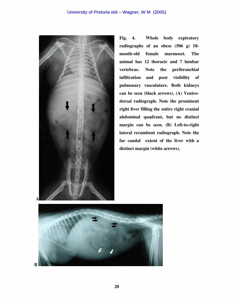
20
A
Fig. 4. Whole body expiratory
radiographs of an obese (506 g) 18-
month-old female marmoset. The
animal has 12 thoracic and 7 lumbar
vertebrae. Note the peribronchial
infiltration and poor visibility of
pulmonary vasculature. Both kidneys
can be seen (black arrows). (A) Ventro-
dorsal radiograph. Note the prominent
right liver filling the entire right cranial
abdominal quadrant, but no distinct
margin can be seen. (B) Left-to-right
lateral recumbent radiograph. Note the
far caudal extent of the liver with a
distinct margin (white arrows).
B
UUnniivveerrssiittyy ooff PPrreettoorriiaa eettdd –– WWaaggnneerr,, WW MM ((22000055))

21
A
B
Fig. 5. Left-to-right lateral recumbent thoracic radiographs of a 4.5-year-old male
marmoset to illustrate the effect of the respiration cycle on the position of the ribs.
Note the prominent right liver, lack of abdominal contrast and the generalized
interstitial/peribronchial infiltration. Because of the difficulty in determining the
cardiac edges and carina, this view is unsuitable for vertebral heart size evaluation.
(A) Inspiration. Note the superimposition of the ribs onto each other allowing better
evaluation of the thorax. (B) Expiration. The ribs are not superimposed onto each
other, hampering thoracic evaluation.
UUnniivveerrssiittyy ooff PPrreettoorriiaa eettdd –– WWaaggnneerr,, WW MM ((22000055))

22
Fig. 6. Left renal ultrasonogram of a 4-year-old female marmoset in a sagittal
plane. The image demonstrates good corticomedullary distinction for this species.
Note also the cranially adjacent hypoechoic spleen (S).
Fig. 7. Right renal ultrasonogram of a 4.5-year-old female marmoset in a sagittal
plane. The margins of the right kidney are indicated by callipers. Corticomedullary
distinction is less than in Fig. 6. Note the extensive cranial contact of the kidney to
the isoechoic liver (L).
UUnniivveerrssiittyy ooff PPrreettoorriiaa eettdd –– WWaaggnneerr,, WW MM ((22000055))
23
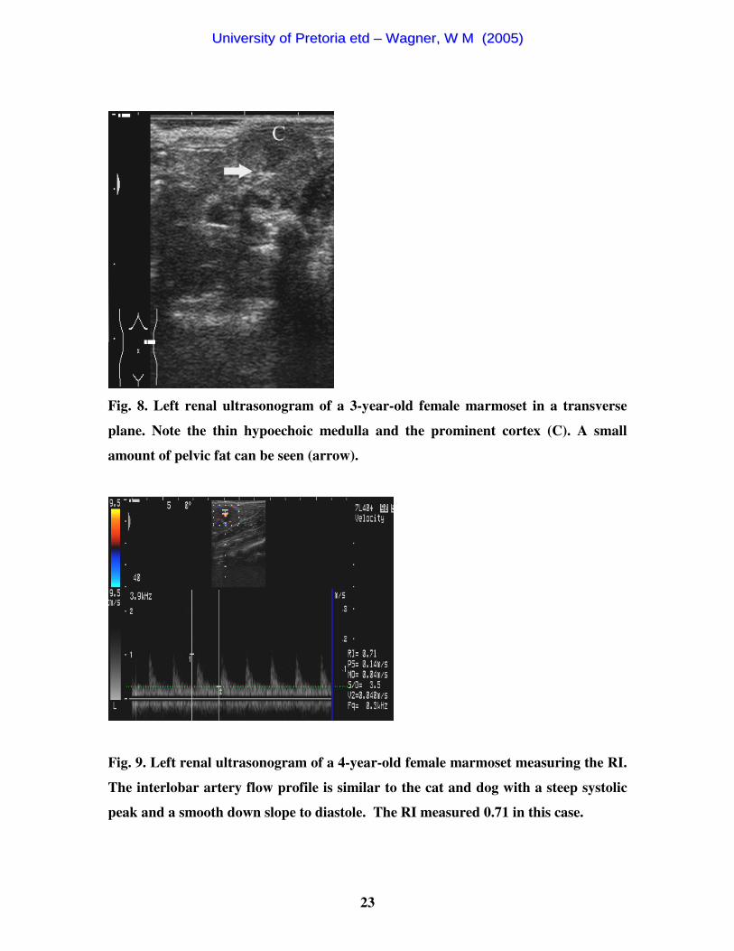
Fig. 8. Left renal ultrasonogram of a 3-year-old female marmoset in a transverse
plane. Note the thin hypoechoic medulla and the prominent cortex (C). A small
amount of pelvic fat can be seen (arrow).
Fig. 9. Left renal ultrasonogram of a 4-year-old female marmoset measuring the RI.
The interlobar artery flow profile is similar to the cat and dog with a steep systolic
peak and a smooth down slope to diastole. The RI measured 0.71 in this case.
UUnniivveerrssiittyy ooff PPrreettoorriiaa eettdd –– WWaaggnneerr,, WW MM ((22000055))
24
Fig. 10. Ultrasonogram of the bladder of a 6-year-old female marmoset in a sagittal
plane. Note the near field artefact ventrally (vertical arrow) and slice thickness
artefact, side lobe artefact and grating artefact (horizontal arrow) dorsally within
the bladder lumen.
Fig. 11. Left adrenal gland ultrasonogram of a 3-year-old female marmoset in a
sagittal plane. Clear corticomedullary distinction of the triangular-shaped adrenal
gland (indicated by callipers) with hypoechoic cortex and hyperechoic medulla. Note
also the caudal adjacent left kidney (K) with poor corticomedullary distinction. The
spleen (S) is just visible.
UUnniivveerrssiittyy ooff PPrreettoorriiaa eettdd –– WWaaggnneerr,, WW MM ((22000055))

25
Fig. 12. Right adrenal gland ultrasonogram of a 7-year-old female marmoset in a
sagittal plane. Clear corticomedullary distinction of the right triangular-shaped
adrenal gland (indicated by callipers) with hypoechoic cortex and hyperechoic
medulla. Caudally the adjacent right kidney (K) with poor corticomedullary
distinction can be seen. The liver (L) with CVC can be seen on the cranial edge of
the image.
A B
Fig. 13. Gallbladder ultrasonograms in sagittal planes. (A) Three-year-old female
marmoset with a multilobed appearance and thin hyperechoic wall and its length
indicated by callipers. Just dorsocaudally to the gallbladder the prominent cystic
duct can be seen (horizontal arrow). (B) Six-year-old female marmoset. Note the
folded appearance of the gallbladder, which illustrates its large variability.
UUnniivveerrssiittyy ooff PPrreettoorriiaa eettdd –– WWaaggnneerr,, WW MM ((22000055))
26
4.6. Tables
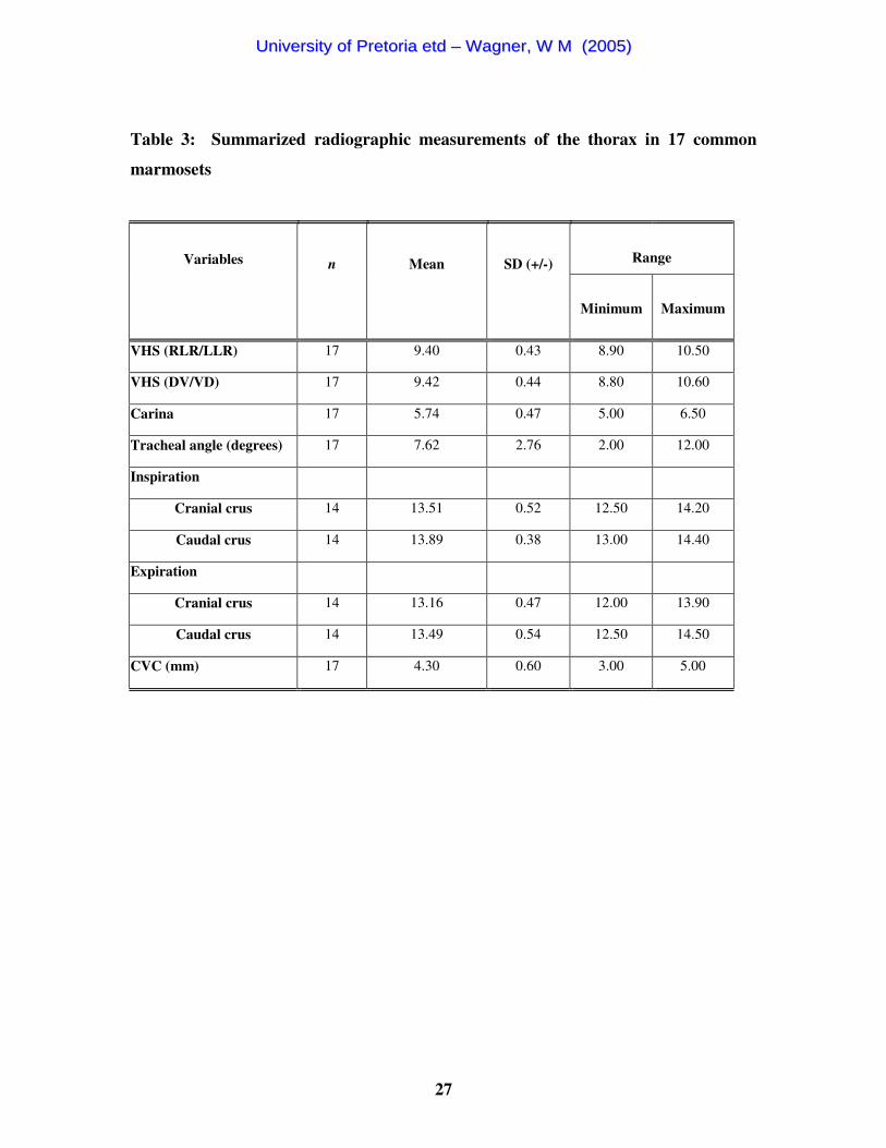
Table 1. Statistical analysis of radiographic findings in 17 common marmosets
Tests Parameter 1 Parameter 2 P-value Male left kidney length Female left kidney length 0.003
Male right kidney length Female right kidney length 0.001
Male left kidney position Female left kidney position 0.238
Male right kidney position Female right kidney position 0.111
Male weight Female weight 0.260
T-tests Male VHS Female VHS 0.112
Male right kidney position Female right kidney position 0.286 Paired t-tests Inspiration Expiration 0.003
Male thoracic vertebrae number Female thoracic vertebrae number 0.309 Mann-Whitney Rank Sum Test Male lumbar vertebrae number Female lumbar vertebrae number 0.528
Table 2: Summarized radiographic findings of the skeletal system in 17 common
marmosets
Range
Variables
n
Mean
SD (+/-)
Minimum
Maximum
Cervical vertebrae 17 7.00 0.00 7 7
Thoracic vertebrae 17 12.59 0.51 12 13
Lumbar vertebrae 17 6.35 0.49 6 7
Sacral vertebrae 17 2.94 0.24 2 3
Sternebrae 17 4.53 0.62 4 6
UUnniivveerrssiittyy ooff PPrreettoorriiaa eettdd –– WWaaggnneerr,, WW MM ((22000055))
27
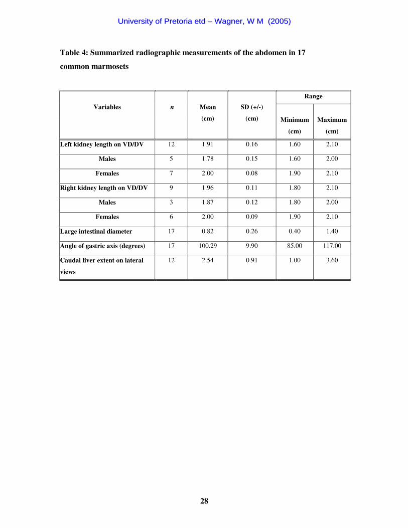
Table 3: Summarized radiographic measurements of the thorax in 17 common
marmosets
Range
Variables
n
Mean
SD (+/-)
Minimum
Maximum
VHS (RLR/LLR) 17 9.40 0.43 8.90 10.50
VHS (DV/VD) 17 9.42 0.44 8.80 10.60
Carina 17 5.74 0.47 5.00 6.50
Tracheal angle (degrees) 17 7.62 2.76 2.00 12.00
Inspiration
Cranial crus 14 13.51 0.52 12.50 14.20
Caudal crus 14 13.89 0.38 13.00 14.40
Expiration
Cranial crus 14 13.16 0.47 12.00 13.90
Caudal crus 14 13.49 0.54 12.50 14.50
CVC (mm) 17 4.30 0.60 3.00 5.00
UUnniivveerrssiittyy ooff PPrreettoorriiaa eettdd –– WWaaggnneerr,, WW MM ((22000055))
28
Table 4: Summarized radiographic measurements of the abdomen in 17
common marmosets
Range
Variables
n
Mean
(cm)
SD (+/-)
(cm)
Minimum
(cm)
Maximum
(cm)
Left kidney length on VD/DV 12 1.91 0.16 1.60 2.10
Males 5 1.78 0.15 1.60 2.00
Females 7 2.00 0.08 1.90 2.10
Right kidney length on VD/DV 9 1.96 0.11 1.80 2.10
Males 3 1.87 0.12 1.80 2.00
Females 6 2.00 0.09 1.90 2.10
Large intestinal diameter 17 0.82 0.26 0.40 1.40
Angle of gastric axis (degrees) 17 100.29 9.90 85.00 117.00
Caudal liver extent on lateral
views 12 2.54 0.91 1.00 3.60
UUnniivveerrssiittyy ooff PPrreettoorriiaa eettdd –– WWaaggnneerr,, WW MM ((22000055))

29
Table 5. Summarised ultrasonographic measurements of abdominal organs in 17
common marmosets
Range
Variables
n
Mean (mm)
SD (+/-)
(mm)
Minimum (mm)
Maximum (mm)
Left kidney Length 17 19.04 1.95 15.60 22.50 Height 17 10.01 1.18 8.20 9.50 Width 14 12.73 2.07 9.30 16.60
Average RI 15 0.75 0.04 0.66 0.83 Right kidney
Length 17 18.21 2.09 14.30 21.20 Height 17 10.14 1.22 7.90 12.20 Width 8 13.00 1.68 11.10 16.20
Average RI 17 0.72 0.05 0.64 0.83 Left adrenal
Length 17 4.80 0.95 3.00 6.70 Height 17 3.82 0.80 2.80 5.40
Right adrenal Length 16 4.79 0.68 3.70 5.90 Height 16 3.94 1.02 2.70 5.30
Bladder Length 12 12.13 9.12 4.10 36.90 Height 12 5.48 3.74 2.70 16.30
Wall thickness 12 0.49 0.25 0.20 1.00 Spleen
Thickness 17 6.65 1.56 4.10 9.80 Liver
Height 14 21.14 2.29 17.80 25.70 Height dorsal to CVC 8 6.24 0.61 5.00 6.80 Right caudal extent 15 13.81 6.39 3.30 28.20
CVC diameter 17 2.89 0.64 2.10 4.10 Hepatic vein diameter 14 1.25 0.34 0.90 2.00 Portal vein diameter 16 2.00 0.41 1.20 2.60
Gallbladder Wall thickness 16 0.36 0.12 0.20 0.60
Cystic duct diameter 12 2.55 0.91 1.30 4.40 Stomach
Length 16 15.24 5.20 6.40 22.60 Wall thickness 15 0.87 0.22 0.60 1.30
Duodenum Wall thickness 16 0.86 0.19 0.50 1.10
Diameter 11 3.35 0.94 2.30 4.90
UUnniivveerrssiittyy ooff PPrreettoorriiaa eettdd –– WWaaggnneerr,, WW MM ((22000055))
30
Table 6. Statistical analysis of ultrasonographic findings in 17 common marmosets
Tests Parameter 1 Parameter 2 P-value
Male left kidney length Female left kidney length 0.006
Male right kidney length Female right kidney length 0.028
Male left adrenal length Female left adrenal length 0.055
Male right adrenal length Female right adrenal length 0.009
T-tests
Male splenic thickness Female splenic thickness 0.220
Paired t-test Left kidney Right kidney 0.010
Weight Left adrenal length 0.173 Linear regression Weight Right adrenal length 0.671
UUnniivveerrssiittyy ooff PPrreettoorriiaa eettdd –– WWaaggnneerr,, WW MM ((22000055))
31
CHAPTER 5
5. DISCUSSION
5.1. Animals
Two marmosets were excluded due to elevated white cell counts since occult infection
could not be ruled out, and an additional marmoset based on aortic mineralization.
Differentials diagnoses in the canine patient15 for the latter would include lymphoma,
renal failure, primary or secondary hyperparathyroidism, arteriosclerosis,
hyperadrenocorticism and hypervitaminosis D, and hence normal health status of this
patient could not be guaranteed.
A 9 year-old male had a markedly deviating VHS of 10.6 compared to the second highest
value of 9.8. Its heart did not appear subjectively enlarged when compared to the other
marmosets. Since no secondary signs of left or right heart failure were seen the animal
was not excluded from this study. Normal extreme values are statistically proven and
hence must be regarded as such. No other radiographic or ultrasonographic parameters of
this animal were out of the normal range. Two years later the animal is still alive and not
showing any signs of cardiac disease. Echocardiography was not performed, as no normal
reference values exist for the common marmoset.
5.2. Radiography of the thorax and abdomen
Since RLR versus LLR and DV versus VD whole body radiographs did not result in a
subjective difference in image quality, and since inspiration gave slightly better images of
the thorax without loss of abdominal contrast, RLR and VD inspiratory whole body
radiographs are recommended for consistency with other exotic animal radiographic
techniques16. For suspect thoracic pathology, additional inspiratory thoracic radiographs
should be considered since their quality was slightly better than whole body radiographs.
However, as a standard view they are not recommended since collimation and
particularly positioning was often difficult due to the dominance of the abdomen, which
UUnniivveerrssiittyy ooff PPrreettoorriiaa eettdd –– WWaaggnneerr,, WW MM ((22000055))
32
makes up 80% of the image, and resulted in thoracic rotation if not positioned correctly
as for a whole body radiograph. For the VD views the arms should be positioned
cranially adjacent to the skull in order to minimize thoracic superimposition resulting in
better cranial lungfield and shoulder joint visibility. Collimated abdominal radiographs
did not provide any additional information compared to whole body radiographs.
Contrary to the dog and cat, positioning made no difference to the diaphragmatic cupula
and crura position or shape.
Male and female body weight did not differ significantly, consistent with another study17.
A strong positive correlation between body weight and fat mass was shown by the same
author, implying that the body weight provided a reliable estimate of fat and fat-free
mass.
5.3. Radiographic anatomy of the thorax and abdomen
5.3.1. Relevant skeletal system
Variation amongst the number and characteristics of the vertebrae existed contrary to the
reference given for Callitrichidae2. The number of cervical vertebrae was constantly 7.
Interestingly, the male almost always had one thoracic vertebra and hence one rib more
than the female, but this could not be statistically proven. The number of the lumbar
vertebrae almost consistently corresponded to the thoracic number resulting in a total of
19 thoracolumbar vertebrae. There was a high incidence of transitional last lumbar
vertebra (10/17) and must be considered species specific. This has also been reported in
the German shepherd dog18 as a predisposing cause of cauda equina syndrome, but this
has not yet been described in marmosets. The prominent accessory processes should not
be confused with herniated mineralized disc material.
5.3.2. Thorax
The VHS should be measured on DV/VD views as landmarks may be indistinct on lateral
views. The mean value was between that of normal dogs14 and cats19.
UUnniivveerrssiittyy ooff PPrreettoorriiaa eettdd –– WWaaggnneerr,, WW MM ((22000055))
33
A generalised interstitial/peribronchial pattern, hampering evaluation of the pulmonary
vasculature, was normally seen independent of respiratory phase. This may be because
animals were anaesthetised with a mask and no positive ventilation could be applied.
Hence it must be considered normal under the described circumstances, and should rather
be referred to as an interstial/peribronchial opacity. Furthermore, a mammography
screen/film combination is designed to optimise short scale contrast and have an optimal
kVp of about 3520. Using long scale contrast techniques similar to thoracic radiology of
dogs and cats in a few additional cases did not appear to be compatible with a
mammography screen/film combination due to a dramatic increase in scatter, and
resultant decreased image quality.
A statistical significant difference between crura position on inspiration and expiration in
each animal could be proven. However, since the inter-individual variation was larger
than the intra-individual variation, it is not possible to reliably determine retrospectively
whether a radiograph was made during expiration or inspiration. Contrary to the dog and
cat, it could not be determined which of the crura was the dependent one, since the CVC
was not visible on the lateral views, and the prominent right liver was believed to allow
only limited positional effects. The superimposition of the ribs on lateral views during
inspiration was believed to be due to the perpendicular position of the ribs to the spine
with maximal inflation. The respiration rate (20-50/min)21 allowed for adequate
inspiratory exposures without motion blur.
Abdominal contrast was not correlated to body weight, which is proportional to the fat17,
nor to the amount of subcutaneous thoracic fat, but was best in marmosets of medium
body weight. Abdominal contrast was generally poor and is not comparable to canine or
feline radiographic contrast. This is also a characteristic of other pet animals’ (such as
rabbit, guinea pig, and bird) radiographs22,23. It can be speculated that fat in these species
has a different composition, more approaching that of soft tissues. It has been shown, that
fatty acid composition of various tissues is affected by different lipid supplemented
diets24 and may thus influence radiographic characteristics. A different lipid metabolism
may also contribute. It should be remembered that the short scale exposure technique
used in this study together with the quite high mAs-settings due to the mammography
system, could also have influenced contrast. Yet in routine clinical cases, using similar
UUnniivveerrssiittyy ooff PPrreettoorriiaa eettdd –– WWaaggnneerr,, WW MM ((22000055))

34
radiographic procedures, marmosets with good abdominal contrast have been seen at
OVAH and the exact reason for this phenomenon requires further investigation.
5.3.3. Abdomen
Due to the poor abdominal contrast, organ location could often only be identified due to
gastrointestinal gas and lack of gas in the liver area. The gastric axis was mostly further
cranially angled than in the dog. The more central position of the pylorus on VD/DV
views was similar to the cat, and was believed to be due to the prominent right liver. Its
central position might also be the reason why no significant positional effects on luminal
gas were noted. Additionally, the radiographic procedures were quickly performed, thus
possibly not allowing enough time for gas to rise to the non-dependent side. Since
differentiation between small and large intestine was not possible on survey radiographs,
small intestinal diameters could not be determined. The largest intestinal diameter was
believed to be large intestine. The diameter of the large intestine was compared to the
length of L2 rather than L7 as in small animals25 since L7 was of inconsistent length and
shorter than the other vertebrae.
The liver could be identified by its homogeneous appearance which was contrasted by the
surrounding small and large intestinal ingesta even when its exact caudal margin could
not clearly be determined. The right cranial abdominal quadrant liver position should not
be misdiagnosed as focal hepatomegaly and/or mass effect. In animals with good or
excellent abdominal contrast the sharp caudoventral tip of the liver could be seen on
lateral, but never on VD/DV views.
The spleen (as in the cat) and bladder could not be identified on any radiographs. The
pancreas, lymph nodes and ureters can also not be seen in normal small animals.
Both kidneys could not be seen in all animals. The left kidney was further cranially
positioned than the right, contrary to small animals, which can be explained by the caudal
extent of the right liver. This must be remembered particularly when interpreting lateral
radiographs. Since there was no statistical difference between female and male body
weight consistent with another study17, the statistical significant difference between renal
UUnniivveerrssiittyy ooff PPrreettoorriiaa eettdd –– WWaaggnneerr,, WW MM ((22000055))
35
length in females and males implies a gender dimorphism. Renal gender dimorphism has
only been described in rats after uninephrectomy26, and needs further investigation.
Considering the often poor abdominal contrast, the benefit of radiology as a worthwhile
diagnostic tool for abdominal pathology in this species may be debatable. However, it
should provide useful information in many instances such as foreign bodies, renal calculi,
dystrophic mineralization, metabolic bone disease, ileus and masses. Additional
abdominal ultrasound should be considered. Other diagnostic imaging techniques such as
contrast studies of the gastrointestinal tract and urogenital system could also be
considered, however transit times have not yet been described.
5.4. Transcutaneous ultrasonography of the abdomen
5.4.1. Technique
Due to the size of the marmoset a 9 MHz transducer for abdominal ultrasonography is
recommended in order to obtain adequate resolution. A small transducer footpad should
be used, particularly close to the pelvic area. Anaesthesia is recommended to minimize
stress and injury risk as well as to optimise the examination. The patient should be
positioned on a heat pad in dorsal recumbency and warmed ultrasound gel used to
minimize heat loss to avoid further compromise to its metabolic state.
5.4.2. Ultrasonographic findings
In this study, abdominal ultrasonographic examination provided good images of kidneys,
adrenal glands, spleen, bladder, liver and the GIT. It is recommended to scan the left
kidney and left adrenal gland prior to the spleen, since the spleen is often difficult to find
initially. The pancreas, lymph nodes, and caecum were not seen in this study but this may
have been due to the time limitations. They may well be seen under optimal conditions.
The comparative echogenicity of the spleen (most hypoechoic), liver, and renal cortex
(most hyperechoic) triad was exactly opposite to that of the dog. The corticomedullary
distinction of the kidneys was generally poor on sagittal planes and an overall increased
UUnniivveerrssiittyy ooff PPrreettoorriiaa eettdd –– WWaaggnneerr,, WW MM ((22000055))
36
renal echogenicity was present. This has been described with congenital renal dysplasia,
chronic inflammatory diseases, and end-stage kidneys from a variety of causes in dogs27
and cats28. Since these animals were considered normal, this poor corticomedullary
distinction and generalised increased renal echogenicity when compared to canine
kidneys must be considered physiological for marmosets. However since captive versus
natural diet differs enormously29, it might have an effect and a comparative study
between captive and wild marmosets would be required for a definitive answer. The thick
hyperechoic cortex and the thin medulla could, particularly on the transverse images, be
misdiagnosed as pyelectasia. When scanning the kidneys, the correct side must be
ensured, since, due to its small abdomen, the opposite kidney can easily be scanned on
the same dorsal imaging plane. The RI values revealed some differences between inter-
and intrarenal measurements, but these were not statistically significant. It must also be
remembered that anaesthesia has an effect on the RI values30. Right RI measurements
were taken 5 min after the left and this may have contributed to some of the variation.
The RI is commonly used in cats and dogs31, as well as horses32, where an increased
value is most commonly associated with acute renal failure (most likely of tubular origin)
or outflow obstruction. The RI is believed to be particularly important for two reasons:
Firstly, renal disease is a common pathological finding in marmosets33,34; and secondly,
since the kidney is hyperechoic and has a poor corticomedullary distinction,
differentiation between healthy animals and those with renal disease would be difficult
based on echotexture alone.
Even though our findings suggest, that the left kidney is larger than the right, it must be
remembered that only the renal length was evaluated. Gaschen12 found in the cynomolgus
monkey, that the left kidney volume estimation were significantly smaller than those of
the right, and both increased significantly with increasing body weight. Renal volume
might be a more representative way of comparing renal size and it might well be linked to
body weight. Renal volume estimates were beyond the scope of this study, particularly
as no formula has been established for the marmoset, and simply applying the canine
one35 without further investigation would not be scientifically correct.
Since there was no statistical difference between female and male body weight consistent
with another study17, the statistical significant difference between renal length in females
UUnniivveerrssiittyy ooff PPrreettoorriiaa eettdd –– WWaaggnneerr,, WW MM ((22000055))

37
and males implies a gender dimorphism. Renal gender dimorphism has been only
described in the rat after uninephrectomy26.
Bladder demonstration depended on its filling status. It was mostly empty, since
marmosets tended to urinate when caught. It is believed that an empty bladder was
difficult to see due to its collapsed state rather than an intrapelvic position, since some
fairly empty bladders were seen intra-abdominally without the acoustic shadow of the
pubic bones interfering at its caudal border. The bladder of females was more often seen,
however this should be interpreted with caution due to the low numbers imaged. Since
females were scanned after the males, this might also be due to improved examiner
experience. The spleen emphasised the importance of a sound species-specific
echoanatomy knowledge, since it was initially difficult to find – not only because of its
small size which was similar to a cat. By simply applying canine echoanatomy a
hyperechoic superficially located organ was anticipated, instead of a central, and cranial
to the left kidney, located hypoechoic organ. Once this misperception was overcome, it
was easily and consistently found. Due to its central left dorsal position, care should be
taken not to confuse it with the left adrenal gland just cranial to the left kidney. Length of
the spleen was not considered to be a representative measurement due to its orientation
thus thickness and width were measured. Due to its large shape variation, thickness must
be carefully measured.
The large adrenal glands compared to body size, enabled easy demonstration without a
landmark approach. Because of their blunted arrow shape, they could easily be
differentiated from adjacent blood vessels. To determine if adrenal gland size was normal
or rather stress-related to captivity can only be determined in a comparative study with
wild marmosets. Interesting was the significant difference between female and male right
adrenal gland length which was not present on the left, but with a p=0.055 a definite
tendency must be suspected. It could be speculated that this gender variation is a
reflection of their social structure, with the female being dominated by the male, and thus
exposed to more stress. New world monkeys appear to be glucocorticoid resistant36 which
could contribute to the prominent adrenals seen. Whitehouse36 suggests that there is a
reduction in adrenal gland ACTH receptor number or affinity, with a high basal
production rate. In vivo monitoring demonstrated elevated plasma cortisol levels36. Other
UUnniivveerrssiittyy ooff PPrreettoorriiaa eettdd –– WWaaggnneerr,, WW MM ((22000055))

38
studies have been done in the marmoset on the hypothalamic-pituitary-adrenal system37-
39, and it would be interesting to link these with adrenal size.
The far caudal extent of the liver on the right should not be confused with marked focal
hepatomegaly or mass effect. The liver had a coarse echotexture and was mostly
hypoechoic to the renal cortex, even though isoechoic livers were seen. Echogenicity
could not be correlated to obesity based on body weight of the study population. The
hepatic vascular pattern corresponded to that of the dog and cat with the exception that
luminal spontaneous contrast was seen. Spontaneous contrast has been described in
normal animals, such as reptiles40 and horses41. In some of these species40 it is speculated
to result from slow blood flow and in the current study anaesthesia may have contributed
to its presence. The author is seeing spontaneous contrast more frequently in healthy
small animals and believes this is due to the improved quality of ultrasound equipment.
The bi- to multilobed appearance of the gallbladder was present in all cases and is
believed to be due to folding of the gallbladder on itself rather than due to a true multi-
compartmental presentation. This is also consistent with the findings in a few post-
mortems performed by the author. The wide partially tortuous cystic duct should not be
interpreted as obstructive disease and was even seen in younger animals without any
history of disease. This is contrary to the cat where a widened common bile duct may be
seen secondary to prior disease42. No echogenic gallbladder sediment was seen which is
contrary to dogs and cats, where it is often an incidental finding, particularly when fasted.
It may be that the marmoset has a different bile acid consistency. However spontaneous
gallbladder stone formation has been reported in marmosets43.
The GIT revealed the typical 5-layered appearance seen in domestic species. The central
pyloric location (similar to the cat) was attributed to the large right liver lobes. The
duodenal wall did not appear to differ significantly from the rest of the small intestine.
The colonic wall could be as thick as the duodenal one. The caecum was never
specifically examined. Due to time limitations, only a limited number of small intestinal
(4/17) and colonic measurements were made and results must thus be interpreted with
caution.
In the Korean study9, measurements of the gallbladder, spleen, both kidneys, bladder,
CVC and PV were made and results differed slightly from our study. They also showed a
UUnniivveerrssiittyy ooff PPrreettoorriiaa eettdd –– WWaaggnneerr,, WW MM ((22000055))

39
good correlation between their live animals and 1 dead specimen. The largest differences
existed between measurements of the gallbladder (which would depend on fasting state),
the spleen, CVC and PV (which would depend on how and where it was measured). For
the bladder, the Korean study only showed a small standard variation compared to the
large variation in our study, which reflected the varying degrees of filling seen. Variation
between the two studies may have been due to interpretation problems as the article was
in Korean, that they used a 5-7 MHz convex array transducer and a smaller sample size
or that the animals used were really of slightly different sizes since they originated from
different breeding colonies.
5.5. Comparison between radiographic and ultrasonographic results
It is tempting to compare the radiographic and ultrasonographic organ values, since both
were obtained in each individual. However not many organs could be directly compared,
due to the inherent limitations of each imaging modality. The outer margins of the CVC
had to be measured at the caudal thoracic area on radiographs, whereas the luminal
diameter was measured within the liver on ultrasound.
Other organs, such as the bladder and spleen, could not be compared since they were not
radiographically visible.
Additionally, on radiographs some other organs were not measured in absolute values,
but rather in a ratio to another body part in order to standardize for individual size
variation and magnification errors. Both were not believed to be significant in the
marmoset, however these established methods were nevertheless applied to be conform
with other canine and feline radiographic interpretation principles.
The caudal extent of the liver could also not directly be compared since different
reference values were used, once again depending on the imaging modality used.
However right liver lobe prominence was obvious in both modalities.
The radiographic renal values suggested, that the right kidney was larger than the left,
whereas the ultrasonographic study suggested that the left kidney was larger than the
right. However, both measurements still correlated well with the mean of the left kidney
being 19.1 mm on radiographs versus 19.04 mm on ultrasound, and for the right 19.6 mm
UUnniivveerrssiittyy ooff PPrreettoorriiaa eettdd –– WWaaggnneerr,, WW MM ((22000055))
40
versus 18.21 mm. Furthermore, it must be kept in mind, that for the radiographic study
only 12 left and 11 right renal measurements could be obtained (Appendix 5), and hence
no direct comparison between the right and left kidney in one individual as in the
ultrasonographic study was guaranteed.
The colonic diameter on radiographs was 0.82 cm versus 0.86 cm on ultrasound with a
similar SD.
5.6. Limitations of the study
No necropsy or biopsy were performed on any animal, and hence could not be compared
to imaging features. The time limitation of 30 min also hampered examination of all
organs. More detailed studies concentrating on one organ at a time need to be considered
for future studies. Ideally all measurements and evaluations should have been performed
by at least two examiners in order to be more representative.
UUnniivveerrssiittyy ooff PPrreettoorriiaa eettdd –– WWaaggnneerr,, WW MM ((22000055))
41
CHAPTER 6
6. CONCLUSION
This study describes a standard thoracic and abdominal radiographic and abdominal
ultrasonographic procedure in the common marmoset. It emphasises that significant
species specific differences exist, and simply applying canine or feline radiographic or
ultrasonographic interpretation knowledge will result in misdiagnosis, even though some
similarities exist to the cat.
Since this study provides a description and reference values for the corresponding normal
radiographic anatomy, including the relevant skeletal system, and normal abdominal
echoanatomy, it is anticipated that diagnostic proficiency will be facilitated.
UUnniivveerrssiittyy ooff PPrreettoorriiaa eettdd –– WWaaggnneerr,, WW MM ((22000055))
42
REFERENCES
1. Montali R J, Bush M 1999 Diseases of the Callitrichidae. In Fowler M (ed) Zoo
and Wildlife Medicine (4th edn). W B Saunders Company, Philadelphia:369-376.
2. Ankel-Simons F 2000 Primate Anatomy (2nd edn). Academic Press,
London:249-276.
3. Hatt J-M, Sainsbury A W 1998 Unusual case of metabolic bone disease in a
common marmoset (Callithrix jacchus). Veterinary Record:143:78-80.
4. Bush M, Beck B B, Dietz J, et al 1996 Radiographic evaluation of diaphragmatic
defects in golden lion tamarins (Leontopithecus rosalia rosalia): Implications for
reintroduction. Journal of Zoo and Wildlife Medicine 27:346-357.
5. Schultea T D, Smallwood J E, Stott G 1983 Radiographic evaluation of
appendicular skeletal maturation in the common marmoset (Callithrix jacchus).
Journal of Medical Primatology 12:8-29.
6. Nassar-Montoya F, Sainsbury A W, Kirkwood J K 1992 Age determination of
common marmosets (Callithrix jacchus) by radiographic examination of skeletal
development. Journal of Medical Primatology 21:259-264.
7. Kohn L A, Olsen P, Cheverud J M 1997 Age of epiphyseal closure in tamarins
and marmosets. American Journal of Primatology 41:129-139.
8. Jaquish C E, Toal R L, Tardie S D, et al 1995 Use of ultrasound to monitor
prenatal growth and development in the common marmoset (Callithrix jacchus).
American Journal of Primatology 36:259-275.
9. Myung-cheol K, Jae-il L, Soo-jin L, et al 2001 Ultrasonography of abdominal
organs in common marmoset (Callithrix jacchus). Journal of Veterinary Clinics
18:354-357.
UUnniivveerrssiittyy ooff PPrreettoorriiaa eettdd –– WWaaggnneerr,, WW MM ((22000055))

43
10. James A E, Brayton J B, Novak G, et al 1976 The use of diagnostic ultrasound in
evaluation of the abdomen in primates with emphasis on the rhesus monkey
(Macaca mulatta). Journal of Medical Primatology 5:160-175.
11. Gaschen L, Schuurman H-J 2001 Renal allograft vasculopathy: ultrasound
findings in a non-human primate model of chronic rejection. British Journal of
Radiology 74:411-419.
12. Gaschen L, Menninger K, Schuumann H 2000 Ultrasonography of the normal
kidney in the cynomolgus monkey (Macaca fascicularis): morphologic and
Doppler findings. Journal of Medical Primatology 29:76-84.
13. Frank E D 2001 Technical aspects of mammography. In Carlton RR, Adler AM
(eds) Principles of Radiographic Imaging (3rd edn). Delmar – division of
Thomson Learning London:566-591.
14. Buchanan J W, Bücheler J 1995 Vertebral scale system to measure canine heart
size in radiographs. Journal of the American Veterinary Medical Association
206:194-199.
15. Dennis R, Kirberger R M, Wrigley R H, Barr F J 2001 Handbook of Small
Animal Radiological Differential Diagnosis. W B Saunders Company,
London:132.
16. Lavin L M 1994 Radiography in Veterinary Technology. W B Saunders
Company, Philadelphia:283.
17. Power R A, Power M L, Layne D G, et al 2001 Relations among measures of
body composition, age, and sex in the common marmoset (Callithrix jacchus).
Comparative Medicine 51:218-223.
18. Morgan J P, Bahr A, Franti C E, et al 1993 Lumbosacral transitional vertebrae as
a predisposing cause of cauda equina syndrome in the German shepherd dog.
Journal of the American Veterinary Medical Association 202:1877-82.
UUnniivveerrssiittyy ooff PPrreettoorriiaa eettdd –– WWaaggnneerr,, WW MM ((22000055))
44
19. Litster A L, Buchanan J W 2000 Vertebral scale system to measure heart size in
radiographs of cats. Journal of the American Veterinary Medical Association
216:210-14.
20. Wentz G 1997 The image. In Wentz G (ed): Mammography for Radiologic
Technologists (2nd edn). McGraw-Hill, Health Professional Division, New
York:17-35.
21. Johnson-Delaney C A 1994 Primates. Veterinary Clinics of North America:
Small Animal Practice 24: 121-152.
22. McMillan M C 1994. Imaging techniques. In Ritchie B W, Harrison G J,
Harrison L (eds) Avian Medicine: Principles and Application. Wingers
Publishing Inc, Lake Worth:246-326.
23. Rübel G A, Isenbügel E, Wolvekamp P 1991 Atlas of Diagnostic Radiology of
Exotic Pets. Schlütersche Verlagsanstalt und Druckerei, Hannover and WB
Saunders Company, Philadelphia:26-137.
24. Charnock J S, Abeywardena M Y, Poletti V M, et al 1992 Differences in fatty
acid composition of various tissues of the marmoset monkey (Callithrix jacchus)
after different lipid supplemented diets. Comparative Biochemistry and
Physiology 101:387-93.
25. O’Brien T R 1981 Radiographic Diagnosis of Abdominal Disorders in the Dog
and Cat. Covell Park Veterinary, Davis:360.
26. Mulroney S E, Woda C, Johnson M, et al 1999 Gender differences in renal
growth and function after uninephrectomy in adult rats. Kidney International
56:944-53.
27. Walter P A, Feeney D A, Johnston G R et al 1987 Ultrasonographic evaluation
of renal parenchymal diseases in dogs: 32 cases (1981-1986). Journal of the
American Veterinary Medical Association 191:999-1007.
UUnniivveerrssiittyy ooff PPrreettoorriiaa eettdd –– WWaaggnneerr,, WW MM ((22000055))

45
28. Walter P A, Johnston G R, Feeney D A et al 1988 Applications of
ultrasonography in the diagnosis of parenchymal kidney disease in cats: 24 cases
(1981-86). Journal of the American Veterinary Medical Association 192:92-98.
29. Gore M A, Brandes F, Kaup F J, et al 2001 Callitrichid nutrition and food
sensitivity. Journal of Medical Primatology 30:179-184.
30. Rivers B J, Walter P A, Letourneau J G, et al 1997 Duplex Doppler estimation
of resistive index in arcuate arteries of sedated, normal female dogs:
implications for use in the diagnosis of renal failure. Journal of the American
Animal Hospital Association 33:69-76.
31. Rivers B J, Walter P A, Polzin D J, et al 1997 Duplex Doppler estimation of
intrarenal Pourcelot resistive index in dogs and cats with renal disease. Journal
of Veterinary Internal Medicine 11:250-260.
32. Hoffman K L, Wood A K, Kirby A C 1997 Use of Doppler ultrasonography to
evaluate renal arterial blood flow in horses. American Journal of Veterinary
Research 58:697-701.
33. Padilla L R, Raymond J R, Montali R J, et al 2000 Glomerulonephritis and
cardiovascular disease in the pygmy marmoset (Callithrix pygmaea).
Proceedings AAZV and IAAAM Joint Conference, New Orleans, Louisiana:173.
34. Tucker M J 1984 A survey of the pathology of marmosets (Callithrix jacchus)
under experiment. Laboratory Animals 18:351-358.
35. Barr F J 1990 Evaluation of ultrasound as a method of assessing renal size in the
dog. Journal of Small Animal Practice 31:174-79.
36. Whitehouse B J, Abayasekara D R 2000 Adrenocortical function in a new world
primate, the marmoset monkey (Callithrix jacchus). General and Comparative
Endocrinology 120:2-7.
UUnniivveerrssiittyy ooff PPrreettoorriiaa eettdd –– WWaaggnneerr,, WW MM ((22000055))

46
37. Saltzman W, Prudom S L, Schultz-Darken N J et al 2000 Reduced
adrenocortical responsiveness to adrenocorticotropic hormone (ACTH) in
socially subordinate female marmoset monkeys. Psychoneuroendocrinology
25:463-477.
38. Saltzman W, Prudom S L, Schultz-Darken N J, et al 2004 Social suppression of
cortisol in female marmoset monkeys: role of circulating ACTH levels and
glucocorticoid negative feedback. Psychoneuroendocrinology 29:141-161.
39. Pryce C R, Palme R, Feldon J 2002 Development of pituitary-adrenal gland
endocrine function in the marmoset monkey: infant hypercortisolism is the
norm. Journal of Clinical Endocrinology and Metababolism 87:691-699.
40. Pennick D G, Stewart J S, Paul-Murphy J, et al 1991 Ultrasonography of the
California desert tortoise (Xerobates agassizi): anatomy and physiology.
Veterinary Radiology 32:112-116.
41. Patterson M 1996 Diagnostic aids in equine cardiology. In Patterson M (ed)
Equine Cardiology. Blackwell Science Ltd, Oxford:70-115.
42. Léveillé R, Biller D S, Shiroma J T 1996 Sonographic evaluation of the common
bile duct in cats. Journal of Veterinary Internal Medicine 10:296-299.
43. Pissinatti A, da Cruz J B, do Nascimento M D, et al 1992 Spontaneous
gallstones in marmosets and tamarins. Folia Primatology 59:44-50.
UUnniivveerrssiittyy ooff PPrreettoorriiaa eettdd –– WWaaggnneerr,, WW MM ((22000055))
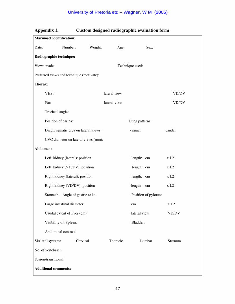
47
Appendix 1. Custom designed radiographic evaluation form
Marmoset identification:
Date: Number: Weight: Age: Sex:
Radiographic technique:
Views made: Technique used:
Preferred views and technique (motivate):
Thorax:
VHS: lateral view VD/DV
Fat: lateral view VD/DV
Tracheal angle:
Position of carina: Lung patterns:
Diaphragmatic crus on lateral views : cranial caudal
CVC diameter on lateral views (mm):
Abdomen:
Left kidney (lateral): position length: cm x L2
Left kidney (VD/DV): position length: cm x L2
Right kidney (lateral): position length: cm x L2
Right kidney (VD/DV): position length: cm x L2
Stomach: Angle of gastric axis: Position of pylorus:
Large intestinal diameter: cm x L2
Caudal extent of liver (cm): lateral view VD/DV
Visibility of: Spleen: Bladder:
Abdominal contrast:
Skeletal system: Cervical Thoracic Lumbar Sternum
No. of vertebrae:
Fusion/transitional:
Additional comments:
UUnniivveerrssiittyy ooff PPrreettoorriiaa eettdd –– WWaaggnneerr,, WW MM ((22000055))
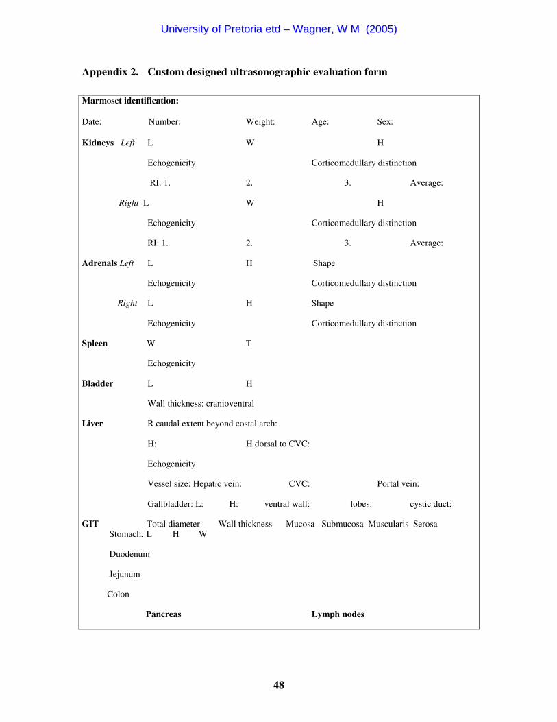
48
Appendix 2. Custom designed ultrasonographic evaluation form
Marmoset identification:
Date: Number: Weight: Age: Sex:
Kidneys Left L W H Echogenicity Corticomedullary distinction RI: 1. 2. 3. Average:
Right L W H Echogenicity Corticomedullary distinction RI: 1. 2. 3. Average: Adrenals Left L H Shape Echogenicity Corticomedullary distinction
Right L H Shape Echogenicity Corticomedullary distinction Spleen W T Echogenicity Bladder L H Wall thickness: cranioventral Liver R caudal extent beyond costal arch: H: H dorsal to CVC:
Echogenicity Vessel size: Hepatic vein: CVC: Portal vein: Gallbladder: L: H: ventral wall: lobes: cystic duct: GIT Total diameter Wall thickness Mucosa Submucosa Muscularis Serosa Stomach: L H W Duodenum Jejunum Colon Pancreas Lymph nodes
UUnniivveerrssiittyy ooff PPrreettoorriiaa eettdd –– WWaaggnneerr,, WW MM ((22000055))

49
Appendix 3. Radiographic findings of the skeletal system in 17 common marmosets
N
Age
Sex
Cer
vica
l ve
rteb
rae
Tho
raci
c ve
rteb
rae
Ster
nebr
ae
Fuse
d st
erne
brae
L
umba
r
vert
ebra
e T
hora
cic
and
lum
bar
vert
ebra
e
Tra
nsiti
onal
(la
st lu
mba
r)
Alig
nmen
t of
last
lum
bar
vert
ebra
Sa
cral
ve
rteb
rae
Fuse
d sa
cral
ve
rteb
rae
M2 9.00 M 7.00 13.00 5.00 5.00 6.00 19.00 No Slopes 3.00 3.00 M4 1.50 M 7.00 12.00 4.00 No 7.00 19.00 Yes Sacral 3.00 2.00 M5 4.50 M 7.00 13.00 5.00 No 6.00 19.00 No Slopes 3.00 3.00 M6 1.50 M 7.00 13.00 5.00 No 6.00 19.00 Yes Sacral 3.00 3.00 M7 3.50 M 7.00 13.00 4.00 No 6.00 19.00 Right Slopes 3.00 3.00 M8 3.50 M 7.00 12.00 4.00 No 7.00 19.00 Yes Sacral 3.00 2.50
M10 2.00 M 7.00 13.00 4.00 No 6.00 19.00 Yes Sacral 3.00 3.00 M11 9.00 M 7.00 13.00 6.00 No 6.00 19.00 No Sacral 3.00 3.00 M12 10.00 F 7.00 13.00 5.00 No 6.00 19.00 No Slopes 3.00 2.00 M13 1.50 F 7.00 12.00 4.00 No 7.00 19.00 Right Slopes 3.00 3.00 M14 1.50 F 7.00 13.00 5.00 No 6.00 19.00 No Lumbar 3.00 3.00 M15 5.00 F 7.00 13.00 4.00 2.00 6.00 19.00 Right Slopes 2.00 2.00 M16 4.50 F 7.00 12.00 4.00 No 7.00 19.00 No Slopes 3.00 3.00 M17 3.00 F 7.00 12.00 5.00 No 7.00 19.00 Yes Sacral 3.00 3.00 M18 4.00 F 7.00 12.00 4.00 No 6.00 18.00 No Lumbar 3.00 3.00 M19 7.00 F 7.00 13.00 5.00 3.00 6.00 19.00 Left Slopes 3.00 3.00 M20 6.00 F 7.00 12.00 4.00 No 7.00 19.00 Yes Slopes 3.00 2.00
Males & females Mean 7.00 12.59 4.53 6.35 18.94 2.94 2.74 SD 0.00 0.51 0.62 0.49 0.24 0.24 0.44 2 x SD 0.00 1.01 1.25 0.99 0.49 0.49 0.87 Range top (2 x) 7.00 13.60 5.78 7.34 19.43 3.43 3.61 Range bottom (2x) 7.00 11.57 3.28 5.37 18.46 2.46 1.86 Range top 7.00 13.10 5.15 6.85 19.18 3.18 3.17 Range bottom 7.00 12.08 3.91 5.86 18.70 2.70 2.30 Males Mean 7.00 12.75 4.63 6.25 19.00 3.00 2.81 SD 0.00 0.46 0.74 0.46 0.00 0.00 0.37 2 x SD 0.00 0.93 1.49 0.93 0.00 0.00 0.74 Range top (2 x) 7.00 13.68 6.11 7.18 19.00 3.00 3.56 Range bottom (2x) 7.00 11.82 3.14 5.32 19.00 3.00 2.07 Range top 7.00 13.21 5.37 6.71 19.00 3.00 3.18 Range bottom 7.00 12.29 3.88 5.79 19.00 3.00 2.44 Females Mean 7.00 12.44 4.44 6.44 18.89 2.89 2.67 SD 0.00 0.53 0.53 0.53 0.33 0.33 0.50 2 x SD 0.00 1.05 1.05 1.05 0.67 0.67 1.00 Range top (2 x) 7.00 13.50 5.50 7.50 19.56 3.56 3.67 Range bottom (2x) 7.00 11.39 3.39 5.39 18.22 2.22 1.67 Range top 7.00 12.97 4.97 6.97 19.22 3.22 3.17 Range bottom 7.00 11.92 3.92 5.92 18.56 2.56 2.17
UUnniivveerrssiittyy ooff PPrreettoorriiaa eettdd –– WWaaggnneerr,, WW MM ((22000055))
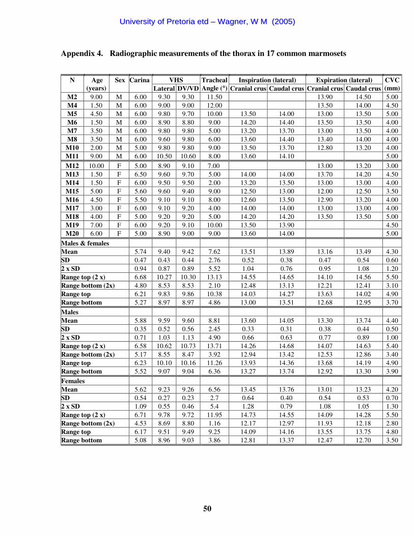
50
Appendix 4. Radiographic measurements of the thorax in 17 common marmosets
VHS Inspiration (lateral) Expiration (lateral) N
Age (years)
Sex
Carina Lateral DV/VD
Tracheal Angle (°°) Cranial crus Caudal crus Cranial crus Caudal crus
CVC (mm)
M2 9.00 M 6.00 9.30 9.30 11.50 13.90 14.50 5.00 M4 1.50 M 6.00 9.00 9.00 12.00 13.50 14.00 4.50 M5 4.50 M 6.00 9.80 9.70 10.00 13.50 14.00 13.00 13.50 5.00 M6 1.50 M 6.00 8.90 8.80 9.00 14.20 14.40 13.50 13.50 4.00 M7 3.50 M 6.00 9.80 9.80 5.00 13.20 13.70 13.00 13.50 4.00 M8 3.50 M 6.00 9.60 9.80 6.00 13.60 14.40 13.40 14.00 4.00
M10 2.00 M 5.00 9.80 9.80 9.00 13.50 13.70 12.80 13.20 4.00 M11 9.00 M 6.00 10.50 10.60 8.00 13.60 14.10 5.00 M12 10.00 F 5.00 8.90 9.10 7.00 13.00 13.20 3.00 M13 1.50 F 6.50 9.60 9.70 5.00 14.00 14.00 13.70 14.20 4.50 M14 1.50 F 6.00 9.50 9.50 2.00 13.20 13.50 13.00 13.00 4.00 M15 5.00 F 5.60 9.60 9.40 9.00 12.50 13.00 12.00 12.50 3.50 M16 4.50 F 5.50 9.10 9.10 8.00 12.60 13.50 12.90 13.20 4.00 M17 3.00 F 6.00 9.10 9.20 4.00 14.00 14.00 13.00 13.00 4.00 M18 4.00 F 5.00 9.20 9.20 5.00 14.20 14.20 13.50 13.50 5.00 M19 7.00 F 6.00 9.20 9.10 10.00 13.50 13.90 4.50 M20 6.00 F 5.00 8.90 9.00 9.00 13.60 14.00 5.00
Males & females Mean 5.74 9.40 9.42 7.62 13.51 13.89 13.16 13.49 4.30 SD 0.47 0.43 0.44 2.76 0.52 0.38 0.47 0.54 0.60 2 x SD 0.94 0.87 0.89 5.52 1.04 0.76 0.95 1.08 1.20 Range top (2 x) 6.68 10.27 10.30 13.13 14.55 14.65 14.10 14.56 5.50 Range bottom (2x) 4.80 8.53 8.53 2.10 12.48 13.13 12.21 12.41 3.10 Range top 6.21 9.83 9.86 10.38 14.03 14.27 13.63 14.02 4.90 Range bottom 5.27 8.97 8.97 4.86 13.00 13.51 12.68 12.95 3.70 Males Mean 5.88 9.59 9.60 8.81 13.60 14.05 13.30 13.74 4.40 SD 0.35 0.52 0.56 2.45 0.33 0.31 0.38 0.44 0.50 2 x SD 0.71 1.03 1.13 4.90 0.66 0.63 0.77 0.89 1.00 Range top (2 x) 6.58 10.62 10.73 13.71 14.26 14.68 14.07 14.63 5.40 Range bottom (2x) 5.17 8.55 8.47 3.92 12.94 13.42 12.53 12.86 3.40 Range top 6.23 10.10 10.16 11.26 13.93 14.36 13.68 14.19 4.90 Range bottom 5.52 9.07 9.04 6.36 13.27 13.74 12.92 13.30 3.90 Females Mean 5.62 9.23 9.26 6.56 13.45 13.76 13.01 13.23 4.20 SD 0.54 0.27 0.23 2.7 0.64 0.40 0.54 0.53 0.70 2 x SD 1.09 0.55 0.46 5.4 1.28 0.79 1.08 1.05 1.30 Range top (2 x) 6.71 9.78 9.72 11.95 14.73 14.55 14.09 14.28 5.50 Range bottom (2x) 4.53 8.69 8.80 1.16 12.17 12.97 11.93 12.18 2.80 Range top 6.17 9.51 9.49 9.25 14.09 14.16 13.55 13.75 4.80 Range bottom 5.08 8.96 9.03 3.86 12.81 13.37 12.47 12.70 3.50
UUnniivveerrssiittyy ooff PPrreettoorriiaa eettdd –– WWaaggnneerr,, WW MM ((22000055))
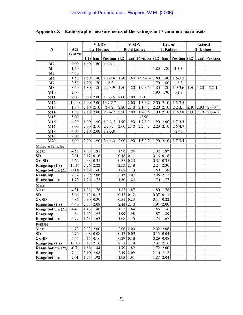
51
Appendix 5. Radiographic measurements of the kidneys in 17 common marmosets
VD/DV VD/DV Lateral Lateral Left kidney Right kidney 1. Kidney 2. Kidney
N
Age
(years) (L2) (cm) Position (L2) (cm) Position (L2) (cm) Position (L2) (cm) Position
M2 9.00 1.60 1.60 1.4-3.2 M4 1.50 1.80 1.80 2-3.5 M5 4.50 M6 1.50 1.80 1.80 1.1-2.8 1.70 1.80 13.5-2.4 1.80 1.80 1.5-3.3 M7 3.50 1.70 1.70 1.2-3 1.70 1.60 1.2-3 M8 3.50 1.80 1.80 2.2-4.9 1.80 1.80 1.9-3.5 1.80 1.80 1.9-3.6 1.80 1.80 2.2-4
M10 2.00 1.90 1.90 1-2.9 M11 9.00 2.00 2.00 1.7-3.5 2.00 2.00 1-3.1 M12 10.00 2.00 2.00 13.7-2.7 2.00 1.3-3.2 2.00 2.10 1.5-3.5 M13 1.50 2.10 2.10 2-4.2 2.20 2.10 2.1-4.2 2.20 2.10 2.2-3.1 2.10 2.00 2.6-3.4 M14 1.50 2.10 2.00 2.2-4.2 2.20 2.00 1.7-3.6 1.90 2.10 1.9-3.8 2.00 2.10 2.0-4.0 M15 5.00 -2.90 M16 4.50 1.90 1.90 1.9-3.5 1.90 1.90 1.7-3.5 1.90 2.00 1.7-3.3 M17 3.00 2.00 2.10 2.2-4.1 2.00 2.10 2.2-4.2 2.20 2.10 2.6-4.7 M18 4.00 2.10 2.00 1.9-3.8 -2.40 M19 7.00 M20 6.00 2.00 1.90 2.4-4.2 2.00 1.90 1.5-3.2 1.90 2.10 1.7-3.4
Males & females Mean 4.53 1.93 1.91 1.98 1.96 1.92 1.95 SD 2.81 0.17 0.16 0.18 0.11 0.16 0.18 2 x SD 5.62 0.33 0.31 0.35 0.23 0.32 0.35 Range top (2 x) 10.15 2.26 2.22 2.33 2.18 2.24 2.30 Range bottom (2x) -1.09 1.59 1.60 1.62 1.73 1.60 1.59 Range top 7.34 2.09 2.06 2.15 2.07 2.08 2.12 Range bottom 1.72 1.76 1.75 1.80 1.84 1.76 1.77 Male Mean 4.31 1.78 1.78 1.83 1.87 1.80 1.78 SD 3.64 0.15 0.15 0.15 0.12 0.07 0.11 2 x SD 4.86 0.30 0.30 0.31 0.23 0.14 0.22 Range top (2 x) 4.43 2.08 2.08 2.14 2.10 1.94 2.00 Range bottom (2x) 4.43 1.48 1.48 1.53 1.64 1.66 1.56 Range top 4.64 1.93 1.93 1.99 1.98 1.87 1.89 Range bottom 4.79 1.63 1.63 1.68 1.75 1.73 1.67 Female Mean 4.72 2.03 2.00 2.06 2.00 2.02 2.08 SD 2.72 0.08 0.08 0.13 0.09 0.15 0.04 2 x SD 5.43 0.15 0.16 0.27 0.18 0.29 0.08 Range top (2 x) 10.16 2.18 2.16 2.33 2.18 2.31 2.16 Range bottom (2x) -0.71 1.88 1.84 1.79 1.82 1.72 2.00 Range top 7.44 2.10 2.08 2.19 2.09 2.16 2.12 Range bottom 2.01 1.95 1.92 1.93 1.91 1.87 2.04
UUnniivveerrssiittyy ooff PPrreettoorriiaa eettdd –– WWaaggnneerr,, WW MM ((22000055))

52
Appendix 6. Radiographic measurements of the rest of the abdomen in 17 common
marmosets
LI Liver extent (cm) N
Age (years)
Sex
Weight (g)
Contrast
Gastric axis angle (°°) (xL2) (cm) Lateral VD/DV
M2 9.00 M 328.00 Poor 93.00 0.40 0.40 M4 1.50 M 328.00 Poor 97.00 0.75 0.60 M5 4.50 M 420.00 Poor 114.00 0.60 0.65 3.40 M6 1.50 M 342.00 Good 90.00 0.50 0.50 2.00 M7 3.50 M 420.00 Poor 95.00 1.20 1.20 1.70 1.70 M8 3.50 M 354.00 Excellent 90.00 1.00 1.00 3.00 3.00
M10 2.00 M 320.00 Poor 89.00 0.70 0.70 1.50 M11 9.00 M 496.00 Poor 95.00 0.80 0.80
M12 10.00 F 330.00 Good 110.00 1.00 1.00 M13 1.50 F 506.00 Excellent 85.00 1.40 1.40 3.60 3.60 M14 1.50 F 350.00 Excellent 95.00 1.00 0.90 3.30 3.00 M15 5.00 F 418.00 Good 103.00 0.80 0.80 1.70 2.50 M16 4.50 F 398.00 Good 117.00 0.90 0.90 M17 3.00 F 402.00 Good 106.00 0.50 0.50 2.70 3.00 M18 4.00 F 428.00 Good 110.00 1.10 1.00 3.00 2.50 M19 7.00 F 446.00 Poor 104.00 1.00 0.90 3.60 M20 6.00 F 398.00 Good 112.00 0.70 0.70 1.00
Males & females Mean 393.18 100.29 0.84 0.82 2.54 2.76 SD 57.74 9.90 0.27 0.26 0.91 0.60 2 x SD 115.48 19.80 0.54 0.52 1.83 1.19 Range top (2 x) 508.66 120.09 1.38 1.34 4.37 3.95 Range bottom (2x) 277.69 80.50 0.31 0.30 0.72 1.56 Range top 450.92 110.19 1.11 1.08 3.45 3.35 Range bottom 335.44 90.40 0.58 0.56 1.63 2.16 Male Mean 376.00 95.38 0.74 0.73 2.32 2.35 SD 62.86 8.05 0.26 0.26 0.83 0.92 2 x SD 125.71 16.10 0.52 0.53 1.67 1.84 Range top (2 x) 501.71 111.48 1.27 1.26 3.99 4.19 Range bottom (2x) 250.29 79.27 0.22 0.20 0.65 0.51 Range top 438.86 103.43 1.00 0.99 3.15 3.27 Range bottom 313.14 87.32 0.48 0.47 1.49 1.43 Female Mean 408.44 104.67 0.93 0.90 2.70 2.92 SD 51.51 9.67 0.25 0.24 1.00 0.45 2 x SD 103.03 19.34 0.51 0.49 1.99 0.91 Range top (2 x) 511.47 124.01 1.44 1.39 4.69 3.83 Range bottom (2x) 305.41 85.33 0.42 0.41 0.71 2.01 Range top 459.96 114.34 1.19 1.14 3.70 3.37 Range bottom 356.93 95.00 0.68 0.66 1.70 2.47
UUnniivveerrssiittyy ooff PPrreettoorriiaa eettdd –– WWaaggnneerr,, WW MM ((22000055))

53
Appendix 7. Ultrasonographic measurements of the kidneys in 17 common
marmosets
Left kidney RI Right kidney RI N
Age (years)
Sex L H W 1. 2. 3. Average L H W 1. 2. 3. Average
M2 9.00 M 15.60 9.40 11.00 0.72 0.72 16.00 9.30 0.67 0.67 0.67 M4 1.50 M 16.00 9.70 0.70 0.75 0.80 0.75 15.40 9.80 0.67 0.67 0.67 0.67 M5 4.50 M 22.50 11.90 21.20 12.20 0.82 0.78 0.70 0.77 M6 1.50 M 16.90 8.30 12.90 0.75 0.80 0.76 0.77 17.10 7.90 0.77 0.82 0.79 0.79 M7 3.50 M 17.70 9.20 16.70 8.50 12.70 0.75 0.72 0.70 0.72 M8 3.50 M 17.20 9.50 10.20 0.80 0.75 0.73 0.76 15.40 9.00 0.70 0.75 0.67 0.71
M10 2.00 M 17.10 8.20 9.70 0.73 0.75 0.80 0.76 14.30 9.20 11.40 0.80 0.84 0.84 0.83 M11 9.00 M 19.10 9.10 12.70 0.70 0.60 0.69 0.66 20.40 11.70 14.30 0.67 0.61 0.64 0.64
M12 5.00 F 20.40 11.80 13.90 0.67 0.79 0.73 18.20 12.20 13.70 0.75 0.80 0.72 0.76 M13 1.50 F 21.00 11.50 13.10 0.75 0.63 0.70 0.69 20.00 10.40 12.50 0.71 0.80 0.73 0.75 M14 1.50 F 20.30 10.30 14.10 0.79 0.71 0.74 0.75 18.20 9.50 16.20 0.68 0.70 0.66 0.68 M15 5.00 F 19.20 9.80 13.10 0.77 0.75 0.77 0.76 17.90 9.90 0.69 0.62 0.69 0.67 M16 4.50 F 20.90 9.40 9.30 0.71 0.79 0.75 18.70 10.40 0.64 0.73 0.75 0.71 M17 3.00 F 19.70 9.80 15.30 0.76 0.80 0.78 20.50 9.90 0.73 0.73 M18 4.00 F 20.00 11.70 12.90 0.71 0.75 0.71 0.72 19.90 10.40 12.10 0.75 0.79 0.75 0.76 M19 7.00 F 20.10 11.10 16.60 0.84 0.82 0.82 0.83 20.10 11.20 0.71 0.71 0.71 0.71 M20 6.00 F 19.90 9.50 13.40 0.83 0.71 0.78 0.77 19.50 10.80 11.10 0.67 0.74 0.75 0.72
Males & females Mean 19.04 10.01 12.73 0.75 0.74 0.75 0.75 18.21 10.14 13.00 0.72 0.73 0.72 0.72 SD 1.95 1.18 2.07 0.05 0.06 0.04 0.04 2.09 1.22 1.68 0.05 0.07 0.05 0.05 2 x SD 3.89 2.36 4.15 0.10 0.13 0.09 0.08 4.19 2.45 3.36 0.10 0.14 0.11 0.10 Range top (2 x) 22.93 12.37 16.88 0.85 0.87 0.84 0.82 22.39 12.58 16.36 0.82 0.87 0.82 0.82 Range bottom (2x) 15.14 7.65 8.58 0.65 0.62 0.67 0.67 14.02 7.69 9.64 0.62 0.60 0.61 0.62 Range top 20.98 11.19 14.80 0.80 0.81 0.80 0.79 20.30 11.36 14.68 0.77 0.80 0.77 0.77 Range bottom 17.09 8.83 10.65 0.70 0.68 0.71 0.71 16.11 8.91 11.32 0.67 0.67 0.67 0.67 Males Mean 17.76 9.41 11.30 0.73 0.73 0.76 0.74 17.06 9.70 12.80 0.73 0.73 0.72 0.72 SD 2.19 1.14 1.45 0.04 0.08 0.05 0.04 2.47 1.50 1.45 0.06 0.08 0.07 0.07 2 x SD 4.38 2.28 2.89 0.08 0.15 0.09 0.08 4.94 3.01 2.91 0.12 0.16 0.14 0.13 Range top (2 x) 22.14 11.70 14.19 0.81 0.88 0.85 0.82 22.00 12.71 15.71 0.85 0.89 0.86 0.86 Range bottom (2x) 13.39 7.13 8.41 0.66 0.58 0.66 0.66 12.12 6.69 9.89 0.61 0.57 0.57 0.59 Range top 19.95 10.55 12.75 0.77 0.81 0.80 0.78 19.53 11.20 14.25 0.79 0.81 0.79 0.79 Range bottom 15.57 8.27 9.85 0.70 0.65 0.71 0.70 14.59 8.20 11.35 0.67 0.65 0.64 0.66 Females Mean 20.17 10.54 13.52 0.76 0.75 0.75 0.75 19.22 10.52 13.12 0.70 0.74 0.72 0.72 SD 0.57 0.98 1.99 0.06 0.06 0.05 0.04 0.98 0.81 1.96 0.04 0.06 0.03 0.03 2 x SD 1.13 1.96 3.98 0.11 0.12 0.09 0.08 1.96 1.61 3.92 0.07 0.12 0.07 0.07 Range top (2 x) 21.30 12.51 17.50 0.87 0.87 0.84 0.83 21.18 12.14 17.04 0.78 0.86 0.79 0.79 Range bottom (2x) 19.04 8.58 9.54 0.65 0.63 0.66 0.68 17.27 8.91 9.20 0.63 0.61 0.65 0.65 Range top 20.73 11.53 15.51 0.82 0.81 0.80 0.79 20.20 11.33 15.08 0.74 0.80 0.75 0.75 Range bottom 19.60 9.56 11.53 0.70 0.69 0.71 0.72 18.24 9.71 11.16 0.67 0.67 0.69 0.69
UUnniivveerrssiittyy ooff PPrreettoorriiaa eettdd –– WWaaggnneerr,, WW MM ((22000055))
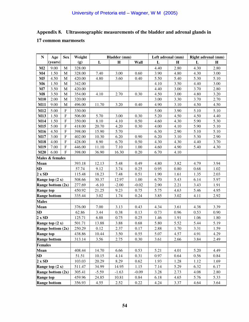
54
Appendix 8. Ultrasonographic measurements of the bladder and adrenal glands in
17 common marmosets
Bladder (mm) Left adrenal (mm) Right adrenal (mm) N
Age (years)
Sex
Weight (g) L H Wall L H L H
M2 9.00 M 328.00 4.40 2.80 4.30 2.80 M4 1.50 M 328.00 7.40 3.00 0.60 3.90 4.80 4.30 3.00 M5 4.50 M 420.00 4.80 3.60 0.40 5.50 5.40 5.30 5.10 M6 1.50 M 342.00 4.10 3.50 4.40 3.00 M7 3.50 M 420.00 4.40 3.00 3.70 2.80 M8 3.50 M 354.00 4.10 2.70 0.30 4.50 3.00 4.80 3.20
M10 2.00 M 320.00 3.00 3.30 3.70 2.70 M11 9.00 M 496.00 11.70 3.20 0.40 4.90 3.10 4.50 4.50
M12 5.00 F 330.00 5.00 3.90 5.10 5.10 M13 1.50 F 506.00 5.70 3.00 0.30 5.20 4.50 4.50 4.40 M14 1.50 F 350.00 8.10 4.10 0.50 4.60 4.30 5.90 5.30 M15 5.00 F 418.00 20.70 4.20 0.30 4.00 4.10 5.90 5.10 M16 4.50 F 398.00 15.90 5.70 6.30 2.90 5.10 5.10 M17 3.00 F 402.00 10.30 6.20 0.90 6.20 3.10 5.30 2.90 M18 4.00 F 428.00 8.90 6.70 0.50 4.30 4.30 4.40 3.70 M19 7.00 F 446.00 11.10 7.10 1.00 4.60 4.90 5.40 4.30 M20 6.00 F 398.00 36.90 16.30 0.20 6.70 4.10
Males & females Mean 393.18 12.13 5.48 0.49 4.80 3.82 4.79 3.94 SD 57.74 9.12 3.74 0.25 0.95 0.80 0.68 1.02 2 x SD 115.48 18.23 7.48 0.51 1.90 1.61 1.35 2.03 Range top (2 x) 508.66 30.37 12.97 1.00 6.70 5.43 6.14 5.97 Range bottom (2x) 277.69 -6.10 -2.00 -0.02 2.90 2.21 3.43 1.91 Range top 450.92 21.25 9.23 0.75 5.75 4.63 5.46 4.95 Range bottom 335.44 3.02 1.74 0.24 3.85 3.02 4.11 2.92 Males Mean 376.00 7.00 3.13 0.43 4.34 3.61 4.38 3.39 SD 62.86 3.44 0.38 0.13 0.73 0.96 0.53 0.90 2 x SD 125.71 6.88 0.75 0.25 1.46 1.91 1.06 1.80 Range top (2 x) 501.71 13.88 3.88 0.68 5.80 5.52 5.44 5.19 Range bottom (2x) 250.29 0.12 2.37 0.17 2.88 1.70 3.31 1.59 Range top 438.86 10.44 3.50 0.55 5.07 4.57 4.91 4.29 Range bottom 313.14 3.56 2.75 0.30 3.61 2.66 3.84 2.49 Females Mean 408.44 14.70 6.66 0.53 5.21 4.01 5.20 4.49 SD 51.51 10.15 4.14 0.31 0.97 0.64 0.56 0.84 2 x SD 103.03 20.29 8.29 0.62 1.93 1.28 1.12 1.69 Range top (2 x) 511.47 34.99 14.95 1.15 7.14 5.29 6.32 6.17 Range bottom (2x) 305.41 -5.59 -1.63 -0.09 3.28 2.73 4.08 2.80 Range top 459.96 24.85 10.81 0.84 6.18 4.65 5.76 5.33 Range bottom 356.93 4.55 2.52 0.22 4.24 3.37 4.64 3.64
UUnniivveerrssiittyy ooff PPrreettoorriiaa eettdd –– WWaaggnneerr,, WW MM ((22000055))

55
Appendix 9. Ultrasonographic measurements of liver and spleen in 17 common
marmosets
Liver (mm) Gallbladder Spleen (mm) H Vessel diameter
N
Age (years)
Sex
Right extent Maximal D toCVC HV PV CVC
L (mm)
H (mm)
Lobes
Wall (mm)
Bile ducts (mm) W T
M2 9.00 M 5.60 23.60 2.00 4.10 16.00 5.80 3.00 0.30 1.80 9.30 6.50 M4 1.50 M 3.30 2.10 3.60 14.80 7.30 3.00 0.50 4.10 M5 4.50 M 1.30 2.10 2.20 17.10 6.60 0.20 14.20 7.60 M6 1.50 M 14.10 6.20 1.10 1.20 3.00 8.90 6.20 0.60 12.90 6.40 M7 3.50 M 7.70 18.80 6.60 1.20 1.80 2.30 14.70 4.50 3.00 0.40 10.00 5.50 M8 3.50 M 28.20 18.60 1.00 1.40 2.10 9.50 4.50 1.00 0.30 2.10 16.60 6.10
M10 2.00 M 15.60 20.70 5.70 1.10 1.40 3.40 9.60 8.10 2.00 0.30 3.10 8.50 4.80 M11 9.00 M 16.00 22.00 6.30 2.00 3.90 12.10 5.30 3.00 0.20 2.00 11.00 8.20 M12 5.00 F 10.40 21.70 5.00 1.40 2.30 2.10 13.50 6.00 3.00 0.30 14.10 6.30 M13 1.50 F 18.10 25.70 6.60 1.60 2.40 3.00 15.50 5.00 3.00 0.40 3.70 17.80 9.80 M14 1.50 F 15.90 18.90 6.80 1.80 2.20 3.10 12.10 5.30 3.00 0.30 4.40 16.50 6.90 M15 5.00 F 16.90 23.40 6.70 2.10 2.40 14.20 7.50 3.00 0.30 1.30 16.00 8.30 M16 4.50 F 8.80 22.60 0.90 1.90 3.40 12.20 5.30 2.10 16.30 6.90 M17 3.00 F 20.80 1.00 1.80 2.10 17.90 5.90 3.00 0.50 1.80 14.00 6.00 M18 4.00 F 22.00 22.20 1.20 2.10 2.70 12.10 5.60 0.30 2.40 9.10 6.00 M19 7.00 F 12.50 17.80 0.90 2.60 2.80 10.60 8.50 3.00 0.50 2.50 9.10 M20 6.00 F 12.10 19.20 1.00 2.60 3.00 13.70 6.10 0.30 3.40 11.70 4.60
Males & females Mean 13.81 21.14 6.24 1.25 2.00 2.89 13.21 6.09 2.75 0.36 2.55 13.20 6.65 SD 6.39 2.29 0.61 0.34 0.41 0.64 2.66 1.17 0.62 0.12 0.91 3.11 1.56 2 x SD 12.77 4.59 1.22 0.68 0.82 1.28 5.32 2.35 1.24 0.23 1.83 6.21 3.12 Range top (2 x) 26.59 25.73 7.46 1.93 2.82 4.18 18.52 8.44 3.99 0.59 4.38 19.41 9.77 Range bottom (2x) 1.04 16.55 5.01 0.57 1.18 1.61 7.89 3.74 1.51 0.13 0.72 6.99 3.54 Range top 20.20 23.44 6.85 1.59 2.41 3.53 15.86 7.26 3.37 0.47 3.46 16.31 8.21 Range bottom 7.43 18.85 5.63 0.91 1.59 2.25 10.55 4.91 2.13 0.24 1.64 10.09 5.09 Males Mean 12.93 20.74 6.20 1.28 1.71 3.08 12.84 6.04 2.50 0.35 2.25 11.79 6.15 SD 8.42 2.13 0.37 0.37 0.38 0.80 3.23 1.28 0.84 0.14 0.58 2.92 1.36 2 x SD 16.83 4.26 0.75 0.73 0.75 1.59 6.47 2.57 1.67 0.28 1.16 5.83 2.72 Range top (2 x) 29.76 25.00 6.95 2.01 2.47 4.67 19.30 8.60 4.17 0.63 3.41 17.62 8.87 Range bottom (2x) -3.90 16.48 5.45 0.55 0.96 1.48 6.37 3.47 0.83 0.07 1.09 5.95 3.43 Range top 21.34 22.87 6.57 1.65 2.09 3.87 16.07 7.32 3.34 0.49 2.83 14.70 7.51 Range bottom 4.51 18.61 5.83 0.92 1.34 2.28 9.60 4.75 1.66 0.21 1.67 8.87 4.79 Females Mean 14.59 21.37 6.28 1.23 2.22 2.73 13.53 6.13 3.00 0.36 2.70 14.44 7.10 SD 4.41 2.48 0.85 0.34 0.28 0.45 2.18 1.15 0.00 0.09 1.04 2.87 1.66 2 x SD 8.81 4.95 1.71 0.68 0.56 0.91 4.35 2.29 0.00 0.18 2.09 5.74 3.33 Range top (2 x) 23.40 26.32 7.98 1.91 2.79 3.64 17.89 8.42 3.00 0.55 4.79 20.18 10.43 Range bottom (2x) 5.77 16.41 4.57 0.54 1.66 1.83 9.18 3.84 3.00 0.18 0.61 8.69 3.77 Range top 18.99 23.84 7.13 1.57 2.50 3.19 15.71 7.28 3.00 0.45 3.74 17.31 8.76 Range bottom 10.18 18.89 5.42 0.88 1.94 2.28 11.36 4.99 3.00 0.27 1.66 11.57 5.44
UUnniivveerrssiittyy ooff PPrreettoorriiaa eettdd –– WWaaggnneerr,, WW MM ((22000055))
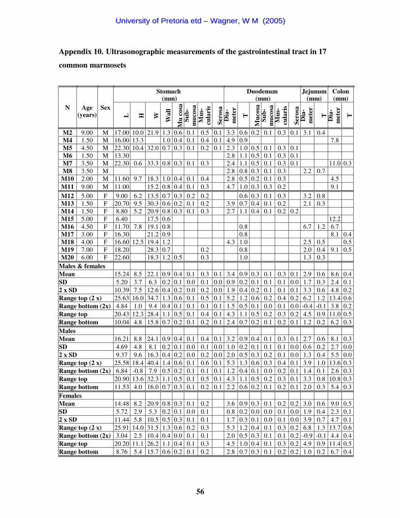
56
Appendix 10. Ultrasonographic measurements of the gastrointestinal tract in 17
common marmosets
Stomach (mm)
Duodenum (mm)
Jejunum (mm)
Colon (mm)
N
Age (years)
Sex
L
H
W
Wal
l
Mu
cosa
Su
b-m
ucos
a M
us-
cula
ris
Sero
sa
Dia
-m
eter
T
Muc
osa
Sub-
muc
osa
Mus
-cu
lari
s
Sero
sa
Dia
-m
eter
T
Dia
-m
eter
T
M2 9.00 M 17.00 10.0 21.9 1.3 0.6 0.1 0.5 0.1 3.3 0.6 0.2 0.1 0.3 0.1 3.1 0.4 M4 1.50 M 16.00 13.3 1.0 0.4 0.1 0.4 0.1 4.9 0.9 7.8 M5 4.50 M 22.30 10.4 32.0 0.7 0.3 0.1 0.2 0.1 2.3 1.0 0.5 0.1 0.3 0.1 M6 1.50 M 13.30 2.8 1.1 0.5 0.1 0.3 0.1 M7 3.50 M 22.30 0.6 33.3 0.8 0.3 0.1 0.3 2.4 1.1 0.5 0.1 0.3 0.1 11.0 0.3 M8 3.50 M 2.8 0.8 0.3 0.1 0.3 2.2 0.7
M10 2.00 M 11.60 9.7 18.3 1.0 0.4 0.1 0.4 2.8 0.5 0.2 0.1 0.3 4.5 M11 9.00 M 11.00 15.2 0.8 0.4 0.1 0.3 4.7 1.0 0.3 0.3 0.2 9.1 M12 5.00 F 9.00 6.2 13.5 0.7 0.3 0.2 0.2 0.6 0.3 0.1 0.3 3.2 0.8 M13 1.50 F 20.70 9.5 30.3 0.6 0.2 0.1 0.2 3.9 0.7 0.4 0.1 0.2 2.1 0.3 M14 1.50 F 8.80 5.2 20.9 0.8 0.3 0.1 0.3 2.7 1.1 0.4 0.1 0.2 0.2 M15 5.00 F 6.40 17.5 0.6 12.2 M16 4.50 F 11.70 7.8 19.1 0.8 0.8 6.7 1.2 6.7 M17 3.00 F 16.30 21.2 0.9 0.8 8.1 0.4 M18 4.00 F 16.60 12.5 19.4 1.2 4.3 1.0 2.5 0.5 0.5 M19 7.00 F 18.20 28.3 0.7 0.2 0.8 2.0 0.4 9.1 0.5 M20 6.00 F 22.60 18.3 1.2 0.5 0.3 1.0 1.3 0.3
Males & females Mean 15.24 8.5 22.1 0.9 0.4 0.1 0.3 0.1 3.4 0.9 0.3 0.1 0.3 0.1 2.9 0.6 8.6 0.4 SD 5.20 3.7 6.3 0.2 0.1 0.0 0.1 0.0 0.9 0.2 0.1 0.1 0.1 0.0 1.7 0.3 2.4 0.1 2 x SD 10.39 7.5 12.6 0.4 0.2 0.0 0.2 0.0 1.9 0.4 0.2 0.1 0.1 0.1 3.3 0.6 4.8 0.2 Range top (2 x) 25.63 16.0 34.7 1.3 0.6 0.1 0.5 0.1 5.2 1.2 0.6 0.2 0.4 0.2 6.2 1.2 13.4 0.6 Range bottom (2x) 4.84 1.0 9.4 0.4 0.1 0.1 0.1 0.1 1.5 0.5 0.1 0.0 0.1 0.0 -0.4 -0.1 3.8 0.2 Range top 20.43 12.3 28.4 1.1 0.5 0.1 0.4 0.1 4.3 1.1 0.5 0.2 0.3 0.2 4.5 0.9 11.0 0.5 Range bottom 10.04 4.8 15.8 0.7 0.2 0.1 0.2 0.1 2.4 0.7 0.2 0.1 0.2 0.1 1.2 0.2 6.2 0.3 Males Mean 16.21 8.8 24.1 0.9 0.4 0.1 0.4 0.1 3.2 0.9 0.4 0.1 0.3 0.1 2.7 0.6 8.1 0.3 SD 4.69 4.8 8.1 0.2 0.1 0.0 0.1 0.0 1.0 0.2 0.1 0.1 0.1 0.0 0.6 0.2 2.7 0.0 2 x SD 9.37 9.6 16.3 0.4 0.2 0.0 0.2 0.0 2.0 0.5 0.3 0.2 0.1 0.0 1.3 0.4 5.5 0.0 Range top (2 x) 25.58 18.4 40.4 1.4 0.6 0.1 0.6 0.1 5.3 1.3 0.6 0.3 0.4 0.1 3.9 1.0 13.6 0.3 Range bottom (2x) 6.84 -0.8 7.9 0.5 0.2 0.1 0.1 0.1 1.2 0.4 0.1 0.0 0.2 0.1 1.4 0.1 2.6 0.3 Range top 20.90 13.6 32.3 1.1 0.5 0.1 0.5 0.1 4.3 1.1 0.5 0.2 0.3 0.1 3.3 0.8 10.8 0.3 Range bottom 11.53 4.0 16.0 0.7 0.3 0.1 0.2 0.1 2.2 0.6 0.2 0.1 0.2 0.1 2.0 0.3 5.4 0.3 Females Mean 14.48 8.2 20.9 0.8 0.3 0.1 0.2 3.6 0.9 0.3 0.1 0.2 0.2 3.0 0.6 9.0 0.5 SD 5.72 2.9 5.3 0.2 0.1 0.0 0.1 0.8 0.2 0.0 0.0 0.1 0.0 1.9 0.4 2.3 0.1 2 x SD 11.44 5.8 10.5 0.5 0.3 0.1 0.1 1.7 0.3 0.1 0.0 0.1 0.0 3.9 0.7 4.7 0.1 Range top (2 x) 25.91 14.0 31.5 1.3 0.6 0.2 0.3 5.3 1.2 0.4 0.1 0.3 0.2 6.8 1.3 13.7 0.6 Range bottom (2x) 3.04 2.5 10.4 0.4 0.0 0.1 0.1 2.0 0.5 0.3 0.1 0.1 0.2 -0.9 -0.1 4.4 0.4 Range top 20.20 11.1 26.2 1.1 0.4 0.1 0.3 4.5 1.0 0.4 0.1 0.3 0.2 4.9 0.9 11.4 0.5 Range bottom 8.76 5.4 15.7 0.6 0.2 0.1 0.2 2.8 0.7 0.3 0.1 0.2 0.2 1.0 0.2 6.7 0.4
UUnniivveerrssiittyy ooff PPrreettoorriiaa eettdd –– WWaaggnneerr,, WW MM ((22000055))