c-anca-positive glomerulonephritis associated with ... · the diagnosis of bcne is difficult and...
TRANSCRIPT
CASE REPORT eISSN 2384-0293https://doi.org/10.12701/yujm.2017.34.1.140Yeungnam Univ J Med 2017;34(1):140-145
140 YUJM VOLUME 34, NUMBER 1, JUNE 2017
C-ANCA-positive glomerulonephritis associated with subacute infective endocarditis caused by Bartonella infection
Min Jeong Kim1, Ha Nee Jang1, Tae Won Lee1, Hyun Seop Cho1,2, Se-Ho Chang1,2, Hyun-Jung Kim1,2
1Department of Internal Medicine, Gyeongsang National University School of Medicine; 2Institute of Health Sciences, Gyeongsang National University, Jinju, Korea
Glomerulonephritis (GN) is sometimes associated with infective endocarditis (IE). Bartonella endocarditis is difficult to diagnose because it is rare and cannot be detected by blood culture. This is the first report of cytoplasmic anti-neutrophil cytoplasmic antibody-positive subacute endocarditis-associated GN caused by Bartonella infection in South Korea. A 67-year-old man was hospitalized due to azotemia. He complained of weight loss and anorexia for 6 months. A diagnosis of IE was made based upon echocardiographic detection of vegetations on the mitral and aortic valves and a Bartonella antibody titer of 1:2,048. Renal histology identified focal crescentic GN. Azotemia and proteinuria improved after doxycycline and rifampin treatment combining with steroid therapy.
Keywords: ANCA-associated glomerulonephritis; Bartonella; Infectious endocarditis
Copyright ©2017 Yeungnam University College of MedicineThis is an Open Access article distributed under the terms of the Creative Commons Attribution Non-Commercial License (http://creative- commons.org/licenses/by-nc/4.0/) which permits unrestricted non-commercial use, distribution, and reproduction in any medium, provided the original workis properly cited.
Received: May 4, 2017, Revised: May 22, 2017Accepted: June 1, 2017
Corresponding Author: Hyun-Jung Kim, Division of Nephrology,Department of Internal Medicine, Gyeongsang National University School of Medicine, 15 Jinju-daero 816beon-gil, Jinju 52727, KoreaTel: +82-55-750-8875, Fax: +82-55-758-9122E-mail: [email protected]
INTRODUCTION
Infective endocarditis (IE) is a life-threatening disease caused
by an infection of the heart tissues. Isolation of the causative organism from blood culture is critical for accurate diagnosis and treatment. When culture results of three independent
blood samples are negative, the putative diagnosis indicates blood culture-negative endocarditis (BCNE) [1]. The incide- nce of BCNE ranges from 5% to 15% in patients with endo-
carditis, depending on the country [2]. BCNE is associated with infection by a number of pathogens, including Granuli- catella, Abiotrophia, HACEK organisms, Coxiella burnetii, and Bartonella spp. [3]. Diagnostic tests for BCNE include
special extended-culture techniques, molecular techniques,
and histopathologic evaluation.Approximately 40-50% of IE cases have an associated kid-
ney disease such as glomerulonephritis (GN) [4]. Crescentic
GN is the most common renal pathology, which can be derived from anti-neutrophil cytoplasmic antibody (ANCA)-associated small-vessel vasculitis. The presence of ANCAs against protei-
nase-3 or myeloperoxidase is a key diagnostic marker [5].Bartonella spp. were first described as a cause of endocardi-
tis in 1993 [6]. Bacteria belonging to the Bartonella genus
are facultative intracellular gram-negative rods that infect er-ythrocytes or endothelial cells [7]. Most reports of Bartonella IE have involved adult patients and more than 70% of cases
were men [8]. The clinical manifestation of Bartonella IE is similar to that of other subacute IE [9]. A diagnosis of Bartonella IE, however, often requires diagnostic tests other
than blood culture to identify the infectious agent (as is the case for other BCNE). In most cases, antibiotic treatment of Bartonella-caused IE is effective. If renal function deterio-
rates despite appropriate antibiotic therapy, other treatments
Glomerulonephritis with Bartonella endocarditis
YUJM VOLUME 34, NUMBER 1, JUNE 2017 141
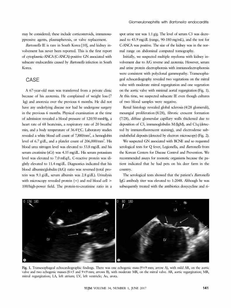
Fig. 1. Transesophageal echocardiographic findings. There was one echogenic mass (9×9 mm; arrow A), with mild AR, on the aorticvalve and two echogenic masses (8×5 and 9×9 mm; arrows B), with moderate MR, on the mitral valve. AR, aortic regurgitation; MR,mitral regurgitation; LA, left atrium; LV, left ventricle; Ao, arota.
may be considered; these include corticosteroids, immunosu- ppressive agents, plasmapheresis, or valve replacement.
Bartonella IE is rare in South Korea [10], and kidney in-
volvement has never been reported. This is the first report of cytoplasmic-ANCA (C-ANCA)-positive GN associated with subacute endocarditis caused by Bartonella infection in South
Korea.
CASE
A 67-year-old man was transferred from a private clinic because of his azotemia. He complained of weight loss (7
kg) and anorexia over the previous 6 months. He did not have any underlying disease nor had he undergone surgery in the previous 6 months. Physical examination at the time
of admission revealed a blood pressure of 120/50 mmHg, a heart rate of 68 beats/min, a respiratory rate of 20 breaths/ min, and a body temperature of 36.4℃C. Laboratory studies
revealed a white blood cell count of 7,880/mm3, a hemoglobin level of 6.7 g/dL, and a platelet count of 206,000/mm3. His blood urea nitrogen level was elevated to 53.8 mg/dL and his
serum creatinine (sCr) was 4.35 mg/dL. His serum potassium level was elevated to 7.0 mEq/L. C-reactive protein was sli- ghtly elevated to 11.4 mg/dL. Diagnostics indicated that his
blood albumin/globulin (A/G) ratio was reversed (total pro-tein was 9.1 g/dL, serum albumin was 2.8 g/dL). Urinalysis with microscopy revealed protein (+) and red blood cell >
100/high-power field. The protein-to-creatinine ratio in a
spot urine test was 1.1 g/g. The level of serum C3 was decre- ased to 43.9 mg/dL (range, 90-180 mg/mL), and the test for C-ANCA was positive. The size of the kidney was in the nor-
mal range on abdominal computed tomography.Initially, we suspected multiple myeloma with kidney in-
volvement due to A/G reverse and azotemia. However, serum
and urine protein electrophoresis with immunoelectrophoresis were consistent with polyclonal gammopathy. Transesopha- geal echocardiography revealed two vegetations on the mitral
valve with moderate mitral regurgitation and one vegetation on the aortic valve with minimal aortal regurgitation (Fig. 1). At this time, we suspected subacute IE even though cultures
of two blood samples were negative.Renal histology revealed global sclerosis (4/28 glomeruli),
mesangial proliferation (8/28), fibrotic crescent formation
(7/28), diffuse glomerular capillary walls thickened due to deposition of C3, immunoglobulin M(IgM), and C1q (detec- ted by immunofluorescent staining), and electrodense sub-
endothelial deposits (detected by electron microscopy) (Fig. 2).We suspected GN associated with BCNE and so requested
serological tests for Q fever, Legionella, and Bartonella from
the Korean Centers for Disease Control and Prevention. We recommended assays for zoonotic organisms because the pa-tient indicated that he had pets on his deer farm in the
country.The serological tests showed that the patient’s Bartonella
IgG antibody titer was elevated to 1:2048. Although he was
subsequently treated with the antibiotics doxycycline and ri-

Min Jeong Kim et al.
142 YUJM VOLUME 34, NUMBER 1, JUNE 2017
Fig. 3. Clinical course after admission. Serum creatinine and pro-teinuria improved after antibiotics were combined with steroidtherapy. sCr, serum creatinine; CRP, C-reactive protein; uPCR,urine protein/creatinine ratio.
Fig. 2. Renal pathologic findings. (A) Periodic acid-Schiff staining under light microscopy (×400). A glomerulus shows global mesangialexpansion with mild thickening of the capillary wall and crescent formation. Tubulointerstitial lymphocytic infiltration and tubularatrophy are also present. (B) Electromicrograph showing some subendothelial electron dense deposits (×10,000).
fampin for 8 weeks, his azotemia and proteinuria did not improve. After combining steroid therapy (0.5 mg/kg) with antibiotic treatment for another 6 weeks, his sCr improved
to 2.10 mg/dL, his proteinuria disappeared (Fig. 3), and his complement C3 level returned to normal. Follow-up transtho- racic echocardiography after 5 months showed that one of
two vegetations on the mitral valve disappeared and the other decreased slightly from 9.0×9.0 mm to 9.3×7.6 mm with mild mitral regurgitation. The size of vegetation on the aortic
valve decreased markedly from 9.0×9.0 mm to 5.3×4.9 mm. He currently receives out-patient support for chronic kidney
disease.
DISCUSSION
The diagnosis of BCNE is difficult and uncommon in South Korea. Nevertheless, a rigorous approach is needed to identi-
fy the specific infectious causes of endocarditis and non-IE, including careful documentation of a case’s history and ask-ing the patient and family about contact with animals in the
case of a potential zoonosis and joint, gastrointestinal, and neurological symptoms [11]. Our patient’s self-reported his-tory of animal contact was very helpful in our diagnosis.
Among the extra-cardiac manifestations associated with IE, renal involvement occurs in about 40-50% of patients [12]. The causes of kidney disease include acute pyelonephritis,
abscess formation from septic emboli, immune complex-medi- ated GN, ANCA-associated GN, and acute tubular necrosis or acute interstitial nephritis arising from renal toxicity caused
by antibiotic treatment [12].Renal biopsy is the only method of diagnosing IE-asso-
ciated GN. Key histopathologic findings include emboli, in-
terstitial nephritis, crescentic GN, immune complex-mediated GN, and acute tubular necrosis [13]. Our patient exhibited focal crescentic GN with focal mesangial GN.
In this case, a diagnosis of IE was made due to the echocar- diographic detection of vegetations on the mitral and aortic

Glomerulonephritis with Bartonella endocarditis
YUJM VOLUME 34, NUMBER 1, JUNE 2017 143
Table 1. Characteristics of infective endocarditis with glomerulonephritis in KoreaCase No. (year of report) 1 (1990) 2 (1999) 3 (2004) 4 (2009) 5 (present)Sex/age M/53 M/24 M/28 F/59 M/67Organism No growth S. viridans S. sanguis E. faecalis Bartonella spp.Laboratory findings CRP (mg/dL) 9.9 NR 8.5 206.5 11.4 Total protein (g/dL) 7.3 5.8 5.2 5.2 9.1 Serum albumin (g/dL) 2.4 2.1 2.4 2.4 2.8 Serum Cr (mg/dL) 7.0 16.2 3.46 3.2 4.35 Proteinuria 2.0 g/day (+++) 1.5 g/day 3.0 g/day 1.1 g/day Serum IgG (mg/dL) NR NR 2,090.85 NR 4,080.2 Serum C3 Decreased Decreased Decreased Decreased Decreased ANA titer - 1:40 - - 1:40 ANCA NR NR - - + Cryoglobulin + - - - - Anti-GBM Ab NR NR + NR -LM Crescent 29% 100% 21% 54% 25% Global sclerosis 8% - 71% - 14% Segmental sclerosis - - 7% - - Mesangial proliferation Almost - - - 29%IF C3/C1q/IgM (+/+/+) (+/-/+) (+/+/+) (+/-/+) (+/+/+) Mesangial/capillary (+/+) (+/+) (+/-) (+/+) (-/+)EM(EDD) Subepithelial NR + - NR - Subendothelial NR + + NR + Mesangial NR - + NR +Treatment Antibiotics + + + + + Surgery TVR MVR VSD repair Not done Not done Renal therapy HD Steroid+HD PCX Not done SteroidOutcome Died CAPD CKD CKD CKDCRP, C-reactive protein; Cr, creatinine; IgG, imnunoglobulin G; ANA, anti-nuclear antibody; ANCA, anti-neutrophil cytoplasmic anti-body; GBM Ab, glomerular basement membrane antibody; NR, not reported; LM, light microscopy; IF, immunofluorescence stain; EM, electron microscopy; EDD, electrodense deposits; TVR, tricuspid valve replacement; MV, mitral valve; VSD, ventricular septal defect; HD, hemodialysis, CAPD, continuous ambulatory peritoneal dialysis; PCX, plasma exchange; CKD, chronic kidney disease.
valves, and a positive serologic test result for Bartonella. Some may argue that serologic testing is insufficient to diag-nose bartonellosis because there is no definitive cut-off titer
for diagnosis and because of the potential for cross-reaction with antibodies to Chlamydia spp. or Coxiella burnetii. How- ever, Edouard et al. showed that an IgG titer greater than
1:800 (in an immunofluorescence antibody assay) or a pos-itive western blot can be considered as major Duke criteria indicating Bartonella endocarditis [14].
The appropriate antibiotic treatment for Bartonella endo-carditis is a 6-week course of amoxicillin, ceftriaxone, or doxy- cycline in combination with aminoglycosides for 2-4 weeks
[15]. Surgical treatment is recommended if the condition is accompanied by heart failure, high embolic risk, and persis-tent sepsis [16]. Therefore, we did not remove the vegetations
and initially prescribed doxycycline with rifampin rather than gentamicin due to the patient’s azotemia.
There are four case reports of IE associated with GN in
Min Jeong Kim et al.
144 YUJM VOLUME 34, NUMBER 1, JUNE 2017
Korea [17-20]. We summarized the characteristics of each in Table 1. Their clinical manifestations and course of infection were not different from cases reported elsewhere. Serological
markers varied: cryoglobulin in case 1, anti-GBM antibody in case 3 [19], and ANCA in our case. Interestingly, all exhi- bited a reversed A/G ratio, which initially led us to suspect
multiple myeloma. We believe that the elevated IgG levels in cases 1 and 5 are related to polyclonal gammopathy and reveal the chronic inflammation associated with IE. All cases
reported hypocomplementemia, azotemia, and mild protei- nuria. The patient in case 1 died [17] and the other patients developed chronic kidney disease. The renal function of our
patient was improving under treatment, but did not recover completely, although his proteinuria disappeared after com-bined steroid and antibiotic therapy. Follow-up echocardiog-
raphy showed an improvement, but he still developed chronic kidney disease.
Diagnosis was delayed because the patient’s chief com-
plaint and his initial clinical findings were vague, and his condition was not rapidly progressive. Interestingly, the symp- toms were very similar to those in a case reported by Van
Haare Heijmeijer et al. [12]. However, this patient diagnosed to Bartonella IE lately after steroid and cyclophosphamide therapy for idiopathic ANCA-associated GN without antibio-
tics. They summarized the renal pathology and therapy for ten cases of Bartonella-caused endocarditis-associated GN. In most cases, their renal pathologies were either focal or diffuse
crescentic GN. Moreover, as for our patient, seven of the patients displayed a positive ANCA result.
In conclusion, it is necessary to carefully evaluate the case
history, conduct a thorough physical examination, and per-form serologic tests to identify BCNE. In addition, if azotemia presents with proteinuria, a renal biopsy is considered to rule
out GN-associated endocarditis. With proper diagnosis and early treatment, a patient may be able to recover renal func-tion and experience a better clinical outcome.
ACKNOWLEDGMENT
The authors thank Dr. Oh-Hyun Cho and Dr. Dong Jun Park, Department of Internal Medicine and Dr. Dae Hyun Song, Department of Pathology, Gyeongsang National Univer-
sity Changwon Hospital, for assistance for this report.
CONFLICT OF INTEREST
Authors have no potential conflicts of interest to disclose.
ORCID
Min Jeong Kim, https://orcid.org/0000-0002-1677-7426
Ha Nee Jang, https://orcid.org/0000-0002-1402-931XTae Won Lee, https://orcid.org/0000-0003-1758-3217Hyun Seop Cho, https://orcid.org/0000-0002-2103-9129
Se-Ho Chang, https://orcid.org/0000-0002-5278-0438Hyun-Jung Kim, https://orcid.org/0000-0001-5883-4735
REFERENCES
1. Raoult D, Casalta JP, Richet H, Khan M, Bernit E, Rovery C, et al. Contribution of systematic serological testing in diag-nosis of infective endocarditis. J Clin Microbiol 2005;43: 5238-42.
2. Fournier PE, Thuny F, Richet H, Lepidi H, Casalta JP, Arzouni JP, et al. Comprehensive diagnostic strategy for blood culture-negative endocarditis: a prospective study of 819 new cases. Clin Infect Dis 2010;51:131-40.
3. Houpikian P, Raoult D. Blood culture-negative endocarditis in a reference center: etiologic diagnosis of 348 cases. Medicine (Baltimore) 2005;84:162-73.
4. Khalighi MA, Nguyen S, Wiedeman JA, Palma Diaz MF. Bartonella endocarditis-associated glomerulonephritis: a case report and review of the literature. Am J Kidney Dis 2014;63: 1060-5.
5. Satake K, Ohsawa I, Kobayashi N, Osaki K, Toyoda H, Hori- koshi S, et al. Three cases of PR3-ANCA positive subacute endocarditis caused by attenuated bacteria (Propionibacteri- um, Gemella, and Bartonella) complicated with kidney injury. Mod Rheumatol 2011;21:536-41.
6. Daly JS, Worthington MG, Brenner DJ, Moss CW, Hollis DG, Weyant RS, et al. Rochalimaea elizabethae sp. nov. iso-lated from a patient with endocarditis. J Clin Microbiol 1993;31:872-81.
7. Anderson BE, Neuman MA. Bartonella spp. as emerging hu-man pathogens. Clin Microbiol Rev 1997;10:203-19.
8. Raoult D, Fournier PE, Vandenesch F, Mainardi JL, Eykyn SJ, Nash J, et al. Outcome and treatment of Bartonella endo- carditis. Arch Intern Med 2003;163:226-30.
9. Durack DT, Lukes AS, Bright DK. New criteria for diagnosis of infective endocarditis: utilization of specific echocardio-graphic findings. Duke Endocarditis Service. Am J Med 1994; 96:200-9.
10. Lim MH, Chung DR, Kim WS, Park KS, Ki CS, Lee NY, et al. First case of Bartonella quintana endocarditis in Korea. J Korean Med Sci 2012;27:1433-5.
Glomerulonephritis with Bartonella endocarditis
YUJM VOLUME 34, NUMBER 1, JUNE 2017 145
11. Tattevin P, Watt G, Revest M, Arvieux C, Fournier PE. Update on blood culture-negative endocarditis. Med Mal Infect 2015;45:1-8.
12. Van Haare Heijmeijer S, Wilmes D, Aydin S, Clerckx C, Labrio la L. Necrotizing ANCA-positive glomerulonephritis secon- dary to culture-negative endocarditis. Case Rep Nephrol 2015;2015:649763.
13. Majumdar A, Chowdhary S, Ferreira MA, Hammond LA, Howie AJ, Lipkin GW, et al. Renal pathological findings in infective endocarditis. Nephrol Dial Transplant 2000;15: 1782-7.
14. Edouard S, Nabet C, Lepidi H, Fournier PE, Raoult D. Barto- nella, a common cause of endocarditis: a report on 106 cases and review. J Clin Microbiol 2015;53:824-9.
15. Gould FK, Denning DW, Elliott TS, Foweraker J, Perry JD, Prendergast BD, et al. Guidelines for the diagnosis and anti-biotic treatment of endocarditis in adults: a report of the Wor- king Party of the British Society for Antimicrobial Chemothe- rapy. J Antimicrob Chemother 2012;67:269-89.
16. Baddour LM, Wilson WR, Bayer AS, Fowler VG Jr, Tleyjeh
IM, Rybak MJ, et al. Infective endocarditis in adults: diag-nosis, antimicrobial therapy, and management of complica-tions: a scientific statement for healthcare professionals from the American Heart Association. Circulation 2015;132:1435- 86.
17. Lee KW, Chung TH, Park SB, Kim HC, Park KK, Lee SS. A case of diffuse proliferative glomerulonephritis in bacterial endocarditis. Korean J Nephrol 1990;9:427-33. Korean.
18. Lee JH, Ma KA, Kim HS, Shin KT, Kim MS, Suh YJ, et al. A case of crescentic glomerulonephritis associated with bacte-rial endocarditis. Korean J Nephrol 1999;18:820-4. Korean.
19. Han KH, Choi SW, Seong IW, Shin YT, Suh KS, Kwon GC, et al. Rapidly progressive glomerulonephritis associated with infective endocarditis: a dramatically improved case after plasmapheresis. Korean J Med 2004;67:73-7. Korean.
20. Kim JK, Lee YK, Oh SE, Cho JR, Noh JW, Nam ES, et al. Recovery from crescentic glomerulonephritis with bacterial endocarditis with antibiotics alone. Korean J Med 2009;76: 358-64. Korean.