c h a p t e r © 2013 pearson education, inc.© annie leibovitz/contact press images tissue: the...
TRANSCRIPT

C H A P T E R
© 2013 Pearson Education, Inc.© Annie Leibovitz/Contact Press Images
Tissue: The Living Fabric:
4

Types of Primary Tissues
• Epithelial tissue– Covers
• Connective tissue– Supports
• Muscle tissue– Produces movement
• Nerve tissue– Controls
04/18/23 2MDufilho

• Brain• Spinal cord• Nerves
Nervous tissue: Internal communication
• Muscles attached to bones (skeletal)• Muscles of heart (cardiac)• Muscles of walls of hollow organs (smooth)
Muscle tissue: Contracts to cause movement
Epithelial tissue: Forms boundaries between different environments, protects, secretes, absorbs, filters• Lining of digestive tract organs and other hollow
organs• Skin surface (epidermis)
• Bones• Tendons• Fat and other soft padding tissue
Connective tissue: Supports, protects, binds other tissues together
Figure 4.1 Overview of four basic tissue types: epithelial, connective, muscle, and nervous tissues.
04/18/23 3MDufilho

Epithelial Tissue (Epithelium)
• Form boundaries
• Two main types (by location)– Covering and lining epithelia
• On external and internal surfaces
– Glandular epithelia• Secretory tissue in glands
04/18/23 4MDufilho
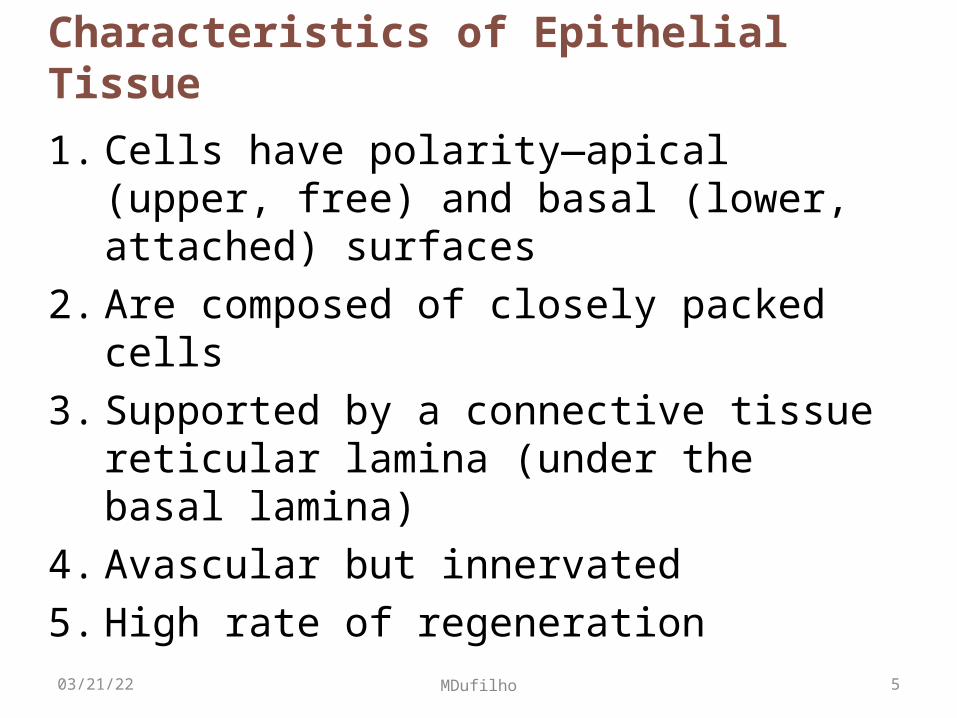
Characteristics of Epithelial Tissue
1. Cells have polarity—apical (upper, free) and basal (lower, attached) surfaces
2. Are composed of closely packed cells
3. Supported by a connective tissue reticular lamina (under the basal lamina)
4. Avascular but innervated
5. High rate of regeneration
04/18/23 5MDufilho

Classification of Epithelia
• Ask two questions:1. How many layers?
1 = simple epithelium
>1 = stratified epithelium
2. What is the shape of the cell• Squamous• Cuboidal• Columnar
04/18/23 6MDufilho

Figure 4.2a Classification of epithelia.
Basal surfaceStratified
Classification based on number of cell layers.
Basal surfaceSimple
Apical surface
Apical surface
04/18/23 7MDufilho

Cells of Epithelial Tissues
• Squamous cells
• Cuboidal cells
• Columnar cells
(If stratified, name according to apical layer of cells)
04/18/23 8MDufilho

Cuboidal
Squamous
ColumnarClassification based on cell shape.
Figure 4.2b Classification of epithelia.
04/18/23 9MDufilho

Overview of Epithelial Tissues
• For each of the following types of epithelia, note:– Description– Function– Location
04/18/23 10MDufilho

Air sacs of lung tissue
Nuclei of squamous epithelial cells
Function: Allows materials to pass by diffusion and filtration in sites where protection is not important; secretes lubricating substances in serosae.
Location: Kidney glomeruli; air sacs of lungs; lining of heart, blood vessels, and lymphatic vessels; lining of ventral body cavity (serosae).
Description: Single layer of flattened cells with disc-shaped central nuclei and sparse cytoplasm; the simplest of the epithelia.
Photomicrograph: Simple squamous epithelium forming part of the alveolar (air sac) walls (140x).
Simple squamous epithelium
Figure 4.3a Epithelial tissues.
04/18/23 11MDufilho

Simple Squamous Epithelium
• Two other locations– Endothelium
• The lining of lymphatic vessels, blood vessels, and heart
– Mesothelium• The epithelium of serous membranes in the ventral
body cavity
04/18/23 12MDufilho

Function: Absorption; secretion of mucus, enzymes, and other substances; ciliated type propels mucus (or reproductive cells) by ciliary action.
Location: Nonciliated type lines most of the digestive tract (stomach to rectum), gallbladder, and excretory ducts of some glands; ciliated variety lines small bronchi, uterine tubes, and some regions of the uterus.
Description: Single layer of tall cells with round to oval nuclei; some cells bear cilia; layer may contain mucus-secreting unicellular glands (goblet cells).
Simple columnar epithelium
Basement membrane
Photomicrograph: Simple columnarepithelium of the small intestine mucosa (660x).
Mucus of goblet cell
Simple columnar epithelial cell
Microvilli
Figure 4.3c Epithelial tissues.
04/18/23 13MDufilho

Pseudostratified columnar epithelium
Function: Secrete substances, particularly mucus; propulsion of mucus by ciliary action.
Description: Single layer of cells of differing heights, some not reaching the free surface; nuclei seen at different levels; may contain mucus-secreting cells and bear cilia.
Photomicrograph: Pseudostratified ciliated columnar epithelium lining the human trachea (800x).
Cilia
Basement membrane
Pseudo-stratified epithelial layer
Location: Nonciliated type in male’s sperm-carrying ducts and ducts of large glands; ciliated variety lines the trachea, most of the upper respiratory tract.
Trachea
Figure 4.3d Epithelial tissues.
04/18/23 14MDufilho

Stratified squamous epithelium
Function: Protects underlying tissues in areas subjected to abrasion.
Description: Thick membrane composed of several cell layers; basal cells are cuboidal or columnar and metabolically active; surface cells are flattened (squamous); in the keratinized type, the surface cells are full of keratin and dead; basal cells are active in mitosis and produce the cells of the more superficial layers.
Basement membrane
Location: Nonkeratinized type forms the moist linings of the esophagus, mouth, and vagina; keratinized variety forms the epidermis of the skin, a dry membrane.
Nuclei
Connective tissue
Stratified squamous epithelium
Photomicrograph: Stratified squamous epithelium lining the esophagus (285x).
Figure 4.3e Epithelial tissues.
04/18/23 15MDufilho

Connective Tissue
• Most abundant and widely distributed of primary tissues
• Four main classes– Connective tissue proper– Cartilage– Bone – Blood
04/18/23 16MDufilho

Table 4.1 Comparison of Classes of Connective Tissues (1 of 2)
04/18/23 17MDufilho

Table 4.1 Comparison of Classes of Connective Tissues (2 of 2)
04/18/23 18MDufilho
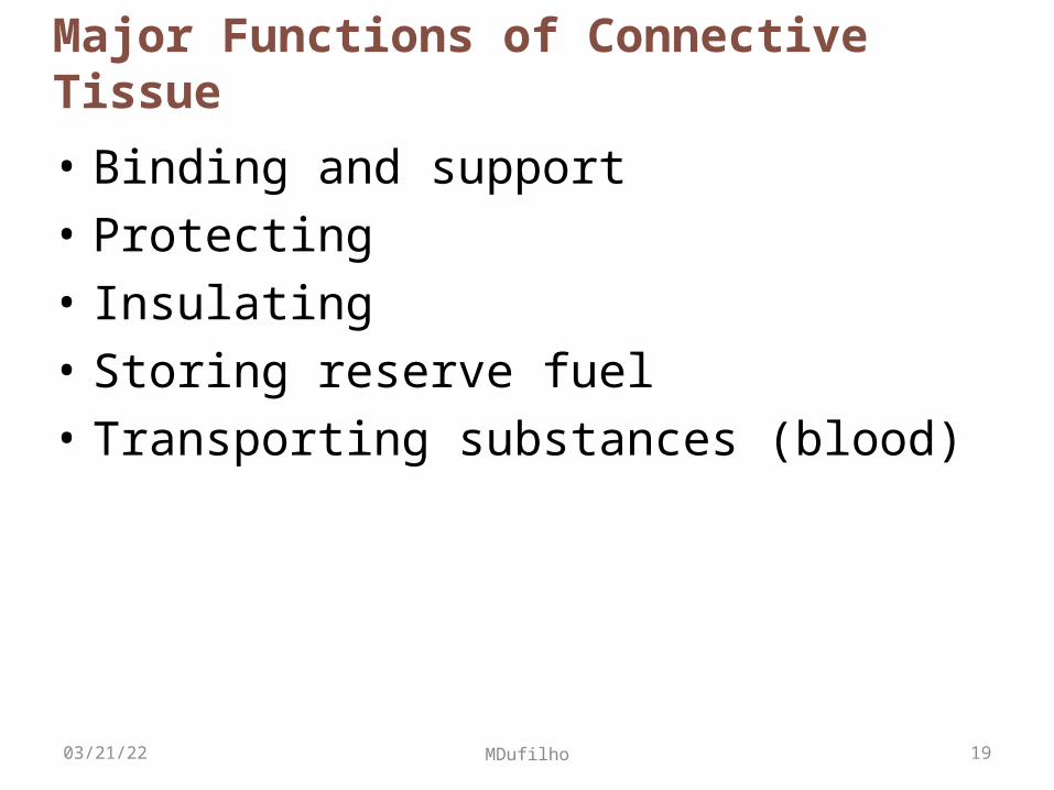
Major Functions of Connective Tissue
• Binding and support
• Protecting
• Insulating
• Storing reserve fuel
• Transporting substances (blood)
04/18/23 19MDufilho

Characteristics of Connective Tissue
• Three characteristics make connective tissues different from other primary tissues– Have mesenchyme (an embryonic tissue) as
their common tissue of origin– Have varying degrees of vascularity (blood
vessels)– Have extracellular matrix
• Connective tissue not composed mainly of cells• Largely nonliving extracellular matrix separates
cells– So can bear weight, withstand tension, endure abuse
04/18/23 20MDufilho

Connective Tissue Fibers
• Three types of fibers provide support– Collagen
– Elastic fibers
– Reticular
04/18/23 21MDufilho

Cells
• "Blasts" cells– Immature forum; mitotically active; secrete ground
substance and fibers – Fibroblasts in connective tissue proper– Chondroblasts in cartilage– Osteoblasts in bone– Hematopoietic stem cells in bone marrow
• "Cyte" cells– Mature form; maintain matrix– Chondrocytes in cartilage – Osteocytes in bone
04/18/23 22MDufilho

Other Cell Types in Connective Tissues
• Fat cells– Store nutrients
• White blood cells– Neutrophils, eosinophils, lymphocytes– Tissue response to injury
• Mast cells– Initiate local inflammatory response against foreign
microorganisms they detect
• Macrophages– Phagocytic cells that "eat" dead cells,
microorganisms; function in immune system
04/18/23 23MDufilho

Extracellular matrixGround substanceFibers• Collagen fiber• Elastic fiber• Reticular fiber
Capillary
Neutrophil
Mast cell
Fat cell
Lymphocyte
Fibroblast
Macrophage
Cell typesFigure 4.7 Areolar connective tissue: A prototype (model) connective tissue.
04/18/23 24MDufilho

Connective tissue proper: loose connective tissue, adipose
Description: Matrix as in areolar, but very sparse; closely packed adipocytes, or fat cells, have nucleus pushed to the side by large fat droplet.
Photomicrograph: Adipose tissue fromthe subcutaneous layer under the skin (350x).
Nucleus of adipose(fat) cell
Function: Provides reserve food fuel; insulates against heat loss; supports and protects organs.
Location: Under skin in subcutaneous tissue; around kidneys and eyeballs; within abdomen; in breasts. Fat droplet
Adipose tissue
Mammary glands
Figure 4.8b Connective tissues.
04/18/23 25MDufilho

Cartilage: hyaline
Description: Amorphous but firmmatrix; collagen fibers form animperceptible network;chondroblasts produce the matrixand when mature (chondrocytes)lie in lacunae.
Function: Supports and reinforces;serves as resilient cushion; resistscompressive stress.
Location: Forms most of theembryonic skeleton; covers theends of long bones in joint cavities;forms costal cartilages of the ribs;cartilages of the nose, trachea, andlarynx.
Costalcartilages Photomicrograph: Hyaline cartilage from
a costal cartilage of a rib (470x).
Matrix
Chondrocytein lacuna
Figure 4.8g Connective tissues.
04/18/23 26MDufilho

Cartilage: elastic
Description: Similar to hyalinecartilage, but more elastic fibersin matrix.
Function: Maintains the shape ofa structure while allowing greatflexibility.
Location: Supports the externalear (pinna); epiglottis.
Photomicrograph: Elastic cartilage fromthe human ear pinna; forms the flexibleskeleton of the ear (800x).
Chondrocytein lacuna
Matrix
Figure 4.8h Connective tissues.
04/18/23 27MDufilho

Cartilage: fibrocartilage
Description: Matrix similar to butless firm than that in hyalinecartilage; thick collagen fiberspredominate.
Function: Tensile strength allowsit to absorb compressive shock.
Location: Intervertebral discs;pubic symphysis; discs of kneejoint.
Photomicrograph: Fibrocartilage of anintervertebral disc (125x). Special stainingproduced the blue color seen.
Collagenfiber
Chondrocytesin lacunae
Intervertebraldiscs
Figure 4.8i Connective tissues.
04/18/23 28MDufilho

Others: bone (osseous tissue)
Description: Hard, calcifiedmatrix containing many collagenfibers; osteocytes lie in lacunae.Very well vascularized.
Function: Supports and protects(by enclosing); provides levers forthe muscles to act on; storescalcium and other minerals andfat; marrow inside bones is thesite for blood cell formation(hematopoiesis).
Location: Bones
Photomicrograph: Cross-sectional viewof bone (125x).
Lamella
Centralcanal
Lacunae
Figure 4.8j Connective tissues.
04/18/23 29MDufilho

Connective tissue: blood
Description: Red and white bloodcells in a fluid matrix (plasma).
Function: Transport respiratorygases, nutrients, wastes, and othersubstances.
Location: Contained within bloodvessels.
Photomicrograph: Smear of human blood(1670x); shows two white blood cellssurrounded by red blood cells.
Plasma
White bloodcells:• Lymphocyte• Neutrophil
Red bloodcells(erythrocytes)
Figure 4.8k Connective tissues.
04/18/23 30MDufilho

Skeletal muscle
Description: Long, cylindrical,multinucleate cells; obviousstriations.
Function: Voluntary movement;locomotion; manipulation of theenvironment; facial expression;voluntary control.
Location: In skeletal musclesattached to bones or occasionallyto skin.
Photomicrograph: Skeletal muscle(approx. 440x). Notice the obvious bandingpattern and the fact that these large cells aremultinucleate.
Striations
Nuclei
Part of musclefiber (cell)
Figure 4.9a Muscle tissues.
04/18/23 31MDufilho

Cardiac muscle
Description: Branching, striated,generally uninucleate cells thatinterdigitate at specializedjunctions (intercalated discs).
Function: As it contracts, itpropels blood into the circulation;involuntary control.
Location: The walls of the heart.
Photomicrograph: Cardiac muscle (900x);notice the striations, branching of cells, andthe intercalated discs.
Striations
Nucleus
Intercalateddiscs
Figure 4.9b Muscle tissues.
04/18/23 32MDufilho

Smooth muscle
Description: Spindle-shapedcells with central nuclei; nostriations; cells arranged closelyto form sheets.
Function: Propels substances orobjects (foodstuffs, urine, a baby)along internal passageways;involuntary control.
Location: Mostly in the walls ofhollow organs.
Photomicrograph: Sheet of smoothmuscle (720x).
Smoothmusclecell
Nuclei
Figure 4.9c Muscle tissues.
04/18/23 33MDufilho

Nervous Tissue
• Main component of nervous system– Brain, spinal cord, nerves– Regulates and controls body functions
• Neurons– Specialized nerve cells that generate and conduct
nerve impulses
• Neuroglia– Supporting cells that support, insulate, and protect
neurons
04/18/23 34MDufilho

Nervous tissue
Description: Neurons arebranching cells; cell processesthat may be quite long extend fromthe nucleus-containing cell body;also contributing to nervous tissueare nonexcitable supporting cells.
Function: Neurons transmitelectrical signals from sensoryreceptors and to effectors (musclesand glands) which control theiractivity; supporting cells supportand protect neurons.
Location: Brain, spinalcord, and nerves.
Photomicrograph: Neurons (350x).
Neuronprocesses
Nuclei ofsupportingcells
Cell bodyof a neuron
Neuron processes Cell body
Axon Dendrites
Figure 4.10 Nervous tissues.
04/18/23 35MDufilho