,c~ liver transplantation in the management …d-scholarship.pitt.edu/4478/1/31735062116466.pdf ·...
TRANSCRIPT

\~C\,c~ LIVER TRANSPLANTATION IN THE MANAGEMENT OF PORTAL HYPERTENSION
VINCENZO MAZZAFERRO, M.D. 5HUNZABURO IWAT5UKI, M.D. THOMAS E. 5T ARZL, M.D., PH.D.
The subjects of liver transplantation and portal hypertension are inseparable. Portal venous hypertension is present in the vast majority of patients who become candidates for liver transplantation. Furthermore, previous operations on the portal venous system, including portosystemic shunts, can greatly complicate the performance of subsequent liver transplantation. Finally, the provision of the new liver with an adequate splanchnic venous flow is a vital objective of all planning in every transplant operation, whether the graft is placed in the natural location (orthotopic) or at an ectopic site (auxiliary transplantation). These specific issues of the portal circulation are considered separately here.
THE RELIEF OF PORTAL HYPERTENSION WITH TRANSPLANT A liON
Discussions are limited in this chapter to those patients whose portal hypertension has been caused by the sinusoidal intrahepatic block that is typical of
)0\1 806
chronic liver disease. Patients with extrahepatic Por tal vein thrombosis and cavernous transformation 0; postsinusoidal block such as Budd-Chiari syndrome may come ~o t~ansplantation, but this is uncommon.
ComplIcatIOns of the venous hypertension that can be palliated by portosysteI?1ic shunts are hemor_ rhage from esophageal vances and intractable ascites. With potent and specific diuretic drugs and with the availability of antialdosterone agents 'truly intractable ascites is extremely uncommon. When it does occur, more appropriate therapy than portosyStemic shunt often will be a peritoneal jugular shunt 1
Thus, the principal problem about which therape~_ tic judgments must be made in planning relief of portal hypertension is hemorrhage from esophageal varices. The preponderance of affected patients have variable degrees of liver failure. A shunting procedure often will make the liver failure worse, even if it is successful in controllin~ t~e variceal hemorrhage.
In contrast, orthotOPIC liver transplantation is a spectacularly effective way to treat portal hyperten_ sion. During the preliminary phases of the operation and during dissection of the portal triad structures bleeding from the thin-walled and often widely rus: tributed varices can be quite alarming. This problem usually can be ameliorated, even during the time when the diseased liver is being removed and the patient is anhepatic. The use of a venovenous bypass during this time provides prompt relief by reducing the portal hypertension (Fig. 1).
If the new liver is a good one, it will readily transmit the portal venous flow, and, within a matter of minutes, the previously high-pressure venous collateral channels are deflated. If very large collateral
000 ····'=~JUJ. 00
o ~
Figure 1 Venovenous bypass allows decompression of the splanchnic and systemic venous beds during the anhepatic phase without the need for heparinization. (Adapted with permission of Griffith et a!. Surg Gynecol Obstet 1985; 160:270-272.)
1. ---La ,
Mi ,
Ril ,
Jw: I
ve th sp be dl
w of bl w 7 ti, oj
1
(

r
Liver Transplantation in the Management of Portal Hypertension / 807
'f.ble 1 Survival Comparison Among Various Treatments for Bleeding Esophageal Varices (Qli]d's Oass A. B, and C) -- No. of Study Patienls 1 year -Langer et aI (1985)
38 80-Selective shunt Nonselective shunt 40 90*
Millikan et aI (1985) 26 85-Selective shunt
Nonselective shunt 29 80-warren et aI (1986)
Sderotherapyt 36 90* Selective shunt; 35 70-
Rikkers et aI (1987) Sclerotherapy 30 77· Selective shunt; 27 75·
lwatsuki et aI (1988) Liver transplantation 302 79
-Value estimated from survival curve. tSclerotherapy failures were rescued by surgical therapy. tTwenly-three selective shunts and four nonselective shunts.
vessels, usually coronary veins, are encountered, these can be ligated, to encourage bepatopetal splanchnic flow, but these extra maneuvers may not be necessary, and in some cases, they may be meddlesome.
Liver substitution has been studied in patients who had severe liver failure in addition to a history of bleeding esophageal varices, or who were actively bleeding.2 The survival rates of 302 such patients were 79 percent at 1 year, 74 percent at 2 years, and 71 percent at 3, 4, and 5 years after liver transplantation. These excellent results were obtained regardless of the cause of cirrhosis, including alcoholism.
The results achieved by liver transplantation were compared with those in four well-studied con-
Survival Rates (96)
2 yetJrs - 3 yetJrS 4 years 5 years
76- 63- 54- 51-85- 70- 56- 56-
77· 65· 60- 55-n- 70- 65- 60-
84- 82- 82-59- 45· 45·
61· 60- so-65· 60- 39-
74 71 71 71
trol trials reported since 1985 (Table I). More than 75 percent of the patients in each report listed in Table 1 had good or fair hepatic function (Child's class A and B) before the shunting procedure, whereas all of the patients treated with liver transplantation had poor or very poor hepatic function (Child's class C). Despite this severe disadvantage of preoperative condition, the survival rates after liver transplantation were better than or similar to those of patients who underwent shunt operation.
When the results were compared only among the patients with advanced hepatic dysfunction (Child's class C), the survival rates with liver transplantation were far better than those achieved by shunt operations (Table 2). These comparative data
Table 2 Survival Comparison Among Various Treatments for Bleeding Esophageal Varices (Child's Oass C, Poor Liver Function)
No. of Study Patients 1 year
Turcotte et aI (1973) Nonselective shunt 50 36
Warren et aI (1982) Selective shunt ? 60-Nonselective shunt ? so-
Rikkers et aI (1984) Shunt and nonshunt operationst 24 45·
Chandler et aI (1985) Shun~ 30 36·
lwatsuki et aI (1988) Liver transplantation 302 79
-Value estimated from survival c:une. tFifteen nonselective shunts. seven selective shunts, and two Donshunt opcntiODS. fBoth selective and nonselective operations.
Survival RaJes (%)
2 years 3 years 4 years 5 years
32 22 20 17
53- 45- 40- 35-40- 37· 20- IS-
35· 30- 20- 17-
30- 25- 20- 13·
74 71 71 71
I i 1

808 / Current Therapy in Vascular Surgery-2
were particularly striking because the liver recipients were considered too advanced in their liver disease to be considered for shunting operations.
However the concept of performing a liver transplantati~n for every patient who has signifi~nt variceal hemorrhage would be a grotesque oversImplification of the meaning of these studies. Instead, the data have stimulated a re-examination of previously used therapy, including shunt operations. Such operations can be used in selected patients, although these operations may influence the ultimate step of transplantation.
THE EFFECT OF PREVIOUS UPPER ABDOMINAL OPERATIONS
Nonshunt Procedures
Somewhat surprisingly, previous operations in the upper abdomen, exclusive of those on the splanchnic circulation, have not precluded good results after subsequent transplantation.3 However, the need for transfusion and the average technical difficulty in these patients are greatly increased.
The performance of operations such as the Kasai procedure that increase adhesions and promote collateral circulation may have an adverse effect on the operability of children with biliary atresia. After portoenterostomies, the portal vein in many children has undergone atresia apparently because the collateral vessels in adhesions "steal" portal flow. When transplantation eventually is performed, the graft portal vein often must be anastomosed to the confluence of the splenic and superior mesenteric veins because the native portal vein is frequently atretic above this level. In such cases, ligation of large collateral vessels, particularly the coronary vein, may be important to ensure good portal blood flow. So far, the mortality after liver transplantation has not been significantly different in children who have undergone portoenterostomy than in those who have not. However, with the acquisition oflarger numbers, it is probable that the 10
percent to 15 percent increased operative mortality in patients with. biliary atresia and Kasai operations will become significant.
operations to interrupt periesophageal collateral vessels for decompression of esophageal varices (Sugiura's operation) may create such vascular adhesions in the upper abdomen that transplantation becomes almost impossible.
Previous splenectomy, an operation used in the past to decrease splanchnic blood flow, was the most common condition associated with portal vein abnormalities encountered during liver transplanta_ tion.· The usual situation was that thrombi had propagated retrograde from the ligated splenic vein, which often extended all the way to the confluence with the superior mesenteric vein.
Previous Shunt Procedures
Before the use of cyclosporine, beginning in 1980, nearly a third of our 170 transplant recipients had undergone previous portosystemic shunt placement. S The most common were end-to-side or sideto-side portacaval shunts in the hilum, but there were a number of examples of proximal or distal splenorenat shunts, H-graft mesocaval shunts, or cavomesenteric (Marion) shunts. The presence of any of these shunts was thought to be a highly disadvantageous condition, probably with justification.
In the cyclosporine era, the number of patients with shunts has been less. Over a period of 9 years, from March 1980 to March 1989, 58 liver recipients had previous shunts (Table 3). The most common variety was the distal splenorenal shunt (DSRS = 18 cases), followed by mesocaval shunt (MeS = 17 cases), portacaval end-to-side shunt (PCE-S = 11 cases), portacaval side-to-side and proximal splenorenal shunt (PCS-S = 5 cases and PSRS = 7 cases) Lerut et at· noted that abnormalities in the portal vein leading to significant complications after transplantation were high in this subset of patients. Patients with previous portosystemic shunt procedures have a complicating set of technical and anatomic factors that render the actual performance of liver
Table 3 Characteristics of 58 Liver Recipients with Previous Portosystemic Shunt
Kind of Previous Shunt
Portal venous-maintaining Distal splenorenai Portal venous-diverting Mesocaval Portacaval end-to-side Portacaval side-to-side Proximal sp\enorenai
OL Tx - Orthotopic liver transplantation.
11 1S&&&&&22
No. of Cases
18
17 11 5 7
Age in years (Median/range)
40.9 (12-61)
38.5 (7-56) 41.6 (4-51) 31.6 (20-64) 21.0 (21-41)
Interval Shunt-OLTx
(years. mean ± SD)
5.6 ±0.9
5.1 ±O.9 7.7 ± 2.5 3.5 ±O.7 7.9 ± 1.9
T ,
SZ$iJ~ .4

liver Transplantation in the Management of Portal Hypertension / 809
transplantation more challenging. However, the results still have been good enough to justify continuing efforts at treatment of these patients.
In our case material there were 26 males and 32 females, with a mean age of 39.2 ± 13 years (range 4 to 64 years).6 Twenty-three patients were Child's class Band 35 were Child's class C at the time of the pre-orthotopic liver transplantation evaluation. All patients were immunosuppressed with cyclosporine and prednisone7 to which azathioprine and OKT3 were added when clinically required. Postnecrotic and primary biliary cirrhosis accounted for 45 percent of the cases. The mean interval between the shunt procedure and orthotopic liver transplantation was approximately 6 years. This averaged 3.5 years in the side-ta-side portosystemic shunt group and 7.9 years in patients belonging to the proximal splenorenal group.
There was not a statistically significant difference in the actuarial 9-year survival between patients who underwent previous portosystemic shunt before transplant and those who did not (Fig. 2). Sixtyseven percent of the portosystemic shunt patients were alive 5 years after orthotopic liver transplantation, whereas the survival at the same time interval was 65 percent in the rest of all liver transplant recipients.
Age and sex did not influence the outcome nor did shunt patency at the time of orthotopic liver transplantation. The presence of an atrophic or sclerotic portal vein or use of vein or arterial graft did not have a statistical impact on either graft or patient survival. The graft survival was significantly different between patients classified as Child's Band Child's C. However, because of timely retransplan-
Figure 2 Survival of portosystemic shunt (PSS) patients (N = 58) versus the entire nonPSS population (N = 1445) transplanted in a 9-year period of cyclosporine immunosuppression. No statistical difference is noted. (Reproduced by pennission of Mazzaferro et al. Am J Surg 1990; 160: 111-116.)
100
80
i 60 Ul
j !
! Cl.
40
20
tation in cases of graft failure, no significant difference was observed in patient survival, which was 74 percent for the Child's B group and 67 percent for the Child's'C group.
The first ten patients of the series (1980- 1982) did not have the advantage of the intraoperative venovenous bypass system (Bia-pump).& In a second group of 24 patients (l983-October 1987), the native hepatectomy was performed under bypass conditions. In those patients at least a single cavacaval bypass was always used, and the portal limb of the system was added whenever allowed by the intraoperative conditions of the portal vein and the previous shunt. In a third group of 24 patients (October 1987 - March 1989) the venous bypass was employed and, in addition, the University of Wisconsin solution was used for graft preservation, replacing the Collins solution previously employed.
The use of venovenous bypass probably reduced intraoperative blood loss and may have contributed to a shorter operative time when compared to the first patients of the series in which bypass was not used.
With the introduction of the University of Wisconsin preservation solution, there was a significantly longer cold ischemia time of the graft, but the postreperfusion liver function was improved.9 The longer and more effective preservation allowed more detailed planning of the operation, with better control of the bleeding and elimination of haste in reperfusing the organ. These facts are reflected in a further decrease in blood loss and intensive care unit stay. The actuarial survival also improved significantly.6
The kind of previous shunt had an impact on
PSS
NON-PSS
o ~--~----~----~----~--~----~----~--~------o 2 .,
" 4
Years after transplantation
(i 7 !j

810 / Current Therapy in Vascular Surgery-2
11 II I
I
no -
I ~ 60 ., i PSRS
!
peE-S J 40
20 -
o ,
o '/·1 h
Years after transplantation
survival (Fig. 3). Previous shunt procedures with no liver hilum dissection were safer. Patients with mesocaval and DSRS patients had 5-year and 9-year survivals of 95 percent and 87 percent, respectively, whereas any other previous shunt was not associated with a survival better than 52 percent at the same time intervals. Those differences were highly significant.
Our cumulative experience suggests that although either previous shunts, portal vein abnormalities, or the need for portal vein reconstruction significantly increase the complexity of the procedure as detennined by the operative time and blood loss, these conditions do not prohibit successful hepatic transplantation.
TRANSPLANTATION VERSUS SHUNTS: A MIDDLE GROUND
Should shunting operations ever be recommended as treatment for variceal hemorrhage, knowing that these procedures can jeopardize, or at least make more difficult, the ultimate step of liver transplantation? Probably uncommonly, because endoscopic sclerosis of varices is an effective alternative. In some patients with Child's class A cirrhosis, a DSR anastomosis may be preferred to relieve portal hypertension. We are using this approach in a small number of highly selected patients. Although the portal vein eventually diminishes in size as a result of the Warren shunts, presumably because of the loss of flow, portal flow can be restored at the end of the transplantation by perfonning splenectomy. which functionally removes the shuDt. 6,10
The obvious limitations of the shunt approach to variceal bleeding have greatly reduced the fre-
Mes
OSRS
pesos
Figure 3 Influence of previous shunts on survival after liver transplantation. Mesocaval and distal splenorenal shunt had a significant positive impact on survival when compared with other shunts. (MCS = mesocaval; DSRS = distal splenorenal; PSRS = proximal splenorenal; PCS-S = portacaval side-ta-side; PCE-S = portacaval end-ta-side). (Reproduced by permission of Mazzaferro et al. Am J Surg 1990; 160: III - 116.)
Quency of portal diversion procedures in Western countries. Instead of shunting, more and more reliance has been placed on sclerotherapy, to the extent that the pendulum may have swung too far. Sclerotherapy has its own special dangers, including esophageal perforation and ulceration or stricture of the distal esophagus.
EXTRA-ANATOMIC PORTAL VEIN REVASCULARIZA TlON
Until recently, thrombosis of the portal vein or of its principal tributaries, the splenic and superior mesenteric veins, was thOUght to be a contraindication to transplantation. However, newly developed vein graft techniques routinely allow liver replacement in such patients II (Fig. 4). The vein grafts are routed from the superior mesenteric vein below the transverse mesocolon, brought anterior to the pancreas, and used for portal anastomosis in the hepatic hilum.
PORTAL DECOMPRESSION THROUGH AN AUXILIARY GRAFT
An option to liver replacement is transplantation of an. extra liver in an ectopic site, or auxiliary transplantation. This operation was envisioned by Welch as treatment for patients with end-stage cirrhosis.12 With the auxiliary operation as originally described in unmodified dogs, the extra liver was placed in the right paravertebral gutter, rearterialized from convenient adjacent vessels, and provided with the portal venous inflow with systemic blood from the recipient iliac vein or inferior vena cavall (Fig.
Reel v.
Figl the graf pyle plar
5). infl iar: we: of
ver Iiv, ge5 sIX tar is I
Iiv spl int air 19 ad ha tal we shl
tio

on 1..0;.:
so. r, 1 .'-
"-" • i'/' val its. ·n· 'aI; ~.-
,~ - ".
by 'l'
rg
n I·
11 Of:
l-
I-
e
r
a QI
liver Transplantation in the Management of Portal Hypertension / 811
Donor portal v
Figure 4 Vein graft from the superior mesenteric vein to the portal vein in a case of portal vein thrombosis. The graft is brought anterior to the pancreas and beneath the pylorus. (Reproduced by permission ofTzakis et aI. Transplantation 1989; 48:530-531.)
5). The graft outflow was drained into the recipient inferior vena cava. It was soon observed that auxiliary grafts were much more severely damaged than were orthotopica1ly placed ones, primarily because of rapid hepatocyte atrophy.14
These adverse effects could be prevented by diverting splanchnic venous flow through the auxiliary liver and away from the recipient's own liver!" suggesting that the splanchnic venous blood contained specific liver-supporting factors. The most important of these so-called portal hepatotropic substances is insulin. 15
The experiments suggested that an auxiliary liver graft could not function optimally without splanchnic venous inflow. The condition of providing such blood to the new liver has been met in almost all of the subsequent clinical trials which, by 1988, included about 60 patients. By providing this advantage to the transplant, the additional objective has been incidentally met of decompressing the portal system through the interposed graft. In other words, the graft has become part of a portosystemic shunt.
Two patients from the distant past had unquestionable prolongation of life after auxiliary trans-
I / ~- -J_
Figure 5 Auxiliary liver transplantation in dogs. The extra liver is provided with the portal inflow with systemic blood from the recipient inferior vena cava. Rapid hepatocyte atrophy occurred in the auxiliary graft because the splanchnic venous flow does not contribute to the graft portal inflow. (Reproduced by permission of StanI TE. Experience in hepatic transplantation. Philadelphia, WB Saunders, 1969.)
plantation. The first, a child with biliary atresia, is alive with a follow-up of more than 16 years. 16 A second patient in Paris lived for more than 8 years after auxiliary transplantation but then died from a hepatocellular carcinoma in the diseased host liver. 17
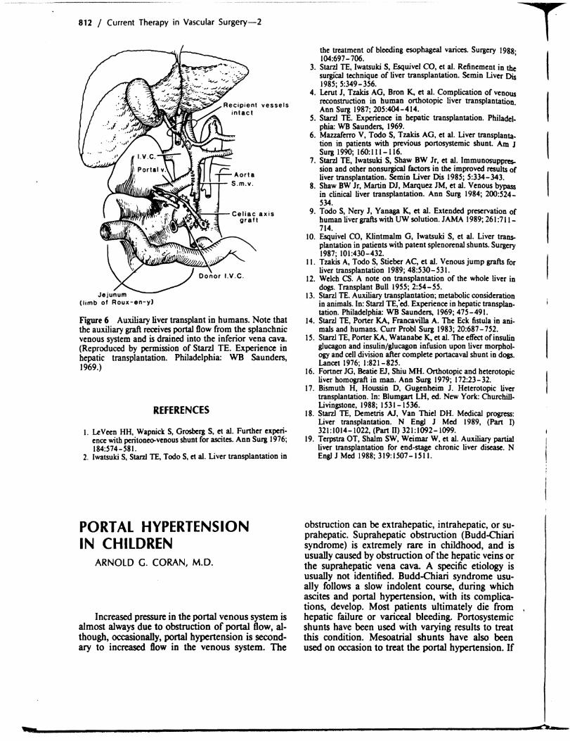
With the increasing success of orthotopic liver transplantation, interest in auxiliary transplantation waned. II The resulting pessimism has been lightened by a recent report of the transplantation of whole livers or liver fragments to the right paravertebral gutter of six adults using essentially the same operation that was tried in earlier times (Fig. 6). At the time of reporting with follow-ups of 5 to 23 months, all six recipients are alive. 19 With these imprOVed results, further cautious trials are certain to be forthcoming.
I
--
812 / Current Therapy in Vascular Surgery-2
-...~-,....- Celiac axis graft
Figure 6 Auxiliary liver transplant in humans. Note that the auxiliary graft receives portal flow from the splanchnic venous system and is drained into the inferior vena cava. (Reproduced by permission of Starzl TE. Experience in hepatic transplantation. Philadelphia: WB Saunders, 1969.)
REFERENCES
I. LeVeen HH, Wapnick S, Grosberg S, et al. Further experi. ence with peritoneo-venous shunt for ascites. Ann Surg 1976; 184:574-581.
2. Iwatsuki S, Stan! TE, Todo S, et al. Liver transplantation in
PORTAL HYPERTENSION IN CHILDREN
ARNOLD C. CORAN, M.D.
Increased pressure in the portal venous system is almost always due to obstruction of portal flow, although, occasionally, portal hypertension is secondary to increased flow in the venous system. The
the treatment of bleeding esophageal varices. Surgery 1988' 104:697 - 706. '
3. Starzl TE, Iwatsuki S, Esquivel CO, et aI. Refinement in the surgical technique of liver transplantation. Semin Liver Dis 1985; 5:349-356.
4. Lerut J, Tzakis AG, Bron K, et aI. Complication of venous reconstruction in human orthotopic liver transplantation. Ann Surg 1987; 205:404-414.
5. Starzl TE. Experience in hepatic transplantation. Philadel. phia: WB Saunders, 1969.
6. Mazzaferro V, Todo S, Tzakis AG, et aI. Liver transplanta. tion in patients with previous portosystemic shunt. Am J Surg 1990; 160:111-116.
7. Starzl TE, Iwatsuki S, Shaw BW Jr, et aI. Immunosuppression and other nonsurgical factors in the improved results of liver transplantation. Semin Liver Dis 1985; 5:334-343.
8. Shaw BW Jr, Martin OJ, Marquez JM, et al. Venous bypass in clinical liver transplantation. Ann Surg 1984; 200:524-534.
9. Todo S, Nery J, Yanaga K, et aI. Extended preservation of human liver grafts with UW solution. JAMA 1989; 261:711_ 714.
10. Esquivel CO, K1intmalm G, Iwatsuki S, et al. Liver transplantation in patients with patent splenorenal shunts. Surgery 1987; 101:430-432.
II. Tzakis A, Todo S, Stieber AC, et al. Venous jump grafts for liver transplantation 1989; 48:530- 531.
12. Welch CS. A note on transplantation of the whole liver in dogs. Transplant Bull 1955; 2:54-55.
13. Starzl TE. Auxiliary transplantation; metabolic consideration in animals. In: Starzl TE;ed. Experience in hepatic transplan. tation. Philadelphia: WB Saunders, 1969; 475-491.
14. Stan! TE, Porter KA, Francavilla A. The Eck fistula in ani. mals and humans. Curr Probl Surg 1983; 20:687 - 752.
15. Starzl TE, Porter KA, Watanabe K, et a1. The effect of insulin glucagon and insulin/glucagon infusion upon liver morphol· ogy and cell division after complete portacaval shunt in dogs. Lancet 1976; 1:821-825.
16. Fortner JG, Beatie EJ, Shiu MH. Orthotopic and heterotopic liver homograft in man. Ann Surg 1979; 172:23-32.
17. Bismuth H, Houssin D, Gugenheim J. Heterotopic liver transplantation. In: Blumgart LH, ed. New York: Churchill· Livingstone, 1988; 1531-1536.
18. Stan! TE, Demetris AI, Van Thiel DH. Medical progress: Liver transplantation. N Engl J Med 1989, (Part I) 321:1014-1022, (Part D) 321:1092-1099.
19. Terpstra OT, Shalm SW, Weimar W, et aI. Auxiliary partial liver transplantation for end-stage chronic liver disease. N Engl J Med 1988; 319:1507-1511.
obstruction can be extrahepatic, intrahepatic, or suprahepatic. Suprahepatic obstruction (Budd-Chiari syndrome) is extremely rare in childhood, and is usually caused by obstruction of the hepatic veins or the suprahepatic vena cava A specific etiology is usually not identified. Budd-Chiari syndrome usually follows a slow indolent course, during which ascites and portal hypertension, with its complications, develop. Most patients ultimately die from hepatic failure or variceal bleeding. Portosystemic shunts have been used with varying results to treat this condition. Mesoatrial shunts have also been used on occasion to treat the portal hypertension. If
I I I I I
.......,,-