c. r. mason, j. e. gomez and t. j. ebner j neurophysiol 86...
TRANSCRIPT

86:2896-2910, 2001. J NeurophysiolC. R. Mason, J. E. Gomez and T. J. Ebner
You might find this additional information useful...
46 articles, 19 of which you can access free at: This article cites http://jn.physiology.org/cgi/content/full/86/6/2896#BIBL
17 other HighWire hosted articles, the first 5 are: This article has been cited by
[PDF] [Full Text] [Abstract]
, January 23, 2008; 28 (4): 880-892. J. Neurosci.S. A. Overduin, A. d'Avella, J. Roh and E. Bizzi
Modulation of Muscle Synergy Recruitment in Primate Grasping
[PDF] [Full Text] [Abstract], February 6, 2008; 28 (6): 1271-1281. J. Neurosci.
P. H. Thakur, A. J. Bastian and S. S. Hsiao Exploration Task
Multidigit Movement Synergies of the Human Hand in an Unconstrained Haptic
[PDF] [Abstract], July 1, 2009; 28 (7): 851-867. The International Journal of Robotics Research
M. T. Ciocarlie and P. K. Allen Hand Posture Subspaces for Dexterous Robotic Grasping
[PDF] [Full Text] [Abstract]
, December 1, 2009; 102 (6): 3310-3328. J NeurophysiolJ. Chen, S. D. Reitzen, J. B. Kohlenstein and E. P. Gardner
Cortex of the Macaque MonkeyNeural Representation of Hand Kinematics During Prehension in Posterior Parietal
[PDF] [Abstract], February 1, 2010; 24 (2): 141-151. Neurorehabil Neural Repair
D. Bensmail, J. Robertson, C. Fermanian and A. Roby-Brami Strategies and Kinematics of Reach-to-Grasp Movements
Botulinum Toxin to Treat Upper-Limb Spasticity in Hemiparetic Patients: Grasp
on the following topics: http://highwire.stanford.edu/lists/artbytopic.dtlcan be found at Medline items on this article's topics
Medicine .. Image Reconstruction Neuroscience .. Central Nervous System
including high-resolution figures, can be found at: Updated information and services http://jn.physiology.org/cgi/content/full/86/6/2896
can be found at: Journal of Neurophysiologyabout Additional material and information http://www.the-aps.org/publications/jn
This information is current as of March 7, 2010 .
http://www.the-aps.org/.American Physiological Society. ISSN: 0022-3077, ESSN: 1522-1598. Visit our website at (monthly) by the American Physiological Society, 9650 Rockville Pike, Bethesda MD 20814-3991. Copyright © 2005 by the
publishes original articles on the function of the nervous system. It is published 12 times a yearJournal of Neurophysiology
on March 7, 2010
jn.physiology.orgD
ownloaded from

Hand Synergies During Reach-to-Grasp
C. R. MASON, J. E. GOMEZ, AND T. J. EBNERDepartment of Neuroscience and Graduate Program in Neuroscience, University of Minnesota,Minneapolis, Minnesota 55455
Received 6 February 2001; accepted in final form 17 August 2001
Mason, C. R., J. E. Gomez, and T. J. Ebner.Hand synergies duringreach-to-grasp.J Neurophysiol86: 2896–2910, 2001. An emergingviewpoint is that the CNS uses synergies to simplify the control of thehand. Previous work has shown that static hand postures for mimedgrasps can be described by a few principal components in which thehigher order components explained only a small fraction of thevariance yet provided meaningful information. Extending that earlierwork, this study addressed whether the entire act of grasp can bedescribed by a small number of postural synergies and whether thesesynergies are similar for different grasps. Five right-handed adultsperformed five types of reach-to-grasps including power grasp, powergrasp with a lift, precision grasp, and mimed power grasp and mimedprecision grasp of 16 different objects. The object shapes were cones,cylinders, and spindles, systematically varied in size to produce alarge range of finger joint angle combinations. Three-dimensionalreconstructions of 21 positions on the hand and wrist throughout thereach-to-grasp were obtained using a four-camera video system. Sin-gular value decomposition on the temporal sequence of the markerpositions was used to identify the common patterns (“eigenpostures”)across the 16 objects for each task and their weightings as a functionof time. The first eigenposture explained an average of 97.36 0.89%(mean6 SD) of the variance of the hand shape, and the secondanother 1.96 0.85%. The first eigenposture was characterized by anopen hand configuration that opens and closes during reach. Thesecond eigenposture contributed to the control of the thumb and longfingers, particularly in the opening of the hand during the reach andthe closing in preparation for object grasp. The eigenpostures and theirtemporal evolutions were similar across subjects and grasps. Thehigher order eigenpostures, although explaining only small amountsof the variance, contributed to the movements of the fingers andthumb. These findings suggest that much of reach-to-grasp is effectedusing a base posture with refinements in finger and thumb positionsadded in time to yield unique hand shapes.
I N T R O D U C T I O N
The hand is a highly complex structure with 27 bones, 18joints, and 39 intrinsic and extrinsic muscles (Kapandji 1970;Tubiana 1981) with over 20 degrees of freedom (Soechting andFlanders 1997). Movement of the fingers requires a coordi-nated interplay of both extrinsic and intrinsic muscles (Lands-meer 1963; Landsmeer and Long 1965; Long et al. 1970). Thisbiomechanical complexity raises the question, how does theCNS control the hand and fingers? There are two divergentviewpoints. The more traditional view has emphasized a strat-egy based on controlling individual muscles and joints togenerate the needed forces (for review see Lemon 1999;
Schieber 1990). Another view has emphasized the need for“simplifying” strategies that reduce the number of degrees offreedom and thereby reduce the complexity of the controlproblem (Arbib et al. 1985; Iberall and Fagg 1996; Santello etal. 1998). Recent psychophysical, anatomical, and physiolog-ical studies have found support for the latter view.
Considerable evidence supports the concept that the fingersact synergistically with other fingers, with the wrist, and withthe arm. Our fingers do not move in isolation of the neighbor-ing fingers (Engel et al. 1997; Flanders and Soechting 1992;Soechting and Flanders 1997) even when the explicit goal is tomake individuated finger movements (Schieber 1991, 1995;Schieber and Poliakov 1998). A striking finding in typing andpiano playing is that almost all the fingers and joints are inmotion simultaneously (Engel et al. 1997; Gordon et al. 1994;Soechting and Flanders 1997). Nor does the hand move inisolation of the arm. Movements of the arm and shaping of thehand during reach-to-grasp are highly coordinated (Bootsma etal. 1994; Chieffi and Gentilucci 1993; Jeannerod 1984; Mar-teniuk et al. 1990; Paulignan et al. 1990, 1991). Therefore thecoordination of the fingers, wrist, and arm indicate that a globalcontrol strategy may be utilized.
The anatomy of the finger muscles may simplify the controlproblem in the primate hand. The low individuation and sta-tionarity of the long fingers shown by Schieber (1991) could bedue to the multi-finger insertions of the communal flexor andextensor muscles or reflect part of the CNS’s strategy to thecontrol the hand (Santello and Soechting 1997, 1998; Santelloet al. 1998). In monkeys, mechanical coupling between fingersby interconnection between tendons and by motor units thatexert tension on more than one tendon prevents movement at asingle finger level (Schieber 1995; Schieber et al. 1997; Serlinand Schieber 1993). The communal flexor and extensor mus-cles also cross multiple joints (Kapandji 1970; Tubiana 1981).Therefore biomechanically the control of the individual jointsor fingers is limited.
Stimulation studies of the primary motor cortex (MI) haveoften been cited as supporting evidence that this structure isorganized to control individual muscles. Cortical stimulation inhumans, apes, and monkeys has yielded the textbook homun-culus (Penfield and Rasmussen 1950) or simiusculus (Leytonand Sherrington 1917; Woolsey 1958). However, as reviewedby Schieber (1990) and Lemon (1999), a strict somatotopicorganization is not consistent either with earlier or more recent
Address for reprint requests: T. J. Ebner, Dept. of Neuroscience, Universityof Minnesota, Lions Research Building, Rm. 421, 2001 Sixth St. SE, Minne-apolis, MN 55455 (E-mail: [email protected]).
The costs of publication of this article were defrayed in part by the paymentof page charges. The article must therefore be hereby marked ‘‘advertisement’’in accordance with 18 U.S.C. Section 1734 solely to indicate this fact.
2896 0022-3077/01 $5.00 Copyright © 2001 The American Physiological Society www.jn.org
on March 7, 2010
jn.physiology.orgD
ownloaded from

stimulation studies. The earliest studies of the hand area withinMI found that the cortical territories from which the surfacestimulation evokes movement of different digits overlap ex-tensively and that stimulation of one site elicits movement ofmultiple digits (Leyton and Sherrington 1917; Penfield andRasmussen 1950; Woolsey 1958; Woolsey et al. 1979). Recentinvestigations using more refined stimulation techniques haveconfirmed that contraction of a particular hand muscle can beevoked from a substantial fraction of MI (Andersen et al. 1975;Sato and Tanji 1989). A similar overlapping organization hasbeen found for finger movement (Gould et al. 1986; Kwan etal. 1978; Strick and Preston 1978). Spike-triggered averagingdemonstrates that many corticomotoneuronal cells facilitate theelectromyographic (EMG) activity of more than one muscle(Buys et al. 1986; Cheney and Fetz 1985; Cheney et al. 1985;Lemon et al. 1986; McKiernan et al. 1998).
Recent inactivation and lesion studies also support the con-cept that M1 is not organized to perform isolated finger move-ments (Poliakov and Schieber 1999). Focal muscimol inacti-vations in the hand region of M1 in the monkey do not disruptthe movements of isolated fingers but instead disrupt move-ments of different finger combinations (Schieber and Poliakov1998). Small infarcts in the hand area of human M1 result inweakness of the fingers, but the deficits are not limited to asingle digit (Schieber 1999). Therefore M1 does not appearorganized around a finely delineated somatotopic map speci-fying the activation of individual muscles or joints.
Last, recent studies have shown that static grasp posture canbe described using a small number of postural synergies (San-tello and Soechting 1998; Santello et al. 1998). These synergiescould be defined as a spatial configuration or “primitive” of thehand shape that is common across the various tasks. In thelatter study, subjects were asked to reach out and grasp imag-inary objects. Even without visual or tactile inputs, the handshapes were distinct (Santello et al. 1998). Using a principalcomponent analysis, the first three components were needed todescribe approximately 90% of the variance, with the first twocomponents explaining approximately 84%. Although the in-dividual contributions were small, the higher order componentswere responsible for more subtle adjustments of the graspposture (Santello et al. 1998). The evolving hand shape duringthe transport phase of reach-to-grasp carries increasing infor-mation that peaks at the actual object grasp when the hand canconform to the object (Santello and Soechting 1998). Thepresence of postural synergies that contributed to the evolvinghand shaping was not examined, yet the results suggest that acommon strategy may control hand shape throughout reach-to-grasp.
Therefore the hand is controlled as a unit at some level andto some degree. The present study asks three questions aboutthese hand synergies. The original description that hand pos-ture can be described by a small number of synergies wasbased on static hand posture (Santello et al. 1998), yet it iswell-known that hand shape evolves throughout reach-to-grasp(Jeannerod 1984; Paulignan et al. 1990). Hence, the first ques-tion was whether the entire behavior can be described by asimilar small set of synergies and whether these synergies weresimilar for different types of grasps. These synergies imply aspatial configuration that is not static but is modulated in timeto allow subjects to grasp objects of different shapes and sizes.Second, the study of Santello et al. (1998) used mimed grasps
of imagined objects. Neither the tactile or visual informationwas available to the subjects. This prompts the question ofwhether tactile input or visual input would dramatically alterthe hand synergies? Last, based on an information theoryanalysis, the higher order postural synergies were shown to beimportant (Santello et al. 1998), but the nature of the contri-bution was not evaluated. Therefore this study examines howthe higher order synergies contributed to the shaping of thehand during reach-to-grasp.
In the present study, subjects performed grasps of 16 objectsconsisting of 3 classes of shapes including cylinders, cones,and spindles. Within each class the sizes of the object weresystematically varied. Five variations of power and precisiongrasps were studied including actual and mimed grasps. Theevolution of the grasp was evaluated in a continuous mannerfrom the initial start position through maintained object contactfor each of the different grasps using singular value decompo-sition (SVD). The results show that the subjects used a basehand shape that explained a large percentage of the variance inhand kinematics throughout reach-to-grasp. This base handshape was independent of the type of grasp or tactile input.However, additional components were necessary to adequatelydescribe the evolution of the grasp. An abstract describingsome of these results has been presented (Mason et al. 1999).
M E T H O D S
Experimental paradigm and procedures
Five adults (3 women and 2 men, age ranging from 21 to 44 yr old)with no known history of neurological or musculoskeletal problems,participated in the study. All were right-handed as determined by theEdinburgh Handedness Inventory (Oldfield 1971) and had normalhand function. The protocol was approved by the Institutional ReviewBoard of the University of Minnesota, and all subjects gave informedconsent.
Each subject performed five different tasks with each object:1)Power Grasp,2) Power Grasp with a Lift,3) Mimed Power Grasp,4)Precision Grasp, and5) Mimed Precision Grasp. In Power Grasp thesubjects were instructed to reach for and grasp the object as if theywere going to lift the object using their whole hand making palmarcontact. Subjects were to maintain the grip without moving the objectuntil the end of the trial. Power Grasp with a Lift was the same asPower Grasp with the inclusion of the lifting the object approximately2 cm off the table surface. In the Mimed Power Grasp, the object wasmoved an additional 40 cm out of the subject’s reach. The subjectswere to reach as if grasping the object at the standard object locationand pantomime a Power Grasp of the object. In Precision Grasp thesubjects were instructed to reach for and grasp the object betweentheir thumb pad and four long finger pads as if they were going to liftthe object. In the Mimed Precision Grasp, the object was moved 40cm further away and subjects were to reach as if grasping the objectat the standard target location and pantomime a Precision Grasp of theobject. During the mimed tasks the subjects had to rely on vision andmemory to shape the hand appropriately for each object.
Subjects were seated at a table with their right arm by their side andthe elbow flexed to 90° so that the hand rested in a comfortableposture on an “X” located near the edge of the table. For tasks inwhich the object was actually grasped, the object was placed 30 cmaway from the table’s edge in the subject’s midsagittal plane. For themimed grasps the object was placed 70 cm away from the table’sedge. At 70 cm the object was beyond the subjects’ comfortablereaching distance yet was within their visual field.
Before each trial, the subject was instructed orally in the desiredgrasp for the upcoming trial. The subject indicated the beginning of a
2897HAND SYNERGIES
J Neurophysiol• VOL 86 • DECEMBER 2001• www.jn.org
on March 7, 2010
jn.physiology.orgD
ownloaded from
trial by pushing the trigger button of the data collection system withthe left hand. The subject reached out with the right hand andperformed the requested grasp, maintaining it until hearing a toneindicating the end of 3 s of data collection. The subject then returnedhis or her hand to the start position. The trials were self-paced. Eachsubject completed 5 repetitions of the 5 experimental tasks for each ofthe 16 randomly presented objects. Object presentation was of a blockdesign with the tasks presented randomly for each object. The subjectwas able to view his or her hand and the object at all times.
Sixteen different wood objects, 12 cm in height, were used (Fig. 1).Object shapes included five cones, five cylinders, and six spindles.The cones had a base diameter of 10 cm and base angles of 67.4, 71.6,76.0, 80.5, and 85.2°. The cylinders were 5, 6, 8, 9, and 10 cm diam.Each spindle had end diameters of 8 cm with central diameters of 4,6, 7, 9, 10, or 12 cm. The mean weight of the objects was 1876 107 g(mean6 SD; range, 31–385 g).
Prior to the initiation of the data collection, reflective markers 4 mmdiam sown to 1-cm2 pieces of nylon fabric were attached with rubbercement to the subject’s right hand to record the kinematics of thereach and grasp. Twenty-one positions on the hand and wrist weremonitored with markers placed on the second through fifth metacar-pophalangeal (MCP) joints, the proximal and distal interphalangeal(IP) joints, and the tips of the fingernail (Fig. 2). In addition, rods withtwo reflective spheres were taped firmly to the skin, extending verti-cally away from the hand at the following locations: 8 cm proximal tothe wrist crease, wrist crease, thumb MCP, thumb IP joint, and the tipof the thumb. The distance from the center of the top sphere to thedesired hand location was measured and entered into the trackingprogram to create virtual markers on the hand. The rods made itpossible to maintain all markers in at least 2 of the 4 camerasthroughout the reach and grasp. All markers and rods remainedadhered to the skin throughout the data recording.
The kinematics of the reach and grasp were recorded using avideo-based motion analysis system (Motion Analysis, Santa Rosa,CA). Prior to each data collection session a two-step calibrationprocedure was completed. The first step utilized a 12-in. cube with 12precisely located markers placed in the middle of the workspace. A60-s data file was collected. The second step utilized a wand withthree reflective markers, the outer two separated by 200 mm. Thewand was moved throughout the workspace so that it was viewed bothin the horizontal and vertical planes by all four cameras for 120 s. Thetracking software utilized the two calibration techniques to establishthe location of each camera and account for any geometric distortionintroduced by the camera lenses. Marker positions were sampled at 60Hz using four video cameras. Using the tracking software the markerpositions were tracked for the 3-s duration of the reach-to-grasp. Eachtrial was checked for correct identification of markers and edited asrequired. The tracked data were then filtered using a Butterworth filter
with the cutoff set at 6 Hz, and exported to SAS (SAS Institute, Cary,NC) for further processing. The velocities of thex, y,andz positionsof the wrist crease virtual marker were determined by numericaldifferentiation. Tangential velocity was the vector summation of theresultantx, y, andz velocities.
Each reach was normalized using movement onset and offset as thebreak points. Movement onset was defined as the time when thetangential velocity of the wrist crease marker exceeded 1 cm/s. Anal-ogously, movement offset was defined as the time when the tangentialvelocity of the wrist crease marker dropped below 1 cm/s. The markerposition data were then interpolated to fill 60 bins for each epoch, theinitial hold position, the reach, and the object grasp for a total of 180bins. The wrist crease marker was defined as the origin (0, 0, 0), andall markers were redefined in relation to the wrist marker. Orientationof the wrist and hand in space was maintained by the preservation ofthe three-dimensional (3-D) position of the markers relative to eachother. The five grasps of each task for each object were then averaged.The averaged grasps for each subject were analyzed by task usingSVD.
Analyses
Singular value decomposition analysis was used to analyze theevolving hand postures throughout the reach-to-grasp (Hendler andShrager 1994). Similar to principal component analysis (Glaser andRuchkin 1976), SVD reduces the data into a linear combination oforthogonal hand postures, referred to as “eigenpostures” in which thevariance explained by each successive eigenposture diminishes pro-gressively. One advantage of SVD is that it also provides informationon the temporal evolution of the hand postures, therefore permitting adetermination across time. Calculation of the SVD was based on thematrix X (2,8803 63) constructed of thex, y, andz positions of the21 hand markers beginning with the 1st bin on the initial hold andcontinuing until the final bin of the object grasp for a total of 180 binsfor each of the 16 objects. Matrix X was then deconvolved into threematrixes, X5 USVT. Matrix U (633 63) consisted of the patterns ofthe marker positions that defined the eigenvectors (i.e., eigenpos-tures). Matrix V (2,8803 2,880) consisted of the temporal weightingsof the eigenpostures, a sequence of values that defined the contribu-
FIG. 1. The 16 objects are shown grouped by shape and increasing in sizefrom theleft to theright. The wooden objects were painted flat black to reducethe glare. The rods with reflective markers extending from each object wereused to provide their location in the work field.
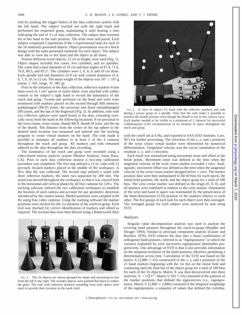
FIG. 2. A view of subject 4’s hand with the reflective markers and rodsduring a power grasp of a spindle. Note that the rods make it possible tomonitor the thumb position even though the thumb is not in this camera view.Each marker needed to be visible to a minimum of 2 cameras for successful3-dimensional (3-D) reconstruction of its location in space throughout thereach and grasp.
2898 C. R. MASON, J. E. GOMEZ, AND T. J. EBNER
J Neurophysiol• VOL 86 • DECEMBER 2001• www.jn.org
on March 7, 2010
jn.physiology.orgD
ownloaded from

tion of each eigenposture throughout the reach-to-grasp. The super-script T denoted the transpose. Last,S (2,8803 63) was a diagonalmatrix consisting of the eigenvalues for the eigenposture-temporalweighting pair in a greatest-to-least order. The eigenvalues indicatethe relative amount of variance explained by each eigenposture-temporal weighting pair. The variance is obtained by squaring theeigenvalues and dividing by the sum of squares. The SVD analysiswas completed for each task separately to be able to explicitly com-pare the eigenpostures generated for the different tasks. The compar-ison was essential to determine whether the same hand synergies weregenerated during the various tasks (e.g., power vs. precision).
Eigenpostures and hand shapes were visualized using 3-D render-ing software [Persistence of Vision Ray Tracer (POVray)] to render3-D images. The images were created by entering thex, y,andzvaluesfor each position as a sphere and linking the appropriate spheres withcylinders to form the hand shapes. Within POVray, it is possible tochange the camera’s perspective and lighting of the 3-D object.However, the same camera perspective and lighting was maintainedfor all of the first eigenpostures and hand reconstructions. The camerawas rotated 180° around thex-axis for improved clarity of the secondeigenposture.
Two methods of comparison of the eigenpostures of the differenttasks and their respective temporal weightings were undertaken. Theroot mean square (RMS) difference for the 21 marker positionsbetween the E1s of the various tasks was calculated to quantify thesimilarities between the eigenpostures. The RMS differences werecalculated for each subject and averaged. A statistical comparison ofthe eigenpostures was based on two sample Student’st-tests of themeans of the marker positions of the eigenpostures between tasks. Thetemporal weightings provide information about the hand shapingthrough time. In addition to plotting the weightings as a function oftime, phase plane plots of the temporal weightings from the first twoeigenpostures were created. Plots of the phase plane trajectories forthe five tasks for different objects were used to address the question ofwhether the different grasps fall within the same or different regionsof the phase plane space.
As shown inRESULTS, the amount of variance explained by succes-sive eigenpostures decreases sharply after the first eigenposture (E1),with higher eigenpostures explaining only small incremental amountsof variance. To determine the nature of the information provided bythe higher order eigenpostures, a series of reduced versions of theoriginal data matrix were constructed using the inverse of the SVDformula. Reduced versions of the original data matrix were calculatedusing only the 1st eigenposture, eigenvalue, and temporal weighting,the 1st and 2nd eigenpostures, eigenvalues and temporal weightings,and so on, up to including the 1st 10 eigenpostures, eigenvalues, andtemporal weightings. The reduced version of the matrix was comparedwith the actual hand posture both visually and by computing the RMSerror as a function of time.
R E S U L T S
SVD analyses across grasps for all subjects and objects
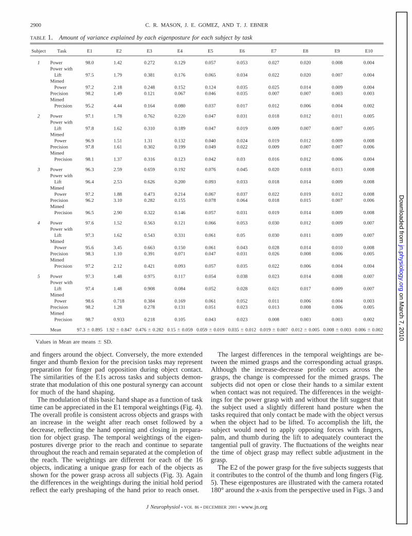
The SVD analyses show that the vast majority of the vari-ability in the hand posture for the entire act of reach-to-graspcan be described by a small number of eigenpostures. The firsteigenposture (E1) accounts for 97.36 0.89% (mean6 SD) ofthe variance of the grasp across tasks and subjects, and E2accounts for 1.96 0.85% (Table 1). The first three eigenpos-tures of any grasp describe over 99.5% of the variance. Ex-tending the earlier findings on static hand posture (Santello etal. 1998), these results demonstrate that hand shape throughoutreach-to-grasp can be described by one dominant eigenpostureand a small number of additional ones.
The first eigenpostures (E1) of the power grasp for the five
subjects are shown in Fig. 3. The E1s are remarkably similarfor the different subjects. E1 consists of an open grasp config-uration in which all the joints are slightly flexed, midposition inthe joint range of motion. Qualitatively E1 appears to be theposition of function as defined by Kapandji (1970). From thisposition the hand can either close for smaller objects or openfor larger objects. There are slight differences that are due inpart to hand size. In Fig. 3 the subjects are ordered by hand sizewith the largest hand on the left and smaller hands to the right.The fingers of the first two subjects are more flexed than thefingers of the other subjects. The fifth subject who has thesmallest hand has the most extended fingers. The gradualdecrease in finger flexion from the largest hand to the smallesthand reflects the excursion of the fingers necessary for thedifferent subjects to grasp the 16 objects. The smaller hands arenear their peak aperture for a greater portion of the objects thanare the larger hands. The similarity in the E1s suggests acommon strategy of an open hand configuration that allows foreasy adjustment for larger or smaller objects.
The temporal weighting profiles of E1 show how the openhand configuration evolves throughout the reach, shaping thehand for the 16 objects (Fig. 3). A common feature across foursubjects is that the weighting increases then decreases through-out the reach in preparation for object grasp. These changes intemporal weighting constitute the larger changes in hand shap-ing that occur during reach. Notice that the change in theweightings begins at the onset of the reach and reflects thepreviously described shaping of the hand in preparation for thegrasping of an object (Jeannerod 1984; Paulignan et al. 1990).The fifth subject tended to rest her hand in a more openposition than the other subjects did. This subject also had thesmallest hand, so there was little additional extension thatoccurred in the reach. This difference is reflected in the weight-ings, which do not show the initial increase. The shape of thisprofile is object-dependent, demonstrating a unique grasp pos-ture for each object. The temporal weightings continue tochange during the beginning of the object grasp period, reflect-ing adjustments in the grasp as the object is contacted.
During the initial hold period the temporal weightings arerelatively constant, indicating that the hand shape was rela-tively constant. The differences in the weightings of the initialhold period reflect the very early initiation of the hand shapingthat precedes the onset of reach as noted previously (Jeannerodand Biguer 1982). The weightings during the object graspperiod are also constant but differ as a function of the object,again an indication of a unique hand shape for each object. Thevariability of the weightings in the three periods suggests thatthe subjects not only controlled this basic hand shape duringthe reach-to-grasp but prior to the reach and after object grasp.
The first eigenposture is similar for the five different grasps.As shown in Fig. 4 forsubject 1,E1 for each type of graspconsists of an open hand configuration with the fingers slightlyflexed and the thumb in opposition to the palm. Again, E1explains the vast majority of the variance (97.26 1.2%) for thefive tasks forsubject 1.The major differences in E1 are afunction of whether the grasp was power or precision, with thepower grasp having more open shape with overall flexion ofthe fingers. Also the thumb is more flexed in the three powergrasps than in the precision grasps. This increased flexion ofthe thumb and long fingers is likely to represent preparation formaking palmar contact with the object by wrapping the thumb
2899HAND SYNERGIES
J Neurophysiol• VOL 86 • DECEMBER 2001• www.jn.org
on March 7, 2010
jn.physiology.orgD
ownloaded from
and fingers around the object. Conversely, the more extendedfinger and thumb flexion for the precision tasks may representpreparation for finger pad opposition during object contact.The similarities of the E1s across tasks and subjects demon-strate that modulation of this one postural synergy can accountfor much of the hand shaping.
The modulation of this basic hand shape as a function of tasktime can be appreciated in the E1 temporal weightings (Fig. 4).The overall profile is consistent across objects and grasps withan increase in the weight after reach onset followed by adecrease, reflecting the hand opening and closing in prepara-tion for object grasp. The temporal weightings of the eigen-postures diverge prior to the reach and continue to separatethroughout the reach and remain separated at the completion ofthe reach. The weightings are different for each of the 16objects, indicating a unique grasp for each of the objects asshown for the power grasp across all subjects (Fig. 3). Againthe differences in the weightings during the initial hold periodreflect the early preshaping of the hand prior to reach onset.
The largest differences in the temporal weightings are be-tween the mimed grasps and the corresponding actual grasps.Although the increase-decrease profile occurs across thegrasps, the change is compressed for the mimed grasps. Thesubjects did not open or close their hands to a similar extentwhen contact was not required. The differences in the weight-ings for the power grasp with and without the lift suggest thatthe subject used a slightly different hand posture when thetasks required that only contact be made with the object versuswhen the object had to be lifted. To accomplish the lift, thesubject would need to apply opposing forces with fingers,palm, and thumb during the lift to adequately counteract thetangential pull of gravity. The fluctuations of the weights nearthe time of object grasp may reflect subtle adjustment in thegrasp.
The E2 of the power grasp for the five subjects suggests thatit contributes to the control of the thumb and long fingers (Fig.5). These eigenpostures are illustrated with the camera rotated180° around thex-axis from the perspective used in Figs. 3 and
TABLE 1. Amount of variance explained by each eigenposture for each subject by task
Subject Task E1 E2 E3 E4 E5 E6 E7 E8 E9 E10
1 Power 98.0 1.42 0.272 0.129 0.057 0.053 0.027 0.020 0.008 0.004Power with
Lift 97.5 1.79 0.381 0.176 0.065 0.034 0.022 0.020 0.007 0.004Mimed
Power 97.2 2.18 0.248 0.152 0.124 0.035 0.025 0.014 0.009 0.004Precision 98.2 1.49 0.121 0.067 0.046 0.035 0.007 0.007 0.003 0.003Mimed
Precision 95.2 4.44 0.164 0.080 0.037 0.017 0.012 0.006 0.004 0.002
2 Power 97.1 1.78 0.762 0.220 0.047 0.031 0.018 0.012 0.011 0.005Power with
Lift 97.8 1.62 0.310 0.189 0.047 0.019 0.009 0.007 0.007 0.005Mimed
Power 96.9 1.51 1.31 0.132 0.040 0.024 0.019 0.012 0.009 0.008Precision 97.8 1.61 0.302 0.199 0.049 0.022 0.009 0.007 0.007 0.006Mimed
Precision 98.1 1.37 0.316 0.123 0.042 0.03 0.016 0.012 0.006 0.004
3 Power 96.3 2.59 0.659 0.192 0.076 0.045 0.020 0.018 0.013 0.008Power with
Lift 96.4 2.53 0.626 0.200 0.093 0.033 0.018 0.014 0.009 0.008Mimed
Power 97.2 1.88 0.473 0.214 0.067 0.037 0.022 0.019 0.012 0.008Precision 96.2 3.10 0.282 0.155 0.078 0.064 0.018 0.015 0.007 0.006Mimed
Precision 96.5 2.90 0.322 0.146 0.057 0.031 0.019 0.014 0.009 0.008
4 Power 97.6 1.52 0.563 0.121 0.066 0.053 0.030 0.012 0.009 0.007Power with
Lift 97.3 1.62 0.543 0.331 0.061 0.05 0.030 0.011 0.009 0.007Mimed
Power 95.6 3.45 0.663 0.150 0.061 0.043 0.028 0.014 0.010 0.008Precision 98.3 1.10 0.391 0.071 0.047 0.031 0.026 0.008 0.006 0.005Mimed
Precision 97.2 2.12 0.421 0.093 0.057 0.035 0.022 0.006 0.004 0.004
5 Power 97.3 1.48 0.975 0.117 0.054 0.038 0.023 0.014 0.008 0.007Power with
Lift 97.4 1.48 0.908 0.084 0.052 0.028 0.021 0.017 0.009 0.007Mimed
Power 98.6 0.718 0.384 0.169 0.061 0.052 0.011 0.006 0.004 0.003Precision 98.2 1.28 0.278 0.131 0.051 0.023 0.013 0.008 0.006 0.005Mimed
Precision 98.7 0.933 0.218 0.105 0.043 0.023 0.008 0.003 0.003 0.002
Mean 97.36 0.895 1.926 0.847 0.4766 0.282 0.156 0.059 0.0596 0.019 0.0356 0.012 0.0196 0.007 0.0126 0.005 0.0086 0.003 0.0066 0.002
Values in Mean are means6 SD.
2900 C. R. MASON, J. E. GOMEZ, AND T. J. EBNER
J Neurophysiol• VOL 86 • DECEMBER 2001• www.jn.org
on March 7, 2010
jn.physiology.orgD
ownloaded from

4 for better viewing of the posture. The proximal and distalphalanges of the thumb are pointing toward the reader (num-bered 1 in Fig. 5) with the MCP of the thumb away from thereader. The MCP of the little finger is toward the reader. Thefingers cross each other so that the second digit is pointingtoward the reader and the fifth digit away from the reader.Likewise, the third and fourth fingers are crossed so that thefingertips are in the inverse order of the MCPs. The sameinversion of the fingertips in relation to the MCPs is present inthe E2 ofsubjects 2, 3,and4. In subject 5’s E2 the crossoveroccurs from the wrist joint. This difference may be due to thesize of this subject’s hand in relation to the other subjects.
The thumbs ofsubjects 2and4 are more prominent than thatof subject 1,suggesting that their thumb positioning maycontribute more to the aperture of the hand than for othersubjects. The thumbs ofsubjects 3and5 are smaller than thethumbs of the other subjects and also shorter relative to theother fingers in their respective eigenpostures; again this mayreflect differences in positioning of the thumb by the subjects.E2 explains a small portion (1.96 0.84%) of the varianceacross subjects and tasks. By definition E2 is orthogonal to E1,and the shape reveals that this is not a natural or physiologicalhand posture. Subjects cannot easily configure their hands tothe postures shown in E2. However, the E2s do provide insightinto the independent control that must be applied to the fingers
and thumb and added to E1 to produce the complete configu-ration of hand postures the subjects achieve.
The temporal profiles of the E2 weightings are similar to theprofiles of E1 for each subject. At the beginning of reach, theweighting increases reflecting the opening of the hand to itsmaximum aperture. Insubjects 1, 3,and5 the temporal weight-ings then decreased as the hand closed in on the object. Insubjects 2and4, the decrease in the temporal weightings aftermaximum aperture were less pronounced. The temporalweightings plateaued during object grasp at different levelsreflecting the hand posture required for the different objects.Therefore even though E2 has more individual variability thanE1, there are similarities in the shapes and the temporal weight-ings.
The E2s across the different tasks are shown for one subjectand explain on average of 2.36 1.25% of the variance (Fig. 6).As described for the E2 of the power grasp ofsubject 1previously, the order of the fingertips is inversed in relation tothe order of the MCPs for the E2 of the power grasp with a lift,precision grasp, and the two mimed grasps. The thumb isprominent for the power grasp, power grasp with a lift, and theprecision grasp and less prominent for the mimed grasps. TheE2s for the two mimed grasp tasks are similar to the grasp ofactual objects (Fig. 6). The E2 temporal weightings follow thesame profile as the E1 weightings (Fig. 4) except for the mimed
FIG. 3. The E1s and temporal weightings for the 5 subjects for power grasp. The temporal weighting profiles of theeigenpostures for each of the 16 objects are shown below the eigenpostures. For ease of comparison, the temporal weightings areseparated by object shapes with cones in the 1st row, and cylinders in the 2nd and spindles in the 3rd row. Above thetop left plotis the task time line including initial hold, reach, and object grasp periods, and the dotted vertical lines provide a similar division.The x-axes are normalized time bins. See text for greater detail.
2901HAND SYNERGIES
J Neurophysiol• VOL 86 • DECEMBER 2001• www.jn.org
on March 7, 2010
jn.physiology.orgD
ownloaded from

precision grasp. For the other four tasks the weights during theinitial hold are constant. The weights increase with onset of thereach then decrease in anticipation of the object grasp. Theweights again are constant during the object grasp. For themimed precision task the weights during the initial hold areconstant. In contrast to the other four tasks, the temporal profileof the E2 weighting for the mimed precision grasp has an initialincrease during the onset of the reach that plateaus and remainsconstant during the object grasp. Again the weighting differ-ences indicate that the grasp posture for each of the 16 objectsis unique.
Among the five tasks, only the power grasp with a liftrequired an efficient grasp with force application adequate toovercome gravity during the lift. In the other tasks the subjectswere requested to grasp the objects as if they were going to liftthem. However, the forces were not monitored, and the sub-jects could have conformed their hands to the objects withoutthe force application. To quantify the similarities between theE1 of power grasp with and without a lift, the RMS differenceswere calculated. The RMS differences between the E1s for thetwo tasks indicate negligible differences considering the dif-ference is across thex, y,andz locations of 21 markers for eacheigenposture. Forsubject 1 the difference was 0.024 mm(Table 2). The average difference across all subjects was0.0246 0.019 (Table 2). Similarly, the differences between theE2s for the two tasks were negligible (Table 2). A two-sample
t-test of the eigenpostures between the two tasks did not showa significant difference (P . 0.05) for either the E1s or the E2s.These small and insignificant differences indicate that theeigenpostures for power grasp with and without a lift are thesame irrespective of the forces utilized.
The average RMS differences between all tasks for E1 andE2 are summarized in Table 3. The differences between theE1s of the five tasks are small, ranging between 0.0246 0.02mm and 0.116 0.07 mm. The differences were greatestbetween the power and precision grasps, particularly betweenthe actual and mimed grasps. However, none of the differenceswere significant (P . 0.05). Similarly, the RMS differencesbetween E2 for the different tasks are small, and the differ-ences between tasks are not significant (P . 0.05). The lack ofsignificant differences between the eigenpostures would implythat the same base posture is used for the five tasks with thehigher order eigenposture adding further shaping information.
To determine whether power and precision grasps are dis-crete postures or fall along a continuum, phase plane plots ofthe temporal weightings of the first two eigenpostures for thefive tasks were constructed. Figure 7 shows representativephase plane plots for the different grasps of the smallest,intermediate, and largest cones (left column), cylinders (middlecolumn), and spindles (right column). The trajectories of thephase plane plots are similar for the different tasks and differ-ent sizes of cones, cylinders, and spindles. For the smallest
FIG. 4. The E1s and the temporal weightings for the 5 tasks for 1 subject. The E1s for the 5 tasks are illustrated in thetop row.Notice the similarities in the hand postures across the task. Below each eigenposture the weightings of the eigenpostures are shownfor each object. Each object has a unique weighting within each task. The weighting are similar for the power grasp and power graspwith a lift, 2 tasks which are essentially the same but are unique for the other tasks. Conventions as in Fig. 3.
2902 C. R. MASON, J. E. GOMEZ, AND T. J. EBNER
J Neurophysiol• VOL 86 • DECEMBER 2001• www.jn.org
on March 7, 2010
jn.physiology.orgD
ownloaded from

cone the trajectories begin in the bottom left quadrant andmove toward the top right quadrant as the hand opens in earlyreach. The maximum aperture of the hand occurs at the pointwhere the trajectories change direction in the top right quad-rant. The trajectories for the three power grasps, power withouta lift (thin black line), power with a lift (dark gray line), andmimed power (dotted line), begin to move back toward thebottom left quadrant as the hand encloses the cone. The tra-jectories of the two precision grasps, precision (thick blackline) and mimed precision (light gray line), do not move as farto the left as the hand does not close to the same degree duringprecision grasp. For the intermediate and large cone the tra-jectories of the power grasps do not move as far toward the leftbottom quadrant after attaining maximum aperture, indicatingthat the fingers flex less to enclose the intermediate and largecones than they did for the smallest cone. All the trajectoriesfor the largest cone are similar, indicating that the hand posturefor the power grasps and precisions grasps approaches thesame aperture forsubject 2.All the trajectories indicate by thesmall movements at the end of the trajectories that final ad-justments in hand shape occur as the object is grasped.
The phase plane plots of the temporal weightings of the firsttwo eigenpostures ofsubject 4for the five tasks in themiddlecolumnand ofsubject 1in theright columnare also similar forthe five tasks for each size cylinder (middle column) or spindle(right column). The trajectories start near the bottom of thegraph, move upward to the right for the hand opening, and
move to the left as the hand closes on the objects. More flexionis indicated during the grasp of the smallest objects by themovement of the trajectories movement further toward the leftthan for the intermediate and large objects. The hand closure inthe power and the precision grasp must be comparable for thecylinders as the trajectories end in the same region of the phaseplane. The clustering and similarities of the temporal weightingprofiles for the five tasks for each object indicates that powerand precision grasps fall along a continuum rather than indiscrete regions of the phase space.
Contribution of higher order eigenpostures
Eigenpostures of order.2 contribute only a small fractionof the variability in the hand posture (Table 1). E3 through E10combined contribute 0.096 0.18% of the variance. On thebasis of information theory, it was shown that these higherorder components are significant (Santello et al. 1998) but thenature of the contribution remained undefined. Therefore weevaluated how hand shape changed as the higher order eigen-postures were successively added and calculated the RMSdifferences between the reconstructed shape and the actualhand shape. In Fig. 8 is shown the reconstructed hand shape forsubject 1performing the power grasp of the smallest spindle. Acomparison of the hand postures in thetop andbottom rowsattwo points in time, bin 15 at the time of peak hand velocity andbin 60 at the beginning of object grasp, illustrates the contri-
FIG. 5. The E2s and the temporal weightings for the 5 subjects for power grasp. By definition E2 is orthogonal to E1. Thecamera perspective has been rotated 180° around thex-axis. The tips of the fingers are numbered beginning with the thumb as 1and ending with the little finger as 5. Other conventions as in Fig. 3.
2903HAND SYNERGIES
J Neurophysiol• VOL 86 • DECEMBER 2001• www.jn.org
on March 7, 2010
jn.physiology.orgD
ownloaded from

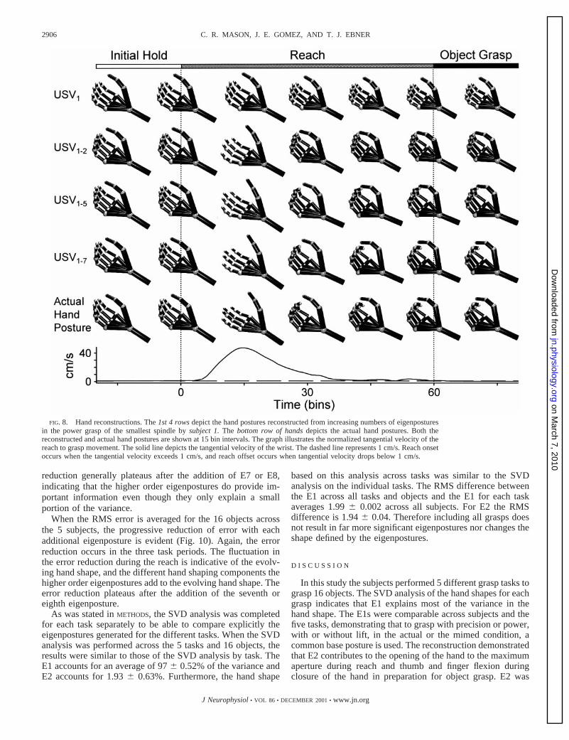
bution of the higher order eigenpostures. At peak velocity thehand posture based on only E1 lacks the finger and thumbextension of the true hand posture illustrated in thebottom row.Note the large increase in thumb and finger extension with theaddition of E2, consistent with the shape of E2 (Figs. 5 and 6).Adding successive eigenpostures further refines the posture ofthe thumb and fingers. Much of the finger extension occurs atthe proximal interphalangeal joints (PIPs) and distal interpha-langeal joints (DIPs) accompanied by some extension of theMCPs. This increase in extension occurs in parallel across thethree rows of joints (MCPs, PIPs, and DIPs for fingers andMCP and IP joint of the thumb) and not at a single joint level.Therefore the additional eigenpostures provide the finer detailsof thumb and finger extension that occurs at the maximumaperture of the true hand posture.
At the onset of object grasp (bin 60), the hand shape basedonly on E1 is lacking the finger and thumb flexion used tograsp the objects. With the addition of E2 the finger and thumbare almost in opposition due to the increased flexion of thethumb and fingers, again showing that E2 is involved in the
control of the thumb and fingers. Adding successively E3 to E7to the reconstruction progressively increases the flexion of theMCPs, PIPs, and DIPs. Again, the increasing flexion occurs inparallel across all joints. After the addition of all the eigenpos-tures up to and including E7, the flexion of the long fingersclosely approximates the flexion observed in the true handposture. Therefore qualitatively E2–E7 provide informationabout the state of the flexion/extension of the MCPs, PIPs, andDIPs, even though these eigenpostures explain only a smallfraction of the variance.
To quantify the importance of the higher order eigenpos-tures, the reduction in the RMS error with the addition ofsuccessive eigenpostures was determined. This reduction inRMS error is shown graphically in Fig. 9 for the five differentgrasps of the smallest spindle bysubject 1.The RMS error forreconstruction based on E1 is constant during the initial hold.The error decreases briefly as the hand begins to open and thefinger extension of the actual hand and the USV1 reconstruc-tion more closely approximate each other. The error increasesmarkedly around peak velocity when the fingers and thumblack adequate extension for the maximum aperture. The errordecreases after the maximum aperture as the fingers begin toflex in preparation for the object grasp. Again the true fingerposture and the USV1 reconstructed hand shape are relativelyclose then the RMS error increases as the reach continues andthroughout the grasp period because the actual fingers flex inpreparation for the grasp and during the grasp but the fingers ofthe USV1 reconstruction do not.
FIG. 6. The E2s and the temporal weightings for the 5 tasks for 1 subject. Finger numbering as in Fig. 5. Other conventions asin Fig. 3. This is the same subject shown in Fig. 4.
TABLE 2. RMS differences between eigenpostures of powerwith and without a lift
Subject 1 Subject 2 Subject 3 Subject 4 Subject 5 Mean
E1 0.024 0.005 0.021 0.057 0.014 0.0246 0.02E2 0.208 0.064 0.113 0.357 0.301 0.2086 0.123
Values in Mean are means6 SD. RMS, root mean square.
2904 C. R. MASON, J. E. GOMEZ, AND T. J. EBNER
J Neurophysiol• VOL 86 • DECEMBER 2001• www.jn.org
on March 7, 2010
jn.physiology.orgD
ownloaded from

The addition of successive eigenpostures reduces the error inall phases of the reach-to-grasp. This error reduction is not byan equivalent amount in all phases, indicating that each eigen-posture is contributing to different aspects of the hand shape.The inclusion of E2 adding the needed finger and thumbextension results in a particularly larger reduction in the errorduring the reach for the five grasps. E2 also contributes sub-stantially to the error reduction in the initial hold period (par-
ticularly in the mimed grasps) and in the object grasp period forthe power grasps. E2 contribution is primarily in the neededflexion of the fingers and thumb during object grasp. Theaddition of successive higher order eigenpostures results in adecrease of the RMS error. The reduction in error plateaus afterthe addition of E7 for the power grasp. The RMS errors for theother grasps show a similar reduction in the RMS error with theaddition of successive higher order eigenpostures. The error
TABLE 3. Summary of RMS differences across subjects
Power Power with Lift Mimed Power Precision Mimed Precision
E1
Power 0Power with Lift 0.0246 0.020 0Mimed Power 0.0666 0.020 0.0826 0.033 0Precision 0.0706 0.028 0.0806 0.021 0.0796 0.020 0Mimed Precision 0.0996 0.075 0.1116 0.070 0.0786 0.054 0.0696 0.049 0
E2
Power 0Power with Lift 0.2096 0.123 0Mimed Power 0.3296 0.226 0.4426 0.316 0Precision 0.2416 0.084 0.3286 0.057 0.3676 0.252 0Mimed Precision 0.3896 0.198 0.4716 0.284 0.2496 0.095 0.3176 0.199 0
Values are means6 SD. RMS, root mean square.
FIG. 7. Phase plane plots of temporal weighting of E1 against the temporal weighting of E2 for the 5 tasks for the smallest (top),intermediate (middle), and largest (bottom) object in each shape. The data in each column are from a different subject. The conesare based on data fromsubject 2,the cylinders fromsubject 4and the spindles fromsubject 1.
2905HAND SYNERGIES
J Neurophysiol• VOL 86 • DECEMBER 2001• www.jn.org
on March 7, 2010
jn.physiology.orgD
ownloaded from
reduction generally plateaus after the addition of E7 or E8,indicating that the higher order eigenpostures do provide im-portant information even though they only explain a smallportion of the variance.
When the RMS error is averaged for the 16 objects acrossthe 5 subjects, the progressive reduction of error with eachadditional eigenposture is evident (Fig. 10). Again, the errorreduction occurs in the three task periods. The fluctuation inthe error reduction during the reach is indicative of the evolv-ing hand shape, and the different hand shaping components thehigher order eigenpostures add to the evolving hand shape. Theerror reduction plateaus after the addition of the seventh oreighth eigenposture.
As was stated inMETHODS, the SVD analysis was completedfor each task separately to be able to compare explicitly theeigenpostures generated for the different tasks. When the SVDanalysis was performed across the 5 tasks and 16 objects, theresults were similar to those of the SVD analysis by task. TheE1 accounts for an average of 976 0.52% of the variance andE2 accounts for 1.936 0.63%. Furthermore, the hand shape
based on this analysis across tasks was similar to the SVDanalysis on the individual tasks. The RMS difference betweenthe E1 across all tasks and objects and the E1 for each taskaverages 1.996 0.002 across all subjects. For E2 the RMSdifference is 1.946 0.04. Therefore including all grasps doesnot result in far more significant eigenpostures nor changes theshape defined by the eigenpostures.
D I S C U S S I O N
In this study the subjects performed 5 different grasp tasks tograsp 16 objects. The SVD analysis of the hand shapes for eachgrasp indicates that E1 explains most of the variance in thehand shape. The E1s were comparable across subjects and thefive tasks, demonstrating that to grasp with precision or power,with or without lift, in the actual or the mimed condition, acommon base posture is used. The reconstruction demonstratedthat E2 contributes to the opening of the hand to the maximumaperture during reach and thumb and finger flexion duringclosure of the hand in preparation for object grasp. E2 was
FIG. 8. Hand reconstructions. The1st 4 rowsdepict the hand postures reconstructed from increasing numbers of eigenposturesin the power grasp of the smallest spindle bysubject 1.The bottom row of handsdepicts the actual hand postures. Both thereconstructed and actual hand postures are shown at 15 bin intervals. The graph illustrates the normalized tangential velocity of thereach to grasp movement. The solid line depicts the tangential velocity of the wrist. The dashed line represents 1 cm/s. Reach onsetoccurs when the tangential velocity exceeds 1 cm/s, and reach offset occurs when tangential velocity drops below 1 cm/s.
2906 C. R. MASON, J. E. GOMEZ, AND T. J. EBNER
J Neurophysiol• VOL 86 • DECEMBER 2001• www.jn.org
on March 7, 2010
jn.physiology.orgD
ownloaded from

more variable across subjects and suggests that there may beindependent control over some aspects of grasp, particularlythumb and finger extension. The higher order eigenposturesadd further shaping information to the hand shape. Theseresults are comparable to earlier findings that over 80% of thevariance of static grasp postures can be explained by twoprincipal components with the higher order components pro-viding additional information (Santello et al. 1998).
The grasp tasks in this study were initially classified as eitherpower or precision as defined by Napier (1956). Napier con-cluded that prehensile movements consisted of two discrete
patterns of movement that identified the grip as either a powergrip, a precision grip, or a combination of the two. The gripwas determined by how the object was to be used (Napier1956). Numerous subsequent studies have proposed even moreelaborate categorical schemes (Cutkowsky and Howe 1990;Kamakura et al. 1980). The similarities of the eigenpostures forthe five different grasps in a subject suggest that only onemovement pattern exists even though the final hand posture isdependent on function (or desired grasp) and object to begrasped. The similarities in the phase plane trajectories of E1and E2 temporal weightings further show that the “classic”Napier precision and power grips were not evident in thegrasps and objects explored. Rather than two or more discretegrasps, hand posture may be composed of a continuum basedon the temporal weighting of a few eigenpostures. In this case,eigenpostures would not be limited to or be expected to cor-
FIG. 10. The average RMS error for the 5 subjects and 16 objects for eachof the grasps. Conventions as in Fig. 9.
FIG. 9. The root mean square (RMS) error for the 5 grasps of the smallestspindle forsubject 1.The top graphcorresponds to the hand reconstructionsobserved in the preceding figure. The solid line indicates the RMS error usingonly E1 in the reconstruction. The numbered arrows indicate the highesteigenposture included in the reconstructed matrix on which the RMS error iscalculated. Note the consistent drop in error as the higher order eigenposturesare sequentially added in the 5 tasks.A: Power Grasp.B: Power Grasp with aLift. C: Mimed Power Grasp.D: Precisions Grasp.E: Mimed Precision Grasp.
2907HAND SYNERGIES
J Neurophysiol• VOL 86 • DECEMBER 2001• www.jn.org
on March 7, 2010
jn.physiology.orgD
ownloaded from
respond to particular grasps. Two caveats must be considered.First, that power and precision grasps are within discrete re-gions of the same manifold and the SVD analysis failed todistinguish between the two types of hand shapes. Second theresults are limited to the set of objects grasped and the graspsstudied. There remains the possibility that if a larger range ofgrasp behaviors was evaluated a more readily apparent divisionwould develop.
The temporal weightings of the eigenpostures demonstratedthat hand shape evolves continuously throughout the reach-to-grasp and is unique for each object/grasp combination. TheSVD analysis is a technique to quantify the modulation of thehand shape that begins prior to the reach-to-grasp and contin-ues through to the object contact in the actual grasps or the endof movement in the mimed grasp. The temporal weightingsmore precisely define the shaping of the fingers for grasp thanjust the linear scaling of the maximum hand aperture to theobject size as previously noted (Chieffi and Gentilucci 1993;Jeannerod 1984; Marteniuk et al. 1990; Paulignan et al. 1990).These results confirm and expand on the finding that handshape evolved gradually throughout the movement (Santelloand Soechting 1998).
A normal open hand posture configuration characterizes E1and is qualitatively similar to the position of function(Kapandji 1970). However, as noted inRESULTS, E2 is not anatural hand posture but an orthogonal posture to E1. With thelarge number of degrees of freedom in the hand and with theequally significant biomechanical constraints, it is difficult toconceive of two orthogonal postures that the hand could readilyassume. The same problem exists for other dimensionalityreduction approaches, and the early study did not show thesecond or higher principal components (Santello et al. 1998).However, E2 and the higher components do provide insightinto the additional finger and thumb movements that are re-quired to shape the hand. The eigenpostures may represent asimplifying scheme used by the CNS to control the hand.
Evolution of synergies throughout reach-to-grasp
The shaping of the hand evolves throughout the reach,beginning with extension of the fingers and thumb followed byflexion in anticipation of object contact (Jeannerod 1984;Paulignan et al. 1990; Santello and Soechting 1998). Thetemporal weightings show that evolution of the synergies wasconsistent irrespective of object shape, grasp, or subject. Ingeneral there was a biphasic pattern to the weightings, firstincreasing during the reach then decreasing as the hand closesin on the object. At maximum hand aperture with thumb andfingers extended, the thumb and fingers begin to flex in prep-aration for object grasp. This general pattern of opening andclosing is further evidence that the hand acts as a functionalunit. There also appears to be coordinating actions across theMCPs, the PIPs, and the DIPs, in which the fingers extend orflex together. Others have also observed a high degree ofcorrelation in joint angles (Santello and Soechting 1998; San-tello et al. 1998). This is further evidence that the hand func-tions as a unit.
Our results indicate that the hand posture, if not constrained,differs for the 16 objects throughout the initial hold period,suggesting that the hand shape preparation begins well inadvance of the reach. This observation concurs with that of
Jeannerod and Biguer (1982), who also reported early initiationof the hand shaping, preceding the onset of reach by 40–120ms. The even earlier preshaping of the hand found in thepresent study may reflect the differences in the paradigms. Inthe previous study the subjects began each trial with their eyesclosed while the object was changed. The subjects then openedtheir eyes and waited for a “go” cues before making a fast andaccurate grasp (Jeannerod and Biguer 1982). In our task thesubjects’ eyes remained open throughout the experiment, andthey viewed the changes in objects prior to receiving instruc-tions as to which grasp to perform. In addition the trials wereself-initiated after receiving the instructions without a timeconstraint. Therefore the subjects had a considerable period oftime in which to adjust hand shape prior to reach.
Actual versus mimed grasps
The E1s of the mimed grasps were similar to the E1s of theactual grasps, suggesting that the subjects used the same con-trol strategy whether or not tactile contact was made. Thethumb and fingers of the E1s for the mimed grasps have thesame flexed posture observed for the actual power grasps andthe more extended posture for the precision grasps. The tem-poral weightings were also similar. Therefore control strategyused for actual grasps generalizes to the control of mimedgrasps, and the results suggest that tactile contact is not adominant factor in the early shaping of the hand.
The temporal weightings for the mimed grasps are lessdivergent than those for the actual grasps, particularly duringthe object hold period. Therefore the mimed grasp hand shapesvaried less throughout the reach-to-grasp than the hand shapesduring actual grasp. This agrees with previous experiments inwhich subjects mimed the grasp adjacent to the object. Thepeak aperture of the mimed was smaller than during actualgrasps of the objects (Goodale et al. 1994). During the actualgrasp the compliance of the hand enables it to mold to theobject contours (Hajian and Howe 1997). In contrast, the handshape during mimed grasp was dependent on visual cues andmemory and lacked the molding to the object. The mimedgrasp lacked the same requirement of the actual grasp, that ofenclosing an object. Therefore the need for accuracy in themaximum aperture and final grasp posture was most likelydiminished. The net result was the more compressed temporalweightings.
Importance of higher synergies
The final goal of the study was to investigate the contribu-tion of the higher order eigenpostures to the formation of thegrasp. It was previously shown that for static grasps the higherprincipal components contributed important information eventhough these components account for only a small percentageof the variance (Santello et al. 1998). The present resultsdemonstrate that the higher order components contribute agreat deal to the overall shape of the hand. The flexion of thePIPs and DIPs increased with the addition of the higher ordereigenpostures. The finger flexion gradually increased acrossthe MCPs, PIPs, or DIPs in parallel until the hand enclosed theobject. Therefore not only did the higher order eigenposturesadd critical details needed for static grasp but also for the actof preshaping during the grasp.
2908 C. R. MASON, J. E. GOMEZ, AND T. J. EBNER
J Neurophysiol• VOL 86 • DECEMBER 2001• www.jn.org
on March 7, 2010
jn.physiology.orgD
ownloaded from

The findings in this study add to the growing evidence thatthe hand is controlled as a unit. There are four specific findings.First the base posture described over 97% of the varianceacross subjects, grasps, and objects. Second, the entire act ofreach-to-grasp is specified by a temporal weighting of this baseposture. Third, adding the higher order eigenpostures did notinfluence hand shape at the single joint or digit level but insteadchanged hand posture across the joints. For example, the in-creased flexion of the PIPs from USV1 to USV1–7 at the timeof object grasp occurred in all long fingers (Fig. 7). Last, themimed grasps can be explained by the same eigenpostures asthe actual grasps indicating a common control strategy. Thesefindings refute the traditional view that the hand is controlledat a single joint, digit, or muscle level and suggest that the CNSuses a simplifying strategy in the control of the hand duringreach-to-grasp. Other control strategies may be used for otherhand movements.
The simplifying control strategy detected by the SVD tech-nique is observed at the output stage. Are these eigenposturesrepresented at the cortical level? The results of stimulation andlesion studies in the motor cortex are consistent with globalcontrol of the hand. Stimulation of one site in M1 evokesresponses in several muscles of the hand (Donoghue et al.1992; Sato and Tanji 1989) or movement around contiguousjoints or fingers (Gould et al. 1986; Kwan et al. 1978; Strickand Preston 1978). Focal inactivations or focal strokes in thehand area of the motor cortex do not disrupt movements ofindividual fingers; rather, movement of different combinationsare effected (Schieber 1999; Schieber and Poliakov 1998).
Single-unit recordings in primary motor and premotor cortexalso suggest that the hand is controlled as a unit. In monkeys asingle neuron in M1 generally discharges in relation to multipleinstructed finger movements (Schieber and Hibbard 1993).Furthermore, the population of cells active with different fingermovements overlaps extensively (Schieber and Hibbard 1993).Motor neurons in the ventral premotor (F5) cortex in monkeyshave been shown to discharge selectively during a specificgrasp such as precision grip, finger prehension, or whole handprehension (Murata et al. 1997; Rizzolatti et al. 1988). Thefiring of these neurons correlated only with the specific graspsand not the individual movements made by the monkeys (Riz-zolatti et al. 1988). These findings suggest that the hand isrepresented as a unit at the premotor cortical unit. The eigen-postures observed at the output stage may be represented in thedischarge of a population of hand-related motor and premotorcortical cells.
The authors thank M. McPhee for technical and graphical assistance.Funding was provided in part by National Institute of Neurological Disor-
ders and Stroke Grants F32 NS-10491, R01 NS-18338, and R01 NS-31530.
REFERENCES
ANDERSENP, HAGAN PJ, PHILLIPS CG, AND POWELL TPS. Mapping by micro-stimulation of overlapping projections from area 4 to motor units of thebaboon’s hand.Proc R Soc Lond B Biol Sci188: 31–60, 1975.
ARBIB MA, I BERALL T, AND LYONS D. Coordinated control programs formovements of the hand.Exp Brain Res Suppl10: 111–129, 1985.
BOOTSMA RJ, MARTENIUK RG, MACKENZIE CL, AND ZAAL FTJM. The speed-accuracy trade-off in manual prehension: effects of movement amplitude,object size, and object width on kinematic characteristics.Exp Brain Res98:535–541, 1994.
BUYS EJ, LEMON RN, MANTEL GW, AND MUIR RB. Selective facilitation ofdifferent hand muscles by single corticospinal neurones in the consciousmonkey.J Physiol (Lond)381: 529–549, 1986.
CHENEY PD AND FETZ EE. Comparable patterns of muscle facilitation evokedby individual corticomotoneuronal (CM) cells and by single intracorticalmicrostimulation in primates: evidence for functional groups of CM cells.J Neurophysiol53: 786–804, 1985.
CHENEY PD, FETZ EE,AND PALMER SS. Patterns of facilitation and suppressionof antagonist forelimb muscles from motor cortex sites in the awake mon-key. J Neurophysiol53: 805–820, 1985.
CHIEFFI S AND GENTILUCCI M. Coordination between the transport and thegrasp components during prehension movements.Exp Brain Res94: 471–477, 1993.
CUTKOWSKY MR AND HOWE RD. Human grasp choice and robotic graspanalysis. In:Dextrous Robot Hands,edited by Venkataraman ST and IberallT. New York: Springer-Verlag, 1990, p. 5–31.
DONOGHUE JP, LEIBOVIC S, AND SANES JN. Organization of the forelimb areain squirrel monkey motor cortex: representation of digit, wrist, and elbowmuscles.Exp Brain Res89: 1–19, 1992.
ENGEL KC, FLANDERS M, AND SOECHTING JF. Anticipatory and sequentialmotor control in piano playing.Exp Brain Res113: 189–199, 1997.
FLANDERS M AND SOECHTING JF. Kinematics of typing: parallel control of thetwo hands.J Neurophysiol67: 1264–1274, 1992.
GLASER EM AND RUCHKIN DS. Principles of Neurobiological Signal Analysis.New York: Academic, 1967.
GOODALE MA, JAKOBSON LS, AND KEILLOR JM. Differences in the visualcontrol of pantomimed and natural grasping movements.Neuropsychologia32: 1159–1178, 1994.
GORDON AM, CASABONA A, AND SOECHTING JF. The learning of novel fingermovement sequences.J Neurophysiol72: 1596–1610, 1994.
GOULD HJ, CUSICK CG, PONS TP, AND KAAS JH. The relationship of corpuscallosum connections to electrical stimulation maps of motor, supplemen-tary motor and the frontal eyefields in owl monkeys.J Comp Neurol247:297–325, 1986.
HAJIAN AZ AND HOWE RD. Identification of the mechanical impedance at thehuman finger tip.J Biomed Eng119: 109–114, 1997.
HENDLER RW AND SHRAGER RI. Deconvolutions based on singular valuedecomposition and the pseudoinverse: a guide for beginners.J BiochemBiophys Methods28: 1–33, 1994.
IBERALL T AND FAGG A. Neural networks for selecting hand shapes. In:Handand Brain: The Neurophysiology and Psychology of Hand Movements,edited by Wing AM, Haggard P, and Flanagan JR. San Diego, CA: Aca-demic, 1996, p. 243–264.
JEANNEROD M. The timing of natural prehension movements.J Mot Behav16:235–254, 1984.
JEANNEROD M AND BIGUER B. Visuomotor mechanisms in reaching withinextrapersonal space. In:Analysis of Visual Behavior,edited by Ingle DJ,Goodale MA, and Mansfield RJW. Cambridge, MA: MIT Press, 1982, p.387–409.
KAMAKURA N, MATSUO M, ISHII H, MITSUBOSHI F, AND MIURA Y. Patterns ofstatic prehension in normal hands.Am J Occup Ther7: 437–445, 1980.
KAPANDJI IA. The Physiology of the Joints. Upper Limb(2nd ed.). London: Eand S Livingstone, 1970, vol. 1, p. 146–202.
KWAN HC, MACKAY WA, MURPHY JT,AND WONG YC. Spatial organization ofprecentral cortex in awake primates. II. Motor outputs.J Neurophysiol41:1120–1131, 1978.
LANDSMEER JMF. The coordination of finger-joint motions.J Bone Joint Surg45A: 1654–1662, 1963.
LANDSMEER JMF AND LONG C. The mechanism of finger control based onelectromyograms and location analysis.Acta Anat60: 330–347, 1965.
LEMON RN. Neural control of dexterity: what has been achieved?Exp BrainRes128: 6–12, 1999.
LEMON RN, MANTEL GW, AND MUIR RB. Corticospinal facilitation of handmuscles during voluntary movement in the conscious monkey.J Physiol(Lond) 381: 529–549, 1986.
LEYTON ASF AND SHERRINGTON CS. Observations on the excitable cortex ofthe chimpanzee, orangutan and gorilla.Q J Exp Physiol11: 137–222, 1917.
LONG C, CONRAD PW, HALL EA, AND FURLER SL. Intrinsic-extrinsic musclecontrol of the hand in power grip and precision handling.J Bone Joint Surg52A: 853–867, 1970.
MARTENIUK RG, LEAVITT JL, MACKENZIE CL, AND ATHENES S. Functionalrelationships between grasp and transport components in a prehension task.Hum Mov Sci9: 149–176, 1990.
2909HAND SYNERGIES
J Neurophysiol• VOL 86 • DECEMBER 2001• www.jn.org
on March 7, 2010
jn.physiology.orgD
ownloaded from

MASON CR, GOMEZ JE,AND EBNER TJ. Simplifying strategies of human wholehand grasp as determined by singular value decomposition.Soc NeurosciAbstr 25: 113, 1999.
MCKIERNAN BJ, MARCARIO JK, KARRER JH, AND CHENEY PD. Corticomo-toneuronal postspike effects in shoulder, elbow, wrist, digit, and intrinisichand muscles during a reach and prehension task.J Neurophysiol80:1961–1980, 1998.
MURATA A, FADIGA L, FOGASSI L, GALLESE V, RAOS V, AND RIZZOLATTI R.Object representations in the ventral premotor cortex of the monkey.J Neu-rophysiol78: 2226–2230, 1997.
NAPIER JR. The prehensile movement of the human hand.J Bone Joint Surg38B: 902–913, 1956.
OLDFIELD RC. The assessment and analysis of handedness: the Edinburghinventory.Neuropsychologia9: 97–113, 1971.
PAULIGNAN Y, JEANNEROD M, MACKENZIE C, AND MARTENIUK R. Selectiveperturbation of visual input during prehension movements 2. The effects ofchanging object size.Exp Brain Res87: 407–420, 1991.
PAULIGNAN Y, MACKENZIE C, MARTENIUK R, AND JEANNERODM. The couplingof arm and finger movements during prehension.Exp Brain Res79: 431–435, 1990.
PENFIELD W AND RASMUSSEN T. The Cerebral Cortex of Man. New York:MacMillan, 1950.
POLIAKOV AV AND SCHIEBER MH. Limited functional grouping of neurons inthe motor cortex hand area during individuated finger movements: a clusteranalysis.J Neurophysiol82: 3488–3505, 1999.
RIZZOLATTI G, CAMARDA R, FOGASSI L, GENTILUCCI M, LUPPINO G, AND
MATELLI M. Functional organization of inferior area 6 in the macaquemonkey. II. Area F5 and the control of distal movement.Exp Brain Res71:491–507, 1988.
SANTELLO M, FLANDERS M, AND SOECHTING JF. Postural hand synergies fortool use.J Neurosci18: 10105–10115, 1998.
SANTELLO M AND SOECHTING JF. Matching object size by controlling fingerspan and hand shape.Somatosens Mot Res14: 203–212, 1997.
SANTELLO M AND SOECHTING JF. Gradual molding of the hand to objectcontours.J Neurophysiol79: 1307–1320, 1998.
SATO KC AND TANJI J. Digit-muscle responses evoked from multiple intracor-tical foci in monkey precentral motor cortex.J Neurophysiol62: 959–970,1989.
SCHIEBER M. How might the motor cortex individuate movements?TrendsNeurosci13: 440–445, 1990.
SCHIEBER M. Individuated finger movements of Rhesus monkeys: a means ofquantifying the independence of the digits.J Neurophysiol65: 1381–1391,1991.
SCHIEBER M. Muscular production of individuated finger movements: the rolesof extrinsic finger muscles.J Neurosci15: 284–297, 1995.
SCHIEBER M. Somatotopic gradients in the distributed organization of thehuman primary motor cortex hand area: evidence from small infarcts.ExpBrain Res128: 139–148, 1999.
SCHIEBER M AND POLIAKOV AV. Partial inactivation of the primary motorcortex hand area: effects of individuated finger movements.J Neurosci18:9038–9054, 1998.
SCHIEBER MH, CHUA M, PETIT J,AND HUNT CC. Tension distribution of singlemotor units in multitendoned muscles: comparison of a homologous digitmuscle in cats and monkeys.J Neurosci17: 1734–1747, 1997.
SCHIEBER MH AND HIBBARD LS. How somatotopic is the motor cortex handarea?Science261: 489–492, 1993.
SERLIN DM AND SCHIEBER M. Morphological regions of the multitendonedextrinsic finger muscles in the monkey forearm.Acta Anat14: 255–266,1993.
SOECHTING JF AND FLANDERS M. Flexibility and repeatability of finger move-ments during typing: analysis of multiple degrees of freedom.J ComputNeurosci4: 29–46, 1997.
STRICK PL AND PRESTON JB. Multiple representation in the primate motorcortex.Brain Res154: 366–370, 1978.
TUBIANA R. Architecture and function of the hand. In:The Hand,edited byTubiana R. Philadelphia, PA: Saunders, 1981, vol. 1, p. 19–93.
WOOLSEYCN. Organization of somatic sensory and motor areas of the cerebralcortex. In:Biological and Biochemical Bases of Behavior,edited by HarlowHF and Woolsey CN. Madison, WI: Univ. of Wisconsin Press, 1958, p.63–81.
WOOLSEYCN, ERICKSONTC, AND GILSON WE. Localization in somatic sensoryand motor areas of human cerebral cortex as determined by direct recordingsof evoked potentials and electrical stimulation.J Neurosurg51: 476–506,1979.
2910 C. R. MASON, J. E. GOMEZ, AND T. J. EBNER
J Neurophysiol• VOL 86 • DECEMBER 2001• www.jn.org
on March 7, 2010
jn.physiology.orgD
ownloaded from