calcific deposit needling in combination with …...original article calcific deposit needling in...
TRANSCRIPT

SICOT-J 2018, 4, 45© The Authors, published by EDP Sciences, 2018https://doi.org/10.1051/sicotj/2018043
Available online at:www.sicot-j.org
ORIGINAL ARTICLE
Calcific deposit needling in combination with extracorporeal shockwave therapy (ESWT): A proposed treatment for supraspinatuscalcified tendinopathyEmilios Pakos1, Ioannis Gkiatas1,*, Georgios Rakkas2, Dimitrios Papadopoulos1, Ioannis Gelalis1, Marios Vekris1,and Anastasios Korompilias1
1 Orthopaedic Department, University Hospital of Ioannina, Ioannina, Greece2 Physiotherapy Department, Salamagka 4, Ioannina, Greece
Received 6 May 2018, Accepted 10 August 2018, Pub
*Correspon
This is anO
lished online 19 October 2018
Abstract -- Background: Calcified tendinopathy of the rotator cuff is one of the most common conditionsconcerning the shoulder pathology. It is characterized by a reactive calcification that affects the tendons beingpart of the rotator cuff. The reported prevalence varies from 2.7% to 22%. Most of the patients can be treatedeffectively with non-operative measures such as anti-inflammatory drugs, subacromial injection of steroid,physiotherapy, extracorporeal shock wave therapy (ESWT) and needle aspiration irrigation. Results of atreatment combining some of these methods have not been reported.Objectives: The purpose of this study is to present the radiological as well as the clinical results of our proposedprotocol which combines drilling of the calcium deposits with xylocaine under ultrasound guidance, with aspecific program of physiotherapy for 1 month without the use of NSAIDs.Methods: Sixty-six consecutive patients (68 shoulders) were treated for calcified tendinitis of supraspinatus,which was diagnosed clinically and radiologically, with needle drilling using xylocaine under ultrasoundguidance. After the drilling the patient followed a physiotherapy protocol with ESWTwhich included five visitswithin a month. After the end of the physiotherapy, the patients were evaluated clinically and radiologically.The Visual Analogue Scale (VAS) for pain and the Disabilities of the Arm, Shoulder, and Hand (DASH) scorewere measured before and after the end of the therapy.Results: All the patients showed clinical improvement of the symptoms at the follow-up. The mean VAS scoreshowed improvement from 8.1 to 3.3 whereas the mean DASH score was 27 and after the end of the therapy 5.Radiologically all but one calcific deposits were disappeared.Conclusions: The ultrasound-guided drilling of the calcific deposit using xylocaine, in combination withphysiotherapy using ESWT provides a reliable alternative treatment for the calcific tendinitis of thesupraspinatus
Key words: Supraspinatus, Calcified tendinopathy, Needling, Extracorporeal shock wave therapy.
Introduction
Calcified tendinopathy of the rotator cuff is one of themost common conditions regarding the shoulder patholo-gy. It is characterized by a reactive calcification thataffects the tendons that are part of the rotator cuff [1]. Thereported prevalence of this condition varies from 2.7% to22%. Women aged between 30 and 50 years old are themost common affected group [2]. The pathogenesisinvolves two proposed different processes leading toformation of the calcium deposits in the cuff. Thedegenerative calcification where the degeneration of the
ding author: [email protected]
penAccess article distributed under the terms of the CreativeComwhich permits unrestricted use, distribution, and reproduction i
tendon fibers precedes calcification and the reactivecalcification where the process of calcification is activelymediated by cells in a viable environment [3].
Concerning the histopathologic findings the calcifiedtendinopathy consists of three stages: the precalcific,where the tendinous tissue is altered into fibrocartilage,the calcific stage, with a phase of formation and anotherone of resorption, and the post-calcific stage [4,5]. Due tothe fact that the disease is multifocal, with several parts ofthe tendon undergoing varying stages of the evolutionaryprocess, there are some classification systems trying topersonalize the disease’s therapy [5].
The management of calcified tendinitis can be dividedinto two major categories: the conservative treatment andthe operative one. The first one includes certain exercises
monsAttribution License (http://creativecommons.org/licenses/by/4.0),n any medium, provided the original work is properly cited.
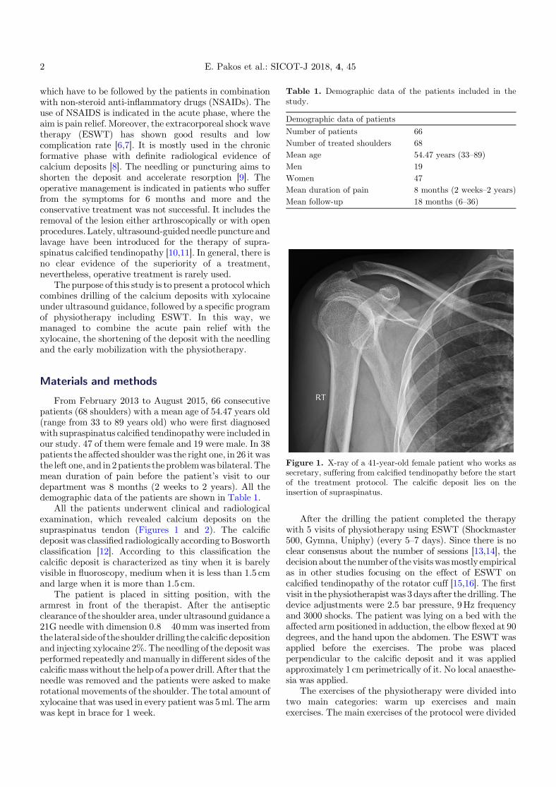
Table 1. Demographic data of the patients included in thestudy.
Demographic data of patientsNumber of patients 66Number of treated shoulders 68Mean age 54.47 years (33–89)Men 19Women 47Mean duration of pain 8 months (2 weeks–2 years)Mean follow-up 18 months (6–36)
2 E. Pakos et al.: SICOT-J 2018, 4, 45
which have to be followed by the patients in combinationwith non-steroid anti-inflammatory drugs (NSAIDs). Theuse of NSAIDS is indicated in the acute phase, where theaim is pain relief. Moreover, the extracorporeal shock wavetherapy (ESWT) has shown good results and lowcomplication rate [6,7]. It is mostly used in the chronicformative phase with definite radiological evidence ofcalcium deposits [8]. The needling or puncturing aims toshorten the deposit and accelerate resorption [9]. Theoperative management is indicated in patients who sufferfrom the symptoms for 6 months and more and theconservative treatment was not successful. It includes theremoval of the lesion either arthroscopically or with openprocedures. Lately, ultrasound-guidedneedle puncture andlavage have been introduced for the therapy of supra-spinatus calcified tendinopathy [10,11]. In general, there isno clear evidence of the superiority of a treatment,nevertheless, operative treatment is rarely used.
The purpose of this study is to present a protocol whichcombines drilling of the calcium deposits with xylocaineunder ultrasound guidance, followed by a specific programof physiotherapy including ESWT. In this way, wemanaged to combine the acute pain relief with thexylocaine, the shortening of the deposit with the needlingand the early mobilization with the physiotherapy.
Figure 1. X-ray of a 41-year-old female patient who works assecretary, suffering from calcified tendinopathy before the startof the treatment protocol. The calcific deposit lies on theinsertion of supraspinatus.
Materials and methods
From February 2013 to August 2015, 66 consecutivepatients (68 shoulders) with a mean age of 54.47 years old(range from 33 to 89 years old) who were first diagnosedwith supraspinatus calcified tendinopathywere included inour study. 47 of them were female and 19 were male. In 38patients the affected shoulder was the right one, in 26 itwasthe left one, and in 2patients the problemwasbilateral.Themean duration of pain before the patient’s visit to ourdepartment was 8 months (2 weeks to 2 years). All thedemographic data of the patients are shown in Table 1.
All the patients underwent clinical and radiologicalexamination, which revealed calcium deposits on thesupraspinatus tendon (Figures 1 and 2). The calcificdeposit was classified radiologically according toBosworthclassification [12]. According to this classification thecalcific deposit is characterized as tiny when it is barelyvisible in fluoroscopy, medium when it is less than 1.5 cmand large when it is more than 1.5 cm.
The patient is placed in sitting position, with thearmrest in front of the therapist. After the antisepticclearance of the shoulder area, under ultrasound guidance a21G needle with dimension 0.8� 40mm was inserted fromthe lateral side of the shoulderdrilling the calcificdepositionand injecting xylocaine 2%.The needling of the deposit wasperformed repeatedly andmanually in different sides of thecalcificmasswithout the help of a powerdrill.After that theneedle was removed and the patients were asked to makerotational movements of the shoulder. The total amount ofxylocaine that was used in every patient was 5ml. The armwas kept in brace for 1 week.
After the drilling the patient completed the therapywith 5 visits of physiotherapy using ESWT (Shockmaster500, Gymna, Uniphy) (every 5–7 days). Since there is noclear consensus about the number of sessions [13,14], thedecision about the number of the visitswasmostly empiricalas in other studies focusing on the effect of ESWT oncalcified tendinopathy of the rotator cuff [15,16]. The firstvisit in the physiotherapistwas 3 days after the drilling.Thedevice adjustments were 2.5 bar pressure, 9Hz frequencyand 3000 shocks. The patient was lying on a bed with theaffected arm positioned in adduction, the elbow flexed at 90degrees, and the hand upon the abdomen. The ESWT wasapplied before the exercises. The probe was placedperpendicular to the calcific deposit and it was appliedapproximately 1 cm perimetrically of it. No local anaesthe-sia was applied.
The exercises of the physiotherapy were divided intotwo main categories: warm up exercises and mainexercises. The main exercises of the protocol were divided

Figure 2. X-ray of a 33-year-old female patient who works as areceptionist. A calcific deposit was found in the insertion of thesupraspinatus tendon.
Figure 3. X-ray of the shoulder of the previous patient after theend of the treatment protocol 25 days after the drilling of thedeposit.
E. Pakos et al.: SICOT-J 2018, 4, 45 3
into 5 different sections: (1) The patient is placed instandingworking on the internal and external rotation. (2)The patient in standing position working on the shoulderflexion. (3) The patient is in standing position working onshoulder’s abduction. (4) The patient is in standingposition, facing a multifunctional bench, holding a barwith both hands close to each other. The patient pulls thebar up, keeping it close to the body, all the way up until itreaches under the chin. The patient tries to keep his elbowsas raised as possible and over the level of the bar. The bar isattached via wire and pulleys to a 5 kg weight. (5) Thepatient is in standing position, slightly forward bendedand supporting his healthy hand on the wall. The treatedhand is hanging down holding a 2.5 kg weight. The patientperforms forward, backward, circular and rotating move-ments in the treated shoulder joint without bending theelbow. The patient is trying not putt effort in all range ofmotion (ROM) instead leave the weight to carry on themovement with a small swing at the beginning of themotion. Purpose of the exercise is to apply some traction inthe shoulder tendons in each direction and as a relaxingfinishing exercise.
After the end of the physiotherapy sessions, the patientunderwent new radiological evaluation and then every 6months. Both Visual Analogue Score (VAS) and theDisabilities of the Arm, Shoulder, andHand (DASH) scorewere measured before the start of the therapeutic protocolas well as after the end of the whole therapy.Moreover, theabduction rate wasmeasured before and after the therapy.The mean follow-up of the patients was 18 months (rangefrom 6 to 36 months).
Results
According toBosworth classification15 calcific depositswere characterized as tiny, 33 as medium and 20 as large.All the patients showed significant improvement of thesymptoms. There was a significant improvement in theROM of the shoulder. The mean abduction improved from81° (rate from 40° to 100°) before the start of the therapy to110° (range from 90° to 160°). The rate of the improvementwas 35.8%. The mean VAS before the start of thetherapeutic protocol was 8.1/10 (6.7–9.8), whereas afterthe end of the therapeutic protocol the mean VAS was3.3/10 (0–7.2). The percentage of the improvement was58.02%. Moreover, at the last follow-up of the patients themeanDASHscore showeda significant improvementwith adecrease from 27(22–32) to 5(2–8). All the results werestatistical significant (p < 0.001). Additionally, 65 of 66patients (98.48%) were able to perform their everydayactivity with no restriction of the movement of theshoulder. In one patient despite the improvement of thepain, he complained that he was still not able to performfreely his everyday activities.
As far as it concerns the radiological aspects, in all butone cases the deposit of calcium in the tendon ofsupraspinatus disappeared in the X-ray that was madeafter the end the of the five physiotherapy visits (Figures 3and 4). During the follow-up 65 of the 66 patients did notshow signs of deterioration.

Figure 4. X-ray of the same patient 30 days after the needlingand immediately after the end of the whole treating protocol.
4 E. Pakos et al.: SICOT-J 2018, 4, 45
Discussion
The real causes of calcified tendinitis of the rotator tillremains a controversial issue. The calcium deposits ofthe rotator cuff mostly occur in the supraspinatus tendonnear its insertion followed by the infraspinatus, teresminor and subscapularis. Calcified tendinitis can alsoaffect more than one tendons at a time [17,18]. Conserva-tive treatment remains the first choice of treatment andmost of the times it has successful results [1,19,20].
There are plenty of studies which report good toexcellent results after conservative treatment of supra-spinatus calcifying tendinitis. Greis et al. [21] report thenon-operative treatment options of calcified tendinopathyof the rotator cuff, which include the NSAIDs, thetherapeutic ultrasound, the ESWT, the physical therapyand injections of corticosteroid. In a study of Moretti et al.[22] using medium-energy shock wave therapy for calcifiedtendinopathy of the rotator cuff, 70% of the patientsreported satisfactory functional results, whereas in 54%there was disappearance of the calcific deposit in theradiological evaluation.
Minimally invasive techniques such as puncture consista treatmentchoice for thephysician.Theultrasound-guidedbarbotage (needling and lavage) has been associated withpromising results. Nevertheless, it seems that the cortico-steroid injection in the subacromial bursa may have thesame impact on the patients in a long-term follow-up [23].
Despite the good reported results of the non-operativetreatment of supraspinatus calcified tendinopathy, thereis the option of operative treatment. Surgical treatment isreserved for patients in which prolonged conservative
therapy has failed and the deposits do not show signs ofspontaneous resolution on radiographs [24–26]. Ranallettaet al. [2] reported that 96.2% of the patients, who wereoperated arthroscopically for the removal of calcificdeposit without acromioplasty, were satisfied with thetreatment. Later, in 2011 [17], all 56 patients whounderwent arthroscopic removal of the calcific depositwere able to return to their level of activity before thebeginning of their complaint. Recently Hashiguchi et al.[27] presented a series of patients who were treated witharthroscopic removal and tendon repair for refractoryrotator cuff calcific tendinitis. The authors propose thatthe accurate determination of the deposit even with three-dimensional computed tomography scan is of highimportance and the deposit can be removed through alongitudinal incision and end-to-side repair of the cuff. Inorder to achieve better visualization of the rotator cuffduring the operative removal, arthroscopic subacromialbursectomy is proposed. Additionally, in cases of impinge-ment acromioplasty should be performed [28].
Despite the fact that surgical treatment is reserved forpatients in which prolonged conservative therapy has failedand thedeposits donot showsignsof spontaneous resolutionon radiographs [24–26], there are controversial issuesconcerning the arthroscopic treatment of these lesions suchas the remove of all calciumdeposits and if the remove of thecalcific material leaves a hole in the tendon. Moreover, thesubacromial decompression is still debated [29–32].
In 2008 Zhu et al. [10] compared the treatment ofcalcifying tendinitis using ultrasound-guided needle punc-ture without aspiration of the calcified deposits, withultrasound-guided needle puncture combined with aspira-tion of the calcified deposits. They reported good to excellentresults in 83%of thepatients and theydidnotfindsignificantstatistical difference when compared with the group ofpatients who were treated with both needle puncture andaspiration. Later, in 2014 Castillo-Gonzalez et al. [11]reported that in 83.78% of the patients treated withultrasound-guided percutaneous needle lavage the calcificdepositswere disappeared after 1 yearwhereas 89.26%of thepatients were pain free. All the patients after 2 years follow-up had a decrease in pain as well as in the size of thecalcification.
In a retrospective comparative study between arthro-scopic surgeryand shockwave therapy for chronic calcifyingtendinitis [33], the authors conclude that shock wavetherapy is equivalent to arthroscopy, and so shock wavetherapy should be preferred due to its noninvasiveness. Inaddition, the authors support that ESWT should beperformedwhen adequate conservative approach has failed.
Despite the different proposed protocols for the treat-ment of calcified tendinopathy in the international litera-ture, the information concerning the epidemiology and theprognostic factors is rare. In 2016, de Witte et al. [34]collected the demographic characteristics and treatment of342patients.According to theauthors the female gender, thedominant arm the bilateral disease, the longer duration pfthe symptoms and the presence ofmultiple calcified depositsare factors which predispose for inferior outcome.

E. Pakos et al.: SICOT-J 2018, 4, 45 5
Our study presents a treatment protocol whichincludes the drilling of the calcific deposit and ESWTwith very promising results. It is the first describedtechnique which takes advantage of both needle punctureas well as of the well-established beneficial effect of theESWT. In our series of patients, approximately 98% weresatisfied with the results and there was a movementimprovement approximately 58%.
Despite the promising results, which are similar withthe results of the current international bibliography, thecurrent study has several limitations. At first, a biggernumber of patients is needed in order to be able to extractsecure results about the proposed protocol treatment aswell as a longer follow-up period for every patient.Moreover, the present study consists a retrospectivestudy, and there is absence of control group as acomparative group in order to assess better the resultsof the presented method.
In conclusion, conservative treatment should be thefirst choice in the treatment of the patients who suffer fromcalcified tendinitis of the supraspinatus with the use ofESWT and operative treatment for the removal of thecalcific deposit should be considered after the failure of theconservative treatment. Based on our study, it seems thata combination of the use of ESWT with the drilling of thedeposit using a 21G needle as well as the xylocaineinjection offers a very promising therapeutic protocol forthese patients and especially for those who favor the non-operative treatment.
Conflict of interest
The authors declare no conflict of interest.
References
1. Chiou H-J, Chou Y-H, Wu J-J, Huang T-F, Ma H-L, HsuC-C, Chang C-Y (2001) The role of high-resolutionultrasonography in management of calcific tendonitis ofthe rotator cuff. Ultrasound Med Biol 27(6), 735–743.
2. Ranalletta M, Rossi LA, Bongiovanni SL, Tanoira I, PiuzziN, Maignon G (2015) Arthroscopic removal and rotator cuffrepair without acromioplasty for the treatment of symp-tomatic calcifying tendinitis of the supraspinatus tendon.Orthop J Sports Med 3(4), 2325967115577957.
3. Uhthoff HK, Loehr JW (1997) Calcific tendinopathy of therotator cuff: pathogenesis, diagnosis, and management. JAm Acad Orthop Surg 5(4), 183–191.
4. Uhthoff HK (1997) Anatomopathology of CalcifyingTendinitis of the Cuff. In: The cuff. Gazielly DF, GleyzeP, Thomas T, Editors. Paris, Elsevier. pp. 144–147.
5. Lam F, Bhatia D, Van Rooyen K, de Beer J (2006) Modernmanagement of calcifying tendinitis of the shoulder. CurrOrthop 20(6), 446–452.
6. Cosentino R, De Stefano R, Selvi E, Frati E, Manca S,Frediani B, Marcolongo R (2003) Extracorporeal shockwave therapy for chronic calcific tendinitis of theshoulder: single blind study. Ann Rheum Dis 62(3),248–250.
7. Daecke W, Kusnierczak D, Loew M (2002) Long-termeffects of extracorporeal shockwave therapy in chroniccalcific tendinitis of the shoulder. J Shoulder Elbow Surg11(5), 476–480.
8. Galasso O, Amelio E, Riccelli DA, Gasparini G (2012)Short-term outcomes of extracorporeal shock wave therapyfor the treatment of chronic non-calcific tendinopathy of thesupraspinatus: a double-blind, randomized, placebo-con-trolled trial. BMC Musculoskelet Disord 13(1), 86.
9. Moutounet J, Chevrot A, Wybier M, Godefroy D (1992) X-ray guided puncture-aspiration of refractory calcifications ofthe shoulder. Ann Radiol 3, 156–159.
10. Zhu J, Jiang Y, Hu Y, Xing C, Hu B (2008) Evaluating thelong-term effect of ultrasound-guided needle puncturewithout aspiration on calcifying supraspinatus tendinitis.Adv Ther 25(11), 1229–1234.
11. Castillo-Gonzalez FD, Ramos-Alvarez JJ, Rodriguez-Fabian G, Gonzalez-Perez J, Calderon-Montero J (2014)Treatment of the calcific tendinopathy of the rotator cuffby ultrasound-guided percutaneous needle lavage. Twoyears prospective study.Muscles Ligaments Tendons J 4(2),220–225.
12. Bosworth BM (1941) Calcium deposits in the shoulder andsubacromial bursitis: a survey of 12, 122 shoulders. J AmMed Assoc 116(22), 2477–2482.
13. Hsu CJ, Wang DY, Tseng KF, Fong YC, Hsu HC, Jim YF(2008) Extracorporeal shock wave therapy for calcifyingtendinitis of the shoulder. J ShoulderElbowSurg 17(1), 55–59.
14. Mouzopoulos G, StamatakosM,Mouzopoulos D, TzurbakisM (2007) Extracorporeal shock wave treatment for shouldercalcific tendonitis: a systematic review. Skeletal Radiol36(9), 803–811.
15. Ioppolo F, Tattoli M, Di Sante L, Attanasi C, Venditto T,Servidio M, Cacchio A, Santilli V (2012) Extracorporealshock-wave therapy for supraspinatus calcifying tendinitis:a randomized clinical trial comparing two different energylevels. Phys Ther 92(11), 1376–1385.
16. Gerdesmeyer L, Wagenpfeil S, Haake M, Maier M, LoewM, Wortler K, Lampe R, Seil R, Handle G, Gassel S,Rompe JD (2003) Extracorporeal shock wave therapy forthe treatment of chronic calcifying tendonitis of therotator cuff: a randomized controlled trial. JAMA 290(19),2573–2580.
17. El Shewy MT (2011) Arthroscopic removal of calciumdeposits of the rotator cuff: a 7-year follow-up. Am J SportsMed 39(6), 1302–1305.
18. Lippmann R (1961) Observations concerning the calcificcuff deposit. Clin Orthop 20, 49.
19. Hurt G, Baker CL, Jr. (2003) Calcific tendinitis of theshoulder. Orthop Clin North Am 34(4), 567–575.
20. del Cura JL, Torre I, Zabala R, Legorburu A (2007)Sonographically guided percutaneous needle lavage incalcific tendinitis of the shoulder: short- and long-termresults. AJR Am J Roentgenol 189(3), W128–W134.
21. Greis AC, Derrington SM, McAuliffe M (2015) Evaluationand nonsurgical management of rotator cuff calcifictendinopathy. Orthop Clin North Am 46(2), 293–302.
22. Moretti B, Garofalo R, Genco S, Patella V, Mouhsine E(2005)Medium-energy shock wave therapy in the treatmentof rotator cuff calcifying tendinitis. Knee Surg SportsTraumatol Arthrosc 13(5), 405–410.
23. de Witte PB, Kolk A, Overes F, Nelissen R, Reijnierse M(2017) Rotator cuff calcific tendinitis: ultrasound-guidedneedling and lavage versus subacromial corticosteroids: five-year outcomes of a randomized controlled trial. Am J SportsMed 45(14), 3305–3314.

6 E. Pakos et al.: SICOT-J 2018, 4, 45
24. Jerosch J, Strauss JM, Schmiel S (1998) Arthroscopictreatment of calcific tendinitis of the shoulder. J ShoulderElbow Surg 7(1), 30–37.
25. Oliva F, Via AG, Maffulli N (2011) Calcific tendinopathy ofthe rotator cuff tendons. Sports Med Arthrosc Rev 19(3),237–243.
26. Porcellini G, Paladini P, Campi F, Paganelli M (2004)Arthroscopic treatment of calcifying tendinitis of theshoulder: clinical and ultrasonographic follow-up findingsat two to five years. J Shoulder Elbow Surg 13(5), 503–508.
27. Hashiguchi H, Iwashita S, Okubo A, Takai S (2017)Arthroscopic removal and tendon repair for refractoryrotator cuff calcific tendinitis of the shoulder. J NipponMedSch 84(1), 19–24.
28. ElShewy MT (2016) Calcific tendinitis of the rotator cuff.World J Orthop 7(1), 55–60.
29. Ark JW, Flock TJ, Flatow EL, Bigliani LU (1992)Arthroscopic treatment of calcific tendinitis of the shoulder.Arthroscopy 8(2), 183–188.
30. Jacobs R, Debeer P (2006) Calcifying tendinitis of therotator cuff: functional outcome after arthroscopic treat-ment. Acta Orthop Belg 72(3), 276–281.
31. Maier D, Jaeger M, Izadpanah K, Bornebusch L, SuedkampNP, Ogon P (2013) Rotator cuff preservation in arthroscop-ic treatment of calcific tendinitis. Arthroscopy 29(5), 824–831.
32. Marder RA, Heiden EA, Kim S (2011) Calcific tendonitis ofthe shoulder: is subacromial decompression in combinationwith removal of the calcific deposit beneficial? J ShoulderElbow Surg 20(6), 955–960.
33. Rebuzzi E, Coletti N, Schiavetti S, Giusto F (2008)Arthroscopy surgery versus shock wave therapy for chroniccalcifying tendinitis of the shoulder. J Orthop Traumatol9(4), 179–185.
34. de Witte PB, van Adrichem RA, Selten JW, Nagels J, Reijnierse M, Nelissen RG (2016) Radiological and clinicalpredictors of long-term outcome in rotator cuff calcifictendinitis. Eur Radiol 26(10), 3401–3411.
Cite this article as: Pakos E, Gkiatas I, Rakkas G, Papadopoulos D, Gelalis I, Vekris M, Korompilias A (2018) Calcific depositneedling in combination with extracorporeal shock wave therapy (ESWT): A proposed treatment for supraspinatus calcifiedtendinopathy. SICOT-J, 4, 45.