calcified right atrial - thoraxthorax.bmj.com/content/thoraxjnl/27/3/373.full.pdf · admitted for...
TRANSCRIPT

Thorax (1972), 27, 373.
Calcified right atrial massReport of a case and discussion of the differential diagnosis
H. A. FLEMING and P. G. I. STOVIN
Regional Cardiac Unit, and Department of Pathology, Papworth Hospital, Cambridge
This is the report of a case of a grossly calcified right atrial mass in a woman of 40 who alsosuffered from systemic hypertension, pyelonephritis, and hepatic cirrhosis. The differentialdiagnosis, surgical treatment, and necropsy findings are described along with a review of theliterature.
Myxoma is the commonest intracardiac tumourand it is well known to present in bizarre ways,particularly when on the right side of the heart.The frequency of serious co-existent disease isstriking and adds to the difficulty of diagnosis.Calcification of the tumour is uncommon, thoughprobably more frequent in right than in left hearttumours; massive calcification is rare. Diagnosiscan be followed by surgery with very satisfactoryresults.The purpose of this paper is to present the case
of an enormous, grossly calcified mass in the rightatrium which extended down into the inferiorvena cava and was associated with large calcifieddeposits in the main pulmonary artery and in thelungs. Although no recognizable myxoma tissueremained there is strong evidence that this was,in fact, a 'burnt-out' myxoma. The patient alsohad chronic pyelonephritis and hepatic cirrhosis.The difficulty of diagnosis is discussed and the
literature on calcified atrial myxomata is reviewed.
CASE HISTORY
Mrs. E. L. was aged 40 years when first seen in 1968.In 1951 she had suffered from severe pre-eclamptic
toxaemia leading to an abortion at five months. Herblood pressure was 190/120 mmHg and on thatoccasion pus cells, red blood cells, and granular castswere found in the urine. She was subsequentlyadmitted for inevitable abortions in May and againin August 1955. These were at approximately threeand two months' gestation respectively.
In June 1968 she was referred for treatment ofmenorrhagia of six months' duration. She was foundto have a blood pressure of 220/160 mmHg and heavyalbuminuria. The uterus was bulky and before adilatation and curettage she was referred for a medicalopinion to Dr. K. D. Allanby who made the followingobservations. She was breathless on climbing stairs.She slept on two pillows and there had been grossankle oedema for six months. A rash and periorbital
oedema had appeared about one week previously andwere attributed to strawberries. The venous pressurewas above the angle of the jaw, oedema of the legswas well marked, and there was hepatomegaly. Acoarse rub, probably of pericardial origin, was heard.A diagnosis of cellulitis of the legs was made, and
she was admitted to Peterborough Memorial Hospitalimmediately. A haemolytic streptococcus was isolatedfrom the throat and she was therefore treated withpenicillin. She was also treated with chlorothiazideand methyldopa, with some improvement. However,at the end of one month she was still in congestivecardiac failure and grossly overweight. The treatmentof her heart failure was intensified but the result wasdisappointing. The 'pericardial rub' varied in intensitybut never disappeared. On occasions she complainedof a feeling of tightness in the chest. The electrocar-diogram showed changes which were thought to becompatible with myocardial infarction but these didnot vary throughout the course of her illness. Thetracings showed sinus rhythm with a long PR interval(0-28 sec), a partial right bundle-branch block withT-wave inversion from VI to V5, and low-voltageventricular complexes. The haemoglobin variedbetween 93% and 113%, and the ESR was 27 mm inone hour. The white cells, platelets, and bone marrowwere normal. No LE cells were found on repeatedsearch, and tests for antinuclear factor were negative.The blood urea was usually within normal limits butit rose to 67 mg/100 ml in August 1968; the electro-lytes were normal. Protein electrophoresis showeda slight reduction in the albumin. The ASO-titre was255 units. The serum levels of cholesterol, creatinine,phosphokinase, B12, folate, and iron, and the serumiron-binding capacity were all normal. The creatinineclearance was 90 ml/min and urinary VMA and5 HIAA excretions were not elevated. The LDH waspersistently raised, being 980 units in July and 1,980units in August. The urine contained cellular castson one occasion but most frequently showed onlyalbuminuria.She was admitted to Addenbrooke's Hospital on
1 November 1968. By then she had somewhatimproved and her blood pressure was 140/100 mmHg.
373
on 26 May 2018 by guest. P
rotected by copyright.http://thorax.bm
j.com/
Thorax: first published as 10.1136/thx.27.3.373 on 1 M
ay 1972. Dow
nloaded from
H. A. Fleming and P. G. 1. Stovin
The 'rub' remained. She was still in heart failure.Gross albuminuria persisted and there were a fewred cells in the urine which was sterile. The followingtests were normal or negative: full blood count, serumelectrolytes, proteins and the electrophoretic pattern,liver function tests, serum calcium, phosphorus, cho-lesterol and iron, protein bound iodine, T3 resinuptake, urine VMA, bleeding time, kaolin-cephalinclotting time, direct Coombs test, LE cells, and theRose-Waaler and Paul Bunnell reactions.The blood urea was by now 57 mg/100 ml, and the
creatinine clearance 30 ml/min, and a 24-hour urinaryprotein was 1-3 g.
RADIOLOGICAL FINDINGS The chest radiograph showedgross cardiac enlargement. Some irregular densities inthe left hilar region were noted and thought to bedue to calcification of the lymph nodes. It was notuntil she was being screened prior to a renal aortogramthat Dr. Gregg noted patches of calcification in theheart and mentioned that these were in the region ofthe right ventricle and pulmonary artery. Tomo-graphic cuts from 12 to 16 cm subsequently showedthat the two chief areas of calcification were in theheart. A survey for metastatic calcification elsewherein the body was negative.
During an infusion pyelogram it was noted that fourgallstones were present. Both kidneys excreted equallybut the contrast was not good. The right kidneymeasured approximately 10-8 cm in its long axis and
the left 12 cm. The examination was otherwiseunremarkable. A renal arteriogram was carried outand this showed no evidence of renal artery stenosis.The intrarenal pattern on both sides suggested anephrosclerotic type of vascular lesion. There wasalso shrinkage of the upper pole of the right kidneyand some reduction of the cortical thickness. Therewas irregularity of the outline, suggesting that theremight be pyelonephritis which appeared to be bilateral.There was a reduction in the arterial flow and alsoin the tubular and excretory phases.Renal biopsy was carried out and the findings were
consistent with chronic pyelonephritis.Rectal biopsy was negative for amyloid.At this stage we had a patient in gross congestive
cardiac failure with systemic hypertension and adiagnosis of chronic pyelonephritis. There were areasof massive calcification within the heart shadow with-out any overt disturbance of calcium or phosphorusmetabolism. The ESR was normal and there was noevidence of a systemic disorder. It was thought thatthe 'pericardial rub' was due to the mechanical effectof the calcium within the heart. The systolic and dias-tolic elements of this were confirmed by phonocardio-graphy.
She was admitted to Papworth Hospital on 18November 1968 for cardiac investigation. On screeningthe heart it was agreed that the inferior calcificationmoved into the right ventricle in early diastole andthe superior calcification moved away from the ven-tricle in systole, i.e., paradoxical movement. On the
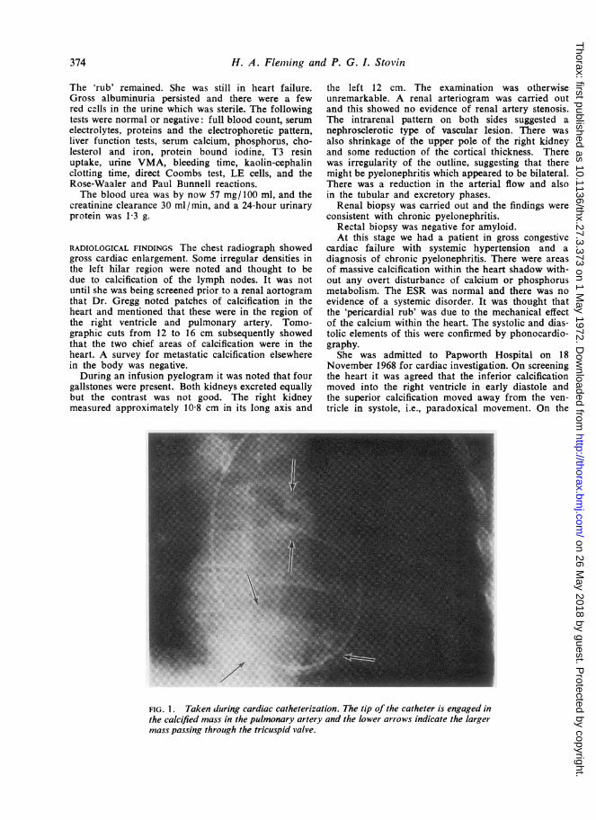
FIG. 1. Taken during cardiac catheterization. The tip of the catheter is engaged inthe calcified mass in the pulmonary artery and the lower arrows indicate the largermass passing through the tricuspid valve.
374
on 26 May 2018 by guest. P
rotected by copyright.http://thorax.bm
j.com/
Thorax: first published as 10.1136/thx.27.3.373 on 1 M
ay 1972. Dow
nloaded from
Calcified right atrialmass37
(A) RIGHT AT RIUM defect was seen to enter the right ventricle duringmmHg..:1.. .T: -: :.I diastole. The right ventricular cavity appeared to be40~~~~~~~~~~~~.. curiously striated, suggesting that there was a great
~~~increase in transverse trabeculation. The relationship30 ~~~~~~~~~~~~~ofthe superior smaller calcified mass to the pulmonary.
......artery was confirmed.20~~~~. ..Anticoagulant therapy with warfarin-sodium was
instituted because of the possibility of thirombus.occurring in the right atrial mass and giving rise to
Jo ~ ~ ~ :::: :- ::--: :::: :::: :::: :, pulmonary embolism.
0~~ ~~~~. SURGERY On 17 December 1968 Mr. Milstein....... explored the heart under cardiopulmonary bypass.
The right atrial mass was palpable when the heart wasexpoed. It could be felt extending into the tricuspid
:::: ::: orifice and for some 4 cm down the inferior venacava. A small mass could be felt in the main pul-monary artery extending down right and left main
() RIGHT VENTRICLE branches. Bypass was started and the right atrium..was found to contain a heavily calcified polypoid
40 - -.. ........ mass measuring 7x5x5 cm (Fig. 3). This mass had~~~no attachment to the right atrium or the atrial septum.
30 - i- Its only attachment was within the inferior vena4'cava. It extended through the destroyed tricuspid
20 .....J.orifice into the right ventricle whence it was delivered- -- ..by traction. With some difficulty the portion remain-ing in the inferior vena cava was removed by sharp
10- - - -~... dissection. It appeared to occlude the inferior vena cavaalmost completely and extended into the hepatic vein.
~~~. .The main pulmonary artery was opened with a ver-1-- - - - ~~~tical incision. A similar tumour was found to be firmly
-- - -~~. ....-........ adherent to the walls of the bifurcation of the main---- - - - - - ~~~~pulmonary artery and its right and left branches. This
was removed piecemeal with sharp dissection. Over ansec ~~~~~~~~areaof about 1 cm high on the atrial septum were
FIG. 2. A ih atrial pressure ple B, ih a number of sessile, pearly nodules which werevetiua,prightr pulse,.ih excised. The tricuspid valve had been completely
eve of cardiac catheterization and angiocardiographythe diagnosis of calcified right atrial myxoma withcalcified metastases in the pulmonary artery was made.At cardiac catheterization the catheter was mani-
pulated very gently into the main pulmonary arterywhere its further passage was interrupted by a calci-fied mass which could be felt on the tip of the catheter(Fig. 1). It was subsequently possible to pass thecatheter past this into the distal pulmonary artery.The pressures were as follows.
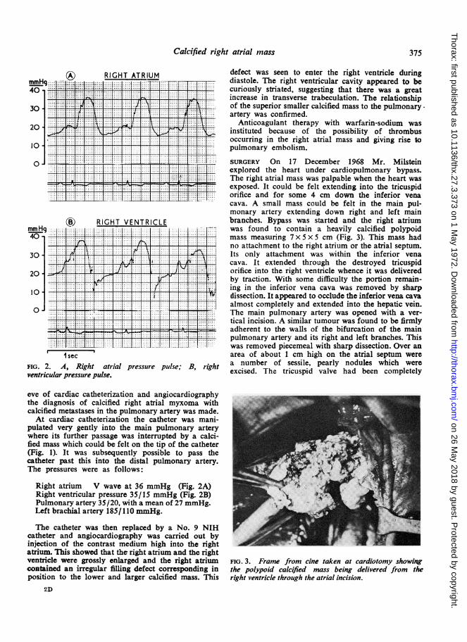
Right atrium V wave at 36 mmHg (Fig. 2A)Right ventricular pressure 35/15 mmHg (Fig. 2B)Pulmonary artery 35/20, with a mean of 27 mmHg.Left brachial arteryl185/110 mmHg.
T'he catheter was then replaced by a No. 9 NIHcatheter and angiocardiography was carried out byinjection of the contrast medium high into the rightatrium. This showed that the right atrium and the rightventricle were grossly enlarged and the right atrium FiG. 3. Frame from cine taken at cardiotomy showingcontained an irregular fillng defect corresponding in the polypoid calcified mass being delivered from theposition to the lower and larger calcified mass. This right ventricle through the atrial incision.
2D
375
on 26 May 2018 by guest. P
rotected by copyright.http://thorax.bm
j.com/
Thorax: first published as 10.1136/thx.27.3.373 on 1 M
ay 1972. Dow
nloaded from

H. A. Fleming and P. G. I. Stovin
destroyed by the larger calcified mass. No cusps,chordae or papillary muscles were left A 3M Starr-Edwards prosthesis was therefore sutured in the tri-cuspid orifice. Epicardial pacing wires were fixed atthe end of the operation. Total bypass time was 2hours 30 minutes.
Following surgery she remained hypotensive andunconscious with a low cardiac output. Despite treat-ment, her circulatory state and the ensuing oliguriafailed to improve and after a series of cardiac arrestsshe died on the fourth postoperative day.
BIOPSY The surgical specimens came from vary-ing sites between the venae cavae and the rightpulmonary artery. The largest single specimencame from the right atrium and was a coiled masswith smooth-surfaced yellow and grey gyri andulcerated calcium in the sulci (Fig. 4). The appear-ance suggested a coiled cylindrical structure, 1 to13 cm diameter and about 13 cm in total length.There appeared to be no uncalcified tissue presentand free iron could not be demonstrated.
Microscopically, in the right ventricle there wasfibrous tissue on the eroded surface of muscleand small coronary artery branches. The intimaand inner media of the pulmonary trunk wereeroded and calcium was embedded in the damagedwall. The atrial septum showed endocardial fibrosisbut no evidence of previous erosion of the under-lying myocardium.The calcified material looked very similar to the
tissue of heavily calcified heart valves, being mostlyfibrous tissue and calcium, with occasional foci ofrecent haemorrhage or fibrin, some areas with
FIG. 4. Radiograph of the main mass of tumourremoved at operation.
thin-walled blood vessels and some with a fewinflammatory cells. Bone formation and myxoma-tous tissue were absent.
NECROPSY The necropsy was performed three daysafter death. The endocardium of the inferior venacaval orifice and the lateral wall of the right atriumwere thickened, rough, and white. There was asmall paravalvular leak lateral to the Starr-Edwards tricuspid prosthesis. Except for the apical2 cm and the conus, the right ventricular cavitywas lined by a granular, opaque, thickened endo-cardium. There were no remnants of tricuspidpapillary muscles. The pulmonary valves werenormal but the endothelium of the medial andposterior surface of the main pulmonary trunkwere roughened. Fragments of calcified materialwere present in the segmental pulmonary arterieswithout thrombosis on the fragments and withoutinfarction of the lung.The liver was cirrhotic and the gall bladder con-
tained four calculi. The brain was macroscopicallynormal and there was an old haematoma aboutthe left kidney.
Microscopically there was widespread endo-cardial thickening in the right heart. This wascomposed of laminated and nodular arrangementsof fibroelastic tissue containing occasional thin-walled blood vessels and a few flecks of fibrintogether with round and stellate and spindle cells,and occasional small foci of lymphocytes andeosinophils. For the most part the process appearedto have been a laying down on a previously intactendocardium but in the left ventricle there werefoci of oedema and stellate and spindle cells sugges-tive of a subendocardial myxomatoid degeneration.There was some residual calcium still present inthe endothelium of the inferior vena cava.With alcian blue at pH 2-5 there were patches
staining intense blue both superficially and deeperin the thickened endocardium. These areas ofalcian blue staining were also the areas of mostintense PAS staining, but the intensity of PASstaining was not as great as that of the alcian blue.The only exception was in the endocardium of thenormal left atrium where the PAS staining wasslightly but definitely stronger than the alcian blue.This indicates a predominant acidic mucopoly-saccharide with a little neutral mucopolysaccharideground substance consistent with immature reactivefibrous tissue and not necessarily implying anoriginal myxoma. The nodal artery in the rightatrium showed a considerable deposition ofstrongly alcian blue and PAS staining mucin in itsadventitia.
376
on 26 May 2018 by guest. P
rotected by copyright.http://thorax.bm
j.com/
Thorax: first published as 10.1136/thx.27.3.373 on 1 M
ay 1972. Dow
nloaded from

Calcified right atrial mass
Stains for fibrin (Mallory's PTAH andLendrum's MSB) showed a little fibrin incorpor-ated in the thickened endocardium of the rightventricle and in a patchy thin film on the endo-cardium of the ventricles and right atrium withoccasional Lambl excrescence-like structureshaving an acellular fibrous or fibroelastic core anda fibrin outer coat which are present in bothventricles. These lesions we would interpret asresults of the abnormal turbulence around thecalcified intracavitary mass.
In *the lungs organized and recanalized oldemboli were present in occasional pulmonaryartery branches with a few calcific emboli. Onecalcific nodule in the right upper lobe apexappeared to have eroded through the arteriolarwall to reach the adventitia. Apart from the oldand recent emboli the lungs showed some increasein medial thickness of pulmonary arteries. In noneof the vessels was any recognizable myxoma tissuepresent.
Occasional calcific and sparsely cellular emboliup to 10 , diameter were present in the coronary,cerebral, and adrenal arterioles.The liver showed a fairly advanced portal type
of cirrhosis rather than that which results fromcardiac failure or hepatic venous occlusion of theBudd-Chiari type.
Patchy neuronal loss and subcortical demyelina-tion were found in the cerebrum.The kidney showed congestion with only
minimal interlobular arterial intimal fibrosis.
DISCUSSION
PATHOLOGICAL The origin of the calcified massin our patient is to some extent speculative as therewas virtually nothing left of the active phase ofthe tumour. The two possibilities which have tobe considered are myxoma and thrombus.
In favour of a myxoma is the presence of acidicmucopolysaccharides in the endocardium of theinferior vena cava, and the lateral wall of theright atrium and of the ventricles. It must be bornein mind that these areas had recently been the siteof surgical trauma and this had probably been thecause of the increased acidic mucopolysaccharidesin the subendocardial parts of the crests of somecolumnae carnae of the left ventricle. Similarmucoid changes are also seen in calcified aorticvalves and in balloon degeneration of the mitralvalve (Bittar and Soas, 1968), and these may indi-cate congenital abnormality in the valve structurebut not necessarily a potential myxoma. Heath(1968) recommended alcian blue as the stain ofchoice for demonstrating myxomas. However, the
relative intensity of staining with alcian blue andPAS is not a reliable guide to myxoma (Fine,Morales, and Horn, 1968) nor is the presence orabsence of elastic fibres. In neither the calcifiedmass nor in the heart was any vasoformative tissueseen, this being the other hall-mark of myxoma(Heath, 1968). Rodbard, Kihoshita, and Montes(1964), in an elegant experiment in dogs, producedapparently similar endocardial reactions by invagi-nating the left auricle over a firm Silastic ball.
In favour of a thrombotic origin is the twistedand coiled appearance of the larger pieces ofexcised calcified mass, the fact that apart from theembolic calcific fragments all other pulmonaryvascular lesions were compatible with old throm-botic emboli, and the absence of myxoma tissue.It must be admitted, however, that the late resultsof myxomatous emboli are unknown. In thereported cases (Emmanuel and Lloyd, 1962;Heath and Mackinnon, 1964; Oliver and Missen,1966; Fluck and Lopez-Bescos, 1968) only recentemboli were seen. The recent small calcific andfibrous and fibrinous systemic emboli in ourpatient were probably small fragments which weredislodged from the inferior vena cava and rightatrium at the time of operation and passedthrough the filter in the Melrose pump oxygen-ator.The observation made at the time of operation
that there were two separate tumour massesattached respectively to the inferior vena cava andto the pulmonary artery could indicate a blood-borne or embolic spread but does not help todistinguish between a myxomatous or thromboticorigin for this patient's unusual tumour. It ispossible that the tumour arose as a hepatic veinthrombus occurring at the time of the pregnancytoxaemia; the portal cirrhosis is consistent withpast eclampsia (Popper and Schaffner, 1957). Thepresence of a little fresh fibrin on the surface ofthe clefts in the calcified mass is not unexpectedand cannot be used as an argument in favour ofa thrombotic origin for the calcified mass.The few Lambl excrescence-like formations
observed microscopically in the ventricles of thispatient were similar to those noted in the heavilycalcified myxoma of Oliver and Missen (1966).The minuteness and the infrequency of theseexcrescences would exclude the calcified mass beinga unique degenerate papilliferous tumour of theheart. These rare papillary tumours are reportedon the left side of the heart and are not calcified(Hudson, 1970).Heath (1968) refers to calcification occurring in
cardiac lipomata. There was no evidence of any
377
on 26 May 2018 by guest. P
rotected by copyright.http://thorax.bm
j.com/
Thorax: first published as 10.1136/thx.27.3.373 on 1 M
ay 1972. Dow
nloaded from

H. A. Flenming and P. G. I. Stovin
residual fatty tissue in our case and the calcificationin lipomata is not a major feature when it occurs.Dean, Pamukcoglu, and Roberts (1969) specu-
lated that the calcified nodules in the right ventricleof their patient might be either the residua ofmural thrombi or of Histoplasma granulomata.These authors did not consider myxoma and thereappears to have been no myxoid change in theendocardium between the calcified endocardialnodules. Although not searched for there was noreason to suspect that this patient had had anhistoplasma infection.A study of the literature indicates that calcifi-
cation is not infrequent in myxomata, especiallythose in the right atrium (Hudson, 1970), though itrarely completely replaces the tumour. Despitetheir much greater frequency, calcification is rarein thrombi in the atria and when it does occur itusually takes the form of a few plaques in thedeeper endocardial part. It is essentially on thiscomparison with other less totally calcified myxo-mata that we venture to call this calcified mass amyxoma, even though its site of origin was theinferior vena cava and even though we have failedto find any convincing myxomatous tissue. We feelthat there is a continuous gradation of cases fromour totally calcified mass through the extensivelycalcified myxomata of Fluck and Lopez-Bescos(1968), the slightly less but still very extensivelycalcified myxoma of Oliver and Missen (1966),and the less calcified myxomata of Buenger, Paul,and Fell (1956) to the typical non-calcifiedmyxoma.CLINICAL Goodwin et al. (1962) emphasized thatthe general systemic effects of atrial myxoma areoften of more diagnostic value than the classicalbut variable local signs. A useful evaluation ofclinical and laboratory manifestations is made byHattler, Fuchs, Cosson, and Sabiston (1970). Theextensive literature on cardiac tumours in generaland on myxomata in particular is well reviewed inthe symposium published in the American Journalof Cardiology (Symposium on Cardiac Tumors,1968), by Goodwin (1968), and by Hudson (1965,1970).Myxomata can occur anywhere in the heart and
have even been described above the aortic valveand arising in the left ventricle (Danta andWilliams, 1969). However, the commonest site oforigin is from the atrial septum. More than threetimes as many tumours are found in the left atriumas in the right. Very occasional bilateral cases arerecorded (Anderson et al., 1970).Our case is unusual for myxoma in that the
'tumour' was grossly calcified and arose in the
inferior vena cava, though Oliver and Missen's(1966) case, which was also calcified, arose closeto the vena caval opening into the right atrium.In Fluck and Lopez-Bescos' (1968) case, a heavilycalcified tumour arose in the fossa ovale andextended into the inferior vena cava. An inferiorvena caval origin was also reported by Matsushita,Kuramochi, Kaneko, and Kuramoto (1968) fortheir haemorrhagic but not calcified myxoma.The confusing presentation is in keeping with
the general experience of myxomata and there isno doubt that right atrial myxoma can present asdisease of any bodily system. Confusion is increasedby the frequent co-existence of other disease, suchas in our case where there was pyelonephritis withsystemic hypertension and hepatic cirrhosis. Diag-nosis can even be missed at cardiac catheterization;angiocardiography is obligatory for completeinvestigation. The diagnosis should be consideredin any case of severe heart failure which does notrespond to treatment.The following review of some of the conditions
that have been mimicked or falsely diagnosed illus-trates these points. Because of the difficulty ofmaking any reasonable classification, these aregiven in approximately chronological order. Thisby no means covers the whole literature. It will benoted that most of these cases were reported after1960. An earlier review was made by Sannerstedtet al. (1962):
Sudden death from tumour embolization of the pul-monary artery
(Chiari, 1931, quoted by Prichard, 1951)Recurrent attacks of heart failure for 43 years
(Strouse, 1938) Carcinoma of rectum and markedatherosclerosis also present
Epilepsy and presumed bacterial endocarditis(Kendall and Symonds, 1952)
Ebstein's anomaly(Coates and Drake, 1958)
Fatigue and intermittent oedema(Campeti, Mahoney, and Yu, 1960)
Severe congestive failure and weight loss of 21 lb(Catt, Denborough, Grigg, and Sloman, 1962)
Polycythemia and raised platelet count regressing afterremoval of the tumour
(Levinson and Kincaid, 1961)Constrictive pericarditis
(Sannerstedt et al., 1962)Short attacks of abdominal pain with extreme nausea,breathlessness and faintness, loss of appetite, andweight loss of 41 lb
(Barlow, Fuller, and Denny, 1962)Pulmonary tuberculosis and later constrictive peri-carditis(Emmanuel and Lloyd, 1962)
378
on 26 May 2018 by guest. P
rotected by copyright.http://thorax.bm
j.com/
Thorax: first published as 10.1136/thx.27.3.373 on 1 M
ay 1972. Dow
nloaded from

Calcified right atrial mass
Tricuspid stenosis, apparently confirmed by cardiaccatheterization
(Morrissey, Campeti, Mahoney, and Yu, 1963)Pulmonary stenosis with pressure gradient measuredacross the pulmonary valve
(Gottsegen, Wessely, Arvay, and Temesvari, 1963)This tumour actually arose in the right ventricle
Recurrent pulmonary emboli with pulmonary hyper-tension
(Heath and Mackinnon, 1964)Symptoms from the age of 26 years to the age of 63years-thought to be tuberculous pericarditis
(Oliver and Missen, 1966) Grawitz tumour of thekidney also present
Diabetic, with Kimmelsteil Wilson syndrome(Stems, Eliot, Varco, and Edwards, 1966)
Joint pains, anaemia, disordered proteins, and heartantibodies diagnosed as rheumatic disorder.
(Currey, Mathews, and Robinson, 1967)Steatorrhoea clearing after removal of tumour
(Webb-Peploe, Goodbody, Johnson, and McMillan,1968)
Pericardial friction rub mimicking acute pericarditis(Greenwood, 1968) Greenwood also mentionscarcinoid and obstruction of the vena cava in thedifferential diagnosis.
Cyanosis and polycythaemia(Miller, Paneth, and Gibson, 1968) This case hada patent foramen ovale.
Tricuspid incompetence(Harvey, 1968) Harvey vividly describes the'wrecking ball' action of the tumour so evident inour case.
Clubbing and cyanosis(Talley, Baldwin, Symbas, and Nutter, 1970)
Diverse manifestations of inflammatory illness sug-gesting D.L.E., myocarditis, bacterial endocarditis oracute rheumatic fever
(Hattler et al., 1970)
SYSTEMIC EFFECrS Fever, anaemia, raised ESR,and disordered plasma proteins were not found inour case, although these are frequently present inrecorded reports. The mechanism of these changesis not clear, but it is possible that the completelyburnt out appearance of our 'tumour' was con-sistent with its lack of reaction. The ESR wasnormal in the heavily calcified tumour of Fluckand Lopez-Bescos (1968) and was not recorded inOliver and Missen's (1966) case. The very highLDH in our case was presumably due to con-tinuing damage to the myocardium and liver.
AUSCULTATION The loud 'rub' was presumablydue to the massive movement of the 'tumour' inand out of the ventricle which Harvey (1968) hasso graphically described as the 'wrecking-ball'action. Matsushita et al. (1968) describe severalsystolic and diastolic clicks and murmurs, varyingwith posture in a non-calcified right atrial myxoma.
THE ELECTROCARDIOGRAM Sinus rhythm is remark-ably common, considering the damage to theatrium. Atrial dysrhythmia is rarely mentionedand only one report of complete atrioventriculardisassociation (Hattler et al., 1970) has been dis-covered. Our patient consistently showed a first-degree heart block with a PR interval of0-28 second. The low voltage of the ventricularcomplexes, together with the partial right bundle-branch block seen in our case, are relativelycommon features.
HAEMODYNAMICS The 'tumour' had completelydestroyed the tricuspid valve and inflicted muchdamage within the right ventricle. This had resultedin the changes found in the Ebstein anomaly-sothat the pressure pulses recorded in the superiorvena cava, the right atrium, and the right ventriclewere very similar to one another, with a systolicpressure of nearly 40 mmHg in all chambers(Fig. 2A and B). The notch on the upstroke of theright ventricular pressure trace has already beennoted by Fluck and Lopez-Bescos (1968) in theircase, and by others in cases of left heart tumours(Penny et al., 1967; Pitt, Pitt, Schaefer, and Criley,1967) and is attributed to the movement of thetumour in and out of the ventricle.
EMBOLI AND METASTASES The occurrence of pul-monary emboli and infarcts with atrial myxomais frequently mentioned (Emmanuel and Lloyd,1962; Heath and Mackinnon, 1964), and calcifiedemboli were found in Oliver and Missen's (1966)case. Heath and Mackinnon (1964) noted that atnumerous sites the adventitia had been invadedand that there were also emboli in the bronchialarteries; they suggested that this demonstrated alimited invasiveness. Similar limited invasivenesshad been recorded with emboli from left atrialmyxoma in the mesenteric and brain vessels(Ringertz, 1942). In our case the main pulmonaryartery was extensively scarred at the site of thelarge calcified deposits but there was no histo-logical evidence to confirm invasion. The mainpulmonary arteries must be an unusual site forlarge myxoma deposits to occur and we have foundno report similar to this. There were, in addition,numerous calcified deposits in the pulmonaryarteries, especially in the right lower lobe.
CALCIFICATION Degenerative changes are com-monly reported in these tumours, but we havefound no previous report of one which appearedto be so completely calcified. As is usual indegenerative areas, minor calcification is commonand bone formation is occasionally recorded
379
on 26 May 2018 by guest. P
rotected by copyright.http://thorax.bm
j.com/
Thorax: first published as 10.1136/thx.27.3.373 on 1 M
ay 1972. Dow
nloaded from

H. A. Fleming and P. G. 1. Stovin
(Gottsegen et al., 1963). Strouse (1938) firstreported calcification in a tumour of 43 years'duration, but gross calcification such as was seenin our case is rare and appears to be relativelymore common in right atrial than in left atrialmyxomata. Once seen, its radiological appearanceis not easily forgotten-particularly the dramaticmovement on fluoroscopy. Buenger et al. (1956)described calcification visible radiologically in aboy aged 16 years. Wight, McCall, and Wenger(1963) described two cases with calcified tumours,one in the right atrium and one in the left. Oliverand Missen (1966) described extensive microscopiccalcification in a tumour removed from a managed 63 whose symptoms dated from the age of 26,and in whom the calcified mass had been notedradiologically from the age of 26. Harvey (1968)also noted calcification in a woman aged 60.Greenwood (1968) says that gross calcification israrely present in myxomata and records that ithas been seen fluoroscopically in two right atrialand one left atrial myxomata. The case of Fluckand Lopez-Bescos (1968) seems to resemble oursin the grossness of the calcification as well as inthe low ESR and the haemodynamic features.
TREATMENT Surgery has been successful in anumber of cases (e.g., Fluck and Lopez-Bescos,1968). In our case surgery was technically success-ful but the right ventricle was so massively damagedthat it did not appear capable of adequate outputto maintain life. The friable nature of the calcifiedtumour also proved a hazard not recognized at thetime of operation. Multiple fragments, too fine tobe arrested by the filter in the pump oxygenator,embolized small vessels in many of the organsexamined. The long period on cardiopulmonarybypass necessary for the removal of the tumourprobably also contributed. Detailed dissection ofthe tumour may have prolonged this time andproduced a number of calcific embdi in the lungs.Although occasional tumour recurrences arereported after surgery (Bahl et al., 1969; Baumannand Clavadetscher, 1969), the results of sim.plelocal excision in a number of series are excellent(Goodwin et al., 1970). The calcified mass in thepulmonary artery was not causing obstruction andMr. Milstein considers it would have been wiserto have left it and to have carried out a simpleresection of the main mass prolapsing into the rightventricle with relief of the inferior vena cavalobstruction.
Our thanks are due to Dr. K. D. Allanby, who firstreferred this patient, to Dr. D. McG. Gregg and Dr.G. 1. Verney, who assisted with the radiological studies,
to Mr. B. B. Milstein for access to his operativefindings and opinion, to Dr. J. Zamler, who carriedout the necropsy, to the Photographic Departmentof the United Cambridge Hospitals for the figures,and to Mrs. Anita Mead and Miss Julia P. Cusden forsecretarial help. The Research Committee of the EastAnglian Regional Hospital Board provided a grant.
REFERENCESAnderson, S. T., Pitt, A., Zimmet, R., Kay, H. B., and
Morris, K. N. (1970). A case of bi-atrial myxomas withsuccessful surgical removal. J. thorac. cardiovasc. Surg.,59, 768.
Bahl, 0. P., Oliver, G. C., Ferguson, T. B., Schad, N., andParker, B. M. (1969). Recurrent left atrial myxoma.Report of a case. Circulation, 40, 673.
Barlow, J., Fuller, D., and Denny, M. (1962). A case of rightatrial myxoma. Brit. Heart J., 24, 120.
Baumann, R. P., and Clavadetscher, P. (1969). Gut- undbosartige Endokardtumoren: sogenanntes Mysom, mitEmbolie in die Aorta-papillares Endotheliom-myxo-matoses Fibrosarkom. Schweiz. med. Wschr., 99, 444.
Bittar, N., and Soas, J. A. (1968). The billowing mitral valveleaflet. Report on 14 patients. Circulation, 38, 763.
Buenger, R. E., Paul, O., and Fell, E. H. (1956). Calcifiedpolyp of the heart. Radiology, 67, 531.
Campeti, F. L., Mahoney, E. B., and Yu, P. N. (1960).Myxoma of the right atrium. Circulation, 22, 730.
Catt, K., Denborough, M. A., Grigg, L., and Sloman, G.(1962). Myxoma of the right atrium. Brit. Heart J., 24,525.
Coates, E. O., and Drake, E. H. (1958). Myxoma of the rightatrium, with variable right-to-left shunt. Clinical andphysiologic observations and report of a case withsuccessful operative removal. New Engl. J. Med., 259,165.
Currey, H. L. F., Mathews, J. A., and Robinson, J. (1967).Right atrial myxoma mimicking a rheumatic disorder.Brit. med. J., 1, 547.
Danta, G., and Williams, D. 0. (1969). Multiple emboli fromleft ventricular myxoma. Brit. Heart J., 31, 799.
Dean, D. C., Pamukcoglu, T., and Roberts, W. C. (1969).Rocks in the right ventricle. A complication of con-genital right ventricular infundibular obstruction asso-ciated with chronic pulmonary parenchymal disease.Amer. J. Cardiol., 23, 744.
Emmanuel, R. W., and Lloyd, W. E. (1962). Right atrialmyxoma mistaken for constrictive pericarditis. Brit.Heart J., 24, 796.
Fine, G., Morales, A., and Horn, R. C. (1968). Cardiacmyxoma. A morphologic and histogenetic appraisal.Cancer (Philad.), 22, 1156.
Fluck, D. C., and Lopez-Bescos, L. (1968). Calcified rightatrial myxoma producing tricuspid incompetence. Proc.roy. Soc. Med., 61, 1115.
Goodwin, J. F. (1968). The spectrum of cardiac tumors.Amer. J. Cardiol., 21, 307., Croxson, R. S., Oakley, C. M., Cleland, W. P., andBentall, H. H. (1970). The long term results of surgeryof atrial myxoma. Abstracts 6th World Congress ofCardiology. Cardiovasc. Res., p. 152., Stanfield, C. A., Steiner, R. E., Bentall, H. H., Sayed,H. M., Bloom, V. R., and Bishop, M. B. (1962). Clinicalfeatures of left atrial myxoma. Thorax, 17, 91.
380
on 26 May 2018 by guest. P
rotected by copyright.http://thorax.bm
j.com/
Thorax: first published as 10.1136/thx.27.3.373 on 1 M
ay 1972. Dow
nloaded from

Calcified right atrial mass
Gottsegen, G., Wessely, J., Arvay, A., and Temesvari, A.(1963). Right ventricular myxoma simulating pulmonarystenosis. Circulation, 27, 95.
Greenwood, W. F. (1968). Profile of atrial myxoma. Amer.J. Cardiol., 21, 367.
Harvey, W. P. (1968). Clinical aspects of cardiac tumors.Amer. J. Cardiol., 21, 328.
Hattler, B. G., Fuchs, J. C., Cosson, R., and Sabiston, D. C.(1970). Atrial myxoma: an evaluation of clinical andlaboratory manifestations. Ann. thorac. Surg., 10, 65.
Heath, D. (1968). The pathology of cardiac tumors. Amer. J.Cardiol., 21, 315.
-, and Mackinnon, J. (1964). Pulmonary hypertensiondue to myxoma of the right atrium; with special refer-ence to the behaviour of emboli of myxoma in the lung.Amer. Heart J., 68, 227.
Hudson, R. E. B. (1965). Cardiac myxoma. CardiovascularPathology, Vol. 2, p. 1565. Edward Arnold, London.
- (1970). Cardiac myxoma. Cardiovascular Pathology,Vol. 3, Supplement, p. 868. Edward Arnold, London.
Kendall, D., and Symonds, B. (1952). Epileptiform attacksdue to myxoma of the right auricle. Brit. Heart J., 14,139.
Levinson, J. P., and Kincaid, 0. W. (1961). Myxoma of theright atrium with polycythaemia. New Engl. J. Med.,264,1187.
Matsushita, S., Kuramochi, M., Kaneko, J., and Kuramoto,K. (1968). Right atrial myxoma mimicking pericarditis.Jap. circulat. J. (En.), 32, 1283.
Miller, G. A. H., Paneth, M., and Gibson, R. V. (1968)Right atrial myxoma with right-to-left shunt and poly-cythaemia. Brit. med. J., 3, 537.
Morrissey, J. F., Campeti, F. L., Mahoney, E. B., and Yu,P. N. (1963). Right atrial myxoma. Report of two casesand review of the literature. Amer. Heart J., 66, 4.
Oliver, G. C., and Missen, G. A. K. (1966). A heavily calcifiedright atrial myxoma. Guy's Hosp. Rep., 115, 37.
Penny, J. L., Gregory, J. J., Ayres, S. M., Gianelli, S., andRossi, P. (1967). Calcified left atrial myxoma, simulatingmitral insufficiency. Circulation, 36, 417.
Pitt, A., Pitt, B., Schaefer, J., and Criley, J. M. (1967).Myxoma of the left atrium. Haemodynamic and phono-cardiographic consequences of sudden tumour move-ment. Circulation, 36, 408.
Popper, H. L., and Schaffner, F. (1957). Liver: Structure andFunction, p. 486. McGraw-Hill, New York.
Prichard, R. W. (1951). Tumors of the heart. Review of thesubject and report of 150 cases. Arch. Path., 51, 98.
Ringertz, N. (1942). Uber sog. Endokardmyxome. Actapath. microbiol. scand., 19, 262.
Rodbard, S., Kihoshita, Y., and Montes, M. (1964). Induc-tion of papillary growths in the heart. Science, 143,1341.
Sannerstedt, R., Varnauskas, E., Paulin, S., Linder, E.,Ljunggren, H., and Werko, L. (1962). Right atrialmyxoma. Report of a case and review of the literature.Amer. Heart J., 64, 243.
Sterns, L. P., Eliot, R. S., Varco, R. L., and Edwards, J. E.(1966). Intracavitary cardiac neoplasms. A review of 15cases. Brit. Heart J., 28, 75.
Strouse, S. (1938). Primary benign tumor of the heart of 43years' duration. Arch. intern. med., 62, 401.
Symposium of Cardiac Tumors (1968). Amer. J. Cardiol.,21, No. 3, 307-466.
Talley, R. C., Baldwin, B. J., Symbas, P. N., and Nutter, D. 0.(1970). Right atrial myxoma: Unusual presentation withcyanosis and clubbing. Amer. J. Med., 48, 256.
Webb-Peploe, M. M., Goodbody, R. A., Johnson, A. M.,and McMillan, I. K. R. (1968). Personal communication.
Wight, R. P., McCall, M., and Wenger, N. K. (1963).Primary atrial tumor. Amer. J. Cardiol., 11, 790.
ADDENDUM
Since this paper was submitted for publicationWaxler, Kawai, and Kasparian (1972) have repor-ted another case of successful operation on aright atrial myxoma.
REFERENCE
Waxler, E. B., Kawai N., and Kasparian, H. (1972). Rightatrial myxoma: echocardiographic, phonocardiographic,and haemodynamic signs. Amer. Heart J., 83, 251.
381
on 26 May 2018 by guest. P
rotected by copyright.http://thorax.bm
j.com/
Thorax: first published as 10.1136/thx.27.3.373 on 1 M
ay 1972. Dow
nloaded from