calcium current in cortical astrocytes: induction by …corey.med.harvard.edu/pdfs/1989 barres corey...
TRANSCRIPT

The Journal of Neuroscience, September 1989, g(9): 31893175
Calcium Current in Cortical Astrocytes: Induction by CAMP and Neurotransmitters and Permissive Effect of Serum Factors
Barbara A. Barres,i,z,3,4 Linda L. Y. Chun,ls* and David P. Corey1,2,3
‘Program in Neuroscience, Harvard Medical School, *Department of Neurology, Massachusetts General Hospital, and 3Howard Hughes Medical Institute, Boston, Massachusetts 02114
Voltage-dependent L-type calcium channels were induced in highly purified, cultured, cortical astrocytes by exposure to substances known to increase their intracellular CAMP: 8-bromo-CAMP, forskolin, isoproterenol, and vasoactive in- testinal peptide. In untreated control cultures, L-type calcium currents were entirely absent. The induction of this calcium current was specific to cortical astrocytes and a closely related astrocyte type in white matter and did not occur in meningeal cells or oligodendrocytes. The ability of forskolin to induce L-type calcium current in astrocytes depended on previous culture of these cells in a permissive lot of serum for at least 48 hr. In addition, certain lots of sera caused expression of a voltage-dependent T-type calcium channel in untreated control cultures. Several possible mechanisms of calcium channel induction by CAMP are consistent with our data: modification of a silent channel already present in the membrane, indirect effect of cytoskeletal alteration, or insertion of channels from submembrane stores. If the en- vironment in viva is permissive, regulation of glial ion chan- nels may be under neuronal control.
Although most functions of astrocytes are still poorly under- stood, one function has been convincingly demonstrated: astro- cytes participate in the regulation of ions in the neuronal mi- croenvironment (Coles and Orkand, 1983; Ballanyi et al., 1987). Recently, astrocytes in culture have been demonstrated to ex- press several kinds of voltage-sensitive ion channels that might mediate this function. In particular, cortical astrocytes in culture express sodium channels (Bevan et al., 1985), chloride channels (Bevan et al., 1985), and several types of potassium channels, including a calcium-dependent potassium channel (Quandt and MacVicar, 1986).
There is also some evidence for a calcium channel. Astrocytes in culture fire slow action potentials when bathed in a solution that would accentuate calcium current and inhibit potassium current (MacVicar, 1984). We have used the whole-cell patch- clamp recording technique to measure calcium current directly and to characterize its properties. We found that cortical astro-
Received Dec. 19, 1988; revised Feb. 16, 1989; accepted Feb. 20, 1989 We thank Michael Goy for his advice and donation of forskolin for the initial
experiments, and Bruce Silverstein for expert technical assistance. This work was supported by National Institute of Health grants F32 NS-07970 (to B.A.B.), ROl NS-22059 (to D.P.C.), and ROl NS-21269 and ROl NS-16367 (to L.L.Y.C.), and by the Howard Hughes Medical Institute.
Correspondence should be addressed to Barbara A. Barres, Wellman 4 14, Mas- sachusetts General Hospital, Boston, MA 02114. Copyright 0 1989 Society for Neuroscience 0270-6474/89/093169-07$02.00/O
cytes did not express a voltage-dependent calcium current in control cultures but that an L-type calcium current was induced by preincubation with agents that raise intracellular CAMP. Fur- thermore, isoproterenol and vasoactive intestinal peptide (VIP), which raise CAMP levels in cortical astrocytes (McCarthy and de Vellis, 1978; Narumi et al., 1978; Rougon et al., 1983), also induced this calcium current. A preliminary report of this work has appeared in abstract form (Chun et al., 1986).
Materials and Methods
Preparation of cortical cell suspensions. Cerebral cortices from postnatal day- 1 Long/Evans rats were stripped of their meninges and dissociated enzymatically to make a suspension of single cells, essentially as de- scribed by Huettner and Baughman (1986). Briefly, the tissue was minced and incubated at 37°C for 75 min in a papain solution (30 units/ml; Worthington) equilibrated with 95% 0, and 5% CO,. This solution also contained Earle’s balanced salts, calcium, magnesium, EDTA, sodium bicarbonate, glucose, and L-cysteine. The tissue was then triturated with a 1 ml pipette in a solution containing ovomucoid (0.2%, Boehringer- Mannheim) and BSA (0.1%; Sigma) to yield a suspension of single cells.
Preparation of cortical astrocyte cultures. Cultures of cerebral cortical astrocytes were prepared from postnatal day 1 rat cell suspensions (as above) with the technique of McCarthy and devellis (1980) and further purified by the procedure of Noble and Murray (1984). Primary cultures of astrocytes were plated at 20 million cells per 75 cm* flask and grown to confluence in a medium containing Dulbecco’s modified Eagle’s me- dium, 10% fetal calf serum, penicillin ( 100 units/ml), streptomycin (100 pg/ml), and L-glutamine (2 mM). The top layer of cells, containing oli- godendrocytes, type 2 astrocytes and progenitor cells, was shaken off manually, and the remaining cells were treated with cytosine arabinoside (25 FM) for 2 days to kill dividing cells. Finally, these cells were tryp- sinized from the flask, and most contaminating cells were destroyed by complement-dependent lysis using monoclonal antibodies directed at galactocerebroside (GC; antibodies were generously provided by Bar- bara Ranscht: Ranscht et al.. 1982) and the A2B5 antigen (Eisenbarth et al., 1979; the A2B5 cell’line was obtained from Amehcan Type Culture Collection). These recognize surface antigens on oligodendro- cytes and on type 2 astrocytes and neurons, respectively (Raff et al., 1983b). The cells were then plated on 13-mm-diameter round coverslips coated with poly-1-lysine. The final cultures were demonstrated to be greater than 98% pure astrocytes, based on immunohistochemical la- beling with markers to glial fibrillary acidic protein (GFAP), A2B5, and GC. The cultures were at least 1 month old before recording.
Electrophysiological recording. Tight-seal, whole-cell patch recording. A piece of glass coverslip bearing cultural cortical astrocytes was placed in the recording chamber, which contained an appropriate bath solution (volume 500-750 ~1). Standard procedures for pipette preparation, seal formation, and whole-cell recording were used (Hamill et al., 1981; Corey et al., 1984). Micropipettes were drawn from hard borosilicate capillary glass (Coming 7052, Gamer Glass), coated with Sylgard to reduce their capacitance, and fire-polished to a bubble number of 4.0- 4.5 (corresponding to an internal tip diameter of about 0.6-0.8 pm) (Corey and Stevens, 1983). Pipette capacitance and series resistance were electronically compensated by the patch clamp, a Yale Mark V. All experiments were done at room temperature, approximately 23°C.
Data acquisition and analysis. Voltage stimuli were generated and
3170 Barres et al. * Induction of a Ca Current in Astrocytes
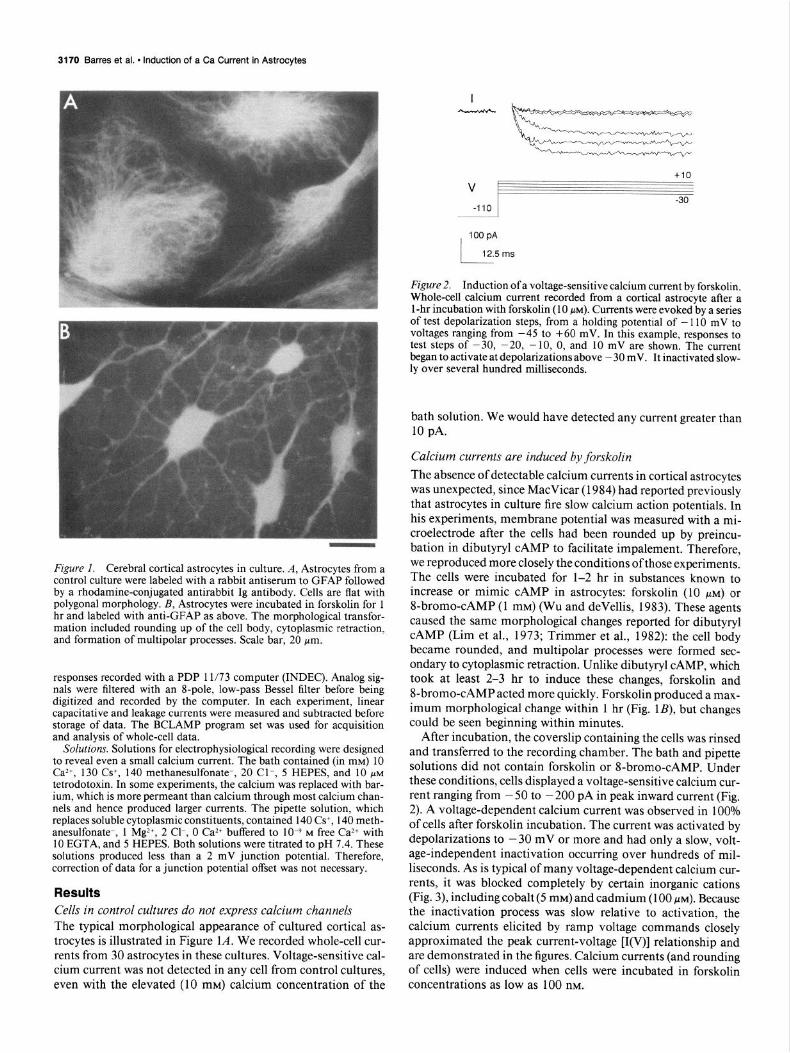
Figure 1. Cerebral cortical astrocytes in culture. A, Astrocytes from a control culture were labeled with a rabbit antiserum to GFAP followed by a rhodamine-conjugated antirabbit Ig antibody. Cells are flat with polygonal morphology. B, Astrocytes were incubated in forskolin for 1 hr and labeled with anti-GFAP as above. The morphological transfor- mation included rounding up of the cell body, cytoplasmic retraction, and formation of multipolar processes. Scale bar, 20 pm.
responses recorded with a PDP 1 l/73 computer (INDEC). Analog sig- nals were filtered with an I-pole, low-pass Bessel filter before being digitized and recorded by the computer. In each experiment, linear capacitative and leakage currents were measured and subtracted before storage of data. The BCLAMP program set was used for acquisition and analysis of whole-cell data.
Solutions. Solutions for electrophysiological recording were designed to reveal even a small calcium current. The bath contained (in mM) 10 Ca2+, 130 Cs+, 140 methanesulfonate-, 20 Cl-, 5 HEPES, and 10 PM tetrodotoxin. In some experiments, the calcium was replaced with bar- ium, which is more perrneant than calcium through most calcium chan- nels and hence produced larger currents. The pipette solution, which replaces soluble cytoplasmic constituents, contained 140 Cs+, 140 meth- anesulfonate-, 1 Mg2+, 2 Cl-, 0 Ca*+ buffered to 1O-9 M free Ca2+ with 10 EGTA, and 5 HEPES. Both solutions were titrated to pH 7.4. These solutions produced less than a 2 mV junction potential. Therefore, correction of data for a junction potential offset was not necessary.
Results Cells in control cultures do not express calcium channels The typical morphological appearance of cultured cortical as- trocytes is illustrated in Figure 1A. We recorded whole-cell cur- rents from 30 astrocytes in these cultures. Voltage-sensitive cal- cium current was not detected in any cell from control cultures, even with the elevated (10 mM) calcium concentration of the
tie v-
-- -30
-110
IOOpA
12.5 rns
Figure 2. Induction of a voltage-sensitive calcium current by forskolin. Whole-cell calcium current recorded from a cortical astrocyte after a 1 -hr incubation with forskolin (10 NM). Currents were evoked by a series of test depolarization steps, from a holding potential of - I10 mV to voltages ranging from -45 to +60 mV. In this example, responses to test steps of -30, -20, - 10. 0, and 10 mV are shown. The current began to activate at depolarizations above - 30 mV. It inactivated slow- ly over several hundred milliseconds.
bath solution. We would have detected any current greater than 10 pA.
Calcium currents are induced by forskolin The absence of detectable calcium currents in cortical astrocytes was unexpected, since MacVicar (1984) had reported previously that astrocytes in culture fire slow calcium action potentials. In his experiments, membrane potential was measured with a mi- croelectrode after the cells had been rounded up by preincu- bation in dibutyryl CAMP to facilitate impalement. Therefore, we reproduced more closely the conditions ofthose experiments. The cells were incubated for l-2 hr in substances known to increase or mimic CAMP in astrocytes: forskolin (10 PM) or 8-bromo-CAMP (1 mM) (Wu and devellis, 1983). These agents caused the same morphological changes reported for dibutyryl CAMP (Lim et al., 1973; Trimmer et al., 1982): the cell body became rounded, and multipolar processes were formed sec- ondary to cytoplasmic retraction. Unlike dibutyryl CAMP, which took at least 2-3 hr to induce these changes, forskolin and 8-bromo-CAMP acted more quickly. Forskolin produced a max- imum morphological change within 1 hr (Fig. lB), but changes could be seen beginning within minutes.
After incubation, the coverslip containing the cells was rinsed and transferred to the recording chamber. The bath and pipette solutions did not contain forskolin or 8-bromo-CAMP. Under these conditions, cells displayed a voltage-sensitive calcium cur- rent ranging from - 50 to - 200 pA in peak inward current (Fig. 2). A voltage-dependent calcium current was observed in 100% of cells after forskolin incubation. The current was activated by depolarizations to - 30 mV or more and had only a slow, volt- age-independent inactivation occurring over hundreds of mil- liseconds. As is typical of many voltage-dependent calcium cur- rents, it was blocked completely by certain inorganic cations (Fig. 3), including cobalt (5 mM) and cadmium (100 PM). Because the inactivation process was slow relative to activation, the calcium currents elicited by ramp voltage commands closely approximated the peak current-voltage [I(V)] relationship and are demonstrated in the figures. Calcium currents (and rounding of cells) were induced when cells were incubated in forskolin concentrations as low as 100 nM.

The Journal of Neuroscience, September 1989, 9(9) 3171
25 ms I I
PA
-200
Control 50 ms
1 I
Figure 3. Blockade of voltage-sensitive calcium current by cadmium. Whole-cell calcium current was elicited with a ramp voltage command that changed from - 100 to + 100 mV over 250 msec. Linear leakage current was subtracted from the current response. Cadmium (100 PM)
completely abolished the current.
The time course of appearance of current was closely corre- lated with the time course of morphological change, although the earliest we studied current was 15 min after incubation. That is, before cells rounded up, calcium currents were absent or small; larger currents were observed only with maximal mor- phological changes. Thus, large calcium currents coincident with morphological changes were induced earlier after onset of for- skolin incubations (about 30 min, with smaller currents present at about 15 min) than with 8-bromo-CAMP (about one hr) or with dibutyryl CAMP (about 2 hr).
The induced current is an L-current This induced conductance thus appears similar to the “L,” or longlasting, calcium channel described in dorsal root ganglion neurons (Nowycky et al., 1985). Like other calcium currents of this type, inward current progressively diminished or “washed- out” over minutes during whole-cell recording (Fig. 4). In the cell of Figure 4, peak inward current was - 200 pA immediately after establishment of the whole-cell configuration but had de- clined to less than - 100 pA 20 min later. This figure also dem- onstrates that during recording, the I(V) relationship shifted progressively in the hyperpolarizing direction, typically by 20 to 30 mV. This phenomenon has been observed previously during whole-cell recording in other cell types, particularly for calcium currents (Marty and Neher, 1983; Corey et al., 1984). As with other L-type calcium channels, barium was also per- meant, and when barium was the current carrier, currents ex- hibited little inactivation (data not shown).
In addition, in 3 of 3 cells the voltage-dependent calcium current was blocked by nifedipine (100 FM), a dihydropyridine calcium antagonist that blocks L-current in other cells (Fig. 5). Other concentrations were not studied, nor were the effects of depolarizing prepulses on nifedipine interactions with the chan- nel. In experiments with cells cultured in certain sera (see below), some cells exhibited another type of calcium current, a “T,” or transient, current with a rapid voltage-dependent inactivation (see below). The nifedipine block was mostly specific for the L channel and not for the T component of current (Fig. 5). Control recordings using solutions containing ethanol (0.5%), used to dissolve the nifedipine, did not block either current. We also observed that diazepam, a benzodiazepine agonist reported to displace nitrendipine binding in cortical astrocytes (Bender and Hertz, 1985) had no effects on calcium currents in concentra- tions as high as 100 MM (3 of 3 cells; studied in the absence of nifedipine).
PA
-400 i
Figure 4. Wash-out of calcium current and shift of the activation potential. Whole-cell calcium currents were elicited by a ramp voltage command from - 100 to + 100 mV over 500 msec. Linear leakage currents were not subtracted. The current responses were obtained im- mediately after establishing the whole-cell configuration and after 20 min of recording. Over time, the peak current declined progressively, and the I(V) relationship shifted in a hyperpolarizing direction.
Calcium current in astrocytes is induced by neurotransmitters Can calcium currents in astrocytes be induced by a physiological substance? Astrocytes in culture express many neurotransmitter and peptide receptors, several of which have been shown to mediate an increase in intracellular CAMP. Among them are p-adrenergic receptors, and @-adrenergic agonists have been found to increase CAMP levels in astrocytes and to induce the morphological changes (McCarthy and devellis, 1978; Mc- Carthy and devellis, 1983; Narumi et al., 1978). Therefore, we tested the ability of isoproterenol to induce a calcium current.
A T
+400
-50 mV I ! ! ! I I I ; I I
7, \
125 ms PA
1 I -800
6
v -15
-100
Figure 5. Nifedipine block of calcium current. Whole-cell current was elicited by a ramp voltage command (A) and by a voltage step to - 15 mV (B) and is shown in the same cell before and after nifedipine (100 PM). The L-type current was decreased significantly. This cell also con- tained a small component of the T-type calcium current, which was blocked less strongly by the nifedipine.
3172 Barres et al. - Induction of a Ca Current in Astrocytes
0 V
-110 -60
100 pA
25 ms
Figure 6. Induction of voltage-sensitive calcium current with isopro- terenol. Preincubation of cultures in isoproterenol(lO0 PM) for 30 min induced a whole-cell current indistinguishable from that induced by forskolin. Currents elicited from a prepulse of - 110 mV to a series of voltage steps of -60, -45, -30, -20, - 10, and 0 mV are shown.
Cells were incubated with isoproterenol(lO0 KM) and recorded from at intervals of 15 min. Voltage-dependent calcium currents were induced, again with a time course that corresponded with morphological rounding changes (Fig. 6). Peak currents after 30 minutes of incubation in isoproterenol ranged from -50 to -400 pA (barium as current carrier; 20 of 20 cells). Prior to 30 min, little rounding of the cells or calcium current was observed. Isoproterenol could induce the current in concentrations as low as 1 PM. Below this concentration, we observed neither rounding of the cells nor expression of the calcium current.
VIP also has been reported to increase CAMP levels in cortical astrocytes in culture (Rougon et al., 1983). We next incubated cultured cortical astrocytes in VIP as described above. Because these incubations were performed in medium containing serum, we included aprotinin (0.1 mg/ml) during the incubation to prevent potential digestion of VIP by proteases present in the serum. Control experiments demonstrated that aprotinin did
Table 1. Effect of serum on expression of calcium currents
Fetal calf serum lot (lot no.) T current” L current”
M.A. Bioproducts (1) M.A. Bioproducts (2) M.A. Bioproducts (3) Hyclone (1) Hyclone (2) Hyclone (3) Hyclone (4) Hyclone (5) Hyclone (6) Flow Labs (1) Flow Labs (2) Irvine Scientific Hazelton (1) Hazelton (2) Gibco (1) Gibco (2) Gibco (3)
- - - + - - - - + - - + - - - - + - - - + - - - + - + + - -
0 Constitutive. h Inducible.
V
-110
33 pA
/ 25 ms
-60
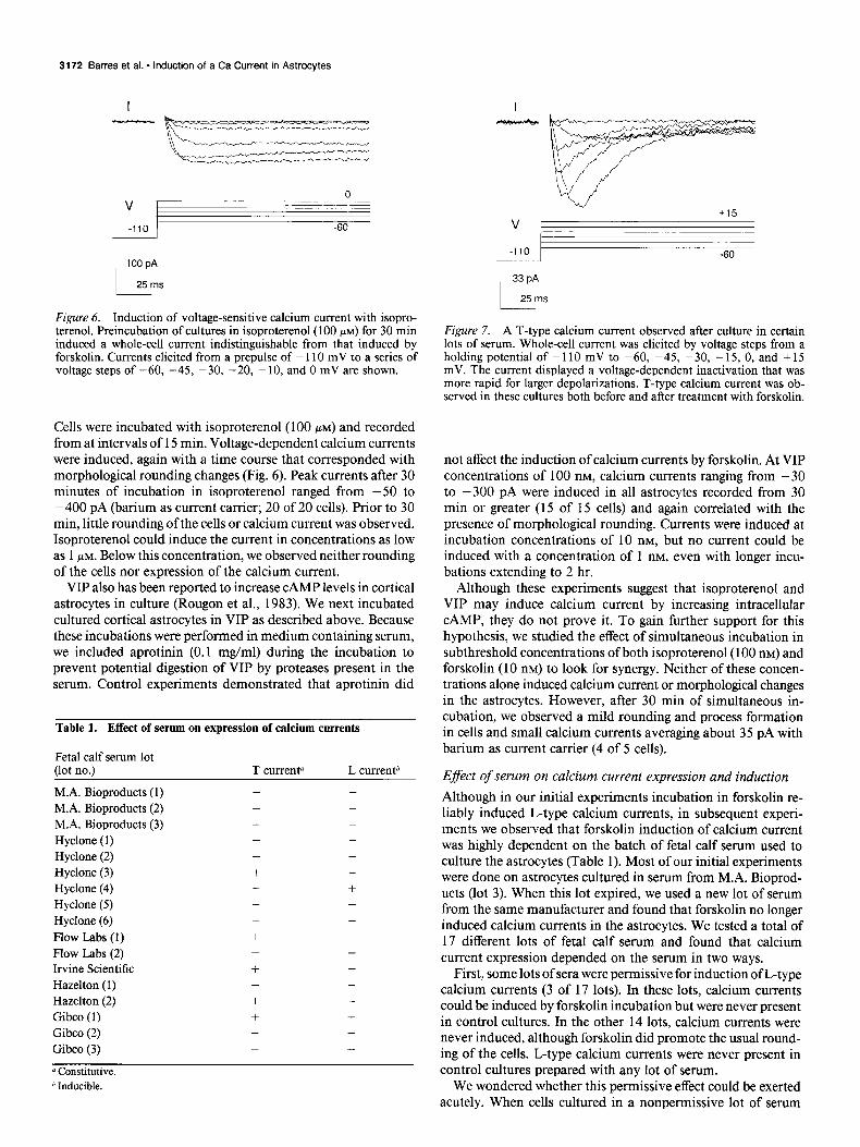
Figure 7. A T-type calcium current observed after culture in certain lots of serum. Whole-cell current was elicited by voltage steps from a holding potential of -110 mV to -60, -45, -30, -15, 0, and +15 mV. The current displayed a voltage-dependent inactivation that was more rapid for larger depolarizations. T-type calcium current was ob- served in these cultures both before and after treatment with forskolin.
not affect the induction of calcium currents by forskolin. At VIP concentrations of 100 nM, calcium currents ranging from -30 to -300 pA were induced in all astrocytes recorded from 30 min or greater (15 of 15 cells) and again correlated with the presence of morphological rounding. Currents were induced at incubation concentrations of 10 nM, but no current could be induced with a concentration of 1 nM, even with longer incu- bations extending to 2 hr.
Although these experiments suggest that isoproterenol and VIP may induce calcium current by increasing intracellular CAMP, they do not prove it. To gain further support for this hypothesis, we studied the effect of simultaneous incubation in subthreshold concentrations of both isoproterenol(lO0 nM) and forskolin (10 nM) to look for synergy. Neither of these concen- trations alone induced calcium current or morphological changes in the astrocytes. However, after 30 min of simultaneous in- cubation, we observed a mild rounding and process formation in cells and small calcium currents averaging about 35 pA with barium as current carrier (4 of 5 cells).
Efect of serum on calcium current expression and induction Although in our initial experiments incubation in forskolin re- liably induced L-type calcium currents, in subsequent experi- ments we observed that forskolin induction of calcium current was highly dependent on the batch of fetal calf serum used to culture the astrocytes (Table 1). Most of our initial experiments were done on astrocytes cultured in serum from M.A. Bioprod- ucts (lot 3). When this lot expired, we used a new lot of serum from the same manufacturer and found that forskolin no longer induced calcium currents in the astrocytes. We tested a total of 17 different lots of fetal calf serum and found that calcium current expression depended on the serum in two ways.
First, some lots of sera were permissive for induction of L-type calcium currents (3 of 17 lots). In these lots, calcium currents could be induced by forskolin incubation but were never present in control cultures. In the other 14 lots, calcium currents were never induced, although forskolin did promote the usual round- ing of the cells. L-type calcium currents were never present in control cultures prepared with any lot of serum.
We wondered whether this permissive effect could be exerted acutely. When cells cultured in a nonpermissive lot of serum

The Journal of Neuroscience, September 1989, 9(9) 3173
were incubated in forskolin for 30-60 min in a new medium containing only permissive serum, calcium currents could not be induced (12 of 12 cells). However, if cells were cultured initially in nonpermissive serum and then cultured at least 48 hr in a permissive serum, forskolin could induce L-type calcium currents (7 of 7 cells).
Second, some lots of serum (5 of 17) allowed the constitutive expression of another type of calcium channel, the “T,” or tran- sient, type channel described by Nowycky et al. (1985). As de- scribed in other cell types, this current had a rapid voltage- dependent inactivation and activated at about -60 mV (Fig. 7). Peak inward current was diminished with depolarizing pre- pulses (not shown). Its properties were not further characterized. With these lots of sera, the T-type calcium current was present before incubation in forskolin. We did not study possible effects of forskolin on the T-type current.
Cell type specijicity of calcium current expression Type 1 astrocytes in the optic nerve are similar to cortical as- trocytes in some respects, including morphology in culture, re- sponse to growth factors, and antigenic phenotype (Raff et al., 1983a; Raff and Miller, 1984). Like cortical astrocytes, type 1 astrocytes did not normally express calcium channels in culture but were induced to express channels after incubation in for- skolin. The response of type 1 astrocytes to forskolin was much less robust; only small currents of 50 pA or less were induced. In these experiments and those described below involving other cell types, we exclusively studied cells that had been cultured in permissive lots of fetal calf serum.
Optic nerve oligodendrocytes in cultures prepared according to the procedures of Raff et al. (1983b) could not be induced to express a calcium current with incubation in forskolin. Type 2 astrocytes (Raff et al., 1983a, b, Miller and Raff, 1984) express large T and L currents before forskolin incubation (Barres et al., 1988).
We also prepared purified cultures of meningeal (pial) cells according to the procedures of Noble and Murray (1984). Like the cortical astrocytes, meningeal cells were flat before incu- bation in forskolin but could be induced to round up and form processes with forskolin incubation. These changes were so pro- nounced that it was difficult to distinguish a forskolin-treated meningeal culture from a similarly treated cortical astrocyte culture by morphology alone. Nevertheless, forskolin never in- duced expression of calcium currents in purified meningeal cells (20 of 20 cells).
Discussion Our findings demonstrate that L-type calcium channels are in- duced in cultured cortical astrocytes by substances known to increase their intracellular CAMP: forskolin, isoproterenol, and VIP. Because 8-bromo-CAMP was sufficient to induce these channels, it is likely that the isoproterenol and VIP effects were mediated by their established ability to increase intracellular CAMP in cortical astrocytes. The induction of the L-type cal- cium current appeared specific to cortical astrocytes and a closely related astrocyte type in white matter but did not occur in men- ingeal cells or oligodendrocytes. The ability of forskolin to in- duce calcium current in astrocytes depended on the previous culture of the cells in a permissive lot of serum for at least 48 hr.
Using 2 microelectrode voltage clamping, Tse and MacVicar (1987; MacVicar and Tse, 1988) also observed the “enhance-
ment” of calcium current in primary cultures of cortical astro- cytes by isoproterenol. The term “induction” is probably pref- erable, since L-type calcium current is entirely absent before incubation.
The induction of calcium current in astrocytes by neurotrans- mitters, such as VIP, is different from direct neurotransmitter gating of ion channels. In the latter case, the binding of the neurotransmitter to its receptor activates the ion channel di- rectly, whereas in the induction mechanism we have described here, the ion channels are activated by voltage, and the effect of the transmitter is to make the channels available for voltage activation. Although many neurotransmitters have been shown to depolarize or hyperpolarize astrocytes, conclusive evidence that neurotransmitters can directly activate glial ion channels is only now coming to light (for review, see Barres et al., 1990).
Possible mechanism of calcium channel induction How is the current induced? The channels may already exist in the plasma membrane in a silent or nonfunctional state that is made functional by phosphorylation. This mechanism is thought to underlie the increase in calcium current seen in myoctes after exposure to isoproterenol (Bean et al., 1984). However, there are two clear differences between the induction in myocytes and astrocytes: in myocytes the current is augmented from a basal level induced in seconds and returns to basal levels within min- utes after washout of the transmitter, whereas in astrocytes the calcium current was completely absent before induction and remained for a long time after washout of the inducing substance (for at least 1 hr; data not shown). Another possibility, protein synthesis of new channels, is unlikely because the current ap- pears within 15 min. Dibutyryl CAMP induces a striking reor- ganization of the actin microfilament cytoskeleton in astrocytes (Opas et al., 1986) and it is possible that the cytoskeletal al- teration itself induces function of calcium channels already pres- ent in the membrane, although it is not clear how that might happen. Finally, it may be that channels are recruited to the plasma membrane by insertion from an intracellular membrane compartment. This mechanism has been shown to explain in- sulin induction of glucose transporters to adipocyte membranes (Kono et al., 1982; Simpson and Cushman, 1985), hormonal recruitment of several ion transporters in epithelia (Schwartz and Al-Awqati, 1986; Wade, 1986), and possibly the recruit- ment of the apical sodium channel found in rabbit urinary blad- der (Lewis and de Moura, 1984). As with the calcium channel in astrocytes, the glucose transporter is largely absent from adi- pocyte plasma membranes, is induced within minutes after in- sulin exposure, and remains in the membrane for several hours (at room temperature) after the insulin is proteolyzed.
An intriguing question left unanswered by the present study is how the timecourse of the rise and fall of intracellular CAMP correlates with the induction of the calcium channel. First, are calcium channels recruited instantly with the rise in CAMP? The timecourse of forskolin induction of calcium current corre- sponds closely with that of forskolin-induced CAMP increase in cortical astrocytes (Wu and devellis, 1983). This question pos- sibly could be addressed in future experiments by inclusion of CAMP (or caged CAMP) in the patch pipette solution. Second, when CAMP falls back to basal levels after withdrawal of the inducer, do the calcium channels persist? Our data suggest that this may the case. Calcium channels are present for at least 1 hr after withdrawing forskolin, yet CAMP has been shown to return to basal levels within 10 min offorskolin withdrawal (Wu and devellis, 1983).

3174 Barres et al. * Induction of a Ca Current in Astrocytes
Are calcium channels expressed by astrocytes in vivo?
Walz and MacVicar (1988) recorded from astrocytes in hip- pocampal slices and failed to induce slow action potentials using a bath solution similar to that which allowed them to observe action potentials in cultures. However, they did not incubate the tissue in dibutyryl CAMP, and the cells were not definitively identified as astrocytes by labeling with GFAP antibodies. Thus, these experiments do not rule out the possibility that calcium currents may be induced in astrocytes in vivo.
It may be possible to determine whether astrocytes express calcium channels in vivo by recording from acutely dissociated cells. Although we have not been able to induce L-type calcium currents in acutely dissociated type 1 astrocytes from optic nerve (unpublished observations), lack of a definitive surface marker has made it difficult to study acutely isolated cortical astrocytes. Thus, it remains unclear whether induction occurs in vivo. Sim- ilarly, it remains to be determined whether astrocytes in vivo express P-adrenergic or VIP receptors.
Potential functional significance of an astrocyte calcium channel If present in vivo, what role could this calcium channel play in normal astrocytic function? Calcium entry could activate par- ticular calcium-dependent enzymes, such as those involved in the glycogenolytic pathway (Ververken et al., 1982) or regulate the permeability of gap junctions that are known to couple glia extensively. It could play a role in activating calcium-dependent channels, such as the calcium-activated potassium channel pres- ent in cortical astrocytes in culture (Quandt and MacVicar, 1986). Calcium entry could be involved in the mediation of a secretory process. Taurine release by astrocytes in culture has been shown to be triggered by ,f3-adrenergic agonists (Shain et al., 1986). Finally, the channel might affect neuronal excitability by de- creasing calcium in the extracellular space.
A recurrent issue in studies of voltage-dependent ion channels in nonexcitable cell types is whether the cell is ever depolarized sufficiently in vivo to allow activation of the ion channel. This is certainly a concern for L-type calcium channels, which acti- vate at - 30 mV. The T-type calcium current, on the other hand, can be activated with only very slight depolarizations, about -60 mV under the conditions of our recording, and perhaps even closer to -70 mV with physiological calcium concentra- tions (that screen surface charge to a lesser extent). Moreover, because of the strong voltage dependence of the T-type calcium current decay, shallow depolarizations evoked relatively sus- tained currents (Fig. 7).
MacVicar (1987) suggested that the morphological transfor- mation induced by CAMP is mediated by the induced calcium entry. In support, he showed that the morphological transfor- mation could be blocked by concentrations of cadmium or co- balt that blocked the calcium currents. However, this hypothesis appears untenable for at least three reasons. First, P-adrenergic agonists have been reported to hyperpolarize astrocytes in cul- ture (Hosli et al., 1982), and this would not allow activation of either T- or L-type voltage-dependent calcium channels. Sec- ond, we have found with nonpermissive sera that the morpho- logical transformation occurs in the absence of voltage-depen- dent calcium current. Third, a similar morphological transformation occurs in meningeal cells, yet these cells are not induced to express calcium currents.
Serum efects on calcium channel expression Serum affected calcium channel expression in astrocytes in two distinct ways. Culture of astrocytes in some lots of sera induced the constitutive expression of a T-type calcium channel. Other lots of sera were permissive for the induction of the L-type channel by forskolin. Most did neither. There appeared to be no correlation between these two effects. Each could exist in isolation, or they could be found together. Because the permis- sive effect could not be exerted acutely but required culture in the permissive serum for at least 48 hr, it is likely that the permissive lots of sera induce the synthesis and expression of a protein. Such a protein could be the calcium channel itself, a regulatory protein, or another protein involved in the induction process. These findings serve to reinforce the need for correlation of in vitro electrophysiological properties with actual in vivo behavior.
References Ballanyi, K., P. Grafe, and G. ten Bruggencate (1987) Ion activities
and potassium uptake mechanisms of glial cells in guinea-pig olfactory cortex slices. J. Physiol. (Lond.) 382: 159-174.
Barres, B. A., L. L. Y. Chun, and D. P. Corey (1988) Ion channel expression by white matter glia: I. Type 2 astrocytes and oligoden- rocytes. Glia I: 10-30.
Barres, B. A., L. L. Y. Chun, and D. P. Corey (1990) Ion channels in vertebrate glia. Annu. Rev. Neurosci. 13: in press.
Bean, B. P., M. C. Nowycky, and R. W. Tsien (1984) fl-adrenergic modulation of calcium channels in frog ventricular heart cells. Nature 307: 371-375.
Bender, A. S., and L. Hertz (1985) Pharmacological evidence that the non-neuronal diazepam binding site in primary cultures of glial cells is associated with a calcium channel. Eur. J. Pharmacol. 110: 287- 288.
Bevan, S., S. Y. Chiu, P. T. A. Gray, and J. M. Ritchie (1985) The presence of voltage-gated sodium, potassium and chloride channels in rat cultured astrocytes. Proc. R. Sot. Lond. [Biol.] 22.5: 299-3 13.
Chun, L. L. Y., B. A. Barres, and D. P. Corey (1986) Induction of a calcium channel in astrocytes by CAMP. Sot. Neurosci. Abstr. 12: 368.4.
Coles, J. A., and R. K. Orkand (1983) Modification of potassium movement through the retina of the drone (A@ mellifera) by glial uptake. J. Physiol. (Lond.) 340: 157-174.
Corey, D. P., and C. F. Stevens (1983) Science and technology of patch-recording electrodes. In Single-Channel Recording, B. Sak- mann and E. Neher, eds., pp. 53-68, Plenum, New York.
Corey, D. P., J. M. Dubinsky, and E. A. Schwarz (1984) The calcium current in inner segments of rods from the salamander. (Ambystoma tigrinum) retina. Jr Physiol. (Lond.) 354: 557-575.
Eisenbarth. G. S.. F. S. Walsh. and M. Nirenberg (1979) Monoclonal antibody to a plasma membrane antigen of neurons. Pr&. Natl. Acad. Sci. USA 76: 49 13-49 17.
Hamill, 0. P., A. Marty, E. Neher, B. Sakmann, and F. J. Sigworth (198 1) Improved patch-clamp techniques for high-resolution current recording from cells and cell-free membrane patches. Pfluegers Arch. 391: 85-100.
Hosli, L., E. Hosli, C. Zehntner, R. Lehman, and T. W. Lutz (1982) Evidence for the existence of alpha- and beta-adrenoceptors on cul- tured glial cells-an electrophysiological study. Neuroscience 7: 2867- 2872.
Huettner, J. E., and R. W. Baughman (1986) Primary culture of iden- tified neurons from the visual cortex of postictal rats. J. Neurosci. 6: 3044-3060.
Kono, T., F. W. Robinson, T. L. Blevins, and0. Ezaki (1982) Evidence that translocation of the glucose transport activity is the major mech- anism of insulin action on glucose transport in fat cells. J. Biol. Chem. 257: 10942-10947.
Lewis, S. A., and J. L. C. de Moura (1984) Apical membrane area of rabbit urinary bladder increases by fusion of intracellular vesicles: An electrophysiologic study. J. Membr. Biol. 82: 123-136.
Lim, R., K. Mitsunobu, and W. K. P. Li (1973) Maturation-stimu-

The Journal of Neuroscience, September 1989, 9(9) 3175
lating effect of brain extract and dibutyryl cyclic AMP on dissociated Raff, M. C., R. H. Miller, and M. Noble (1983a) A glial progenitor embryonic cells in culture. Exp. Cell. Res. 79: 243-246. cell that develops in vitro into an astrocyte or an oligodendrocyte
MacVicar, B. A. (1984) Voltage-dependent calcium channels in glial depending on culture medium. Nature 303: 390-396. cells. Science 226: 1345-l 347.
MacVicar, B. A. (1987) Morphological differentiation of cultured as- trocytes is blocked by cadmium or cobalt. Brain Res. 420: 175-l 77.
MacVicar, B. A., and F. W. Tse (1988) Norepinephrine and CAMP enhance a nifedipine-sensitive calcium current in cultured rat astro- cytes. Glia 1: 359-365.
Marty, A., and E. Neher (1983) Tight-seal whole-cell recording. In Single-Channel Recording, B. Sakmann and E. Neher, eds., pp. 107- 122, Plenum, New York.
McCarthy, K. D. (1983) An autoradiographic analysis of beta-adre- nergic receptors on immunohistochemically defined astroglia. J. Pharm. Exp. Ther. 226: 282-290.
McCarthy, K. D., and J. devellis (1978) Alpha-adrenergic receptor modulation of beta-adrenergic, adenosine and prostaglandin El in- creased 3:5-cyclic monophosphate levels in primary cultures of glia. J. Cyclic Nucleotide Res. 4: 15-26.
Ralf-M. C.,E. R. Abney, J. Cohen, R. Lindsay, and M. Noble (1983b) Two types of astrocytes in cultures of developing rat white matter: Differences in morphology, surface gangliosides, and growth char- acteristics. J. Neurosci. 3: 1289-1300.
Raff, M. C., E. R. Abney, and R. H. Miller (1984) Two glial cell lineages diverge prenatally in rat optic nerve. Dev. Biol. 106: 53-60.
Ranscht, B., P. A. Clapshaw, J. Price, M. Noble, and W. Seifert (1982) Development of oligodendrocytes and Schwann cells studied with a monoclonal antibody against galactocerebroside. Proc. Natl. Acad. Sci. USA 79: 2709-27 13.
Rougon, G., M. Noble, and A. W. Mudge (1983) Neuropeptides mod- ulate the /3-adrenergic response of purifed astrocytes in vitro. Nature 305: 715-717.
Schwartz, G. J., and Q. Al-Awqati (1986) Regulation oftransepithelial H+ transport by exocytosis and endocytosis. Annu. Rev. Physiol. 48: 153-161.
McCarthy, K. D., and J. devellis (1980) Preparation of separate as- trogial and oligodendroglial cell cultures from rat cerebral tissue. J. Cell. Biol. 85: 890-902.
Miller, R. H., and M. C. Raff (1984) Fibrous and protoplasmic astro- cvtes are biochemicallv and develonmentallv distinct. J. Neurosci. 4: 585-592.
Miller, R. H., S. David, R. Patel, E. R. Abney, and M. C. Raff (1985) A auantitative immunohistochemical studv of macroalial cell devel- opment in the rat optic nerve: in vivo evidence for two distinct as- trocyte lineages. Dev. Biol. I1 I: 35-4 1.
Narumi, S., H. K. Kimelberg, and R. S. Bourke (1978) Effects of norepinephrine on the morphology and some enzyme activities of primary monolayer cultures from rat brain. J. Neurochem. 31: 1479- 1490.
Noble, M., and K. Murray (1984) Purified astrocytes promote the in vitro division of a bipotential glial progenitor cell. EMBO J. 3: 2243- 2247.
Nowycky, M. C., A. P. Fox, and R. W. Tsien (1985) Three types of neuronal calcium channel with different calcium agonist sensitivity. Nature 316: 440-443.
Opas, M., V. I. Kalnins, and S. Fedoroff (1986) Spectrin does not redistribute with actin during dBcAMP-induced changes in astrocytes in vitro. Dev. Br. Res. 25: 314-3 17.
Quandt, F. N., and B. A. MacVicar (1986) Calcium activated potas- sium channels in cultured astrocytes. Neuroscience 19: 29-41.
Raff, M. C., and R. H. Miller (1984) Glial cell development in the rat optic nerve. TINS 7: 469-472.
Shain, W., V. Madelian, D. L. Martin, H. K. Kimelberg, M. Perrone, and R. Lepore (1986) Activation of beta-adrenergic receptors stim- ulates release of an inhibitory transmitter from astrocytes. J. Neuro- them. 46: 1298-1303.
Simpson, I. A., and S. W. Cushman (1985) Regulation of glucose transporter and hormone receptor cycling by insulin in the rat adipose cell. Curr. Top. Memb. Trans. 24: 459-503.
Trimmer, P. A., P. J. Reier, T. H. Oh, and L. F. Eng (1982) An ultrastructural and immunocytochemical study of astrocytic differ- entiation in vitro. J. Neuroimmunol. 2: 235-260.
Tse, F. W., and B. A. MacVicar (1987) Isoproterenol enhances calcium current in cultured astrocytes. Sot. Neurosci. Abstr. 13: 330.12.
Ververken, D., P. V. Van Veldhoven, C. Proost, H. Carton, and H. DeWulf (1982) On the role of calcium ions in the regulation of glycogenolysis in mouse brain cortical slices. J. Neurochem. 38: 1286- 1295.
Wade, J. B. (1986) Role of membrane fusion in hormonal regulation of epthelial transport. Annu. Rev. Physiol. 48: 2 13-223.
Walz, W., and B. MacVicar (1988) Electrophysiological properties of glial cells: Comparison of brain slices with primary cultures. Brain Res. 443: 321-324.
Wu, D. K., and J. devellis (1983) Effect of forskolin on primary cultures of astrocytes and oligodendrocytes. J. Cyclic Nucleotide Res. 9: 59-67.