calmodulin-dependentproteinkinasemediatesglucose … · 2 experimental diabetes research...
TRANSCRIPT

Hindawi Publishing CorporationExperimental Diabetes ResearchVolume 2012, Article ID 829758, 11 pagesdoi:10.1155/2012/829758
Research Article
Ca+2/Calmodulin-Dependent Protein Kinase Mediates GlucoseToxicity-Induced Cardiomyocyte Contractile Dysfunction
Rong-Huai Zhang,1 Haitao Guo,2 Machender R. Kandadi,3
Xiao-Ming Wang,1 and Jun Ren1, 2, 3
1 Department of Geriatrics, Xijing Hospital, The Fourth Military Medical University, Xi’an 710032, China2 Department of Physiology, The Fourth Military Medical University, Xi’an 710032, China3 Division of Pharmaceutical Sciences, University of Wyoming, College of Health Sciences, Laramie, WY 82071, USA
Correspondence should be addressed to Xiao-Ming Wang, [email protected] and Jun Ren, [email protected]
Received 22 March 2012; Accepted 29 March 2012
Academic Editor: Yingmei Zhang
Copyright © 2012 Rong-Huai Zhang et al. This is an open access article distributed under the Creative Commons AttributionLicense, which permits unrestricted use, distribution, and reproduction in any medium, provided the original work is properlycited.
(1) Hyperglycemia leads to cytotoxicity in the heart. Although several theories are postulated for glucose toxicity-inducedcardiomyocyte dysfunction, the precise mechanism still remains unclear. (2) This study was designed to evaluate the impactof elevated extracellular Ca2+ on glucose toxicity-induced cardiac contractile and intracellular Ca2+ anomalies as well as themechanism(s) involved with a focus on Ca2+/calmodulin (CaM)-dependent kinase. Isolated adult rat cardiomyocytes weremaintained in normal (NG, 5.5 mM) or high glucose (HG, 25.5 mM) media for 6-12 hours. Contractile indices were measuredincluding peak shortening (PS), maximal velocity of shortening/relengthening (±dL/dt), time-to-PS (TPS), and time-to-90%relengthening (TR90). (3) Cardiomyocytes maintained with HG displayed abnormal mechanical function including reduced PS,±dL/dt, and prolonged TPS, TR90 and intracellular Ca2+ clearance. Expression of intracellular Ca2+ regulatory proteins includingSERCA2a, phospholamban and Na+-Ca2+ exchanger were unaffected whereas SERCA activity was inhibited by HG. Interestingly,the HG-induced mechanical anomalies were abolished by elevated extracellular Ca2+ (from 1.0 to 2.7 mM). Interestingly, the highextracellular Ca2+-induced beneficial effect against HG was abolished by the CaM kinase inhibitor KN93. (4) These data suggestthat elevated extracellular Ca2+ protects against glucose toxicity-induced cardiomyocyte contractile defects through a mechanismassociated with CaM kinase.
1. Introduction
Hyperglycemia in individuals with diabetes mellitus usuallycompromises myocardial contractile function and energymetabolism independent of macro- and microvascular coro-nary anomalies [1–4]. Hyperglycemia-associated patholog-ical changes in the heart are characterized by myocardialdamage, cardiac hypertrophy, overt fibrosis, structural andfunctional changes of myocardium, and cardiac autonomicneuropathy [4]. A number of theories have been postulatedfor hyperglycemia-induced myocardial dysfunction includ-ing direct glucose toxicity, impaired glucose metabolism, dis-rupted energy metabolism, oxidative stress, and interruptedintracellular Ca2+ homeostasis [4–8]. Despite the fact thatthese factors may contribute to cardiac contractile anomalies
and tissue damage in hyperglycemic condition, the ultimatecause responsible for the hyperglycemia- and diabetes-triggered myopathic change remains elusive. Recent evidencehas suggested a role of impaired energy metabolism andenergy reserve in hyperglycemia-associated cardiac contrac-tile impairment [9, 10]. Along the same line, reports fromour laboratory as well as others have revealed that the cellenergy fuel AMP-dependent protein kinase (AMPK) andthe essential energy substrate pyruvate protect against car-diomyocyte contractile dysfunction under hyperglycemic ormetabolic derangement conditions [11–13]. However, theprecise nature behind AMPK and pyruvate-improved cardiaccontractile function under hyperglycemic or diabetic condi-tion remains unclear.

2 Experimental Diabetes Research
Ca2+/calmodulin-dependent protein kinase II (CaMKII),a serine-threonine protein kinase implicated in a variety ofcardiovascular regulation, was found with high activity inbrains under diabetes, possibly contributes to neuronal celldeath [14, 15]. Interestingly, CaMKII also promotes cellsurvival in response to various stresses [16, 17], thus makingCaMKII an important point of intersection for differentpathways involved in diseases. To this end, this study wasdesigned to examine the impact of elevated extracellularCa2+ levels mimicking a higher cardiac energy supply oncardiomyocyte contractile function and intracellular Ca2+
handling in cardiomyocytes maintained in normal glucose(NG) or high glucose (HG) environment. Levels and activ-ity of the intracellular Ca2+ regulatory proteins includingsarco(endo)plasmic reticulum Ca2+-ATPase (SERCA), phos-pholamban, and Na+/Ca2+ exchanger were examined.
2. Methods
2.1. Isolation and Culture of Rat Cardiomyocytes. The exper-imental procedures used in this study were approved bythe Animal Use and Care Committee at the University ofWyoming (Laramie, WY, USA). In brief, adult male Sprague-Dawley rats (200–250 g) were anesthetized using ketamine/xylazine (5 : 3, 1.32 mg/kg i.p.). Hearts were rapidly removedand perfused (at 37◦C) with the Krebs-Henseleit bicarbon-ate (KHB) buffer (mM: NaCl 118, KCl 4.7, MgSO4 1.2,KH2PO4 1.2, NaHCO3 25, N-[2-hydro-ethyl]-piperazine-N′-[2-ethanesulfonic acid] (HEPES) 10, glucose 11.1; pH7.4). The heart was perfused for 20 min with KHB containing176 U/mL collagenase II (Worthington Biochemical Corp.,Freehold, NJ, USA) and 0.5 mg/mL hyaluronidase. Afterperfusion, left ventricles were removed and minced. Thecells were further digested with 0.02 mg/mL trypsin beforebeing filtered through a nylon mesh (300 µm). ExtracellularCa2+ was added incrementally back to 1.25 mM. Isolated car-diomyocytes were cultured on glass coverslips precoated withlaminin (10 µg/mL) and maintained for 6 or 12 hours in adefined medium consisting of Medium 199 with Earle’s saltscontaining 25 mM HEPES and NaHCO3 supplemented withalbumin (2 mg/mL), L-carnitine (2 mM), creatine (5 mM),taurine (5 mM), insulin (0.1 µM), penicillin (100 U/mL),streptomycin (100 µg/mL), and gentamicin (100 µg/mL).This medium also contained either normal glucose (NG:5.5 mM) or high glucose (HG: 25.5 mM) concentrations. Thehigh glucose concentration is comparable to serum glucoselevels in severely diabetic rats [18]. Subsets of NG and HGmedia were supplemented with the glycation inhibitoraminoguanidine (1 mM) [19] or the translation inhibitorcycloheximide (60 µm) [20]. DMSO was used as a solventwith a final concentration less than 0.5%. These inhibitorswere added at the time when myocytes were placed inmedium, and cells were incubated for 12 hours at 37◦C under100% humidity and 5% CO2 [21].
2.2. Cell Shortening/Relengthening. Mechanical properties ofcardiomyocytes were assessed using a video-based edge-detection system (IonOptix Corporation, Milton, MA, USA)as described previously [21]. In brief, cardiomyocytes were
superfused at 25◦C with a buffer containing (in mM)131 NaCl, 4 KCl, 1 CaCl2, 1 MgCl2, 10 glucose, and 10HEPES, at pH 7.4. The cells were field stimulated at 0.5 Hz.Cardiomyocytes were displayed on a computer monitorusing a Pulnix variable speed camera, which rapidly scansimage areas every 8.3 msec such that the amplitude ofshortening/relengthening is recorded with good fidelity.A cohort of cardiomyocytes was recorded in the above-mentioned recording buffer with the exception of a higherextracellular Ca2+ level at 2.7 mM in the absence or presenceof the CaM kinase inhibitor 2-[N-(2-hydroxyethyl)]-N-(4-methoxybenzenesulfonyl)amino-N-(4-chloro-cinnamyl)-N-methylbenzylamine (KN93, 10 µM) [20].
2.3. Intracellular Ca2+ Transients. Isolated cardiomyocyteswere loaded with fura-2/AM (0.5 µM) for 10 min. Fluores-cence intensity was recorded using a dual-excitation fluores-cence photomultiplier system (IonOptix). Cardiomyocyteswere placed onto an Olympus IX-70 inverted microscope andimaged through a Fluor 40x oil objective. Cells were exposedto light emitted by a 75 W lamp and passed through eithera 360 or a 380 nm filter while being stimulated to contractat 0.5 Hz. Fluorescence emissions were detected between 480and 520 nm, and qualitative change in fura-2 fluorescenceintensity was inferred from the fura-2 fluorescence intensityratio at the 2 wavelengths (360/380). Fluorescence decay timewas calculated as an indicator of intracellular Ca2+ clearance[22].
2.4. Western Blotting. Protein samples for western blottingwere prepared as described [22]. Briefly, cardiomyocyteswere homogenized and sonicated in a lysis buffer con-taining 20 mM Tris (pH 7.4), 150 mM NaCl, 1 mM eth-ylenediaminetetraacetic acid (EDTA), 1 mM ethylenegly-coltetraacetic acid (EGTA), 1% Triton, 0.1% SDS, and 1%protease inhibitor cocktail. Equal amounts (50 µg) of pro-teins were separated on 10%–12% SDS-polyacrylamidegels in a minigel apparatus (Mini-PROTEAN II, Bio-RadLaboratories Inc., Hercules, CA, USA) and were thentransferred electrophoretically to nitrocellulose membranes.The membranes were blocked with 5% milk in Tris-bufferedsaline before overnight incubation at 4◦C with anti-SERCA2a(1 : 1,000), anti-Na+-Ca2+ exchanger (1 : 1,000), anti-phos-pholamban (1 : 1,000), and anti-GAPDH (1 : 1,000) anti-bodies. Membranes were then incubated for 1 hr withhorseradish peroxidase- (HRP-) conjugated secondary anti-body (1 : 5,000). All other antibodies were obtained fromthe Cell Signaling Technology (Beverly, MA, USA). Afterimmunoblotting, the film was scanned, and the intensity ofimmunoblot bands was detected with a Bio-Rad CalibratedDensitometer. GAPDH was used as the loading control.
2.5. SERCA Activity Measured by 45Ca2+ Uptake. Cardiomy-ocytes were sonicated and solubilized in a Tris-sucrosehomogenization buffer consisting of 30 mM Tris-HCl, 8%sucrose, 1 mM PMSF and 2 mM dithiothreitol, pH 7.1. Todetermine SERCA-dependent Ca2+ uptake, samples weretreated with and without the SERCA inhibitor thapsigargin

Experimental Diabetes Research 3
(10 µM) for 15 min. The difference between the two read-ings was deemed the thapsigargin-sensitive uptake throughSERCA. Uptake was initiated by the addition of an aliquotof supernatant to a solution consisting of (in mM) 100 KCl,5 NaN3, 6 MgCl2, 0.15 EGTA, 0.12 CaCl2, 30 Tris-HCl pH7.0, 10 oxalate, 2 ATP, and 1 µCi 45CaCl2 at 37◦C. Aliquots ofsamples were injected onto glass filters on a suction manifoldand washed 3 times. Filters were then removed from themanifold, placed in scintillation fluid and counted. SERCAactivity was expressed as cpm/mg protein [23].
2.6. Statistical Analysis. For each experimental series, dataare presented as Mean ± SEM. Statistical significance (P <0.05) for each variable was estimated by analysis of variance(ANOVA).
3. Results
3.1. Effect of Short-Term Culture on Adult Rat CardiomyocyteMechanical Properties. Our data shown in Figure 1 revealedthat 6 hours of incubation of high extracellular glucose didnot affect any of the cell mechanics evaluated. Interestingly,prolonged culturing in normal glucose medium signifi-cantly diminished peak shortening (PS) amplitude withoutaffecting the resting cell length, maximal velocity of short-ening/relengthening (±dL/dt), time to PS (TPS), and timeto 90% relengthening (TR90). Following 12 hours of incu-bation, high glucose medium significantly decreased PS and±dL/dt, as well as prolonged TPS and TR90.
3.2. Impact of Glycation or Translation Inhibition on HighGlucose-Induced Cardiomyocyte Mechanical Anomalies. Toexamine the potential mechanism(s) behind the highglucose-induced cardiomyocyte contractile abnormalities,the glycation inhibitor aminoguanidine (1 mM), or thetranslation inhibitor cycloheximide was coincubated withadult rat cardiomyocytes for 12 hours maintained in eithernormal or high glucose medium. Our data depicted thatboth aminoguanidine and cycloheximide significantly atten-uated or mitigated the high glucose-induced cardiomyocytemechanical anomalies (without affecting resting cell length).Neither inhibitor affected cardiomyocyte contractile proper-ties by itself (Figure 2). These data depicted a potential role ofglycation and protein translation in glucose toxicity-inducedcardiomyocyte contractile defects.
3.3. Impact of Elevated Extracellular Ca2+ Concentration onHigh Glucose-Induced Cardiomyocyte Mechanical and Intra-cellular Ca2+ Handling Derangements. The above-mentioneddata were collected under an extracellular Ca2+ concentra-tion of 1 mM. To evaluate if facilitated intracellular Ca2+
dynamics with elevated extracellular Ca2+ levels on highglucose-induced response in cardiomyocyte mechanics, Ca2+
concentration was raised from 1 mM to 2.7 mM in therecording contractile buffer. Data shown in Figure 3 revealedthat elevated extracellular Ca2+ significantly improved car-diomyocyte contractile capacity in cells maintained in bothnormal and high glucose environments. PS and ±dL/dt wereovertly elevated in response to elevated extracellular Ca2+
level associated with unchanged resting cell length, TPS, andTR90. Interestingly, high glucose-induced abnormalities incardiomyocyte contractile abnormalities seen at the extracel-lular Ca2+ level of 1 mM were abolished when extracellularCa2+ levels were raised to 2.7 mM. To evaluate the potentialmechanism of action underlying high extracellular Ca2+-induced beneficial effect against glucose toxicity-inducedcardiomyocyte mechanical responses, intracellular Ca2+ han-dling was evaluated using the fura-2 fluorescence dye.Our data indicated that high extracellular Ca2+ promotedintracellular Ca2+ handling in both normal and high glucoseenvironments [elevated resting, peak and change of fura-2fluorescence intensity (FFI)] associated with unchangedintracellular Ca2+ decay. Similar to the mechanical response,elevated extracellular Ca2+ levels effectively attenuated ornullified glucose toxicity-induced derangement of intracellu-lar Ca2+ handling (delayed intracellular Ca2+ clearance withunchanged baseline, peak and increase in FFI) (Figure 4).
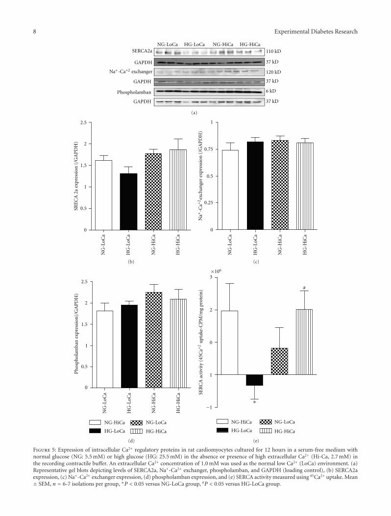
3.4. Impact of Elevated Extracellular Ca2+ on High Glucose-Induced Changes in Intracellular Ca2+ Regulatory Proteinsin Cardiomyocytes. To further evaluate elevated extracellularCa2+ and high glucose-induced changes in intracellularCa2+ handling, expression of intracellular Ca2+ regulatoryprotein was evaluated. Our results revealed that neitherhigh extracellular Ca2+ nor high glucose, or both, producedany notable changes in the levels of SERCA2a, Na+-Ca2+
exchanger and phospholamban. However, measurement ofSERCA activity revealed that high extracellular glucosesignificantly dampened the thapsigargin-sensitive 45Ca2+
uptake under low extracellular Ca2+ environment, the effectof which was nullified by elevated extracellular Ca2+. Lastbut not least, increase in extracellular Ca2+ concentrationitself did not affect expression or activity of intracellular Ca2+
regulatory proteins (Figure 5).
3.5. Effect of CaM Kinase Inhibition on Elevated ExtracellularCa2+-Induced Protection against Glucose Toxicity. Elevatedextracellular Ca2+ levels are known to activate CaM kinase.To evaluate if CaM kinase plays a role in the elevatedextracellular Ca2+ concentration-induced beneficial effectagainst glucose toxicity, cardiomyocytes maintained in nor-mal or high glucose medium were recorded in low or highextracellular Ca2+ environments in the absence or presence ofthe CaM kinase inhibitor KN93 (10 µM). Our data revealedthat KN93 effectively abolished high extracellular Ca2+-offered protection against high glucose-induced cardiomy-ocyte contractile anomalies (decrease in PS and ±dL/dt aswell as prolonged TPS and TR90). In addition, KN93 alsoabolished high extracellular Ca2+-induced cardiomyocytecontractile responses (increased PS and ±dL/dt along withunchanged TPS and TR90) under low extracellular Ca2+
environment (Figure 6).
4. Discussion
Result from our study revealed that elevated extracellularCa2+ level mitigated the glucose toxicity-induced cardiomy-ocyte mechanical and intracellular Ca2+ anomalies. Glucose

4 Experimental Diabetes Research
0
25
50
75
100
125
NG
-6h
r
HG
-6h
r
NG
-12
hr
HG
-12
hr
Res
tin
gce
llle
ngt
h(µ
m)
(a)
NG
-6h
r
HG
-6h
r
NG
-12
hr
HG
-12
hr
Peak
shor
ten
ing
(%ce
llle
ngt
h)
0
5
10
15
∗#∗
(b)
0
25
50
75
100
NG
-6h
r
HG
-6h
r
NG
-12
hr
HG
-12
hr
∗#
+dL
/dt
(µm
/s)
(c)
−dL
/dt
(µm
/s)
−100
−75
−50
−25
0
NG
-6h
r
HG
-6h
r
NG
-12
hr
HG
-12
hr
∗#
(d)
0
25
50
75
100
NG
-6h
r
HG
-6h
r
NG
-12
hr
HG
-12
hr
∗#
TP
S(m
/s)
(e)
0
25
50
75
100
125
150
175
TR
90(m
/S)
NG
-6h
r
HG
-6h
r
NG
-12
hr
HG
-12
hr
#∗
(f)
Figure 1: Mechanical property of adult rat cardiomyocytes cultured for 6 and 12 hours in a serum-free medium with normal glucose (NG:5.5 mM) or high glucose (HG: 25.5 mM). (a) Resting cell length, (b) peak shortening (PS) amplitude normalized to cell length, (c) maximalvelocity of shortening (+dL/dt), (d) maximal velocity of relengthening (−dL/dt), (e) time to PS (TPS), and (f) time to 90% relengthening(TR90). Mean ± SEM, n = 24 and 41-42 cells per group at 6 and 12 hours, respectively, ∗P < 0.05 versus NG-6 Hr group, #P < 0.05 versusNG-12 Hr group.
toxicity or elevated extracellular glucose level has been shownto compromise cardiomyocyte contractile function andintracellular Ca2+ homeostasis following short-term expo-sure [24, 25], suggesting a role of hyperglycemia in cardiaccontractile anomalies. Interestingly, our finding revealed thatthe high extracellular Ca2+-induced benefit effect againstglucose toxicity was abolished by the CaM kinase inhibitorKN93, suggesting a role of CaM kinase in elevated extracel-lular Ca2+-induced beneficial effect against glucose toxicity.
Hyperglycemia has been shown to be one of the mostdevastating predisposing factors for the onset and devel-opment of diabetic cardiomyopathy [2–4, 24]. Reducedcardiac contractility, depressed maximal velocity of contrac-tion/relaxation, and prolonged contraction/relaxation areconsidered landmarks of hyperglycemia or diabetes-inducedmyopathic alterations [2, 4, 7, 8, 24]. Findings from thisstudy confirmed our earlier observations that cardiomy-ocytes maintained in high glucose medium for 12 hours (but

Experimental Diabetes Research 5
0
20
40
60
80
100
120
NG
HG
NG
-am
inog
uan
idin
e
HG
-am
inog
uan
idin
e
NG
-cyc
loh
exam
ide
HG
-cyc
loh
exam
ide
NG
+D
MSO
Res
tin
gce
llle
ngt
h(µ
m)
(a)
0
5
10
15
NG
HG
NG
-am
inog
uan
idin
e
HG
-am
inog
uan
idin
e
NG
-cyc
loh
exam
ide
HG
-cyc
loh
exam
ide
NG
-DM
SO
Peak
shor
ten
ing
(%ce
llle
ngt
h)
(b)
0
20
40
60
80
100
120
+dL
/dt
(µm
/s)
NG
HG
NG
-am
inog
uan
idin
e
HG
-am
inog
uan
idin
e
NG
-cyc
loh
exam
ide
HG
-cyc
loh
exam
ide
NG
+D
MSO
##
∗
(c)
−125
−100
−75
−50
−25
0
NG
HG
NG
-am
inog
uan
idin
e
HG
-am
inog
uan
idin
e
NG
-cyc
loh
exam
ide
HG
-cyc
loh
exam
ide
NG
+D
MSO
-dL
/dt
(µm
/s)
∗
# #
(d)
0
10
20
30
40
50
60
70
80
90
NG
HG
NG
-am
inog
uan
idin
e
HG
-am
inog
uan
idin
e
NG
-cyc
loh
exam
ide
HG
-cyc
loh
exam
ide
NG
-DM
SO
∗∗∗
##
TP
S(m
/s)
(e)
0
50
100
NG
HG
NG
-am
inog
uan
idin
e
HG
-am
inog
uan
idin
e
NG
-cyc
loh
exam
ide
HG
-cyc
loh
exam
ide
∗∗# ∗#
NG
-DM
SO
TR
90(m
/s)
(f)
Figure 2: Mechanical property of adult rat cardiomyocytes cultured for 12 hours in a serum-free medium with normal glucose (NG: 5.5 mM)or high glucose (HG: 25.5 mM) in the absence or presence of the glycation inhibitor aminoguanidine (1 mM) or the translation inhibitorcycloheximide (60 µm). DMSO was used as a solvent with a final concentration less than 0.5%. (a) Resting cell length, (b) peak shortening(PS) amplitude normalized to cell length, (c) maximal velocity of shortening (+dL/dt), (d) maximal velocity of relengthening (−dL/dt); (e)time to PS (TPS), and (f) time to 90% relengthening (TR90). Mean± SEM, n = 22–29 cells per group, ∗P < 0.05 versus NG group, #P < 0.05versus HG group.
not 6 hours) compromised cardiomyocyte mechanical prop-erties manifested as depressed peak shortening and maxi-mal velocity of shortening/relengthening, along with pro-longed duration of contraction and relaxation, in a manner
similar to in vivo diabetes [6, 24, 25]. The subtle difference incontractile capacity in NG-maintained cardiomyocytes (Fig-ures 1 and 6) may be largely due to interculture and/or indi-vidual murine variations. Earlier studies from our laboratory

6 Experimental Diabetes Research
NG
-LoC
a
HG
-LoC
a
NG
-HiC
a
HG
-HiC
a
0
50
100
Res
tin
gce
llle
ngt
h(µ
m)
(a)
0
2.5
5
7.5
10
NG
-LoC
a
HG
-LoC
a
NG
-HiC
a
HG
-HiC
a
Peak
shor
ten
ing
(%ce
llle
ngt
h) ∗
∗#
(b)
0
20
40
60
80
100
120
140
NG
-LoC
a
HG
-LoC
a
NG
-HiC
a
HG
-HiC
a
+dL
/dt
(µm
/s)
#
∗
∗ ∗
(c)
−140
−120
−100
−80
−60
−40
−20
0
NG
-LoC
a
HG
-LoC
a
NG
-HiC
a
HG
-HiC
a
∗
∗ ∗#
-dL
/dt
(µm
/s)
(d)
∗
0
10
20
30
40
50
60
NG
-LoC
a
HG
-LoC
a
NG
-HiC
a
HG
-HiC
a
#
TP
S(m
/s)
(e)
∗
#
NG
-LoC
a
HG
-LoC
a
NG
-HiC
a
HG
-HiC
a
0
50
100
TR
90(m
/s)
(f)
Figure 3: Mechanical property of adult rat cardiomyocytes maintained for 12 hours in a serum-free medium with normal glucose (NG:5.5 mM) or high glucose (HG: 25.5 mM) in the absence or presence of high extracellular Ca2+ (Hi-Ca, 2.7 mM) in the contractile buffer. Anextracellular Ca2+ concentration of 1.0 mM was used as the normal low Ca2+ (LoCa) environment. (a) Resting cell length (b) peak shortening(PS) amplitude normalized to cell length; (c) Maximal velocity of shortening (+dL/dt); (d) Maximal velocity of relengthening (−dL/dt), (e)time to PS (TPS), and (f) time to 90% relengthening (TR90). Mean ± SEM, n = 38–42 cells per group, ∗P < 0.05 versus NG-LoCa group,#P < 0.05 versus HG-LoCa group.
have established that normal cardiomyocytes may displaydiabetes-like phenotype of cardiac mechanical dysfunctionssimulating in vivo diabetes after only 12–24 hours of culturein a serum-free high glucose medium [18, 24, 25]. The use ofisolated cardiomyocyte model allows the precise control ofthe extracellular milieu in which cardiomyocytes reside, thusmaximally eliminating the possible interference from fibrob-lasts, endothelial metabolism, and diffusion barriers [25–27].Intriguingly, the glycation inhibitor aminoguanidine and
the translation inhibitor cycloheximide attenuated glucosetoxicity-induced cardiomyocyte contractile defects withouteliciting any effect by themselves. These findings suggest pos-sible involvement of glycation and protein translation in glu-cose toxicity-induced cardiac contractile anomalies. Earlierfindings from our laboratory as well as others have depicteda pivotal role of glycation in hyperglycemia and diabetes-induced cardiac dysfunctions [5, 6, 28, 29]. Furthermore, ourdata revealed that glucose toxicity-induced cardiomyocyte

Experimental Diabetes Research 7
NG
-LoC
a
HG
-LoC
a
NG
-HiC
a
HG
-HiC
a
0
0.5
1
1.5
∗∗B
asel
ine
FFI
(360
/380
rati
o)
(a)
NG
-LoC
a
HG
-LoC
a
NG
-HiC
a
HG
-HiC
a
0
0.5
1
1.5
Peak
FFI
(360
/380
rati
o)
∗∗
(b)
NG
-LoC
a
HG
-LoC
a
NG
-HiC
a
HG
-HiC
a
0
0.1
0.2
0.3
FFI
(360
/380
rati
o)Δ
∗∗
(c)
0
50
100
150
200
#∗
∗
NG
-LoC
a
HG
-LoC
a
NG
-HiC
a
HG
-HiC
a
Intr
acel
lula
r C
a+2de
cay
(m/s
)
(d)
Figure 4: Intracellular Ca2+ handling property of adult rat cardiomyocytes cultured for 12 hours in a serum-free medium with normalglucose (NG: 5.5 mM) or high glucose (HG: 25.5 mM) in the absence or presence of high extracellular Ca2+ (Hi-Ca, 2.7 mM) in thecontractile buffer. An extracellular Ca2+ concentration of 1.0 mM was used as the low Ca2+ (LoCa) environment. (a) Resting intracellular Ca2+
levels shown as baseline fura-2 fluorescent intensity (FFI), (b) peak FFI, (c) electrically stimulated rise of FFI (ΔFFI), and (d) intracellularCa2+ decay rate. Mean ± SEM, n = 29–42 cells per group, ∗P < 0.05 versus NG-LoCa group, #P < 0.05 versus HG-LoCa group.
anomalies may be abolished with elevated extracellular Ca2+
levels (from the physiological level of 1.0 mM to 2.7 mM),the effect of which may be abolished by inhibition of CaMkinase using KN93. Several mechanisms may be speculatedfor elevated extracellular Ca2+-induced protection againstglucose toxicity. Elevated extracellular Ca2+ levels are capableof facilitating cytosolic Ca2+ cycling and dynamics thuspromoting Ca2+ influx and resequestration, regardless ofthe extracellular glucose environment. This is supported bythe increased rise of intracellular Ca2+ in response to elec-trical stimuli, facilitated intracellular Ca2+ decay, improvedSERCA activity (although not expression), and shortenedTR90 under high extracellular Ca2+ environment. Ourobservation of depressed cardiomyocyte contractile capac-ity and maximal velocity of contraction/relaxation alongwith prolonged duration of contraction and relaxation inhigh glucose-treated cardiomyocytes is supported by com-promised intracellular Ca2+ handling under high glucoseenvironment, consistent with the findings from in vivo dia-betic condition [8, 26]. Another scenario may be related to
changes in cardiac energy dynamics through CaM kinasesignaling cascade. Ca2+ ions promote cell survival througha CaM kinase-dependent activation of Akt [30], in linewith the finding from our current study. If energy stateswere the sole determinant of cardiac contractile function,then normal glucose-cultured cardiomyocytes should displayimproved cardiac contractile performance, consistent withour experimental finding. One of the main mechanisms thatextracellular Ca2+ improves cardiac performance is throughincreased phosphorylation state. It may be speculated thatCa2+ ions promote contractile inotropism first by bolsteringcardiac energy state, which occurs rapidly within minutes.The CaM kinase cascade is essential to many physiologicalprocesses, the defect of which can lead to a variety of diseasestates. Calmodulin acts as a primary receptor for Ca2+ in alleukaryotic cells whereas Ca2+/CaM kinase functions as anallosteric activator of a host of enzymatic proteins [15]. Thearea CaM-dependent kinase cascade is just on the horizon.Further investigation on the cellular machineries in par-ticular cardiac excitation-transcription coupling regulated
8 Experimental Diabetes Research
SERCA2a
GAPDH
Phospholamban
GAPDH
GAPDH
NG-LoCa HG-LoCa NG-HiCa HG-HiCa
37 kD
110 kD
120 kD
37 kD
6 kD
37 kD
Na+-Ca+2 exchanger
(a)
0
0.5
1
1.5
2
2.5H
G-L
oCa
NG
-HiC
a
NG
-LoC
a
SRE
CA
2aex
pres
sion
(/G
AP
DH
)
HG
-HiC
a
(b)H
G-L
oCa
NG
-HiC
a
NG
-LoC
a
HG
-HiC
a
0
0.25
0.5
0.75
1
Na+
-Ca+
2ex
chan
ger
expr
essi
on(/
GA
PD
H)
(c)
0
0.5
1
1.5
2
2.5
HG
-LoC
a
NG
-HiC
a
NG
-LoC
a
HG
-HiC
a
HG-LoCa
NG-HiCa NG-LoCa
HG-HiCa
Ph
osph
olam
ban
expr
essi
on(/
GA
PD
H)
(d)
∗
HG-LoCa
NG-HiCa NG-LoCa
HG-HiCa
#
×106
−1
1
0
2
3
SER
CA
act
ivit
y (4
5Ca+
2u
ptak
e-C
PM
/mg
prot
ein
)
(e)
Figure 5: Expression of intracellular Ca2+ regulatory proteins in rat cardiomyocytes cultured for 12 hours in a serum-free medium withnormal glucose (NG: 5.5 mM) or high glucose (HG: 25.5 mM) in the absence or presence of high extracellular Ca2+ (Hi-Ca, 2.7 mM) inthe recording contractile buffer. An extracellular Ca2+ concentration of 1.0 mM was used as the normal low Ca2+ (LoCa) environment. (a)Representative gel blots depicting levels of SERCA2a, Na+-Ca2+ exchanger, phospholamban, and GAPDH (loading control), (b) SERCA2aexpression, (c) Na+-Ca2+ exchanger expression, (d) phospholamban expression, and (e) SERCA activity measured using 45Ca2+ uptake. Mean± SEM, n = 6-7 isolations per group, ∗P < 0.05 versus NG-LoCa group, #P < 0.05 versus HG-LoCa group.

Experimental Diabetes Research 9
NG
-LoC
a
HG
-LoC
a
NG
-HiC
a
HG
-HiC
a
NG
-HiC
a-K
N93
HG
-HiC
a-K
N93
0
50
100
Res
tin
gce
llle
ngt
h(µ
m)
(a)
∗ #∗
0
2
4
6
8
10
12
14
NG
-LoC
a
HG
-LoC
a
NG
-HiC
a
HG
-HiC
a
NG
-HiC
a-K
N93
HG
-HiC
a-K
N93
∗
Peak
shor
ten
ing
(%ce
llle
ngt
h)
(b)
0
50
100
150
200
250
300
NG
-LoC
a
HG
-LoC
a
NG
-HiC
a
HG
-HiC
a
NG
-HiC
a-K
N93
HG
-HiC
a-K
N93
∗∗
∗ †
#
+dL
/dt
(µm
/s)
(c)
−250
−150
−50
NG
-LoC
a
HG
-LoC
a
NG
-HiC
a
HG
-HiC
a
NG
-HiC
a-K
N93
HG
-HiC
a-K
N93
†∗
∗#∗
-dL
/dt
(µm
/s)
(d)
TP
S(m
/s)
0
20
40
60
80
NG
-LoC
a
HG
-LoC
a
NG
-HiC
a
HG
-HiC
a
NG
-HiC
a-K
N93
HG
-HiC
a-K
N93
†
#
∗
(e)
NG
-LoC
a
HG
-LoC
a
NG
-HiC
a
HG
-HiC
a
NG
-HiC
a-K
N93
HG
-HiC
a-K
N93
0
50
100
150 †
#
∗
TR
90(m
/s)
(f)
Figure 6: Mechanical property of rat cardiomyocytes cultured for 12 hours in a serum-free medium with normal glucose (NG: 5.5 mM)or high glucose (HG: 25.5 mM) in the absence or presence of high or low extracellular Ca2+ (Hi-Ca = 2.7 mM; LoCa = 1.0 mM) in therecording contractile buffer. A cohort of HG-cultured cardiomyocytes was recorded in high Ca2+ environment in the presence of the CaMkinase inhibitor KN93 (10 µM). (a) Resting cell length, (b) peak shortening (PS) amplitude normalized to cell length, (c) maximal velocityof shortening (+dL/dt), (d) maximal velocity of relengthening (−dL/dt), (e) time to PS (TPS), and (f) time to 90% relengthening (TR90).Mean ± SEM, n = 10 cells per group, ∗P < 0.05 versus NG-LoCa group, #P < 0.05 versus HG-LoCa group, †P < 0.05 versus NG-HCa-KN93group.

10 Experimental Diabetes Research
by this intracellular Ca2+-initiated signaling cascade shouldhold considerable promise for the future of disease-relatedresearch [31].
In summary, the findings from the present study revealedimproved cardiac contractile and intracellular Ca2+ home-ostasis against glucose toxicity. Considering the unique roleof CaM kinase in regulation of cardiac energy metabolismand survival, understanding the mechanism of action ofCaM kinase in myocardial contractile regulation underpathological conditions such as hyperglycemia should beessential in the elucidation of CaM kinase in diabetic com-plications.
Acknowledgments
This work was supported in part by NIH/NCRR5P20RR016474, the National Natural Science Foundation ofChina (NSFC 30570758, 30770847, 81070127), and theScientific Research Foundation for the Returned OverseasChinese Scholars, State Education Ministry (SRF for ROCS,SEM no. HG2904). R.-H. Zhang and H. Guo contributedequally to this work.
References
[1] L. Li, Y. Hua, and J. Ren, “Short-chain fatty acid propionatealleviates Akt2 knockout-induced myocardial contractile dys-function,” Experimental Diabetes Research, vol. 2012, ArticleID 851717, 2012.
[2] W. H. Tang, W. T. Cheng, G. M. Kravtsov et al., “Cardiaccontractile dysfunction during acute hyperglycemia due toimpairment of SERCA by polyol pathway-mediated oxidativestress,” American Journal of Physiology, vol. 299, no. 3, pp.C643–C653, 2010.
[3] M. E. Cooper and A. El-Osta, “Epigenetics: mechanisms andimplications for diabetic complications,” Circulation Research,vol. 107, no. 12, pp. 1403–1413, 2010.
[4] C. Voulgari, D. Papadogiannis, and N. Tentolouris, “Diabeticcardiomyopathy: from the pathophysiology of the cardiacmyocytes to current diagnosis and management strategies,”Vascular Health and Risk Management, vol. 6, pp. 883–903,2010.
[5] H. Ma, S. Y. Li, P. Xu et al., “Advanced glycation endproduct(AGE) accumulation and AGE receptor (RAGE) up-regulationcontribute to the onset of diabetic cardiomyopathy,” Journal ofCellular and Molecular Medicine, vol. 13, no. 8, pp. 1751–1764,2009.
[6] J. Ren and A. F. Ceylan-Isik, “Diabetic cardiomyopathy: dowomen differ from men?” Endocrine, vol. 25, no. 2, pp. 73–83,2004.
[7] J. Ren, L. Pulakat, A. Whaley-Connell, and J. R. Sowers,“Mitochondrial biogenesis in the metabolic syndrome andcardiovascular disease,” Journal of Molecular Medicine, vol. 88,no. 10, pp. 993–1001, 2010.
[8] L. E. Wold, A. F. Ceylan-Isik, and J. Ren, “Oxidative stress andstress signaling: menace of diabetic cardiomyopathy,” ActaPharmacologica Sinica, vol. 26, no. 8, pp. 908–917, 2005.
[9] H. Nakamura, S. Matoba, E. Iwai-Kanai et al., “p53 Promotescardiac dysfunction in diabetic mellitus caused by excessivemitochondrial respiration-mediated reactive oxygen speciesgeneration and lipid accumulation,” Circulation, vol. 5, no. 1,pp. 106–115, 2012.
[10] S. K. Kota, S. K. Kota, S. Jammula, S. Panda, and K. D. Modi,“Effect of diabetes on alteration of metabolism in cardiacmyocytes: therapeutic implications,” Diabetes Technology &Therapeutics, vol. 13, no. 11, pp. 1155–1160, 2011.
[11] Z. Xie, K. Lau, B. Eby et al., “Improvement of cardiac functionsby chronic metformin treatment is associated with enhancedcardiac autophagy in diabetic OVE26 mice,” Diabetes, vol. 60,no. 6, pp. 1770–1778, 2011.
[12] J. Ren, “Paradoxical effects of pyruvate on cardiac contractilefunction under normal and high glucose in ventricularmyocytes,” Pharmacological Research, vol. 48, no. 1, pp. 25–29,2003.
[13] K. T. Chambers, T. C. Leone, N. Sambandam et al., “Chronicinhibition of pyruvate dehydrogenase in heart triggers anadaptive metabolic response,” The Journal of Biological Chem-istry, vol. 286, no. 13, pp. 11155–11162, 2011.
[14] S. K. Bhardwaj and G. Kaur, “Effect of diabetes on cal-cium/calmodulin dependent protein kinase-II from rat brain,”Neurochemistry International, vol. 35, no. 4, pp. 329–335, 1999.
[15] J. Colomer and A. R. Means, “Physiological roles of theCa2+/CaM-dependent protein kinase cascade in health anddisease,” Sub-Cellular Biochemistry, vol. 45, pp. 169–214, 2007.
[16] W. Fan, X. Li, and N. G. Cooper, “CaMKIIalphaB mediates asurvival response in retinal ganglion cells subjected to a glu-tamate stimulus,” Investigative Ophthalmology & Visual Scie-nce, vol. 48, no. 8, pp. 3854–3863, 2007.
[17] H. Takeda, Y. Kitaoka, Y. Hayashi et al., “Calcium/calmodulin-dependent protein kinase II regulates the phosphorylationof CREB in NMDA-induced retinal neurotoxicity,” BrainResearch, vol. 1184, no. 1, pp. 306–315, 2007.
[18] J. Ren, L. J. Dominguez, J. R. Sowers, and A. J. Davidoff,“Troglitazone attenuates high-glucose-induced abnormalitiesin relaxation and intracellular calcium in rat ventricularmyocytes,” Diabetes, vol. 45, no. 12, pp. 1822–1825, 1996.
[19] V. Jakus, M. Hrnciarova, J. Carsky, B. Krahulec, and N.Rietbrock, “Inhibition of nonenzymatic protein glycation andlipid peroxidation by drugs with antioxidant activity,” LifeSciences, vol. 65, no. 18-19, pp. 1991–1993, 1999.
[20] B. G. Mockett, D. Guevremont, M. Wutte, S. R. Hulme,J. M. Williams, and W. C. Abraham, “Calcium/calmodulin-dependent protein kinase II mediates group I metabotropicglutamate receptor-dependent protein synthesis and long-term depression in rat hippocampus,” Journal of Neuroscience,vol. 31, no. 20, pp. 7380–7391, 2011.
[21] A. J. Davidoff, M. M. Mason, M. B. Davidson et al., “Sucrose-induced cardiomyocyte dysfunction is both preventable andreversible with clinically relevant treatments,” American Jour-nal of Physiology, vol. 286, no. 5, pp. E718–E724, 2004.
[22] T. A. Doser, S. Turdi, D. P. Thomas, P. N. Epstein, S. Y. Li, and J.Ren, “Transgenic overexpression of aldehyde dehydrogenase-2rescues chronic alcohol intake-induced myocardial hypertro-phy and contractile dysfunction,” Circulation, vol. 119, no. 14,pp. 1941–1949, 2009.
[23] S. Y. Li, X. Yang, A. F. Ceylan-Isik, M. Du, N. Sreejayan, andJ. Ren, “Cardiac contractile dysfunction in Lep/Lep obesityis accompanied by NADPH oxidase activation, oxidativemodification of sarco(endo)plasmic reticulum Ca2+-ATPaseand myosin heavy chain isozyme switch,” Diabetologia, vol. 49,no. 6, pp. 1434–1446, 2006.
[24] A. J. Davidoff and J. Ren, “Low insulin and high glucoseinduce abnormal relaxation in cultured adult rat ventricularmyocytes,” American Journal of Physiology, vol. 272, part 2, no.1, pp. H159–H167, 1997.

Experimental Diabetes Research 11
[25] J. Ren, L. J. Dominguez, J. R. Sowers, and A. J. Davidoff,“Metformin but not glyburide prevents high glucose-inducedabnormalities in relaxation and intracellular Ca2+ transientsin adult rat ventricular myocytes,” Diabetes, vol. 48, no. 10, pp.2059–2065, 1999.
[26] L. E. Wold and J. Ren, “Mechanical measurement of con-tractile function of isolated ventricular myocytes,” Methods inMolecular Medicine, vol. 139, pp. 263–270, 2007.
[27] J. Ren and L. E. Wold, “Measurement of cardiac mechanicalfunction in isolated ventricular myocytes from rats and miceby computerized video-based imaging,” Biological ProceduresOnline, vol. 3, no. 1, pp. 43–53, 2001.
[28] Y. Ti, G. L. Xie, Z. H. Wang et al., “TRB3 gene silencingalleviates diabetic cardiomyopathy in a type 2 diabetic ratmodel,” Diabetes, vol. 60, no. 11, pp. 2963–2974, 2011.
[29] U. Rajamani and M. F. Essop, “Hyperglycemia-mediatedactivation of the hexosamine biosynthetic pathway results inmyocardial apoptosis,” American Journal of Physiology, vol.299, no. 1, pp. C139–C147, 2010.
[30] S. Yano, H. Tokumitsu, and T. R. Soderling, “Calciumpromotes cell survival through CaM-K kinase activation of theprotein-kinase-B pathway,” Nature, vol. 396, no. 6711, pp.584–587, 1998.
[31] D. M. Bers, “Ca2+-calmodulin-dependent protein kinase IIregulation of cardiac excitation-transcription coupling,” HeartRhythm, vol. 8, no. 7, pp. 1101–1104, 2011.

Submit your manuscripts athttp://www.hindawi.com
Stem CellsInternational
Hindawi Publishing Corporationhttp://www.hindawi.com Volume 2014
Hindawi Publishing Corporationhttp://www.hindawi.com Volume 2014
MEDIATORSINFLAMMATION
of
Hindawi Publishing Corporationhttp://www.hindawi.com Volume 2014
Behavioural Neurology
EndocrinologyInternational Journal of
Hindawi Publishing Corporationhttp://www.hindawi.com Volume 2014
Hindawi Publishing Corporationhttp://www.hindawi.com Volume 2014
Disease Markers
Hindawi Publishing Corporationhttp://www.hindawi.com Volume 2014
BioMed Research International
OncologyJournal of
Hindawi Publishing Corporationhttp://www.hindawi.com Volume 2014
Hindawi Publishing Corporationhttp://www.hindawi.com Volume 2014
Oxidative Medicine and Cellular Longevity
Hindawi Publishing Corporationhttp://www.hindawi.com Volume 2014
PPAR Research
The Scientific World JournalHindawi Publishing Corporation http://www.hindawi.com Volume 2014
Immunology ResearchHindawi Publishing Corporationhttp://www.hindawi.com Volume 2014
Journal of
ObesityJournal of
Hindawi Publishing Corporationhttp://www.hindawi.com Volume 2014
Hindawi Publishing Corporationhttp://www.hindawi.com Volume 2014
Computational and Mathematical Methods in Medicine
OphthalmologyJournal of
Hindawi Publishing Corporationhttp://www.hindawi.com Volume 2014
Diabetes ResearchJournal of
Hindawi Publishing Corporationhttp://www.hindawi.com Volume 2014
Hindawi Publishing Corporationhttp://www.hindawi.com Volume 2014
Research and TreatmentAIDS
Hindawi Publishing Corporationhttp://www.hindawi.com Volume 2014
Gastroenterology Research and Practice
Hindawi Publishing Corporationhttp://www.hindawi.com Volume 2014
Parkinson’s Disease
Evidence-Based Complementary and Alternative Medicine
Volume 2014Hindawi Publishing Corporationhttp://www.hindawi.com