canadian cll meeting abstracts - university of manitoba · 10thcanadian cll meeting table of...
TRANSCRIPT

i

i
10thCanadian CLL Meeting
Table of Contents
1.
Fanous M, Liu F, Farren T, Agrawal S* Investigating the disease specific expression profile of the HVEM/LIGHT/BTLA/CD160 co-signaling immune axis in chronic B-cell malignancies ..............................1
2.
Alzaki A*, Gerrie AS, Gillan TL, Huang SJ, Ahmed M, Toze CL, Leitch HA, Ramadan KM Autoimmune cytopenia in chronic lymphocytic leukemia: effect on outcome and survival, a population based analysis in British Columbia, Canada ..............................2
3.
Alzaki AA*, Ramadan KM Positive direct antiglobulin test in chronic lymphocytic leukemia: effect on survival and outcome ..............................4
4.
Bouchard E*, Gehrke I, Poeppl A, Johnston J, Gibson S, Banerji V FK866 causes loss of CLL cell viability by disrupting mitochondrial function ..............................6
5.
Dielschneider R*, Eisenstat H, Xiao W, Johnston JB, Gibson SB Chronic Lymphocytic Leukemia cells are susceptible to lysosome-mediated cell death ..............................7
6.
Goy J*, Gillan TL, Huang SJT, Bruyere H, Hrynchak M, Karsan A, Ramadan K, Smith A, Toze CL, Gerrie AS Clinical outcomes for Chronic Lymphocytic Leukemia (CLL) patients with 11q deletion in British Columbia (BC), Canada: Results of an observational population-based cohort ..............................8
7.
Hewitt D*, Poeppl A, Squires M, McKenzie-Matwiy E, Gibson S, Johnston J, Banerji V Genetic epidemiology of Chronic Lymphocytic Leukemia (CLL) in Manitoba ............................11
ii
8. Huang S*, Gerrie A, Hrynchak M, Karsan A, Ramadan K, Gillan T, Toze C
Characterization of treatment patterns and relation to outcome in a population-based cohort of Chronic Lymphocytic Leukemia patients in British Columbia, Canada ............................12
9.
Kost SEF*, Liang WS, Banerji V, Gibson SB, Katyal S, Johnston JB Improving the activity of bendamustine in chronic lymphocytic leukemia through drug interactions ............................15
10.
Lang R*, Peoppl A, Peltier C, Banerji V Isoform-selective inhibition of GSK-3 in Chronic Lymphocytic Leukemia ............................16
11.
Lee LJ*, Gerrie AS, Bruyere H, Gillan TL, Huang SJT, Toze CL, Ramadan K
Comparison of outcomes in Chronic Lymphocytic Leukemia (CLL) with the addition of rituximab to initial treatment: A comparative effectiveness analysis in the province of British Columbia (BC), Canada ............................17
12.
Li H*, Wu X, Hou S, Jayachandran N, Makondo K, Du O, Noh E, Wilkins J, Johnston J. Gibson S, Lin F, Marshall A Lipid messenger PI(3,4)P2 regulates migratory signaling in malignant B lymphocytes ............................19
13.
Omar T*, Mansour M, Sanders S, Sabloff M, Zanke B, Sheppard D Life-threatening toxicity following fludarabine-based therapy for chronic lymphocytic leukemia: genomic predictors ............................20
14.
Peltier CP*, Lang R, Hicks W, Poeppl A, Banerji V Targeting Glycogen Synthase Kinase-3 (GSK-3) as a potential CLL treatment ............................21
15.
Reimer S*, Streu E Richter’s Transformation in CLL: The Manitoba nursing experience ............................23
iii
16. Schmidt R*, Yang L, Henson E, Gibson SB, Johnston JB Telomere length and cyclin-dependent kinase 1 levels in patients with chronic lymphocytic leukemia ............................24
17.
Sinha S*, Boysen J, Secreto C, Warner SL, Kay NE, Ghosh AK Chronic Lymphocytic Leukemia B-cells express constitutively active fibroblast growth factor receptors linked to Axl signaling pathway: Implications for CLL therapy ............................25
18.
Squires M*, Deneka A, Natividad N, Hunt K Biobank Clinical Data Management: ATiM and Caisis Systems ............................27
19.
Swic SJ*, MacPhail AGT, Dalal CB, Huang SJT, Gerrie AS, Nevill TJ, Sutherland HJ, Broady R, Nantel SH, Narayanan S, Song KW, AbouMourad YR, Hogge DE, Power MM, Barnett MJ, Forrest DL, Young SS, Gillan TL, Toze CL Prognostic factors and outcomes in allogeneic hematopoietic stem cell transplant vs. non-transplant Chronic Lymphocytic Leukemia (CLL) patients: A comparative analysis with the Leukemia/BMT Program of British Columbia (BC) and the BC Provincial CLL Database ............................28
20.
Streu, Erin* The development of a nurse-led pilot program for Subcutaneous Immunoglobulin (SCIG)therapy for patients with Chronic Lymphocytic Leukemia (CLL) and secondary immune deficiencies at CancerCare Manitoba ............................31
21.
Yan X-J*, Scarfo L, Fazi C, Li W, Grant C, Kolitz JE, Barrientos J, Allen SL, Rai KR, Ghia P, Chiorazzi N Gene profile difference between MBL to normal B cells and CLL B cells ............................32
22.
Yang L*, Beiggi S, ZhangY, SchmidtR, Gibson SB, Johnston JB Clinical impact of telomere shortening in normal and leukemia cells in Chronic Lymphocytic Leukemia ............................34

1
Investigating the disease specific expression profile of the HVEM/LIGHT/BTLA/CD160 co-signaling immune axis in chronic B-cell malignancies
1Mariam Fanous, 1Fenting Liu, 1Timothy Farren and 1,2Samir Agrawal*. 1Pathology Group, Blizard Institute, Queen Mary University of London, London, UK; 2Department of Haemato-Oncology, Barts Health NHS Trust, St. Bartholomew’s Hospital, London UK. Background: Chronic B-cell lymphoproliferative disorders (B-LPDs) are heterogeneous in both clinical and biological behaviour. Recently, there has been considerable interest in targeting immune mechanisms for the treatment of haematological malignancies. HVEM (herpes virus entry mediator) and its distinct ligands - CD160, BLTA and LIGHT – represent an important network regulating immune activation and has been linked to tumour progression. We investigated the expression of HVEM and its ligands in B-LPDs. Methods: 103 patients with chronic B-cell malignancies had peripheral blood and bone marrow samples analysed within 24 hours for HVEM, CD160, BTLA and LIGHT expression A cocktail of antibodies was created for each ligand each containing CD2-FITC, CD5-APC, CD19-PerCy7 (BD Biosciences) and an antibody corresponding to each ligand: CD160-PE (Immunotech), LIGHT-PE, BTLA-PE (BD Biosciences) and HVEM-APC (R&D systems). A final lymphocyte concentration of 4x10⁹/L cells of each sample was achieved and labeled with the appropriate antibody cocktail. The percentage expression of each ligand on the malignant B cells was assessed against normal healthy controls (n=15). CLL cases, n = 65; mantle cell lymphoma (MCL), n = 13; follicular lymphoma (FL), n = 4, B-LPD not otherwise specified = 21 (incorporating suspected lymphoplasmacytic and splenic marginal zone lymphoma). Results: CD160 expression was upregulated in CLL cells (mean positivity 64.65%) compared with both B-cells from patients with non-CLL malignancies and healthy donors (mean 11.51% (p<0.01) and 0.30%, p<0.01 respectively). HVEM also appeared upregulated in CLL cells (mean 94.09%) versus other B-LPDs (79.32%, p=0.03), but slightly downregulated from healthy controls (mean 98.75, p=0.04). A highly significant difference was noted between HVEM from patients with FL (n=4, mean 0.70%, p<0.01) and all other malignancies. BTLA was expressed by all malignant B-cells, regardless of the subtype, although expression was higher in CLL over other B-LPDs (mean 93.60% and 81.36% respectively, p=0.03) - however, BTLA expression was highest on B-cells from healthy controls (99.01%). Of interest, BTLA expression was lowest in MCL (mean 63.1%) over other B-LPDs (>84%). There were no significant differences in the expression of LIGHT on CLL cells (mean 0.96%), B-cells from other B-LPD (1.61%) and healthy controls (0.75%). Using a hierarchical clustering model of the expression of these molecules, it was possible to cluster 65% of cases of CLL, 46% of MCL and 100% of FL. Conclusion: The upregulation of CD160 in CLL cells versus other malignant and healthy B-cells makes this an ideal candidate as a CLL-specific and a tumour specific antigen. The hierarchical clustering suggests the HVEM axis may have a role in the diagnosis of B-LPDs, as well as differential effects on the biology of these malignancies. This data highlights the complexity of understanding the consequences of manipulating HVEM and its ligands, with multiple potential interactions in cis, as well as trans.

2
Autoimmune cytopenia in chronic lymphocytic leukemia: Effect on outcome and survival, a population based analysis in British Columbia, Canada
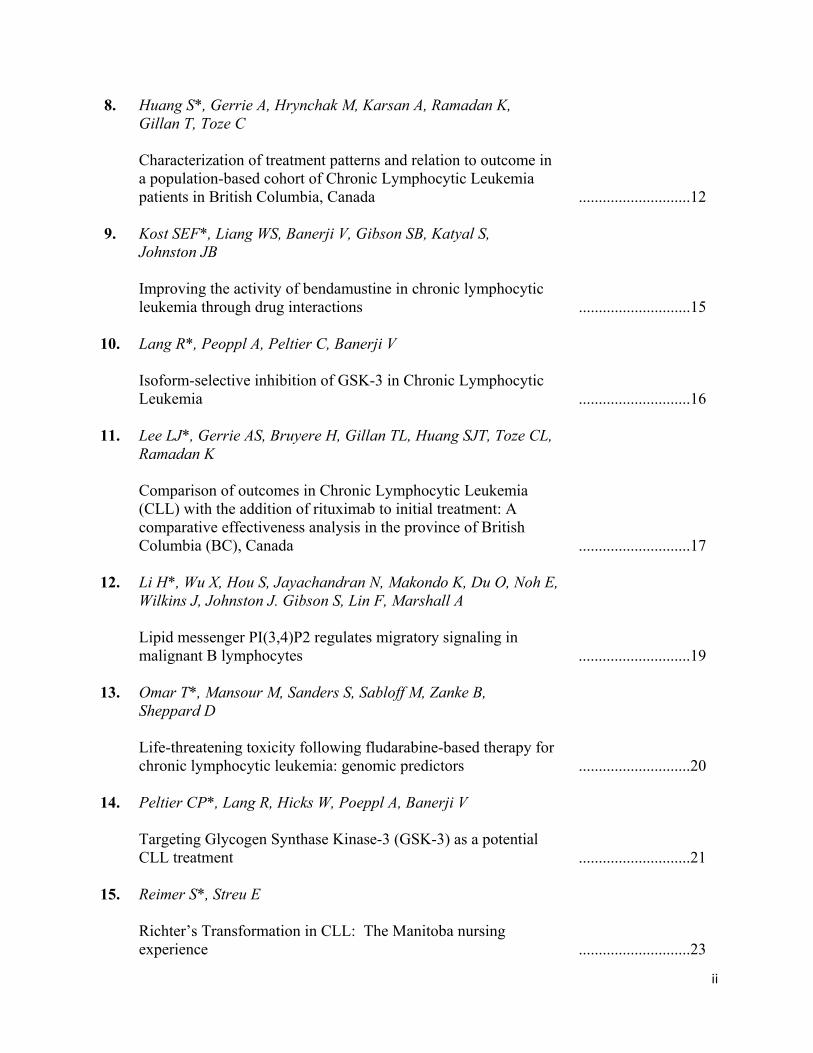
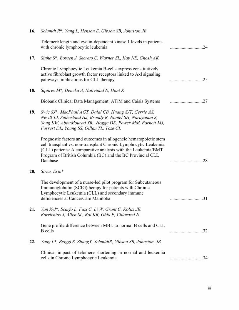
Alaa A. Alzaki1, MBBS, Alina S. Gerrie1,2,3, MD, FRCPC, Tanya L. Gillan4, PhD, FCCMG, Steven J.T. Huang2, BMLSc, FRCPC, Miriam Ahmed5, Cynthia L. Toze1,2, MD, FRCPC, Heather A. Leitch1,5, MD, FRCPC, Khaled M Ramadan1,5, MBBCh, MRCPI, FRCPath, FRCP, FRCPC. 1Department of Medicine, University of British Columbia, Vancouver, BC, Canada; 2Hematology, Leukemia-BMT Program of BC, 3British Columbia Cancer Agency, BC, Canada, 4Pathology and Laboratory Medicine, Vancouver General Hospital and University of BC, Vancouver, BC, Canada 5Division of Hematology, St. Paul’s Hospital, Vancouver, BC, Canada Background: Among Chronic Lymphocytic Leukemia (CLL) patients (pts), 4-10% are diagnosed with autoimmune cytopenias (AC) at some point during the course of their disease. This is less common than cytopenias related to bone marrow infiltration (10-20%). Infiltrative cytopenias(IC) are clearly a poor prognostic factor. However, the effect of AC on survival and prognosis of CLL pts remains understudied. Objectives: To determine the prevalence of AC and IC among CLL pts and their effect on overall survival (OS) and time to first treatment (TTFT) compared to pts without cytopenias. Furthermore, the effect of different treatment modalities including chemotherapy and chemo-immunotherapy as well as hypogammaglobulinemia at diagnosis on the disease course was evaluated in pts with AC. Methods: A population-based retrospective analysis through an electronic search of pts within the Providence Health Care CLL database between 1978 and 2013 was carried out. The diagnostic criteria for autoimmune hemolytic anemia (AIHA) were positive direct antiglobulin test and laboratory evidence of hemolysis,for immune thrombocytopenia (ITP) the exclusion of other etiologies of thrombocytopenia and for pure red cell aplasia (PRCA) anemia with low reticulocyte count and bone marrow evidence of decreased erythropoiesis. Infiltrative cytopenia diagnosis was confirmed by bone marrow biopsy. Anemia was defined as hemoglobin < 100 g/L. Thrombocytopenia was defined as platelets <100 x 109/L. Baseline features of pts with AC and IC were compared using Chi-squared analysis for categorical and the Kruskal-Wallis test for continuous variables. Overall survival was calculated from the date of initial treatment to the date of death from any cause. Time to first treatment (TTFT) was defined as the time interval between the date of diagnosis and date of first CLL treatment. Survival analysis was performed by the Kaplan– Meier method usingSPSS statistics V22.0 for windows. Results: Among 754 pts with CLL, 80 (10.6%) developed cytopenias (anemia and thrombocytopenia). Of those, 50 (6.6%)had IC and 30 (4%) had AC. There was no significant difference between the 2 groups in terms of age, gender, hemoglobin, platelets, LDH, WBC and lymphocyte count at diagnosis. The time to development of cytopenias for the IC and AC groups was similar with median of 3 and 4 years (yrs) from diagnosis, respectively. Within the AC group 16 pts had AIHA, 8 had ITP, 5 had both (Evan’s Syndrome) and 1 had PRCA. The median OS was 12.2 yrs (5.9–18.3) and 13 yrs (1.6-24.3) for IC and AC, respectively (p=0.260). However, when compared to CLL pts without cytopenias (median not reached), the AC group had worse OS (p< 0.005) (Fig 1). For the IC and AC groups, the median TTFT was 6.5 yrs (4.5-8.5) and 8.2 yrs (4.1–12.3), respectively (p=0.191). For the CLL pts without cytopenias TTFT was 8.1 yrs (2-12.2), similar to the AC group (p=0.88) (Fig 2). For AC pts, the OS was not
3
significantly different based on treatment received: alkylator based therapy vs. chemo-immunotherapy (p= 0.885).Of the 30 pts with AC, 26 had the results of serum protein electrophoresis at the time of diagnosis available. Of those, 10 (38.5%) had normal results and 16 (61.5%) had low gammaglobulin levels (IgG < 6 g/L); the mean OS was 18.1 years and 15.7 years respectively (median not reached), (P =0.433). Conclusion: The prognosis of pts with autoimmune and infiltrative cytopenias was similar. However, CLL pts with AC had worse OS compared to those without cytopenias. There was no significant difference in TTFT between AC and IC or when compared to CLL pts without cytopenias. For the AC group, neither treatment with chemotherapy vs. chemo-immunotherapy nor having concomitant hypo-gammaglobulinemia had an effect on outcome. To our knowledge, there are limited population based studies addressing the importance of determining the etiology of cytopenias in CLL pts and the effect of AC on survival and outcome. CLL immune complications need to be studied further especially in the context of novel agents and their possible effects on immune reconstitute
nocytopenia autoimmune cytopenia
OS (months)
Figure1. comparison of OS for CLL pts with autoimmune cytopenia vs. No cytopenia
TFTT (months)
nocytopenia autoimmune cytopenia
Figure2.comparison of TTFT for CLL pts with autoimmune cytopenia vs. no cytopenia

4
Positive direct antiglobulin test in chronic lymphocytic leukemia: Effect on survival and outcome
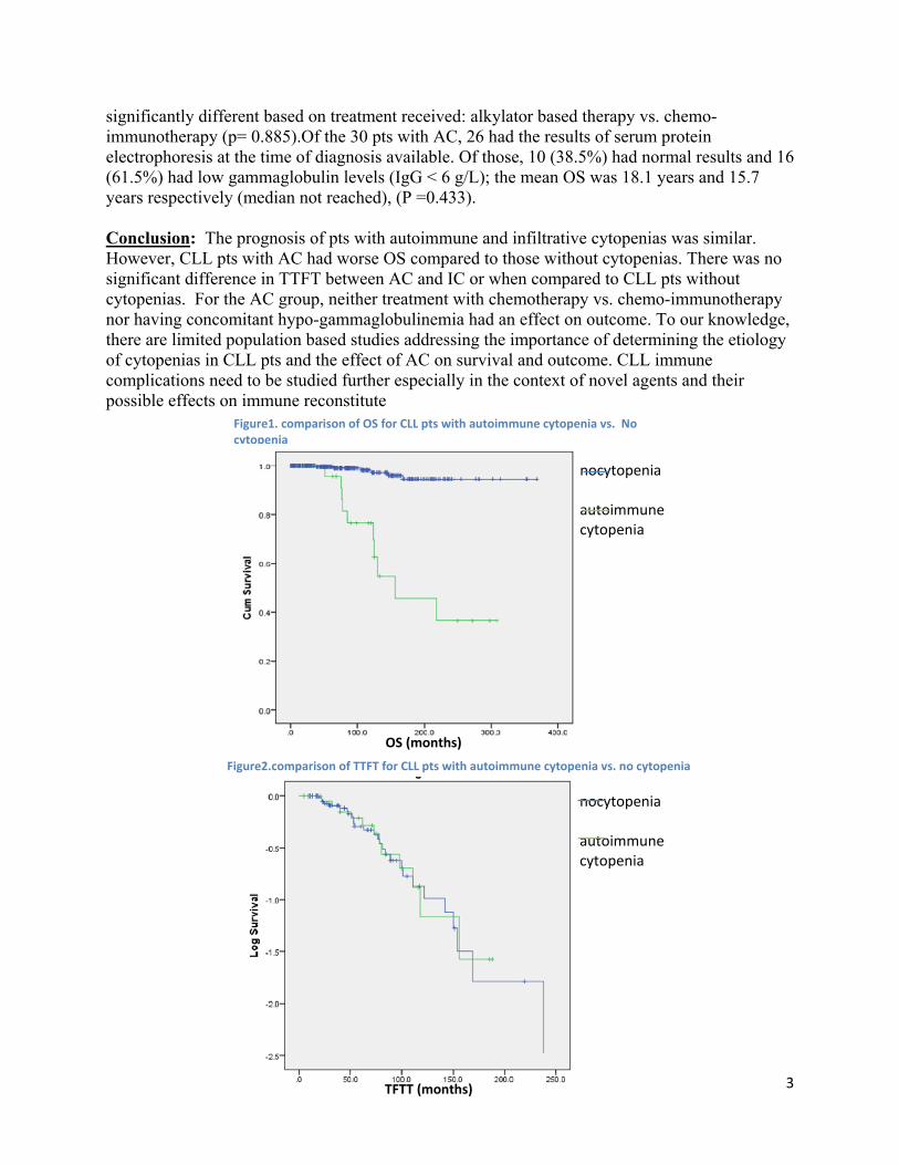
Alaa A. Alzaki1*, MBBS, Khaled M Ramadan1,2, MBBCh, MRCPI, FRCPath, FRCP, FRCPC. 1Department of Medicine, University of British Columbia, Vancouver, BC, Canada; 2Division of Hematology, St. Paul’s Hospital, Vancouver, BC, Canada Background: Autoimmune associated phenomena have been frequently observed in patients with CLL. Direct antiglobulin test (DAT) found to be positive at any stage of the disease in up to 35%.The effect of DAT positivity in natural CLLhistory is under investigated. Objectives: The purpose of this study was to determine the effect of DAT positivity on the time to first treatment (TTFT) and the overall survival (OS) among patients with CLL. We also compared the basic clinical risk factors including age, gender, Rai stage, hemoglobin, lymphocyte count, and lactate dehydrogenase (LDH) at the time of diagnosis between patients with negative and positive DAT. Methods: A retrospective analysis through an electronic search of patients within the St Paul’s hospital CLL data base between 1978 and 2013 was carried out (n=754). The DAT was performed using the anti-IgG, -C3d antibodies according to the hematopathology labs protocol. Patients were divided into 2 categories according to DATpositivity. Survival analysis was performed by Kaplan – Meier method using SPSS. Results: Among 754 with CLL, 249 patients were tested for DAT. Of those, 47(18.9%) had positive DAT while 202 (81.1%) had negative test. There was no significant difference in age, median 67 and 65.5 for the two groups. However, patients with positive DAT had significantly lower median hemoglobin at the time of diagnosis (94.5 vs. 124 g/L), higher median lymphocyte count (88 vs. 23), higher LDH (557 vs. 350). Male: female ratio was 2:1 among DAT positive patients. The OS was worse in patient with positive DAT (p 0.031, mean; 19 vs. 23 years, median was not reached). Among those patients who developed AIHA (n=21, 44.7%) had worse OS compared to those who didn’t develop AIHA (15 vs. 16.5 years, p 0.021). The TTFT was similar between patients with positive and negative DAT (median; 2.8 and 3 years respectively). A multivariate analysis for the covariates age (mean: 64), gender, Rai stage and CD38+ revealed no significant predictive factors for DAT positivity. Conclusion: CLL patients with positive DAT had worse prognosis than those who didn’t. This is more pronounced when they develop AIHA.
5
Figure 1 comparison of OS for DAT positive pt with or without AIHA
__ Neg DAT __Pos DAT, No AIHA __Pos DAT & AIHA

6
FK866 causes loss of CLL cell viability by disrupting mitochondrial function Eric Bouchard1,2*, Iris Gehrke2, Armando Poeppl2, James Johnston1,2, Spencer Gibson1, Versha Banerji1,2 1University of Manitoba, 2CancerCare Manitoba, Winnipeg, MB, Canada Background: The survival and proliferation of Chronic Lymphocytic Leukemia (CLL) cells is known to depend on external stimuli, primarily present in the lymph nodes and bone marrow. CLL cells in these microenvironments also show strong upregulation of NAMPT, the rate limiting enzyme in NAD salvage. FK866 is a novel therapeutic which inhibits NAMPT, selectively reduces cancer cell viability in solid tumors and has shown efficacy against several hematological cells lines and primary CLL cells in vitro. Its antitumour activity has been attributed to apoptosis in some cases and autophagy in others, but its mechanism of action (the molecular events linking NAMPT inhibition to loss of cellular viability) remains largely unknown. This study aims to characterize the molecular mechanisms of FK866-induced cell death in CLL, in order to inform novel drug combinations and treatment strategies. Methods: CLL cells were isolated from patient blood samples by density gradient centrifugation and cultured with 1 to 100 nM FK866, NAD and ATP content were evaluated by luminescent assays (Promega), cell viability was determined by flow cytometry with annexin V-FITC/PI or annexin V/7AAD staining, and apoptotic signaling was detected by western blot and by Caspase3/7-Glo assay (Promega). Mitochondrial function was assessed by flow cytometry with the reactive oxygen species (ROS) indicator dihydroethidium (DHE), the mitochondrial membrane potential (MMP) indicator tetramethylrhodamine methyl ester (TMRM) and by oxygen consumption rates measured by XF24 Extracellular Flux Analyser (Seahorse Bioscience). Results: FK866 potently reduced cellular NAD content at the low concentration of 10 nM as early as day one after treatment. ATP depletion followed by day two and viability was reduced by day three. Overnight pre-treatment with exogenous NAD rescued these effects. Dose and time dependant induction of apoptotic signaling was observed by day three, but inhibition of caspase activity by the pan-caspase inhibitor Z-VAD-fmk did not prevent FK866-induced loss of cellular viability. Heightened ROS production and loss of MMP were also detected, beginning on day two. Extracellular flux analysis revealed no significant differences between CLL cells and control B-lymphocytes in terms of basal mitochondrial stress, respiratory control or coupling efficiency. Conclusion: We have shown that the antitumour activity of FK866 on CLL cells induces, but does not require caspase-dependant apoptosis. Instead, the observed ATP depletion, ROS increase and loss of MMP point to disruption of mitochondrial function as the initiating event in FK886-induced CLL death. Future studies will rely on the XF24 Extracellular Flux Analyser (Seahorse Bioscience) for more sensitive measure of mitochondrial function in order to confirm the central role of mitochondrial inhibition in this process.
7
Chronic Lymphocytic Leukemia cells are susceptible to lysosome-mediated cell death
Rebecca Dielschneider1,2*, Hannah Eisenstat2,Wenyan Xiao2, James B Johnston1,2, Spencer B Gibson1,2. 1University of Manitoba; 2Manitoba Institute of Cell Biology *Presenting author Introduction: Chronic lymphocytic leukemia (CLL) is the most common adult leukemia in North America. Despite many therapeutic advances over the past decade, drug resistance and disease recurrence are common. This trend emphasizes the requirement for novel therapeutic approaches. One novel target identified in cancers such as breast cancer and acute myeloid leukemia is the lysosome. Intransformed cancerous cells, lysosomes were found to be sensitive to permeabilization by lysotropic agents in a process called lysosome membrane permeabilization. Permeabilization of lysosomes releases their acidic and proteolytic contents into the cytoplasm initiating lysosome-mediated cell death. The therapeutic strategy of targeting lysosomes has yet to be determined in CLL. Methods: Three different lysosome-targeting drugs were tested: a quinolone, a fluoroquinolone antibiotic, and a cationic drug (CAD).To determine the mechanism of action, various dyes were used to stain lysosomes, mitochondria, and reactive oxygen species. Fluorescence was visualized under the confocal microscope and quantified using flow cytometry. To determine the role of reactive oxygen species (ROS) the antioxidants α-tocopherol, lycopene, N-acetyl cysteine, and glutathione were added to cells. To determine the role of proteases the inhibitors zVADfmk, Ca-074-Me, Chymostatin, and E64 were added to cells. The chemotherapeutic agent etoposide was used as a positive control. Results: All the lysotropic agents except the antibiotic effectively killed primary patient CLL cellsin vitro. The CAD had the greatest activity and was significantly more cytotoxic to the CLL cells as compared to patient T cells and normal peripheral blood mononuclear cells. Treatment was equally effective in p53-proficient and p53-deficient CLL cells, demonstrating that the most aggressive and drug-resistant CLL cells were sensitive to this CAD. Mechanistic studies revealed that lysosome membrane permeabilization occurred within minutes and led to an increase in ROS and loss of mitochondrial membrane potential. The permeabilization of lysosomes was further confirmed by the translocation of transcription factor EB (TFEB) into the nucleus indicating promotion of lysosomal biogenesis. Lipid ROS were necessary to induce cell death, as only lipophilic antioxidants prevented cell death. Lipophilic antioxidants did not prevent lysosomal permeabilization, but did prevent downstream mitochondrial dysfunction. Inhibitors of caspases and lysosomal cathepsins failed to prevent cell death in CLL cells. Conclusions: Results show that the lysosome-targeting quinolone and CAD effectively permeabilize lysosomes and induce lysosome-mediated cell death in primary human CLL cells in a lipid ROS-dependent but protease-independent mechanism. Overall, targeting lysosomes may be an effective strategy to selectively kill CLLcells regardless of p53 expression. Future studies are focused on the lysosomal differences in B cells and CLL cells.

8
Clinical outcomes for Chronic Lymphocytic Leukemia (CLL) patients with 11q deletion in British Columbia (BC), Canada: Results of an observationalpopulation-based cohort
Jennifer Goy1*, Tanya L. Gillan2, Steven J.T. Huang 2, Helene Bruyere2 , Monica Hrynchak3, Aly Karsan4, Khaled Ramadan5,Adam Smith4, Cynthia L. Toze1, 6, Alina S. Gerrie1, 6 1Division of Hematology, University of British Columbia; 2Pathology and Laboratory Medicine, Vancouver General Hospital; 3Cytogenetics Laboratory, Royal Columbian Hospital;4Pathology and Laboratory Medicine, British Columbia Cancer Agency; 5Division of Hematology, St. Paul’s Hospital and University of British Columbia; 6Leukemia/BMT Program of BC, University of British Columbia; Background: The 11q22.3 deletion (11q-) encompassing the ATM locusdetected by fluorescence in situ hybridization (FISH) is present in up to 20% of CLL patients (pts), and is associated with an aggressive disease course and poor response to treatment (tx). However, there are few clinical studies that have specifically focused on pts with 11q-.Prognostic factors that may explain the variable outcome in this group have not been investigated, nor have survival outcomes in a population-based setting. Furthermore, few clinical studies have describedpractice patterns within the 11q- group. This study aims to improve our understanding of the prognosis, management and clinical course of CLL pts with 11q-. Methods: The BC Provincial CLL Database, which includes all pts who have undergone CLL FISH testing in a provincially validated laboratory since 2004, was used to identify all pts in BC (population 4.5 million) with a confirmed 11q->10% found at any point during the disease course, with or without other detectable FISH abnormalities. Overall survival (OS), treatment-free survival (TFS) (time from diagnosis to first tx/death) and time to second-line treatment (tx) were the main outcomes of interest, and were investigated in relationship to age (<60 vs ≥60 years [yrs]), sex, Rai stage (3-4 vs 0-2), CD38 status, % 11q-, and presence of other recurrentFISH abnormalities (17p deletion [17p-], trisomy 12, 13q deletion, and IGH translocation). Multivariate analysis (MVA) was performed using Cox proportional hazard models with a backwards stepwise selection process to determine predictors of OS/TFS. Results: 125 out of 1044 (12%)pts were identified with 11q- detected at any point. Median age at CLL diagnosis was 61 yrs (range, 35-80). At a median follow-up of all patients of 6.3 yrs (range 0.5 - 26.2), median OS and TFS for the entire cohort are12.6 yrs (standard error [SE] = 1.5 yrs) and 2.5 yrs (SE = 0.5 yrs), respectively. In MVA, advanced Rai stage (HR 3.7, 95% CI 1.28-10.8, P=.015), age ≥ 60 yrs (HR 2.2, 95% CI 1.2-4.3, P=.012) and male gender (HR 2.4, 95 % CI 1.5-5.3, P=.024) were significant predictors of OS. Clonal evolution (CE) to 11q- and/or other FISH abnormalities was documented in 12 pts. In 9/12 cases, 11q- developed after one or more tx courses whereas, in 3/12 cases, 11q- developed prior to tx initiation at median 5 yrs (range, 3.8-6.5) from initial FISH test. Sixty-nine pts had a documented 11q- pre-tx. At median follow-up of 5.0 yrs (range, 0.5 to 18.7), this group had median OS and TFS of 14.7 (SE =1.8) and 2.5 yrs (SE= 0.5), respectively (Fig 1). Pts with presence of 11q- and 17p- had a markedly worse prognosis compared to those without, with median OS 4.9 vs. 14.7 yrs (P< .001) and TFS of 0.2 vs. 2.5 yrs (P=0.31). In MVA, co-presence of 17p- (HR = 11.3, 95% CI 2.4-52.5, P=.02) and age ≥ 60 yrs (HR 3.8, 95% CI 0.83-17.1, P=.05) were adverse prognostic markers for OS. Age ≥ 60 yrs (HR =2.0, 95% CI

9
1.0-3.9, P=.03), presence of > 75% 11q- (vs. < 75 % 11q-) (HR = 1.8, 95%CI 1.0-2.3, P=.03) and advanced Rai stage (HR = 7.1, 95%CI 2.4-21.0, P=.01) were associated with shorter TFS. Of this group of 69, 52 pts (75%) received at least one tx course during follow-up. Thirty-three pts received fludarabine +/- rituximab (FR) as first line, 14 pts received alkylatorbasedtx (FC +/- R in 8; CVP +/- R in 5; CHOP + R in 1) and 5 pts received chlorambucil-based +/- R. Pts treated with FR alone had median OS 12.8 yrs (SE=1.0), which was not statistically different from those treated with alkylators , where median OS was 6.8 yrs (SE=2.1), P=.35 (Fig 2). In MVA, receipt of fludarabine vs alkylator-based treatment was not associated with any significant difference in OS (P=.52) or time to 2nd tx (P=.65). Conclusion: This study is to our knowledge the largest population-based report of CLL pts with 11q- and further enhances our understanding of the clinical course of such pts in a real world setting. Although median TFS of 11q- pts is short at 2.5 yrs, OS remains long at 12.6 yrs. The importance of combined 11q- and 17p- as adverse prognostic markers in CLL is highlighted. Most pts in BC with 11q- received FR as first line therapy. There is retrospective trial data suggesting that first line tx containing alkylators (i.e. cyclophosphamide ) can overcome the adverse prognostic features of 11q-. However, most of this trial data is from the pre-R era. Though limited in numbers, our study does not suggest that first line tx without alkylators is associated with negative outcomes.

10
� �
11
Genetic epidemiology of Chronic Lymphocytic Leukemia (CLL) in Manitoba Donna Hewitt*, Armando Poeppl, Mandy Squires, Eilean McKenzie-Matwiy, Spencer Gibson, James Johnston, Versha Banerji Manitoba Institute of Cell Biology, Winnipeg, MB, Canada CancerCare Manitoba, Winnipeg, MB, Canada Department of Biochemistry and Medical Genetics, University of Manitoba, Winnipeg, MB, Canada Background: Molecular epidemiology is an important tool to further understand populations. CLL patients are an ideal population to study as new diagnoses are catalogued locally by flow cytometry and clinical features. There is a strong suggestion that in CLL; genetics contributes to the etiology with an 8 fold risk of CLL in first degree relatives. The Manitoba Blood and Marrow disorder Bank is collaborating with the Mayo Clinic who have developed a study to investigate this genetic basis through the use of biological materials from high risk CLL families. This study aims to understand the role of genetics in susceptibility and describe the interrelation of genetics and environment in this disease. It also has the potential to identify precursor states of malignancy in relatives of CLL patients through the use of flow cytometry. Methods: Patients are identified through the CLL clinic, informed consent is obtained as per the local ethics. From the index case 4 tubes of blood are drawn and a saliva sample obtained. Environmental risk factors and family history questionnaires are filled out. The family information is entered into a secure database at the Mayo Clinic and used to draw a pedigree. The pedigree is used to detail family history of cancer as well as identify unaffected family members. Unaffected family members are then contacted by the index case and given opportunity to participate. After consent is obtained the unaffected family members donate three tubes of peripheral blood and an Oragene saliva sample sent to the National Cancer Institute in Maryland and a the fourth blood tube is sent to Mayo Clinic for flow cytometry. Their environmental exposure is also captured. Results: Manitoba has a considerable number of affected families with CLL. Of the 900 consented patients in the CLL Bank 7.5 % have familial CLL. In the third year of our participation in this study we have further developed our infrastructure to facilitate smoother collection and communication. We have increased our family participation to 39 families. Conclusion: Western Canada and Northern Ontario is a geographically challenging region in its size and accessibility of courier services. This has led to challenges related to logistics for shipping samples in rural Canadian towns. Many do not have courier coverage making it impossible to get the sample to major centres for overnight delivery or require an expensive mode of shipping. We have worked with rural hospitals and local couriers in order to accrue viable samples. We have been resourceful and have tried to meet the participant’s need, even finding private phlebotomists to draw and ship samples. We have identified Cancer centres and other types of health care centres in other provinces and have gained their assistance in drawing and shipping samples for this project. We have moved to a more automated system of generating shipping airway bills and facilitate communication between the study participants, the research team and laboratory centres where analysis is to be performed.

12
Characterization of treatment patterns and relation to outcome in a population-based cohort of Chronic Lymphocytic Leukemia patients in British Columbia, Canada
Steven Huang*1, 2, Alina Gerrie1, Monica Hrynchak3, Aly Karsan4, Khaled Ramadan5, Tanya Gillan2, Cynthia Toze1
1Division of Hematology, Leukemia/BMT Program of BC, University of British Columbia; 2Pathology and Laboratory Medicine, Vancouver General Hospital; 3Cytogenetics Laboratory, Royal Columbian Hospital; 4Pathology and Laboratory Medicine, British Columbia Cancer Agency; 5Division of Hematology, St. Paul’s Hospital and University of British Columbia Background: There has been a lack of studies evaluating treatment (tx) patterns for chronic lymphocytic leukemia (CLL) in a population-based setting. In the province of British Columbia (BC), population 4.6 million, CLL patients (pts) are evaluated and treated according to centrally derived standard protocols thus allowing analysis of tx patterns and outcomes in a large uniformly managed CLL cohort. The aim of this study is to characterize current tx patterns for CLL in BC in 1st, 2nd and further lines and to compare patient characteristics and outcomes based on initial therapy. Methods: The BC Provincial CLL Database, which includes all CLL patients in BC who had fluorescence in situ hybridization performed since 2004, was used to identify pts with at least one database entry between January 1, 2003 and December 31, 2012. Only pts whose tx status was known were included. Primary outcomes were to estimate the proportion that had tx vs. no tx; the proportion being treated in 1st, 2nd and subsequent lines of tx; and the distribution of pts treated with fludarabine (Flud) vs. non-Flud based tx. Results: 996 pts with a CLL diagnosis (dx) and an entry in the database were identified between 2003 and 2012. 19 pts with no tx data available at this time were excluded. Yearly CLL incidence from 2003-2012 was a median (med) 1.78/100,000 (range, 1.16-2.29/100,000). Proportion of pts receiving tx was 58% (584/996) vs. 42% (412/996) no tx. Proportion requiring new tx in a given year was med 24% (range, 20-27%) per year. Med treatment-free survival (TFS) from dx to 1sttx was 2.5 years (yrs) (range, 0-20.6 yrs) and 1.9 yrs (range, 0.1-21.5 yrs) from 1st to 2ndtx. 399 pts (68%) were treated with Flud vs. 185 pts (32%) without Flud in 1st line tx. There were no differences in baseline characteristics between pts treated with Flud vs. non-Flud except for lymphocyte count at dx, which was med 15x109/L vs. 10x109/L (P<0.001), respectively. 327 out of 996 pts (33%) received 2nd line tx and 41% of treated pts received more than 2 lines of tx.Untx patients had longer overall survival (OS) than tx patients, med OS not reached vs. 14.7 yrs, respectively (P<0.001). Requiring tx remained an independent adverse predictor for OS after adjusting for age, sex, Rai stage, lymphocyte count, deletion 17p and deletion 11q (P=0.001, HR 1.92, 95% CI 1.29-2.86). However, no difference in OS or TFS was seen between pts treated with Flud vs. non-Flud, med OS 14.6 vs. 15.8 yrs respectively (P=0.7); med TFS 2.4 vs. 2.8 yrs (P=0.48). Conclusion: The proportions of pts being treated and receiving 1st line tx were stable on a per year basis in this large cohort over an 11 year period. It was not uncommon for pts to receive multiple lines of tx. This highlights the need to consider both the benefits and long-term risks of each tx. A significant proportion of treated pts (68%) received Flud in 1st line tx. However, no

13
differences in OS or TFS were found between pts treated with Flud or non-Flud. Our data indicates that a fairly constant proportion of patients move from the untx into the tx cohort per year. Moreover, it is of interest that need for tx is associated with decrease in expected survival. Longitudinal genomic studies could therefore be performed to identify underlying changes that occur in pts who acquire need for tx. Ideally a signature could be identified that differs between the stable, untreated state and the advancing ‘need for tx state’ that would be evident prior to the currently used clinical treatment criteria. Knowledge of this underlying genomic shift may permit application of therapy, both in terms of type and timing, which may alter the natural history of CLL. Figure 1.Overall survival of patients treated vs. untreated
403020100
Overall Survival from date of diagnosis (y)
1.0
0.8
0.6
0.4
0.2
0.0
Cum
Sur
viva
l
Treatment, n=584No treatment, n=412
P < 0.001

14
Figure 2. Overall survival of patients treated with fludarabine vs. no fludarabine in first-line therapy
Figure 3. Treatment free survival of patients treated with fludarabine vs. no fludarabine in first-line therapy
403020100
Overall Survival (y)
1.0
0.8
0.6
0.4
0.2
0.0
Cum
Sur
viva
l
Fludarabine tx, n=399
Non-fludarabine tx,n=185
P = 0.696
2520151050
Treatment Free Survival (y)
1.0
0.8
0.6
0.4
0.2
0.0
Cum
Sur
viva
l
Fludarabine tx, n=399
Non-fludarabine tx,n=185
P = 0.480

15
Improving the activity of bendamustine in Chronic Lymphocytic Leukemia through drug interactions
Sara E.F. Kost1*, William S. Liang2, Versha Banerji1,2, Spencer B. Gibson1,SachinKatyal, and James B. Johnston1,2 1Manitoba Institute of Cell Biology, CancerCare Manitoba, Winnipeg MB 2Department of Internal Medicine, University of Manitoba, Winnipeg MB Background: Bendamustine is a promising new agent for chronic lymphocytic leukemia (CLL) and is believed to have the properties of an alkylating agent (e.g. chlorambucil) although it has some structural similarities to a nucleoside analogue (e.g. fludarabine). Synergy has been observed between bendamustine and fludarabine in primary CLL cells in vitro, presumably related to inhibition of bendamustine DNA cross-linking by fludarabine. However, both bendamustine and fludarabine are marrow-toxic, making this combination unlikely to be clinically useful. The adenosine deaminase inhibitor pentostatin is a nucleoside analog thatis less marrow-suppressive than fludarabine. As a result it has been evaluated as a substitute for fludarabine in the elderly. However, whether like fludarabine it also produces synergy with bendamustine is unknown. Methods: MTT assays were used to determine the cytotoxicity of bendamustine against CLL cells in vitro. Flow cytometry for annexin V and 7-ADD was used to determine the effectiveness of bendamustine compared to or in combination with other drugs such as chlorambucil, fludarabine, orpentostatin/deoxyadenosine at various concentrations and time points.γ-H2AX staining and comet assays were used to measureDNA breaks. Results: Bendamustine treatment produced a linear dose-response curve and a narrow range of effective drug concentrations, similar to other alkylating agents. Apoptosis was induced in CLL cells following in vitro treatment with bendamustine, chlorambucil, and fludarabine. All three drugs showed equivalent toxicities 18 hours post treatment with greater killing potential at later time points. At equivalent drug doses, fludarabine produced a high number of double strand breaks (DSB), but this was not seen with bendamustine or chlorambucil. The combination of bendamustine or chlorambucil with fludarabine, resulted in greater killing than these agents alone. However, no synergy was seen with bendamustine and chlorambucil. Conclusion: Bendamustine has equivalent antitumor activity as chlorambucil or fludarabine against CLL cells in vitro, with properties that are more typical of an alkylating agent. The combination of bendamustine with pentostatin has clinical potential and is currently being evaluatedin vitro.

16
Isoform-selective inhibition of GSK-3 in Chronic Lymphocytic Leukemia Rebecca Lang1*, Armando Peoppl4, Cheryl Peltier3, Versha Banerji1,2,3,4 1Manitoba Institute of Cell Biology, 2Department of Internal Medicine, University of Manitoba,3Department of Biochemistry and Medical Genetics, University of Manitoba, 4CancerCare Manitoba Background: Chronic lymphocytic leukemia (CLL) is the most common hematological malignancy in North America and Europe with a median age of onset of 72. Due to the advanced age of most patients with CLL, many are unable to tolerate standard treatments and receive sub-optimal care. Despite the availability of various treatment options, CLL remains incurable. Glycogen synthase kinase-3 (GSK-3) is a metabolic enzyme dysregulated in CLL. It has two isoforms: GSK-3α and β. Our hypothesis is that GSK-3 is a critical enzyme for CLL cell survival and therefore a target that can be exploited for therapeutic benefit. Methods: The effect of chemical pan-GSK-3 inhibition on β-catenin stabilization was observed in lymphoid cell lines after 24 hour of treatment with 15 μM SB. Cell viability was assessed by observing the levels of ATP as measured by luminescence after 48 and 72 hours of treatment with LiCl and SB. Cell viability was also observed by AnnexinV/PI staining using flow cytometry. Nine shRNAs were validated by sequencing. Knockdown of GSK-3α and GSK-3β was confirmed by western blot in U937 cells, a myeloid cell line in which this technique has already been confirmed. ShRNAs that resulted in the most consistent knockdown were selected for subsequent experiments. JVM3 cells treated with 15 μM SB served as a positive control for β-catenin stabilization. Results: We observed β-catenin stabilization with SB compared to DMSO control following treatment with SB. I-83 cells demonstrated a time and dose dependent loss of ATP as measured by luminescence at 48 and 72 hours compared to untreated and vehicle controls. Loss of cell viability was also observed by Annexin V/PI staining. Isoform-specific loss of both GSK-3α and GSK-3β were validated in I-83, Nalm-6, JVM3 and BJAB cell lines without stabilization of β-catenin. Conclusions: The phenotypic effects of isoform-specific loss of GSK-3 have not yet been characterized in CLL. Characterizing these effects may aid in the understanding and development of isoform-specific inhibitor of GSK-3.

17
Comparison of outcomes in Chronic Lymphocytic Leukemia (CLL) with the addition of rituximab to initial treatment: A comparative effectiveness analysis in the province of
British Columbia (BC), Canada Lauren J. Lee, MD1*, Alina S. Gerrie, MD, FRCPC2, Helene Bruyere, MD, FCCMG3, Tanya L. Gillan, PhD, FCCMG3, Steven J.T. Huang, BMLSc3, Cynthia L. Toze, MD, FRCPC2, Khaled Ramadan, MBBCh, MRCPI, FRCPath, FRCP, FRCPC4
1Department of Medicine, University of British Columbia, Vancouver, BC, Canada; 2Hematology, Leukemia/BMT Program of BC, British Columbia Cancer Agency, University of British Columbia, Vancouver, BC, Canada; 3Pathology and Laboratory Medicine, Vancouver General Hospital, University of British Columbia, Vancouver, BC, 4Division of Hematology, St. Paul’s Hospital, University of British Columbia, Vancouver, BC Background: Clinical trials report that chemoimmunotherapy with rituximab (R) improves overall survival (OS) and progression-free survival in the treatment (tx) of symptomatic CLL patients (pts). R has been available for first-line CLL tx in BC, population 4.5 million, since 2004. We compared clinical outcomes with and without addition of R to chemotherapy in a large unselected provincial cohort of pts treated for CLL, to determine the "real world" effectiveness of addition of R to standard chemotherapy. Methods: Three large provincial databases (db) were used to identify eligible pts: the BC Provincial CLL db, the BC Lymphoid Cancer db, and the Providence Hematology CLL db. All pts who received minimum 1 cycle of first-line tx for confirmed CLL were included. Pts with > 1 hematologic malignancy (n=8) were excluded. Baseline features of pts treated with (+R) or without R (No R) were compared using Chi-squared for categorical and Kruskal-Wallis test for continuous variables. OS was calculated from date of initial tx to date of death from any cause. Treatment-free survival (TFS) was calculated from date of initial tx to date of next tx/death from any cause. Multivariate analysis (MVA) was performed using Cox proportional hazard models to evaluate the effect of R on OS/TFS, after controlling for covariates including age (≥60 yrs vs <60 yrs), Rai stage (3-4 vs 0-2), CD38 status (pos vs neg), presence of 17p (17p-) and 11q (11q-) deletions, and first-line tx with purine analogs (PAs). Results: A total of 1784 pts diagnosed with CLL from 1973-2014 were identified, of which 726 pts (41%) received tx in follow-up. Of treated pts, 393 (54%) received R and 333 (46%) received chemotherapy alone. Among the No R group, tx included: chlorambucil 56%; fludarabine (F) 34%; cyclophosphamide (C)-based 8%; cladrabine 2%. Among the +R group, tx included: FR 75%; C-based + R 17%; FCR 7%; chlorambucil + R 1%. 103 pts underwent bone marrow transplant (BMT) during their tx course (19% No R vs 10% +R, P=.002). Median age at diagnosis (dx) and tx between groups were not statistically different (No R vs +R: 60.6 vs 60.8 yrs and 64.7 vs 63.9 yrs, respectively). There were no clinically significant differences in diagnostic parameters including % with elevated LDH, lymphocyte count >20x109/L , Rai stage 3-4. Median follow-up time in survivors was longer in the No R group (13.0 vs 6.8 yrs, P<.001). Among 467 pts with known CD38 status, CD38 pos was more common in +R vs No R groups (47 vs 36%, P=.02). FISH was performed in 586 pts, with no significant differences in abnormalities between tx groups. Poor-risk FISH, 17p- or 11q-, were present in 29% (No R) and 27% (+R).

Median t+R groupSignifica3.1 yrs, P(median yrs, P= .0 MVA coof mortalafter con2.77, 95%whereas 95%CI: 1 Conclusiaddition lower risalso demsubsequeknowledg[Danese,line treat
time from dxps (P=.84). Oant improvemP<.0001), wiOS 9.4 vs 6.004), Fig. 2,
nfirmed thatlity (HR 0.66trolling for c
% CI 1.87-4.for TFS, pre1.03-1.68, P
ion: In this of R to chemk of overall
monstrated thent therapy, age. This studBlood 2011ment of CLL
x to initial txOS was longments in OS ithout 17p- (.4 yrs, P=.00 and in those
t the addition6, 95% CI: 0covariates. O.10, P<.001)esence of 17p
P=.025) were
large, populmoimmunothmortality (9at the additioa finding notdy complem1], demonstraL and shows
x was 2.8 yrsger in the +R
were also se(median OS 001). Mediane without 17
n of R to che0.44-0.98, P=Other indepe) and presencp- (HR 2.08
e independen
lation based herapy as ini95% CI, 2% ton of R to fit previously ents clinicalating benefits the general
s (range 0-20R cohort (medeen in pts <69.3 vs 5.2 yrn TFS was lo7p- (median T
emotherapy =.04) and TF
endent predicce of 17p- (H, 95%CI: 1.4
nt negative p
cohort of ptsitial tx signifto 66%) afteirst-line therareported in
l trial [Hallekt of the additizability of s
0.6) in No R dian OS 11.8
60 yrs of agers, P<.0001)onger in pts TFS 3.1 vs 1
remained a FS (HR 0.6, ctors of OS iHR 1.23, 95%49-2.91, P<.predictors.
s treated for ficantly imprer controllingapy significaa populationk, Lancet 20tion of R to such results
R vs 2.5 yrs (r8 vs 7.1 yrs,
e at tx (media), and treatedtreated with
1.3 yrs, P<.0
strong indep95%CI: 0.4
included age% CI 1.62-3.001) and CD
r CLL, we coroves OS, reg for covariaantly delays n based settin010] and US standard thein a real wo
range 0-22.7 P<.001), Fian OS 11.3 vd with PAs h R (3.3 vs 2001).
pendent pred46-0.79, P<.0e ≥60 yrs (H3.76, P<.001D38+ (HR 1
onfirm that thesulting in a ates. We havthe time to ng to our Registry dat
erapy for firsrld setting.
18
7) in ig. 1. vs
.3
dictor 001)
HR ), .32,
he 44%
ve
ta st-

19
Lipid messenger PI(3,4)P2 regulates migratory signaling in malignant B lymphocytes Hongzhao Li*, Xun Wu, Sen Hou, Nipun Jayachandran, Kennedy Makondo, Qiujiang Du, Edward Noh, John Wilkins, James Johnston, Spencer Gibson, Francis Lin and Aaron Marshall. Department of Immunology, University of Manitoba, Winnipeg, MB, Canada Background: Cell migration is critical to a wide range of physiological and pathological events and is central to disease progression of B lymphocyte (B cell)-derived leukemia and lymphoma as well as many other types of cancer. Malignant B cells express functional chemokine receptors and are capable of directional migration (chemotaxis) by following gradients of chemokines. This facilitates their infiltration and retention in bone marrow and other organs, where they disrupt normal physiological functions, such as hematopoiesis, as well as find supportive niches that promote their survival, expansion and resistance to therapeutic drugs. Cell migration is extensively controlled by phosphoinositide 3-kinase (PI3K), which generates PI(3,4,5)P3 (PIP3) and PI(3,4)P2, lipid messengers that recruit pleckstrin homology (PH) domain-containing signaling proteins. While PIP3 is known to regulate cell migration, it remains a major unanswered question in the field whether PI(3,4)P2 is also implicated in this cellular function. Methods: Lentiviral vector-mediated overexpression or knock-down (KD) of PI(3,4)P2-specific phosphatases or binding proteins (or mutant proteins) in malignant B cells; Transwell and microfluidic chemotaxis assays; Time series and 3D confocal microscopy. Results: By depleting PI(3,4)P2 using its specific phosphatase INPP4, we provide evidences that PI(3,4)P2 mediates motility and directionality in chemotaxing B lymphocytes (B cells). In this cellular context, PI(3,4)P2 depletion led to enhanced phosphorylation of the PI(3,4)P2-binding protein, Akt (or protein kinase B, also binds to PIP3), while the activity of Akt was found here to promote migration. In addition, 3D imaging of chemotaxing cells demonstrated strong colocalization on the plasma membrane between PI(3,4)P2 and lamellipodin/RAPH1 (Lpd), regulator of F-actin and lamellipodia dynamics, depending on the Lpd PH domain. It also revealed a local enrichment of PI(3,4)P2 and Lpd (PH domain-dependent) at the front facing the direction of migration. Consistent with this pattern, LpdKD rescue experiments indicated that PI(3,4)P2 controls directionality through Lpd, while Lpd also promotes motility independently of its PH domain. Conclusion: This work establishes the PI(3,4)P2 pathway as a novel branch of the PI3K signaling network controlling cell migration and suggests that PI(3,4)P2 may integrate diverse downstream migratory pathways to impact on cell migration. The present and follow-up studies may also lead to the potential identification of new therapeutic intervention targets for blocking the dissemination, expansion and drug resistance of malignant B cells.

20
Life-threatening toxicity following fludarabine-based therapy for Chronic Lymphocytic Leukemia: Genomic predictors
Tahmina Omar1,2*, Marlene Mansour2, Sheilagh Sanders2, Mitchell Sabloff1,2, Brent Zanke1,2 and Dawn Sheppard1,2 1. Clinical Epidemiology, Ottawa Hospital Research Institute. 2. Division of Hematology, Department of Medicine, University of Ottawa Background: Chronic lymphocytic leukemia (CLL)/small lymphocytic lymphoma (SLL) is the most common blood cancer in adults. Fludarabine, a nucleoside analog, alone or in combination with other chemotherapeutic agents, has become the most common first-line agent for patients with CLL. Although, fludarabine is demonstrated to be highly effective in the treatment of CLL, it has resulted in unforeseen prolonged bone marrow toxicity. At the Ottawa Hospital, a pilot stuffy demonstrated this complication in 19 – 43% of fludarabine-treated patients. A significant proportion of these patients developed serious complications resulting from prolonged myelosuppression, including serious infection, inability to receive further chemotherapy, and death. Currently, this complication is unpredictable. Objective: To conduct a candidate gene study to determine whether there are genomic associations with the development of prolonged, clinically significant hematologic toxicity following fludarabine-based therapy Methods: All the patients treated with fludarabine were identified at the Ottawa Hospital and a chart review was performed to determine who had first cycle hematological toxicity of grades 3 or 4. From the Gene Ontology Database, genes implicated in fludarabine metabolism were identified. To efficiently capture all genetic variation in this site, a small number of SNPs that are in linkage disequilibrium with others throughout these regions were identified using the software application Tagger. Buccal swabs from the Hematologic Malignancies Tissue Bank were identified from patients treated with fludarabine and were sent for DNA extraction and SNP typing using SequenomeiPLEX Gold system at Genome Quebec. Results: SNPs in ribonucleoside-reductase M1 (RRM1) gene had significant p-values in patients that experienced grade 3 and 4 toxicity due to fludarabine treatment. Specifically, SNPs in the RRM1 gene with linkage disequilibrium with SNP rs54816 were used to complete a second typing. Conclusion: From the SNP typing, a compressed list of significant SNPs within RRM1 gene was identified. A validation cohort with samples and clinical data provided by the Hematologic Malignancies Tissue Bank and the Manitoba Tumor Bank will be used to determine if the identified SNPs are valid genomic predictors for fludarabine induced bone marrow toxicity.
21
Targeting Glycogen Synthase Kinase-3 (GSK-3) as a potential CLL treatment Cheryl P. Peltier*, Rebecca Lang, Wayne Hicks, Armando Poeppl and VershaBanerji(all associated with the Manitoba Institute of Cell Biology, 675 McDermot Avenue, Winnipeg, MB, Canada R3E 0V9) Background: Chronic Lymphocyte Leukemia (CLL) afflicts the elderly with the average age of patients in the early 70s. They often are suffering from additional pre-existing conditions making treatment difficult since they can be quite frail and have a hard time withstanding the toxicity of the standard drugs administered. New types of chemotherapies are being researched to provide better patient care. One such avenue to explore involves a metabolic enzyme called Glycogen Synthase Kinase-3 (GSK3). It is a constitutively active serine/threonine kinase involved in differentiation, proliferation, cell signaling and cell cycle. There are 2 isoforms, GSK-3 alpha (GSK-3α) and GSK-3 beta (GSK-3β) that share 98% homology in the kinase domains. GSK-3 is part of the β-catenin destruction complex. It phosphorylates β-catenin that becomes degraded via ubiquitination. If both isoforms of GSK-3 are inhibited, β-catenin is stabilized and can enter the nucleus to activate TCF/LEF-1. If 1 isoform is inhibited, then β-catenin would become degraded. GSK-3 is an important agent in the survival of Acute Myeloid Leukemia (AML) and CLL. In AML, if it is inhibited, the cells differentiate, go into apoptosis and eventually die. Our hypothesis is that in CLLGSK-3 inhibition will lead to cell death. Methods: For our studies, we were able to obtain inhibitors of GSK-3 either selective for GSK-3 α or β or non-selective (pan). These were synthesized at the Broad Instituteof MIT. B-cells from CLL patients were obtained through the Manitoba Blood and Marrow Bank and used for all experiments. Briefly, on day 0, cells were treated with 1, 5, 10, 15 or 20μM of the inhibitor and allowed to remain in culture for up to 6 days. Cells were tested over time for viability with 2 different assays: Promega’s CellTiter-Glo (an ATP measurement assay) and flow cytometry staining with annexin V and propidium iodide (for apoptosis). Western blotting was implemented to observe the protein status of the cells. The AML cell line, U937, was also used for comparison and followed the same inhibitor treatment as for the CLL cells. Results: The pan and β inhibitors demonstrated a swift response in a dose dependent manner in the CLL and U937 cells with adecrease in cell viability after only 1-2 days of treatment. The CLL cells were not consistently responsive to the α inhibitors as seen with either of the cell viability results. The U937 cells showed some response to the α inhibitors although it was only after 4-5 days of treatment. Western blotting showed a decrease in GSK-3 phosphorylation at the active site and a stabilization of β-catenin in a dose dependent manner for the CLL cells and U937 cells treated with the pan and β inhibitors. The α treated CLL cells displayed a small change in phosphorylation of GSK3’s active site but did not show any stabilization of β-catenin. Conclusion: The GSK-3 pan and β inhibitors appeared to be more effective in the CLL patient samples than the α inhibitors as shown in a decrease in cell viability. The pan inhibitors showed stabilization of β-catenin. When only 1 isoform is inhibited, β-catenin does not get stabilized and will be degraded. The β selective inhibitor shows decreased cell viability butalsoshows β-catenin stabilization instead of degradation.This implies that off-target effects are occurring with the involvement of GSK-3α. The α selective inhibitors didn’t decrease cell viability or express

22
any stabilization of β-catenin. In a perfect world, an isoform selective GSK-3 inhibitor would inhibit the correct isoform without any or at the very least, minimal off-target effects. Our studies have shown that more work needs to be done on synthesizing isoform selective inhibitors of GSK-3 as well as understanding the individual roles of each isoform.
23
Richter’s Transformation in CLL: The Manitoba nursing experience Sara Reimer RN BN* & Erin Streu RN MN CON(C); CancerCare Manitoba Background: Richter's transformation (RS) is the development of high grade non-Hodgkin lymphoma in patients with CLL, most commonly diffuse large B cell lymphoma (DLBCL) but may also include Hodgkin lymphoma, prolymphocytic leukemia or acute leukemia. RS occurs in approximately 5% of patients, with a median onset of 4 years from initial CLL diagnosis, but may also present concomitantly with CLL. The true incidence is unknown as many patients are misdiagnosed as having CLL progression. The clinical work up and treatment of RS differs from CLL and prognosis varies depending whether the cell of origin is related to the CLL clone.Patients with RS provide a unique challenge to the specialized nurses in our CLL clinic because of their dual diagnoses. As patients live and cope with their CLL diagnosis they slowly gain knowledge and expertise of their disease. This new development can cause a shift from "experts" in CLL to "novice" in DLBCL with uncertainty being the underlying emotion. Methods: A review of the medical and nursing literature was conducted confirming our underlying belief that little is known about the RS patient experience, incidence and outcomes, further highlighting the need for inquiry. The CLL database at CCMB provided preliminary statistics for transformation of CLL to DLBCL for 2 time periods: the past 10 years and the past 1 year.
Results: There was an increased occurrence of RS in the CLL clinic in Manitoba last year with 6 patients transforming to DLBCL. This finding contrasts data from 2002-2012 when 13 patients were diagnosed with a DLBCL transformation (n= 1060 patients in the CLL database).
Conclusions: The clinical course and journey for patients with RS is different from CLL as patients face increasing uncertainty from their new diagnosis, an increased number of referrals and diagnostic procedures needed to make an accurate diagnosis. During this time patients and family members require additional emotional support and care. Nurses must be knowledgeable and prepared to adapt and respond to these needs. There is an opportunity for nursing to explore the Richter’s phenomenon, patient experience and identify specific learning needs and nursing role. Our CLL database provides the opportunity to describe the clinical course, treatment history and outcomes for this unique population.

24
Telomere length and cyclin-dependent kinase 1 levels in patients with Chronic Lymphocytic Leukemia
Schmidt R1,2*, Yang L1,3, Henson E1,3, Gibson SB1,3, and Johnston JB1,4 1Manitoba Institute of Cell Biology, Winnipeg, MB, Canada 2College of Medicine, Faculty of Health Sciences, University of Manitoba, Winnipeg, MB, Canada 3Department of Biochemistry and Medical Genetics, University of Manitoba, Winnipeg, MB, Canada 4Department of Internal Medicine, University of Manitoba, Winnipeg, MB, Canada Background: The clinical course of patients with chronic lymphocytic leukemia (CLL) is highly variable, and identifying genetic abnormalities that lead to aggressive disease is of prognostic and therapeutic importance. Telomere length may be shortened in the CLL cells of some patients. This is associated with poor prognosis. In addition, cyclin-dependent kinase 1 (CDK1) mRNA and protein levels may also be increased in CLL. While the traditional role of CDK1 is in cell cycle control, recent studies have indicated that, along with telomerase, CDK1 plays a key role in maintaining telomere length. Typically, telomerase is increased in CLL cells with short telomeres, to prevent further telomere shortening and cell death; however, whether CDK1 is also required for this activity has been unknown and was the focus of the present study. Methods: The Tumour Bank at CancerCare Manitoba collected and cryopreserved B cells from the peripheral blood of consenting patients with CLL.CDK1 mRNA is measured in 63 randomly selected patients by reverse transcriptase quantitative PCR (RT-qPCR). CDK1 protein is measured by immunoblotting in 62 randomly selected patients’ samples. Patient characteristics are compared to mRNA and protein levels by a Chi-Square analysis. Results: We have shown that the CDK1 protein is increased in 52% of CLL patients and particularly in males (n=62 p<0.02), a subgroup that is known to have a poor prognosis. The protein levels of CDK1 did not correlate with mRNA levels demonstrating that additional factors controlled protein expression. Importantly, the increase in CDK1 protein did not appear to be related to cell proliferation, as it did not correlate with CD38, a surrogate marker of cell division. However, the increase did correlate with the presence of short telomeres (n=16 p<0.05). Conclusion: These results demonstrate that CDK1 protein expression in CLL is controlled by factors apart from mRNA expression and that that the increase in CDK1 protein may be a response to telomere shortening.

25
Chronic Lymphocytic Leukemia B-cells express constitutively active fibroblastgrowth factor receptors linked to Axlsignaling pathway:
Implicationsfor CLL therapy
1Sutapa Sinha*, 1Justin Boysen, 1Charla Secreto, 2Steven L. Warner, 1Neil E. Kay and 1Asish K. Ghosh 1Division of Hematology, Mayo Clinic, Rochester, MN, USA and 2Tolero Pharmaceuticals Inc., Lehi, UT, USA
B-cell chronic lymphocytic leukemia (CLL) is an incurable disease and represents a significant health problem in the western world. We and others have reported that primary CLL B-cells spontaneously produce increased levels of the proangiogenic basic fibroblast growth factor (bFGF)in vitro and that CLL plasma contains elevated levels of bFGF. However, the precise role of bFGF in CLL pathobiology is not clearly understood. In this study we investigated the functional implication of the FGF/FGF receptor (FGFR)signaling axis in CLL B-cell biology. We detected expression of FGFR1 and FGFR3 with comparatively higher levels of the latter receptor, but no or notably low levels ofFGFR2/FGFR4,by flow cytometry and Western blot analysis, in primary CLL B-cells.Both FGFR1 and 3 were constitutively phosphorylated in CLL B-cells.However,in vitro stimulation of FGFR with recombinant bFGF was unable to increase phosphorylation levels of FGFRsin CLL B-cells.Further analysis using a bFGF neutralizing antibody suggested that FGFR-phosphorylation in CLL B-cells is likely independent of bFGF ligation. Earlier we had shown that Axl is a critical RTK in CLL B-cells since it actsas a docking site for multiple cellular kinases/lipase. Given this, Axl is likely capable of cross talk with other RTKs including FGFRs in the regulation of CLL B-cell survival. Therefore, in an effort to determine whether Axl is functionally associated with FGFR,we examined ifthese two RTKs exist in the same molecular complex in CLL B-cells. Indeed using immunoprecipitation we found that Axl forms a complex with FGFR3 in CLL B-cells.Thus it is likely that Axl is functionally linked to the FGFR signaling. In this regard we found that Axl inhibition using a high-affinity Axl inhibitor (TP-0903; Tolero Pharmaceuticals) resulted in significant reduction of FGFR phosphorylationin CLL B-cells.Additionally, siRNA-mediated partial depletion of Axlin CLL B-cells reduced FGFR phosphorylation. In contrast,inhibition of FGFR phosphorylation using a high-affinity FGFRinhibitor could not alter phosphorylation levels on Axl in CLL B-cells. Together, these findings suggest that Axl regulates FGFR signaling in CLL B-cells. To find out if inhibition of FGFR can induce apoptosis in CLL B-cells we used a specific inhibitor for FGFR (TKI-258; Novartis) to treat CLL B-cells. Here we found a substantial level of apoptosis induction in the leukemic B-cells with a mean LD50 dose of ~2.5 μM. Interestingly, Axl inhibition by TP-0903 induced a robust level of apoptosis in CLL B-cells in nanomolar dose range with a mean LD50 dose of 0.14�M. It appears that Axl inhibition exerted a massive cytotoxic effect on CLL B-cell survival likely targeting both Axl and FGFR signaling pathways via Axl inhibition. In conclusion, we have detected expression of constitutively active FGFR1 and 3 in primary CLL B-cells and that inhibition of FGFR signaling induces considerable CLL B-cell apoptosis. Interestingly, our findings here suggest that Axl forms an active RTK complex with FGFR and that Axl inhibition modifies FGFR phosphorylation levels. Thus it is likely that Axl RTK can regulate FGFR signaling in the CLLB-cells. In total these findingssuggest that targeting dual

26
RTK signaling pathways (Axl and FGFR) in CLL B-cells warrants further investigation as a way to develop a more effective and efficient therapeutic intervention for CLL patients.
27
Biobank Clinical Data Management: ATiM and Caisis Systems Mandy Squires*, Angela Deneka, Mary Natividad, Kristin Hunt Manitoba Institute of Cell Biology, Winnipeg, MB, Canada CancerCare Manitoba, Winnipeg, MB, Canada Department of Biochemistry and Medical Genetics, University of Manitoba, Winnipeg, MB, Canada Background: ATiM and Caisis are two data information systems that are both valuable in the management of clinical data for tumor banks. ATiM is a Biobank Information Management System (BIMS) designed and developed by the Canadian Tumor Repository Network (CTRNet). ATiM is customizable to each centers needs, has a full audit trail and several tools to aid researchers with case selection for research projects. Caisis is a web-based, chronological, patient data management system that integrates research with patient care. It is used to improve data quality and aid in patient care. It is PHIA and HIPAA compliant. Caisis is designed to evolve and can be adapted to the needs of an individual facility. Methods: Patients are identified by the CLL clinic staff and patient consent is obtained for biological samples and medical information by the research nurse. Each patient is given a unique identification code to de-identify the patient. Blood samples are collected at the patient’s clinic visits under that identification code and samples are isolated and stored in the Manitoba Tumor Bank (MTB).The ATiM database is then used to link the de-identified samples to select clinical data for each patient. Caisis is a program that is used to collect patient history, tracking clinical treatments and procedures for all CLL patients throughout their care. Results: ATiM has 900 consented CLL patients who have donated 8400 samples and 800 buccal swabs to the MTB since 2004. Caisis contains clinical information on all CLL patients who access the CLL clinic in Manitoba. Conclusion: We continue to maintain the ATiM and Caisis databases to ensure that all sample and clinical data is current and readily available to individuals accessing samples from the MTB. ATiM and Caisis assist in the advancement of patient care and identification of potential prognostic factors and preventative measures for CLL patients.

28
Prognostic factors and outcomes in allogeneic hematopoietic stem cell transplant vs. non-transplant Chronic Lymphocytic Leukemia (CLL) patients:
A comparative analysis with the Leukemia/BMT Program of British Columbia (BC) and the BC Provincial CLL Database
Sebastian J. Swic1*, Alexander G. T. MacPhail1*, Chinmay B. Dalal, MD1*, Steven J.T. Huang, BMLSc2*, Alina S. Gerrie, MD, FRCPC1, Thomas J. Nevill, MD1, Heather J. Sutherland, MD, PhD, FRCPC1, RaewynBroady, MBChB1, Stephen H. Nantel, MD, FRCPC1, Sujaatha Narayanan, MBBS, MRCP, FRCPath1, Kevin W. Song, MD, FRCPC1, Yasser R AbouMourad, MD, FRCPC1, Donna E. Hogge, MD, PhD, FRCPC1, Maryse M. Power, MD, MRCPI, FRCPath1, Michael J. Barnett, BMBS, FRCPC, FRCP, FRCPath1, Donna L. Forrest, MD1, Sean S Young, PhD, DABMG, FACMG, FCCMG3*, Tanya L. Gillan, PhD, FCCMG4* and Cynthia L. Toze, MD, MHSc, FRCPC1 1Leukemia/Bone Marrow Transplant Program of British Columbia, Divisions of Hematology, Vancouver, BC, Canada, Vancouver General Hospital, British Columbia Cancer Agency and University of British Columbia, Vancouver, BC, Canada; 2Cytogenetics Laboratory, Department of Pathology and Laboratory Medicine, Vancouver General Hospital and University of British Columbia, Vancouver BC, Vancouver, BC, Canada; 3Department of Pathology and Laboratory Medicine, Cancer Genetics Laboratory, British Columbia Cancer Agency and University of British Columbia, Vancouver, BC, Canada; 4Pathology and Laboratory Medicine, Vancouver General Hospital, University of British Columbia, Vancouver, BC, Canada Background: Chronic Lymphocytic Leukemia (CLL) patients (pts) have significant (sig) heterogeneity; survival ranges from decades to <5 years (yrs). Allogeneic hematopoietic stem cell transplantation (allo-HSCT) is promising treatment (tx) for high-risk pts. Ideally, predictive (pred) tools would allow clinicians to recognize such pts early, permitting transplant performance to maximize benefit and minimize procedure associated risk. Factors with significant (sig) pred capacity are not, however, entirely clarified. Moreover, limited studies compare CLL pts who have/have not received HSCT in terms of differences (diff) in characteristics (char) at diagnosis (dx), population (pop) composition and outcomes. This study evaluates pred factors for outcomes post allo-HSCT, and compares dx char between (bn) tx CLL pts who did /did not receive HSCT by evaluating a large pop-based CLL cohort (n= 1044) from the BC Provincial CLL Database (BCPCD). Methods: 102 CLL pts (71M, 31F) had consecutive allo-HSCT (01-91 to 03-13, L/BMT Program of BC). Median (med) age (range) at dx:HSCT was 50 (26-65):57 (32-68) yrs; med interval dx to HSCT 5.8 yrs (0.5 to 29). Most pts (78, 76%) received non-ablative therapy; (n=61 [60%] reduced-intensity fludarabine /busulfan [flu/bu] based [RIC], n=17 [17%] non-myeloablative flu-cyclophosphamide based [NMA]); 24 pts had myeloablative (MA) conditioning (CON). Donor status was 50% unrelated (UD) (51UD:51RD); 73M, 28 F. Results: With median (med) follow up (FU) (range) post allo-HSCT of 2 yrs (0.5-18); post dx of 9 yrs (1-38), 67 (50%) pts survive. 70 (69%) achieved CR post-HSCT a med of 187 (28-1274) days (d). 27 had CLL PROG a med of 339 (25-4367) d; 18 of 27 (67%) survive a med of 3 (0.5-18) yrs post HSCT. Factors pred OS post HSCT (KM p=; UVA HR=) (p<0.05) were: pre-HSCT FISH deletion 17p (del 17p) (0.005; 2.9), Dohner rank (0.02), HSCT specific comorbidity index (CoI) ≥3 vs. 0-2 (0.04; 2.5), HLA mismatched (MM) donor (0.03;2.3), pre-HSCT tx with alemtuzumab (alem) (0.005;3.0), CON (MA vs NMA or RIC) (0.046; 3.0), acute (A) graft vs host disease (GVHD) grade (g) 3-4 vs 0-2. (<0.001; 4.5), dnchim<90% (0.001; 5.2), abn FISH

not clear post HSCat dx (2.5HSCT (4rituximab(1.8; 0.03 PROG: RCoI ≥3 v0.03); NRCON (M0.04), an Comparimore delFISH); lenon-HSC96), p<0.survival (frequent 17.6, SE (p=0.03) Conclusitime to 1increasedmilestonestrategiesbased BPplateau. yield bes
post-HSCT CT (<0.001; 5; 0.004), CO4.6; 0.004); Cb (R) pre-HS3), S to last tRichter's tran
vs. 0-2 (6.9; 0RM: pre-HS
MA vs. rest) (d no CR (6.5
son bnallo-H 17p (23% vess +12 (13%CT pts (n=95.001; lympho(TFS) from in HSCT pts4.5, CI 95%.
ion: CLL alsttx, and high
d post HSCTes are crucias are successPCDB cohorThis data in
st long-term
(0.009; 2.6)10.5).The fo
ON (RIC vs.CR post-HSCSCT (2.5; 0.tx pre-HSCTnsformation0.006), no RCT alem (2.3.0; 0.007), 5; <0.001).
HSCT and Bvs.11%) and % vs. 17%), 52); Age at docyte (lymphdx to 1sttx shs (8%) vs. no
% 8.8-26) com
llo-HSCT pther risk FISH
T CR. Strategal to best outsful and desert indicate imndicates earlyoutcome.
), yr of HSCollowing sig. NMA, (2.6CT: B symp001), clear F
T (1.7; 0.03)n ( Rich transR pre-HSCT
7; 0.03), CoAGVHD g
BCPCD CLL11q (23% vdel 13q (24%
dx (med, rangh) count highorter at 0.7on (3%), p=0mpared to no
ts have sig dH abn. 17p gies to optimtcome. PROerve further
mproved OS y recognition
CT (pre- vs pg pred for (O6; 0.006); cleptoms at dx (FISH abn(2.5), CON (MAs) pre-HSCT(6.7; 0.01),
oI ≥3 vs. 0-2 3-4 vs. 0-2
L pts showed vs. 9%) in all% vs.41%) oge) was low
gher (14, 1-305 (0-9.3) vs.0.015.OS waon (n=494) (
diff than non del remains
mize post-HSOG post-HSC
study. Compfor allo-HSCn of high-ris
ost-2010) (0OR; p=): >90ear FISH abn(0.4; 0.02), 5; 0.01), flu
A vs. RIC or T (3.5; 0.008CON (MA v(2.7; 0.049)(5.9; <0.001
d sig diff at dlo-HSCT ptsor normal (22
wer in HSCT 00 vs.11, 1-6. 2.86 (0-20.as sig better (med 14.4, S
including hihigh-risk w
SCT CR andCT does not parison of thCT tx CLL psk CLL patie
0.03; 3.13) a% dnchim: n post-HSCT<=1 FISH asensitivity (NMA) (3.2;
8), graft failuvs. NMA or ), HLA MM1), FISH abn
dx for Dohnes (n=84 with2% vs 18%)(50, 26-65) 662, p=0.036) yrs. Rich for HSCT p
SE 1.1, CI 95
igher lymphwith allo-HSCd dnchim are
confer worshis large allopts vs. other,ents for HSC
and lack of Cno B sympto
T: CR post-abn (1.7; 0.0(S) pre-HSC; <0.001); ure (3.3; 0.00RIC), (0.2;
M (2.8; 0.01),n not clear (2
er FISH rankh pre-HSCT ), p<0.001 thvs non (62, ), tx-free trans was m
pts (n=103) (5% 12.1-16.6
h at dx, shortCT. Pre-HSC important; t
se OS; rescueo HSCT and , with a surv
CT is likely t
29
CR oms
045), CT
08),
2.6;
k:
han 25-
more (med 6)
ter CT R these e pop-
vival o

30
31
The development of a nurse-led pilot program for Subcutaneous Immunoglobulin (SCIG) therapy for patients with
Chronic Lymphocytic Leukemia (CLL) and secondary immune deficiencies at CancerCare Manitoba
Erin Streu RN MN CON(C), Clinical Nurse Specialist, CancerCare Manitoba Background: Individuals with CLL may experience infectious complications secondary to their underlying disease and/or chemotherapy treatments. In Manitoba, the standard treatment has been intravenous immunoglobulin (IVIG) therapy administered monthly through our cancer treatment rooms across the province. Treatments are prescribed by the CLL clinical team and response monitored at routine follow up clinic visits. As the demand for cancer related services continues to rise, clinicians are being challenged to examine their current practice and better utilize existing resources in a new and innovative way. Subcutaneous immune globulin (SCIG) therapy is safe and well tolerated, a less technically demanding route of administration, and yields both comparable and more consistent gammaglobulin levels. This route is associated with fewer systemic adverse reactions, improved patient related quality of life, does not require venous access, and may be more convenient for the well, ambulatory cancer patient. SCIG has traditionally been used for primary immune deficiency (PID) patients and those patients with CLL who have been transitioned to SCIG have to be managed by immunology programs rather than within their own cancer centres. The use of SCIG in CLL and oncology populations is relatively new and is has been poorly documented in the medical and nursing literature. Methods: An initial analysis of CLL patients receiving IVIG at one site revealed variability in dosing, a lack of formal monitoring and analysis, and heavy use of chemotherapy chair and nursing resources to deliver treatment. A literature review further supported transitioning CLL patients treated with IVIG to SCIG and changing our standard practice. Results: Through the grant support of CSL Behring, CancerCare Manitoba is implementing a new, nurse-led, pilot program that will transition IVIG patients to self-administer their treatment at home using Hizentra, a subcutaneous, immunoglobulin (SCIG) preparation. This new route of administration will also become the new standard of care at our centre. An integral component of this program will be evaluating, analyzing and reporting outcomes and the proposed savings and benefits to the patient, cancer centre, and healthcare system as a whole. An overview of the project will be described in this presentation.
32
Gene profile difference between MBL to normal B cells and CLL B cells Xiao-Jie Yan1*,Lydia Scarfo2, Claudia Fazi2, Wentian Li3, Crystal Grant1,Jonathan E. Kolitz4,5, Jacqueline Barrientos4,5, Steven L. Allen4,5, Kanti R. Rai4,5,Paolo Ghia2and Nicholas Chiorazzi1,5 1Experimental Immunology, The Feinstein Institute for Medical Research, North Shore-Long Island Jewish Health System, Manhasset, NY; 2Laboratory of B cell Neoplasia, Division of Molecular Oncology, IstitutoScientifico San Raffaele, Milano, Italy; 3Genetics, The Feinstein Institute for Medical Research, Manhasset, NY; 4Department of Medicine, NorthShore University Hospital and Long Island Jewish Hospital, Lake Success and New Hyde Park, NY; 5Department of Medicine, Hofstra North Shore-LIJ School of Medicine, Hempstead, NY Background: Chronic lymphocytic leukemia (CLL) develops in a stepwise fashion from a restricted set of normal B lymphocytes that clonally expand, presumably due to antigen stimulation, reaching levels exceeding normal but less than needed for a diagnosis of CLL. Immunologic, genetic, and epidemiologic studies suggest these clonal expansions, termed monoclonal B lymphocytosis (MBL), are requisite precursors of CLL. Although ~5% of normal subjects over 60 exhibit MBL, only ~1% evolves to overt CLL each year. Hence, it is likely the genetic factors leading to MBL from normal B cells are not sufficient to automaticallylead to CLL and additional genetic lesions are needed for conversion to leukemia. To understand the development of MBL and its evolution to CLL, we investigated gene expression profiles(GEPs) of normal blood-B, MBL-B, and CLL-Bcells. Methods: We sorted CD5+CD19+CD20dimIgL-restricted cells from 22 MBLs (MBL), CD5-
CD19+CD20+Igκ+Igλ+B cells from 11 healthy donors (NB) and CD5+CD19+B cells from 12 CLLs (CLL). RNA was purified and microarrayassays were performed using Illumina HumanHT12 BeadChips. Genes differentially expressed between the populations were identified by anova(22 MBLvs.11 NB or 22 MBL vs. 12 CLL).Genesat or exceeding cutoff values (P < 0.01, fold change (FC) > 2 / < -2) were analyzedbyIngenuity Pathway Analysis (IPA). Results: ComparingGEPs of 22 MBL to 11 NBrevealed 330significantly differentially expressed genes.By IPA analysis the majority of these associated with cell death and survival (22%), cellular growth and proliferation (18%), and cellular movement (17%).Within the cellular growth and proliferation category were 5 upregulated genes of theWnt/β-catenin signaling pathway: LEF1(FC 9.0), TCF4(FC 3.0), WNT3(FC 2.8), TGFBR3 (FC 2.5) and HDAC1(FC 2.0). In the cell death and survival category, 4 genes associated with death receptor signaling were upregulated: BCL2, CFLAR, PARP4 and TNFRSF1B. Several downregulated genes (FOS, JUN, NFKB1, NFKB2, NFKBIA, NFKBIE, PIK3C2B and TNFAIP3) in MBL were associated with other signaling pathways (TNFR2, CD27, IL-17A, iNOS, CD40L). Next we compared GEPs of 22 MBL to 12 CLL, finding only 42differentially expressed genes, 38 genes upregulated and 4 downregulated. These genes associated with IFN signaling and inflammatory responses and included S100A9, S100A8, IL8, and small nuclear RNA genes SNORD3A andRNU1-3. Several of the IFN signaling genes overlapped with a known set of such genes found over-expressed in systemic lupus erythematosus. Summary and Conclusions: The MBL GEP is closer to CLL than normal B cells (significant genes 42 vs. 330). Genes significantly upregulated in MBL vs. NB involve the Wnt/�-cateninand interferon pathways, hinting such signaling is involved in the amplification of MBL
33
clones and associating CLL precursors with autoimmunity. Genes significantly different between MBL and CLL also associate MBL with autoimmunity and the small nuclear ribonucleoproteins involved in splicing are often autoantibody targets in SLE.
34
Clinical impact of telomere shortening in normal and leukemia cells in Chronic Lymphocytic Leukemia.
Lin Yang, MD, PhD1*, Sara Beiggi, PhD2, Yunli Zhang3, Robert Schmidt3, Spencer B Gibson, PhD3 and James B. Johnston, MD3. 1Department of Internal Medicine, University of Manitoba, Winnipeg, MB, Canada; 2Mayo Clinic, Department of Health Science Research, Rochester, MN, USA; 3Manitoba Institute of Cell Biology, CancerCare Manitoba, Winnipeg, MB, Canada. Introduction: In chronic lymphocytic leukemia (CLL), short telomere length in the leukemia cells predicts poor prognosis.However, it is not known whether telomere length in normal tissues also predicts patient outcome and can improve theprognostic value of CLL telomere length. Prognosis in CLL is heterogeneous with the primary cause of death being cancerand infections, thought related to immunosuppression. The elderly, males and those with comorbidities have a particularlypoor relative survival. Whether this reflects differences in the biology of CLL in these patient groups, or to extrinsic factors such as increased immunosuppression or fragility, is unknown. In normal individuals, the telomeres in somatic cells (i.e. buccal cells (BC)) shorten with aging and prior comorbidities; short telomeres being predictive of early mortality related toinfections, cancer and cardiovascular disease. In the present study, we have evaluated BC telomere length in patients withCLL, to determine if this is predictive of survival, and can enhance the predictive value of leukemia cell telomere length. Methods: The Manitoba CLL tumor bank contains 235 samples from newly diagnosed CLL patients between 2007-2011;with a median follow-up of 2 years. One-quarter of these patients required chemotherapy and one-tenth have died.Genomic DNA was extracted from CLL cells and buccal cells (BC) collected at diagnosis. Telomere length wasestablished by multiplex quantitative real-time PCR. Telomere/standard (t/s) ratio was calculated using the beta-globulingene as the standard. Statistical Analysis Software (SAS) and Prism were used for analysis. Results: The median adjusted telomere length was much shorter in CLL cells than in BCs being 0.53 and 2.01,respectively. In BCs, telomere length significantly shortened with increasing age (OR 1.04, CI 95% (1.00-1.078)) and atthis short follow-up time, correlation with second malignancies was approaching significance (p=0.06). However, BCtelomere length was not reflective of the number of comorbidities or survival. In contrast, telomere length in CLL cells wasindependent of age (p=0.44) and sex (p=0.75) confirming that these factors did not influence the cellular biology of thedisease. Telomere length also correlated with other biological markers with short telomeres correlating with unmutated IgHV status (p<0.0001), Zap-70 positivity (p=0.05), and CD38 positivity (p=0.003). These patients with short telomeres alsohad clinical markers of poor prognosis including short lymphocyte doubling time (p=0.004), higher Rai stage (p=0.02) andan earlier time to treatment (p<0.0001). The prognostic value of CLL telomere length was not enhanced by the addition ofBC telomere length. Conclusions: These results demonstrate that BC telomere length in CLL patients shorten withage, and short telomeresmay predict subsequent second malignancies. Telomere length in CLL cells correlates with biological and clinical markersof aggressive disease, but it does not explain the poor prognosis seen in the elderly and male patients. While BCtelomere length does not seem to influence the initial clinical course of CLL, ongoing studies are evaluating whether it

35
ispredictive of the long-term risk of infections and second malignancies. In addition, whether BC telomere length in CLL isshorter than in the normal population and whether shortening correlates with specific comorbidities is presently beingdetermined.