canadian urological association guideline: management …€¦ · cua guideline: management of...
TRANSCRIPT
CUA Guideline: Management of Ureteral Calculi Approved 02-Sep-2015 Page 1
Canadian Urological Association
Guideline: Management of Ureteral Calculi
Michael Ordon1, Sero Andonian
2, Brian Blew
3, Trevor Schuler
4, Ben Chew
5 and
Kenneth T. Pace1
1Division of Urology, Department of Surgery, University of Toronto
2Division of Urology, Department of Surgery, McGill University
3Division of Urology, Department of Surgery, University of Ottawa
4Division of Urology, Department of Surgery, University of Alberta
5Department of Urology, University of British Columbia
Approval Date: September 2, 2015
CUA Guideline: Management of Ureteral Calculi Approved 02-Sep-2015 Page 2
Introduction
A number of factors must be considered to determine the optimal treatment for
patients with renal or ureteral calculi. These factors may be grouped into four broad
categories: stone factors (location, size, composition, presence and duration of
obstruction), clinical factors (symptom severity, patient’s expectations, associated
infection, obesity, coagulopathy, hypertension and solitary kidney), anatomic factors
(horseshoe kidney, ureteropelvic junction obstruction and renal ectopia) and technical
factors (available equipment, expertise and cost)1. When intervention is indicated, the
factors above need to be considered in helping to select the treatment that will achieve
maximal stone clearance with minimal morbidity to the patient. In many cases, more than
one treatment option will be suitable and the ultimate treatment decision will be based on
the patients’ preferences with respect to the balance between invasiveness and morbidity
of the procedure versus the likelihood of achieving stone-free status. Access to necessary
equipment and technical expertise may also play a key role in the treatment options
offered to patients.
The focus of this guideline is the management of ureteral stones. Specifically, the
topics covered include: conservative management, medical expulsive therapy, active
intervention with either shockwave lithotripsy (SWL) or ureteroscopy (URS), factors
affecting SWL treatment success, optimizing success, and special considerations (e.g.,
pregnancy, urinary diversion). By performing extensive literature reviews for each topic
evaluated, we have generated an evidence-based consensus on the management of
ureteral stones. The objective of this guideline is to help standardize the treatment of
ureteral stones to optimize treatment outcomes.
Methods/Guideline Development Process
Separate reviews of the literature were performed for each of the major topic areas
covered. English language publications from 2000-2014 were identified from Medline.
For each topic, two authors independently performed the extensive literature review to
ensure completeness. The International Consultation on Urologic Disease (ICUD)/WHO
modified Oxford Center for Evidence-Based Medicine grading system was used to grade
the quality of evidence for each topic assessed. Importantly, all recommendations were
CUA Guideline: Management of Ureteral Calculi Approved 02-Sep-2015 Page 3
based on expert review of the literature and represent the consensus of all authors of these
guidelines.
Conservative Management
1. Observation/Spontaneous Passage
Conservative management to allow spontaneous stone passage is preferred provided
that passage is likely in a reasonable time frame, with acceptable patient symptoms and a
low risk of complications. Conservative management is not appropriate in the face of
infectious symptoms, intolerable patient symptoms or a threat to renal function.
Numerous case series have described rates of spontaneous passage based on stone
size and location. Ninety-five percent of ureteral stones 2 to 4 mm in size will pass
spontaneously. This drops to 50% for stones greater than 5 mm2. Stones greater than 6
mm have a lower rate of spontaneous passage3. Duration of stone passage may be as long
as 40 days2. Meta-analysis of five series with a total of 224 patients with stones less than
or equal to 5 mm included in the 2007 AUA/EAU ureteral stone guidelines demonstrated
a stone passage rate of 68% decreasing to 47% for stones 5 to 10 mm in diameter4.
In determining stone size, the axial diameter (i.e., width) of the stone on unenhanced
CT, as opposed to the length, is closely correlated with stone passage rate5. Furthermore,
if coronal reconstruction images are available, they can provide additional information
with respect to the maximal stone diameter6. Also of note, ultrasound has been shown to
overestimate stone size, particularly for stones ≤ 5mm, compared with CT scan7 and so if
available, CT-based measurement of stone size should be relied upon for determining a
treatment plan.
Recommendation: Spontaneous passage of stones less than 5 mm in size in the distal
ureter have a >90% chance of spontaneous passage within 40 days and are
appropriate for an attempt at conservative management provided there are no
infectious symptoms, intolerable patient symptoms or a threat to renal function.
Stones above 5 mm in diameter are less likely to pass spontaneously and patients
should be counselled about treatment options. (Level of Evidence 4, Grade C)
CUA Guideline: Management of Ureteral Calculi Approved 02-Sep-2015 Page 4
2. Medical Expulsive Therapy
Calcium channel blockers and alpha-receptor antagonists have been studied as
adjuncts to improve rates of ureteral stone passage and shorten time to stone passage.
The 2007 AUA/EAU ureteral stone guidelines performed a meta-analysis of medical
expulsive therapy trials using these agents. Calcium channel blockers did not demonstrate
a statistically significant improvement in stone passage whereas significantly more (29%;
CI: 20% to 37%) patients passed their stones with alpha-blocker therapy than did patients
receiving a placebo4. A Cochrane collaboration meta-analysis demonstrated a higher
stone-free rate (RR 1.48, 95% CI 1.33 to 1.64), a shorter time to stone passage (2.91 days
less), with a decreased number of pain episodes, analgesic requirements and
hospitalizations for patients with ureteral stones less than 10mm treated with alpha-
receptor antagonists compared to placebo8. Conversely, a recent large randomized
controlled trial failed to show any benefit from the use of tamsulosin or nifedipine to
promote stone passage9.
Ultimately, when reviewing all of the available literature there is likely to be a benefit
for alpha blockers in treating distal ureteral calculi less than 10mm. However, clinicians
should always weigh the risks and benefits of therapy. Since the risks of alpha-blockers
are low, they likely remain an important aspect of medical expulsive therapy.
Recommendation: Medical expulsive therapy with alpha-receptor antagonists
potentially shortens the duration and increases the likelihood of spontaneous stone
passage. Consideration should be given to offering it to patients with distal ureteral
stones less than 10mm in size. (Level of Evidence 1a, Grade A)
Comparative Outcomes of SWL vs. URS
SWL and URS are the two main modalities presently utilized in the treatment of
ureteral calculi in an attempt to achieve the goal of maximal stone clearance with
minimal morbidity to the patient. Below, the stone-free rates and complications of SWL
and URS are reviewed with results stratified by stone location and size.
In 2007 the AUA and EAU joined forces to publish the 2007 Guideline for the
Management of Ureteral Calculi4, which represented a synthesis of the best available
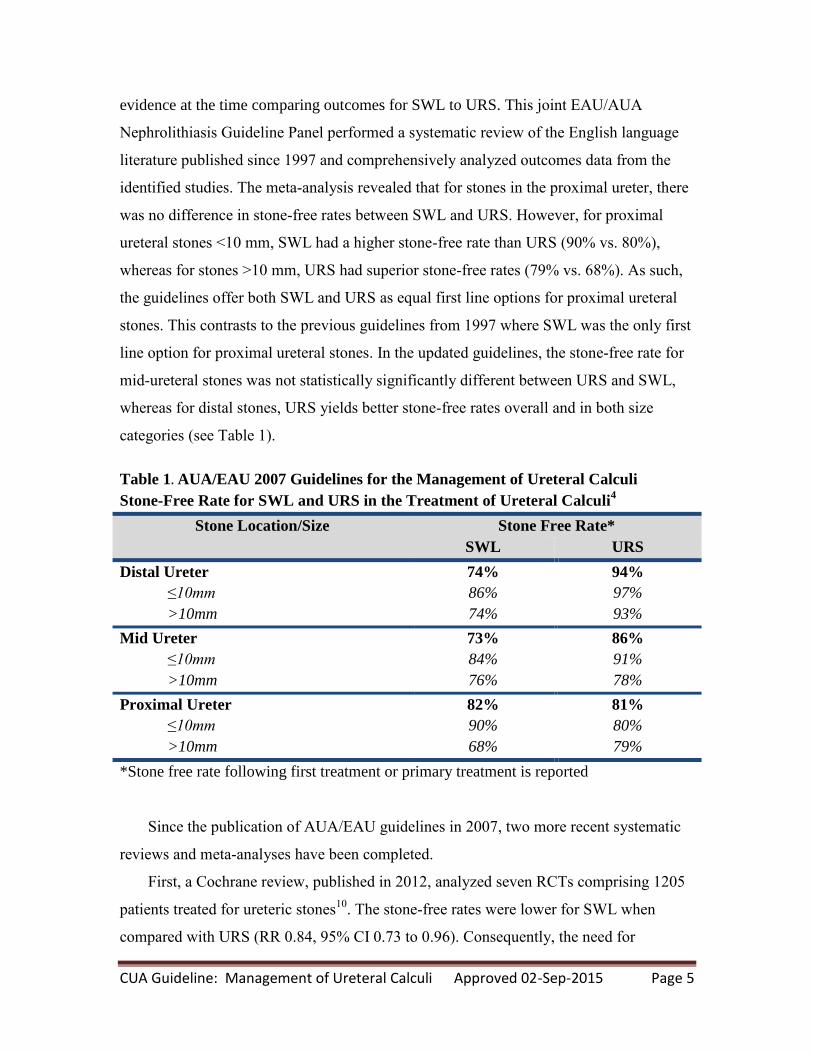
CUA Guideline: Management of Ureteral Calculi Approved 02-Sep-2015 Page 5
evidence at the time comparing outcomes for SWL to URS. This joint EAU/AUA
Nephrolithiasis Guideline Panel performed a systematic review of the English language
literature published since 1997 and comprehensively analyzed outcomes data from the
identified studies. The meta-analysis revealed that for stones in the proximal ureter, there
was no difference in stone-free rates between SWL and URS. However, for proximal
ureteral stones <10 mm, SWL had a higher stone-free rate than URS (90% vs. 80%),
whereas for stones >10 mm, URS had superior stone-free rates (79% vs. 68%). As such,
the guidelines offer both SWL and URS as equal first line options for proximal ureteral
stones. This contrasts to the previous guidelines from 1997 where SWL was the only first
line option for proximal ureteral stones. In the updated guidelines, the stone-free rate for
mid-ureteral stones was not statistically significantly different between URS and SWL,
whereas for distal stones, URS yields better stone-free rates overall and in both size
categories (see Table 1).
Table 1. AUA/EAU 2007 Guidelines for the Management of Ureteral Calculi
Stone-Free Rate for SWL and URS in the Treatment of Ureteral Calculi4
Stone Location/Size Stone Free Rate*
SWL URS
Distal Ureter
≤10mm
>10mm
74%
86%
74%
94%
97%
93%
Mid Ureter
≤10mm
>10mm
73%
84%
76%
86%
91%
78%
Proximal Ureter
≤10mm
>10mm
82%
90%
68%
81%
80%
79%
*Stone free rate following first treatment or primary treatment is reported
Since the publication of AUA/EAU guidelines in 2007, two more recent systematic
reviews and meta-analyses have been completed.
First, a Cochrane review, published in 2012, analyzed seven RCTs comprising 1205
patients treated for ureteric stones10
. The stone-free rates were lower for SWL when
compared with URS (RR 0.84, 95% CI 0.73 to 0.96). Consequently, the need for
CUA Guideline: Management of Ureteral Calculi Approved 02-Sep-2015 Page 6
retreatment, defined as a subsequent intervention for the stone using the same therapeutic
technique as the initial treatment, was higher in SWL patients (RR 6.18, 95% CI 3.68 to
10.38)10
. Procedure-related complications were lower for SWL compared to URS patients
(RR 0.54, 95% CI 0.33 to 0.88)10
.
Second, a meta-analysis by Matlaga et al. (2012)11
stratified their analysis of SWL
versus URS based on stone location in the distal versus proximal ureter. They also
specifically compared URS both to SWL with the Dornier HM3 and to other lithotripters.
Considering no HM3 lithotripters are in use in Canada, we present only the results of
URS versus other lithotripters. For distal ureteral stones, analysis of six studies revealed a
55% greater probability of being stone-free at first follow-up with semi-rigid URS
compared with SWL (RR 1.55). However, with time, SWL approached the stone-free rate
of semi-rigid URS due to re-treatment of SWL cases. Accordingly, patients treated with
semi-rigid URS were less likely to require re-treatment than patients treated with SWL
for distal ureteral stones (RR 0.14). A similar number of complications occurred in both
the semi-rigid URS and SWL groups (pooled RR 1.28, 95% CI 0.94–1.81). For proximal
ureteral stones, there was a greater probability of being stone-free with semi-rigid URS vs.
SWL (RR 1.15) and a lower risk for retreatment (RR 0.08, 95% CI 0.02– 0.32). However
in this meta-analysis, no studies directly compared semi-rigid URS to SWL for proximal
ureteral stones and as such this is a comparison of outcomes across different studies,
which is methodologically undesirable.
Unfortunately, the marked heterogeneity of the existing evidence in terms of study
design, stone location, types of ureteroscope, intracorporeal lithotripsy devices, policy
variations in stenting after ureteroscopy, and time to follow-up limit the conclusion that
can be drawn from both of the aforementioned meta-analyses. Accordingly, it is difficult
to provide a definitive recommendation for use in clinical decision-making.
Ultimately, the size and location of stone(s), the urologist’s expertise and the
availability and access to resources and appropriate technologies remain the principal
criteria to inform treatment choice for the management of ureteric stones. Large, multi-
centre, well designed RCTs and high quality reporting are lacking in the medical
literature.
CUA Guideline: Management of Ureteral Calculi Approved 02-Sep-2015 Page 7
Recommendation: Both SWL and URS are safe and efficacious treatment options
for ureteral stones. Based on the available evidence, patients who undergo URS have
a higher likelihood of achieving stone-free status, especially for distal stones, at the
expense of a greater risk of complications. Patients should be offered both options
when suitable and available, and educated on the benefits and risk of each
treatment modality. (Level of Evidence 2a, Grade B)
Other Factors Affecting SWL Treatment Success
Beyond location in the ureter, other stone-related factors including composition,
density of the stone, and skin-to-stone distance on CT may influence the treating
physician and patient’s discussion regarding the choice to proceed with either SWL or
URS.
1. Composition
The majority of stones, composed of calcium oxalate, will fragment well with SWL
treatment. There exist certain stone compositions, such as cystine, calcium oxalate
monohydrate and brushite, that are more resistant to SWL and may be better served by
ureteroscopic management12
. Moreover, in the case of cystine and calcium oxalate
monohydrate, the fragments created by SWL may be large which can result in poor
clearance. Uric acid stones, while fragile in the face of SWL, present a challenge with
respect to localization for SWL treatment. Either the use of ultrasound or pyelography
(IVP or retrograde) is required to target the stone. In addition, follow-up cannot be
completed in the conventional fashion with plain radiography, and requires the use of
either ultrasound or, more often, CT scanning to ensure the patient is successfully treated.
In many instances the exact stone composition will not be known prior to treatment,
or in the case of recurrent stone formers, it may have changed over time13
. Non-contrast
CT scans using dual energy can distinguish some types of stones in vivo. Uric acid
stones can be differentiated from calcium stones; however, there is significant overlap in
the attenuation of calcium-based stones which makes determining the exact composition
difficult. Recent publications on dual energy CT scanning support that different calcium
CUA Guideline: Management of Ureteral Calculi Approved 02-Sep-2015 Page 8
stone compositions can be determined, however this modality is not readily available in
clinical practice14, 15
.
2. Stone Density
As a surrogate for composition, several authors have postulated that the
fragmentation of stones with SWL could be predicted based on the measurement of stone
density on CT expressed in Hounsfield units (HU). A linear relationship exists between
increased stone density and poor stone fragmentation with a threshold of 1000 HU, above
which stones are less likely to be successfully fragmented with SWL16, 17
. Two
prospective studies reinforced these findings with respect to stone densities greater than
1000 HU and 970 HU18, 19
.
When measuring HU it is best to maximally magnify the image on the stone, use
bone windows and draw an ellipse within the stone.
3. Skin-to-Stone Distance (SSD)
In addition to providing information on stone size and density, CT scans can also
allow for measurement of SSD. Several groups have reported reduced SWL success in
patients with a greater SSD and high stone density. A large retrospective Canadian series
including renal and ureteral stones demonstrated on multivariate analysis that a SSD of
greater than 11 cm (OR = 0.49, CI: 0.31-0.78) and density greater than 900 HU (OR
=0.49, CI: 0.32-0.75) were significant predictors of SWL failure20
. A second large
retrospective review of 1282 SWL treatments also demonstrated on multivariate analysis
that SSD greater than 10 cm was associated with lower stone-free rates21
.
Recommendation: Stone composition, stone density and skin-to-stone distance
should be used when possible to counsel patients regarding the success of SWL
treatment for patients presenting with ureteral stones. Known cystine, calcium
oxalate monohydrate, and brushite stones are likely best treated with URS. Patients
with ureteral stones with a density greater than 1000 HU or SSD greater than 10 cm
are more likely to fail SWL and may be better served with URS. (Level of Evidence
2b, Grade B)
CUA Guideline: Management of Ureteral Calculi Approved 02-Sep-2015 Page 9
Optimizing Treatment Outcomes
Shockwave Lithotripsy (SWL)
Despite the advances in ureteroscopes, holmium laser, and endoscopic
instrumentation, SWL remains a first-line treatment modality for ureteral calculi. SWL
outcomes can be directly influenced by case selection, surgeon technique, and modifiable
parameters to enhance safety and maximize successful outcomes. Most of the data for
SWL outcomes is derived from patients with renal (rather than ureteric) calculi, but these
findings should be generalizable to ureteric stones, particularly for those in the upper
ureter, where renal parenchyma is included in the blast path of the shock wave energy.
1. Coupling
Coupling of the SWL generator head to the patient in an air-tight manner, with
minimization of gas and air bubbles in the coupling media, is critical to maximizing
energy delivery to the stone. Failure to recognize breaks in coupling can lead to failure
of stone fragmentation. Changes in lithotripter design have led to a move away from
water bath coupling (as was seen with the original HM3 design) to the use of a smaller
coupling interface. Coupling can be influenced by the type of SWL machine, type of gel
used at the patient-generator interface (a greater volume of lower-viscosity gel being
preferable), method of application of that gel (best to apply to shock head first), and
patient factors (patient movement during treatment, lifting of the back off the generator
leading to “decoupling” and introducing air bubbles into the coupling interface)22-26
.
Recommendation: SWL operators should ensure proper patient coupling to reduce
air bubbles in the SWL blast path, particularly near the centre of the blast path.
Patients should receive adequate anesthesia and analgesia to prevent patient
movement and “decoupling” during treatment. (Level of Evidence 4, Grade C)
2. Targeting
Proper stone targeting is vital for SWL success. Whether fluoroscopic or ultrasound
targeting is superior is an ongoing debate, and varies with urologist expertise, SWL
CUA Guideline: Management of Ureteral Calculi Approved 02-Sep-2015 Page 10
machine type, and stone composition27
. Real-time, in-line imaging is generally
considered superior, however in-line (or coaxial) ultrasound imaging is not available with
all units. Respiratory excursion hinders targeting by reducing the time that the stone is
within the SWL focal zone, even with ideal targeting. Shock wave triggering based on
respiration has been abandoned because of increased treatment time, but compression
belts reduce renal movement with respiration. Targeting should be confirmed at regular
intervals throughout treatment28
. Greater use of fluoroscopy time can lead to improved
outcomes29, 30
.
Recommendation: SWL targeting (whether fluoroscopic or ultrasonic) should occur
at regular intervals throughout the treatment. Compression belts may help reduce
renal (and ureteric) excursion with treatment. (Level of Evidence 4, Grade C)
3. Dose escalation/pause
SWL energy should be maximized during treatment in order to maximize stone
comminution. This is particularly true for mid and distal ureteral stones, where the renal
parenchyma is not included in the blast path and thus the risk of renal injury is negligible.
However, particularly for upper ureteric stones, SWL energy should be increased
gradually, rather than beginning at maximum energy. This allows for better patient
accommodation to the sensation of treatment (when treatment is performed under
intravenous sedation). This also reduces renal injury by inducing renal vasoconstriction,
which is protective in reducing the rate of renal hematomas31-35
. An alternative strategy
is to pre-treat the kidney with a series of low energy shocks and then pause treatment for
a short period of time before resuming at higher energy levels31
. Of note, if fragmentation
is seen at lower energies it is not necessary to increase the energy any further.
Recommendation: Patients with upper ureteric stones should initially receive low-
energy shocks, with gradual voltage escalation up to maximum energy. (Level of
Evidence 1b and 4, Grade C)
4. Number of Treatments
CUA Guideline: Management of Ureteral Calculi Approved 02-Sep-2015 Page 11
Not all SWL treatments of ureteric stones will be successful and render the ureter
stone free. When treatment is unsuccessful, a decision must be made whether to retreat
with SWL or to switch to endourologic treatment (retrograde or antegrade URS). This
decision-making is often influenced by the degree of fragmentation with the initial SWL
session, and by patient factors (patient preferences, impending travel, importance of
being rendered stone free quickly, dislike of ureteric stent and prior patient treatment
experience). In general, if SWL fails it can be repeated, but the incremental benefit of
more than two SWL treatments for the same ureteric stone is small36, 37
. Accordingly,
after two unsuccessful SWL treatments, consideration should be given to alternative
treatment with ureteroscopy. The optimal time interval between SWL treatments is
unclear, but can be as short as within two days for mid and distal ureteric stones.
Recommendation: If SWL is unsuccessful, the urologist may elect to treat the stone
a second time with SWL in consultation with the patient. More than two SWL
treatments to the same ureteric stone have little incremental benefit and URS should
be considered. (Level of Evidence 4, Grade C)
5. Treatment Rate
A number of randomized trials have indicated that reducing shock wave rate from
120 shocks/min can improve stone fragmentation, particularly for stones larger than 1
cm38-43
. This may also reduce the degree of renal injury, which may be an issue for upper
ureteric stones, but is likely less relevant for mid and distal ureteral stones. Slowing
treatment rate does increase treatment times. The optimal treatment rate is not clear;
however, studies suggest that SWL at 60 to 90 shocks/min leads to better fragmentation
than 120 shocks/min, particularly for larger stones43-45
. Most studies were performed
with renal calculi; however, improved outcomes have been demonstrated for upper
ureteric stones as well39
.
Recommendation: Patients with upper ureteric stones >1cm, or stones that have
failed prior treatment, should be treated with a SWL rate of less than 120
shocks/min. (Level of Evidence 1b, Grade A)
CUA Guideline: Management of Ureteral Calculi Approved 02-Sep-2015 Page 12
6. Alpha blockers
The benefits of oral alpha-blockers to enhance the spontaneous passage of ureteral
stones, known as medical expulsive therapy, is well established and recommended in the
AUA/EAU guidelines on the management of ureteral stones4. Given this, several authors
have studied the effect of alpha blockers administered as an adjunct to SWL in order to
improve stone-free rates. Their work has been summarized in two meta-analyses with
similar findings. The first in 2009 combined the results of four studies, which randomized
patients to receive medical expulsive therapy versus placebo or standard of care post
treatment. Two of the four studies used tamsulosin, while one used a calcium channel
blocker and the other an herbal agent (phyllanthus niruri)46-50
. The use of medical
expulsive therapy was associated with a 17% increase in SWL success rates with a
number needed to treat of six. A more recent meta-analysis focused solely on the use of
alpha blockers post SWL and had similar findings. They identified seven trials that met
the inclusion criteria and found that the use of alpha-blockers, which was tamsulosin in
all seven studies, improved SWL success by 16%47, 51-57
. The authors also reported
reduced time to fragment passage, reduced pain, and less analgesic use.
Alpha blockers are well tolerated, inexpensive, and familiar to urologists. The use of
alpha blocker therapy as an adjunct to SWL should result in increased fragment passage
and a reduction in the need for repeat SWL or more invasive treatments, such as URS.
Additional benefits with respect to less pain and reduced need for analgesic use may also
be realized.
Recommendation: Alpha blockers, in particular tamsulosin, should be prescribed
to patients after SWL for ureteral stones to improve treatment success rates. (Level
of Evidence 1a, Grade A)
7. Number of Shocks
The optimal number of shocks to administer has not been definitively established. In
principle, urologists must balance treatment efficacy with adverse effects (particularly
renal damage). For mid to distal ureteric stones, where the renal parenchyma is not
CUA Guideline: Management of Ureteral Calculi Approved 02-Sep-2015 Page 13
affected by SWL energy, treatment can safely be carried out up to 4000 or more shocks37
.
However, the incremental benefit of treating ureteric stones beyond 4000 shocks has not
been established. For upper ureteral stones the range is from 2000 to 3500 shocks37
. In
general, urologists should follow their lithotripter manufacturer’s recommendations for
the optimal maximum number of shocks.
Recommendation: An adequate number of shocks should be administered to ensure
adequate treatment of ureteric stones. This number varies based on
recommendations from the specific SWL machine manufacturers, but generally
ranges from 2000 to 4000 shocks for ureteric stones. (Level of Evidence 4-5, Grade
D)
8. Stenting
There is good evidence to show that ureteral stenting is not necessary in SWL58
and
does not improve the success rate or passage of fragments.59
In fact, having a stent may
impede the passage of fragments following SWL. A trial consisting of patients with
ureteral stones between 4 to 10 mm undergoing SWL were randomized to a stent or no
stent.60
The stone free rate was much lower in stented patients (68.6%) than non-stented
patients (83.7%, p=0.026). Consequently, stented patients required significantly more
adjuvant procedures to render them stone free compared to non-stented patients. On
multivariate analysis, the authors noted that the location of the stone, size of stone and
presence of a stent were the three factors that significantly affected stone free rate.
Further supporting this, Argyropoulos and Tolley looked at SWL of ureteral stones with a
mean size of 8.5 mm in diameter and found that the stone free rate in stented patients was
significantly lower (71%) compared to those who were not stented (93%).61
In addition, based on the available evidence, stents do not appear to decrease the risk
of steinstrasse or infection following SWL62-64
.
However, consideration should still be given to placing a stent prior to SWL in
patients with a solitary kidney.
CUA Guideline: Management of Ureteral Calculi Approved 02-Sep-2015 Page 14
Recommendation: Ureteral stents do not improve the stone-free rates in SWL and
actually impede the passage of fragments resulting in lower stone free rates. (Level
of Evidence 1a, Grade A) They should be used prior to SWL to treat obstruction,
acute kidney injury, intolerable pain, sepsis, and in those with a solitary kidney. If
inserted for sepsis, a course of antibiotics should be given prior to SWL and the
patient should not be exhibiting signs of sepsis at the time of treatment. (Level of
Evidence 5, Grade D) Stents do not decrease the risk of steinstrasse or infection
following SWL. (Level of Evidence 1a, Grade A)
Ureteroscopy
1. Lithotrite (Laser vs. Electrohydraulic vs. Pneumatic)
Common methods of intracorporeal ureteroscopic lithotripsy include pneumatic,
eletrohydraulic, and Holmium:YAG (Ho:YAG) laser. Treatment of ureteral stones with
Ho:YAG lithotripsy is superior (p<0.05) to pneumatic lithotripsy when comparing stone
free rate (95-98.6% vs. 80-86%) 65-68
, operative time (15 20min vs. 25-33mins)68, 69
, and
need for auxiliary treatment, such as SWL or repeat URS (2-2.5% vs. 14% - 17.5%)65, 66
.
When compared with electrohydraulic lithotripsy, Ho:YAG laser lithotripsy was
demonstrated to have superior stone free rates for stones larger than 15mm (100% vs.
67%) and faster operative time for stones less than 15mm (72 vs. 102min)70
. Available
studies are not sufficiently powered to conclude if a significant difference exists in
complication rates such a ureteral perforation, stone migration, or delayed ureteric
stricture.
Recommendation: Holmium:YAG laser lithotripsy offers superior stone
fragmentation, stone free rates and minimizes the need for auxiliary procedures. It
should be considered the method of choice for intracorporeal lithotripsy of ureteral
stones. (Level of Evidence 2b, Grade B)
2. Ureteral Access Sheath
The use of a ureteral access sheath (UAS) has traditionally been advocated at the time
of flexible URS for renal stones for several reasons, including: 1) facilitating flexible
CUA Guideline: Management of Ureteral Calculi Approved 02-Sep-2015 Page 15
URS by allowing easy multiple entry and re-entry to the upper urinary tract and renal
collecting system; 2) decrease in intrarenal pressure, which could potentially diminish
kidney injury71, 72
; 3) improved irrigation flow thus optimizing vision71
; and 4) the
potential to improve stone-free rates by allowing passive egress or active retrieval of
fragments. However, the impact of UAS on stone-free rates is unclear, as the evidence is
very limited73-75
.
The effect of UAS use on the ureter is also unclear. It has been demonstrated in
animal models that the UAS can induce transient ureteral ischemia and promote an acute
inflammatory response in the ureter76
. Furthermore, a recent prospective study has
questioned the safety of the UAS, demonstrating the potential for ureteral wall injury in
46.5% of patients77
. However, no randomized trials exist comparing the incidence of
ureteral stricture with and without a UAS. Retrospective studies show no increased risk
of stricture formation.
Ultimately and unfortunately, much of the current data with respect to UAS use are
limited by the fact they come from non-randomized studies that are largely retrospective
in nature with short follow-up. This limits the recommendations that can be made.
Recommendation: Further studies are needed to confirm safety, define cost-
effectiveness, and determine the clinical impact of the reduction of ureteral and
intrarenal pressures during sheath deployment before any definitive
recommendation on the use of the UAS can be made. Nevertheless, the UAS remains
a highly useful tool in the armamentarium of the urologist during flexible URS.
(Level of Evidence 4, Grade C)
3. Ureteroscope Size
The outer tip diameter of ureteroscopes typically vary between 4.5 and 8.5Fr. for
semi-rigid ureteroscopes and 6.75 to 8.7Fr. for flexible ureteroscopes. Recently, digital
flexible ureteroscopes have come into more widespread use providing excellent
visualization, but some have a larger diameter (8.7Fr. tip with 9.9Fr. shaft), which can
make insertion into the non-dilated ureter more difficult. Furthermore, the durability of
flexible digital ureteroscopes compared to fibreoptic ureteroscopes remains to be seen.
CUA Guideline: Management of Ureteral Calculi Approved 02-Sep-2015 Page 16
Semi-rigid Ureteroscopes (SR)
Semi-rigid (SR) ureteroscopes represent the mainstay for treating most ureteric
stones in light of the superior optics, excellent irrigant flow and size of the working
channel. Stone free rates are equivalent between small SR ureteroscopes (4.5-7.5Fr tip)
and larger SR ureteroscopes (8.5-10Fr tip)78, 79
. Larger SR ureteroscopes may require
more ureteric dilation and increase minor complications of mucosal abrasion or
postoperative hematuria79
.
Flexible Ureteroscopes
Flexible fibreoptic ureteroscopes range in size from 7.4 to 9.0Fr in diameter and have
a progressively higher rate of insertion failure into the undilated ureteral orifice,
increasing from 0.9% to 37%, respectively, dependent on size80
. With the introduction
of flexible digital ureteroscopes, with typical tip diameters of 8.4 to 8.7Fr broadening to a
9.9Fr shaft, there is an increasing need for ureteral orifice dilation and access sheath use81
.
In addition, the greater diameter may result in a higher likelihood of being unable to
access and treat stones in the proximal ureter or renal pelvis/calyces. However, based on
the available studies comparing larger digital flexible ureteroscopes with fibreoptic
flexible ureteroscopes, the larger diameter did not affect stone free rates and the digital
ureteroscope resulted in shorter operative times82, 83
.
Recommendations: Within the range of commercially available semi-rigid and
flexible ureteroscopes, the available evidence suggests stone-free rates and
complication rates are similar. When available, use of smaller ureteroscopes may
lessen the need for ureteral dilation and slightly reduce minor postoperative
complications such as hematuria. (Level of Evidence 4, Grade C)
4. Stenting
Pre-stenting prior to ureteroscopy
Ureteral stents are often placed at the completion of a ureteroscopic case. However,
this discussion addresses “pre-stenting” of a patient prior to a planned URS. Ureteral
CUA Guideline: Management of Ureteral Calculi Approved 02-Sep-2015 Page 17
stents are known to provide drainage, as well as passively dilate the ureter. Accordingly,
pre-stenting prior to URS has been shown to ease the insertion of ureteroscopes and UAS.
Pre-stenting did not affect stone-free rates in patients with stones less than 1 cm, but in
patients with stones greater than 1 cm, the stone-free rate was significantly better (95.8%)
after a single treatment84
. The same study also performed a cost analysis and in those
patients with a stone greater than 1 cm, there was a decrease in overall costs to
successfully treat the patient from $27,806 (not pre-stented) to $17,706 (pre-stented). Pre-
stented patients required less adjuvant procedures to render them stone free, which
accounted for the cost savings. Another study found that pre-stented patients had
significantly higher stone free rates for ureteral stones 5 mm or greater (99% vs. 90%,
P=0.048)85
. There was no difference in stone free rates for ureteral stones smaller than 5
mm and no difference in complication rates for stones of any size.
Pre-stenting can also be effective in situations where the ureter is narrow and
insertion of a UAS or ureteroscope is difficult. In these instances, placing a ureteral stent
to help passively dilate the ureter and re-attempting URS at a later date is highly
recommended to improve the rate of ureteral access and reduce the rate of complications.
Balloon dilation and URS has been shown to be safe and effective in one sitting, but it
must be recognized that if this does not work, stenting and performing URS after passive
dilation is necessary86
.
In a tertiary referral centre examining 119 consecutive patients, the rate of failure to
access leading to a ureteral stent with delayed URS was 8%.87
A study of 41 patients with
this scenario showed that 71% underwent secondary URS with ease while 12 patients
(29%) had continued resistance88
. Of the 12 patients with continued resistance, nine
underwent URS in the secondary setting and two of these patients subsequently
developed a ureteral stricture. Overall, in this series of patients who underwent stenting
for initial resistance in passing a ureteroscope, 98% had successful subsequent URS.
Care should be taken to avoid continuing despite resistance as this can lead to subsequent
ureteral stricture, particularly with one-step dilation using a UAS77
. Pre-stenting before
the use of a UAS decreased the rate of complication by sevenfold in this particular
study77
. Preoperative discussion in consenting patients should include the potential of
failed access, placement of a ureteral stent and delayed URS at another date.
CUA Guideline: Management of Ureteral Calculi Approved 02-Sep-2015 Page 18
Stenting post ureteroscopy
Stenting post ureteroscopy is not always necessary. The first description and
randomized trial of stent versus stentless URS were both performed in Canada. Hosking
et al. was the first to describe stentless URS in 93 patients undergoing URS for distal
ureteral stones without any further intervention or requirement for subsequent stents or
nephrostomy tubes89
. Denstedt et al performed the first prospective randomized trial of
stent versus no stent following URS90
. At one week following URS, patients without a
stent had significantly less flank pain, abdominal pain, and dysuria compared to stented
patients. There were no complications in those who did not have a stent. A subsequent
meta-analysis showed an absolute lower risk of complications in those patients who were
stented, however, this became insignificant on multivariate-analysis91
. Many other studies
have shown no complications and less symptoms in those who did not receive a ureteral
stent92
. Stenting after uncomplicated URS did not alter the stone free rate, complications,
urinary tract infection, unplanned medical visits, or fever.
Even when the ureteric orifice has been balloon-dilated to 18Fr, stenting has not been
shown to be beneficial. A randomized trial of 144 stented and 142 non-stented patients
following rigid URS in which all patients were dilated to 18 Fr. was undertaken93
. These
researchers found there were no differences in complications or strictures; however, they
did find that stented patients had more irritative voiding symptoms (dysuria and urgency).
Another study undertaking SR URS using pneumatic lithotripsy showed that stented
patients actually did better than non-stented patients94
. Non-stented patients were more
than twice as likely to visit the emergency department following discharge and they also
took a longer time to discharge from the hospital on the day of surgery. Narcotic use was
also significantly higher in the non-stented group in the first five days after surgery. This
study shows evidence that stented patients were actually more comfortable and required
less medical attention and narcotics following URS.
If bilateral URS is performed, depending on the situation, consideration should be
given to stenting at least one side, to prevent the possibility of bilateral ureteric
obstruction post-operatively.
CUA Guideline: Management of Ureteral Calculi Approved 02-Sep-2015 Page 19
Stenting following use of a UAS
If a UAS is used during URS, there is good data to support placing a ureteral stent in
those cases. In a retrospective study of 102 patients, where 51 had no stent following
UAS use and the other half were stented, stented patients were less likely to have
unscheduled emergency visits and had lower pain scores compared to their non-stented
counterparts95
. This was also corroborated by Canadian data where patients who did not
have a ureteral stent were more likely to have an emergency room visit in the
postoperative period (37% vs. 14%, p=0.04)96
.
Duration of stenting:
There is no prescribed indwelling time to leave a ureteral stent. The literature is scant
regarding this issue. One study retrospectively analyzed 125 patients and found that
stents that remained in less than 14 days had less adverse events, such as fever and
lumbago, and they advocated less than two weeks of stenting following uncomplicated
URS97
.
Recommendation: Stenting following uncomplicated URS is still a controversial
topic and there is evidence to support both sides. There is good evidence that
ureteral stents should be left in place following use of a ureteral access sheath with
URS. Stenting does not affect stone-free rates or long-term complications such as
strictures, but may result in less emergency room visits and narcotic use in the
postoperative period. Stenting prior to URS is helpful to improve stone-free rates in
stones greater than 1 cm. Stenting prior to URS also facilitates access to the ureter
due to passive dilation. (Level of Evidence 2a-2b, Grade B)
Special Considerations
1. Pregnancy
No level 1 evidence exists regarding the treatment of ureteral stones during pregnancy.
Retrospective case series provide some guidance on how to manage this situation. The
first diagnostic step in suspected nephrolithiasis during pregnancy should be ultrasound
due to the lack of radiation; however, ultra low dose computed tomography (CT) or
CUA Guideline: Management of Ureteral Calculi Approved 02-Sep-2015 Page 20
magnetic resonance imaging (MRI) are good alternatives with very little or no radiation98,
99. A special protocol involving magnetic resonance urography (MRU) involves a half
fourier single-shot turbo spin-echo (HASTE) which is better at imaging ureteral stones
than other MRU protocols100
.
The majority of ureteral stones will pass spontaneously and the first option in
management is conservative therapy including hydration and analgesia101
. In a recent
study, conservative management was successful in 67% of patients, who had
symptomatic obstructing ureteral stones with an average stone size of 8.8 mm102
.
Immediate causes for intervention are the same as those in non-pregnant situations (signs
of sepsis, renal failure, and unrelenting pain, etc.), but also include induction of
premature labour (contractions, fetal distress etc.) in the pregnant patient103
. The most
immediate method of intervention is nephrostomy tube or ureteral stent insertion.
Failing conservative management, ureteroscopic treatment of stones using laser
lithotripsy with either flexible or SR URS is feasible and safe104
. In fact, if ultrasound
imaging is non-diagnostic and low-dose CT or MRI is unavailable, URS can also be used
for both diagnostic and therapeutic purposes102
. A number of studies have demonstrated
that URS is a viable technique for the treatment of stones in pregnancy102, 105, 106
. Post-
operative stenting following URS in this situation is recommended in an attempt to
reduce post-operative complications102
. Ideally ureteroscopic treatment should be
performed in the second trimester, as teratogenic effects and risks of anaesthesia are
higher in the first trimester103
.
With regards to intraoperative imaging, if URS or ureteral stent insertion is
undertaken, then a lead apron or shield should be put between the x-ray fluoroscopy
source and the fetus to shield it from radiation107
. These authors describe inverting the
fluoroscopy C-arm so that the energy source is above the supine patient and placing two
thyroid collars on the anterior portion of the patient’s abdomen to shield the fetus. This
can also be achieved by placing the lead shield or apron underneath the patient if the C-
arm energy source comes from below the table; this method also reduces radiation and
scatter to operating theatre personnel. Alternatively, URS or ureteral stent insertion can
be performed under ultrasound guidance alone, avoiding radiation exposure.
CUA Guideline: Management of Ureteral Calculi Approved 02-Sep-2015 Page 21
Pregnancy is a contraindication to SWL and although there have been reports of the
inadvertent treatment of pregnant patients with SWL, with no adverse sequelae to the
fetus108
, it should be avoided. Similarly, percutaneous nephrolithotomy (PCNL), if
necessary, should be delayed until after birth as the procedure requires prolonged
anaesthesia and radiation exposure.
Recommendation: First line diagnostic testing for stones in pregnancy is ultrasound,
but low-dose CT or MRI can also be used. In some instances, URS can also be
diagnostic, as well as therapeutic. Obstructing ureteral stones are typically
managed conservatively in the absence of fever, leukocytosis or positive urine
culture. In those patients presenting with signs of sepsis, antibiotics and urinary
decompression via a nephrostomy tube or ureteral stent are of primary importance.
Definitive therapy should be delayed until the infection is treated. URS and laser
lithotripsy is safe in pregnancy, however SWL and PCNL are contraindicated in
pregnancy. (Level of Evidence Level 4, Grade C)
2. Anti-coagulation
There is a paucity of literature regarding surgical management of stone patients with
coagulopathies or on anticoagulation therapy. SWL, laparoscopic, percutaneous and open
surgeries are contraindicated in these patients109, 110
. This is because there is 20 to 40 fold
increased risk of peri-renal hematomas and hemorrhagic complications in patients with
uncorrected coagulopathies undergoing SWL when compared with patients with a normal
bleeding profile111, 112
. Therefore, in consultation with a haematologist or a cardiologist,
bleeding coagulopathies need to be corrected and anticoagulation therapy appropriately
withheld peri-operatively113
. In addition, patients with increased risk of thromboembolic
disease could be managed by bridging with subcutaneous low molecular weight heparin
while oral anticoagulation is held114
. In a retrospective series of 27 anti-coagulated
patients who underwent PCNL with bridging, the stone-free rate was 93% while 7%
developed significant bleeding and 4% had thromboembolic complications115
. The only
prospective SWL study of patients on anti-platelet agents is that of Zanetti and
CUA Guideline: Management of Ureteral Calculi Approved 02-Sep-2015 Page 22
colleagues116
. In this level 2b study, 23 patients were stratified to being at low-risk or at
high-risk of thromboembolic events. Low-risk patients had their antiplatelet agents
withheld for eight days prior to SWL, whereas high-risk patients received unfractionated
heparin 5000 units thrice daily while anti-platelet agents were held. In both groups anti-
platelet agents were re-started within 10-14 days of withdrawal and patients were
followed with abdominal ultrasound and serial hemoglobin/hematocrit measurements.
There were no hematomas or thromboembolic events in either group116
.
Recent advances in manufacturing small-calibre ureteroscopes and introduction of
Ho:YAG laser energy in lithotripsy have made it possible for patients with
coagulopathies to safely undergo URS and laser lithotripsy while anticoagulated110, 117-119
.
However, this is associated with lower stone-free rates and increased risk of post-
operative gross hematuria necessitating admission and bladder irrigation111, 120
. Therefore,
risks and benefits of withholding anti-coagulation or proceeding with URS while anti-
coagulated should be discussed with the patient and his/her cardiologist or hematologist.
Recommendations: SWL and PCNL are contraindicated in patients with
uncorrected coagulopathies. When possible, coagulopathies should be corrected
after consulting with a cardiologist and/or hematologist. However, when risks of
withholding anti-coagulants outweigh the benefits, proceeding with URS and laser
lithotripsy, while anti-coagulated, is an acceptable option. (Level of Evidence 2b,
Grade B)
3. Urinary Diversion
Urinary diversions can be classified anatomically into abdominal (such as ileal
conduits and catheterizable pouches), urethral (such as orthotopic neobladder) and
uretero-sigmoidostomy, with ileal conduit representing the majority (84%) of cases121, 122
.
Patients with urinary diversions are at high risk of stone formation in light of
numerous risk factors including metabolic abnormalities (i.e., metabolic acidosis,
hypocitraturia, hyperoxaluria and hypercalciuria), recurrent infections with urease-
splitting organisms (e.g., Proteus), prolonged urinary stasis, prolonged exposure of urine
CUA Guideline: Management of Ureteral Calculi Approved 02-Sep-2015 Page 23
to non-absorbable materials (e.g., staples), anatomical changes following diversion, and
reflux of mucous into the upper tract 123
.
The reported incidence of upper tract calculi in patients with urinary diversion is 1-
11% depending on the type of diversion, uretero-intestinal anastmosis and the follow up
period. The most common stone types are magnesium ammonium phosphate (struvite)
and calcium phosphate stones124
.
The established anatomical changes in these patients necessitate accurate preoperative
assessment by CT scan to determine whether there are overlying bowel loops, especially
if percutaneous access is contemplated125
. Ultrasound-guided access is recommended in
these cases to avoid intervening bowel loops126
.
Dealing with the stones in these patients represents a challenge to the urologist. Many
factors need to be considered when choosing a certain approach. These factors include:
stone size, location, patient performance status, availability of advanced SWL machines
and flexible ureteroscopes with Ho:YAG laser lithotripsy, and finally, surgeon experience
in dealing with these structural changes in the urinary tract127
. In addition, PCNL in these
patients is associated with higher rates of postoperative fever or sepsis (8% vs. 0%,
p<0.05) and higher rates of second-look nephroscopy (36% vs. 16%, p<0.05) compared
to those with normal anatomy126
.
Minimally invasive modalities, such as SWL, URS and PCNL, that produce the best
stone-free rates should be utilized in these patients since these would avoid open surgical
management and are associated with lower morbidity, early convalescence and shorter
hospital stay124
. When performing antegrade URS for management of ureteral stones, a
ureteral access sheath can be placed in an antegrade fashion to improve irrigation and
facilitate access with a flexible ureteroscope122
.
Close follow up in these patients is mandatory because the risk of re-growth and
recurrence is as high as 63% at 5 year follow-up128
.There are many important factors that
may prevent or at least prolong the period before recurrence. These include increased
fluid intake, timed voiding or frequent clean intermittent catheterization, and frequent
irrigation of reservoirs129
. Metabolic work-up for identification of the correctable risk
factors, medical management of metabolic consequences of diversion, and long-term
CUA Guideline: Management of Ureteral Calculi Approved 02-Sep-2015 Page 24
antibiotic prophylaxis against recurrent infections are also important to reduce
recurrence125
.
Also of importance, the urologist should consider the possibility of anastomotic
stricture or recurrent malignancy when stones are lodged near the uretero-enteric
anastomosis.
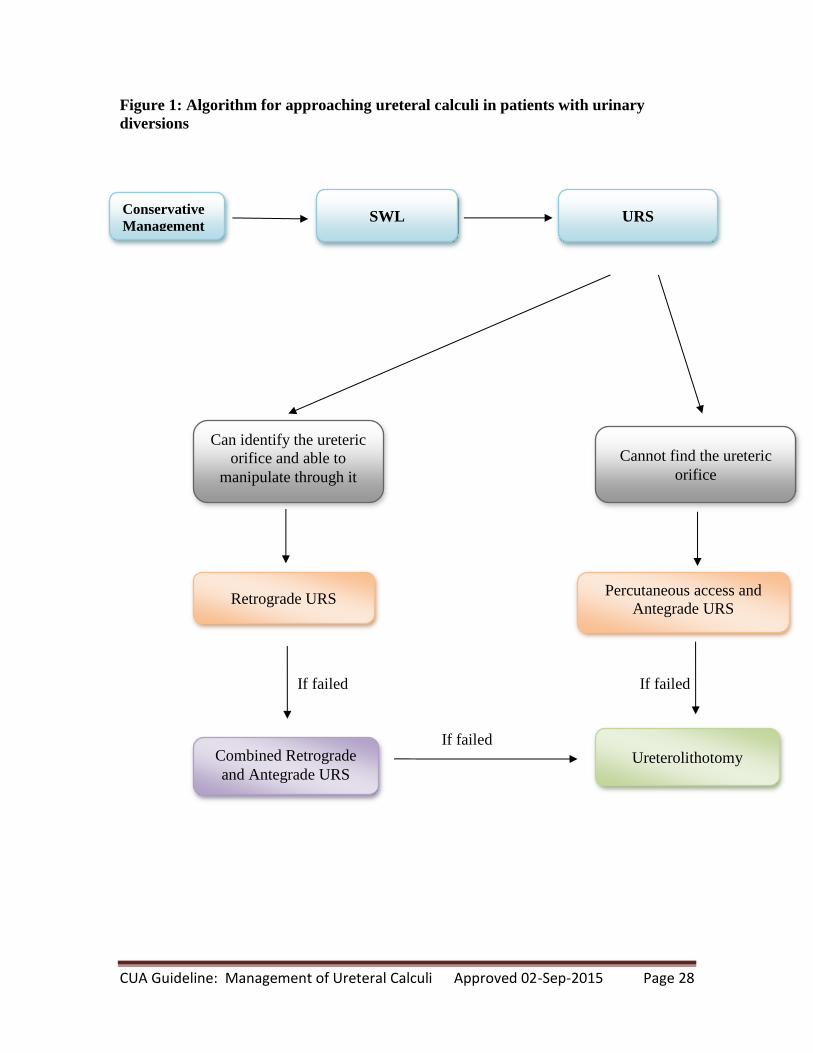
Recommendations: Ureteral calculi in patients with urinary diversions are
challenging. While small, non-obstructive, asymptomatic stones could be managed
conservatively, SWL could be attempted for obstructive stones (Level of Evidence 4,
Grade C). If SWL fails, then retrograde URS with laser lithotripsy could be
attempted if the ureteral orifice can be accessed (Figure 1). However, the most
effective modality for clearing large obstructive ureteral stones is percutaneously
through antegrade URS. (Level of Evidence 4, Grade C) It should be noted, however,
that percutaneous renal surgery in patients with urinary diversion is associated with
higher rates of post-operative sepsis and higher rates of second-look nephroscopy.
(Level of Evidence 2b, Grade B) When percutaneous procedures fail,
ureterolithotomy is the last option in these patients. (Level of Evidence 4, Grade C)
4. Antegrade URS and Ureterolithotomy
In select circumstances, a percutaneous antegrade approach may be necessary instead
of a retrograde endoscopic approach. As discussed above, prior urinary diversion
represents one of these situations. Antegrade URS can also be considered a treatment
option in the following situations: 1) in select cases with a large, impacted proximal
ureteral stone(s); 2) when performed in conjunction with renal stone removal; 3) in select
cases following failure of a retrograde ureteroscopic attempt for a large, impacted
proximal ureteral stone130
; and 4) when the ureteral stone is in a transplant kidney131
.
For large (>15 mm), impacted proximal ureteral stones the stone-free rate with
antegrade URS has been shown to range from 98.5-100% with low risk for
complications130, 132-136
.
CUA Guideline: Management of Ureteral Calculi Approved 02-Sep-2015 Page 25
Laparoscopic, robotic or open ureterolithotomy may be considered when
ureteroscopic and percutaneous procedures have failed or concomitant surgery is
required137
.
Recommendations: Percutaneous antegrade URS should be considered in the
treatment of stones in patients with urinary diversion and select large, impacted
proximal ureteral stones, especially when prior retrograde URS has failed. (Level of
Evidence 4, Grade C) Ureterolithotomy is a salvage option when endoscopic
procedures have failed. (Level of Evidence 2b, Grade B)
5. Uric Acid Stones
Uric acid (UA) urolithiasis is a multi-factorial disease. Persistently low 24-hour
urinary pH (≤5.5) is regarded as the most important factor in formation of UA stones. UA
stones constitute about 10% of urolithiasis in the general population, but this percentage
increases up to 34 % of stones in patients with metabolic syndrome and up to 52.2% of
stones in patients with gout138, 139
. UA stones are typically radiolucent on plain
radiographs and of low attenuation values (<500HU) on non-contrast CT scans of the
abdomen140
. Recently, dual-energy CT scanning has been found to be superior to the
conventional “single-energy” non-contrast CT scanning in differentiating non-uric acid
stones from UA stones141
. However, dual-energy CT scans are not widely available.
Laboratory evaluation should include serum creatinine, potassium, uric acid, and
when renal colic subsides, twenty-four hour urine collection should be obtained and
checked for urine volume, urinary pH and uric acid excretion142
.
When there are no signs of impending renal failure or sepsis, treatment of UA calculi
depends primarily on increased water intake, reduction in the consumption of non-diary
animal protein (low purine diet), and urinary alkalinisation using alkalinising agents such
as potassium citrate or sodium bicarbonate leading to stone dissolution138, 142-144
.When
these agents are given to patients after ESWL or PCNL, they facilitate the dissolution of
residual stones <4mm and prevent stone re-growth and recurrence145
. In addition, the
combination of alpha blockers (such as tamsulosin) and potassium citrate have been
associated with significantly higher stone-free rates when compared with either therapy
CUA Guideline: Management of Ureteral Calculi Approved 02-Sep-2015 Page 26
alone or with placebo in patients with distal ureteric UA stones (84.4%, 68.8%, 58.7%,
and 26.1%, respectively)146
.
Although UA stones are easy to fragment by SWL, they are difficult to localize due
to their radiolucency. Contrast media injection either through the intravenous route,
retrograde ureteropyelography through a ureteral catheter, or antegrade administration
through a nephrostomy tube can be used to localize the radiolucent UA stones. The latest
generation of lithotripters come equipped with ultrasonographic targeting, which could be
used to localize UA stones. URS and laser lithotripsy is also very effective at treating UA
stones in the ureter.
Recommendations: UA stones should be suspected when the stone is radiolucent on
plain radiograph, the density is <500 HU on non-contrast CT scan, and it is
associated with acidic urine (pH ≤5.5). (Level of Evidence 2b, Grade B)
Alkalinization with potassium/sodium citrate or sodium bicarbonate can be used in
conjunction with medical expulsive therapy, such as tamsulosin, or endourologic
procedures, such as SWL, URS or PCNL, to increase stone-free rates of UA stones.
(Level of Evidence 1b, Grade A)
6. Infected Obstructing Ureteral Stones
The basic tenet of treating any infected area or abscess is drainage followed by
antibiotics. An obstructing ureteral stone in the setting of infection constitutes a
requirement for urgent urologic treatment. Drainage of the obstructed renal unit is
paramount and can be performed either by insertion of a ureteral stent or a percutaneous
nephrostomy tube. It is generally agreed that definitive treatment of the obstructing stone
should not be undertaken until the system has been drained, adequate antibiotics have
been administered and the infection treated. In the only prospective randomized trial,
patients presenting with a fever >38oC, leukocytosis, and obstructing stone smaller than
15 mm were randomized to receive a ureteral stent or a nephrostomy tube insertion via
interventional radiology147
. There was no difference in time to defervescence, hospital
stay, resolution of obstruction, or overall clinical improvement. Others have also found
no difference between decompression via nephrostomy or ureteral stent148-150
.
CUA Guideline: Management of Ureteral Calculi Approved 02-Sep-2015 Page 27
The method of decompression should be tailored to each centre and its available
resources. Placement of a nephrostomy tube is not on the list of core competencies for all
radiologists and therefore may not be available at all centres. One survey found that only
44% of hospitals in the UK are capable of nephrostomy tube insertion151
. Ureteral
stenting should be more widely available, but does require cystoscopy or an operating
theatre setting, fluoroscopy or ultrasound, and trained staff. Broad spectrum antibiotics
should be started early upon diagnosis; when starting within 1 hour of diagnosis, the
survival rate is greater than 80% and each hour delay results in decreased survival (8%
per hour)152
.
Recommendation: Obstructing ureteral stones resulting in urosepsis and infection
require emergent drainage. The two methods of decompression, ureteral stenting or
nephrostomy tube placement, are equivalent in outcomes and the method chosen
will depend on availability of resources at each particular hospital. It is important to
start broad-spectrum antibiotics early. Definitive stone treatment should be delayed
until decompression and adequate antibiotics have been administered to treat the
infection. (Level of Evidence Level 2b, Grade B)
CUA Guideline: Management of Ureteral Calculi Approved 02-Sep-2015 Page 28
Figure 1: Algorithm for approaching ureteral calculi in patients with urinary
diversions
If failed If failed
If failed
URS SWL Conservative
Management
Can identify the ureteric
orifice and able to
manipulate through it
Cannot find the ureteric
orifice
Retrograde URS
Combined Retrograde
and Antegrade URS Ureterolithotomy
Percutaneous access and
Antegrade URS
CUA Guideline: Management of Ureteral Calculi Approved 02-Sep-2015 Page 29
References
1. Matlaga, B. R., Lingeman, J.E.: Surgical Management of Upper Urinary Tract Calculi. In: Campbell-Walsh Urology, 10th ed. Edited by A. J. Wein, Kavoussi, L. R., Novick, A. C., Partin, A. W., and Peters, C. A. Philadelphia: Elsevier Saunders, vol. 2, pp. 1357-1410, 2012
2. Miller, O. F., Kane, C. J.: Time to stone passage for observed ureteral calculi: a guide for patient education. The Journal of urology, 162: 688, 1999
3. Hubner, W. A., Irby, P., Stoller, M. L.: Natural history and current concepts for the treatment of small ureteral calculi. Eur Urol, 24: 172, 1993
4. Preminger, G. M., Tiselius, H. G., Assimos, D. G. et al.: 2007 Guideline for the management of ureteral calculi. European urology, 52: 1610, 2007
5. Coll, D. M., Varanelli, M. J., Smith, R. C.: Relationship of spontaneous passage of ureteral calculi to stone size and location as revealed by unenhanced helical CT. AJR Am J Roentgenol, 178: 101, 2002
6. Berkovitz, N., Simanovsky, N., Katz, R. et al.: Coronal reconstruction of unenhanced abdominal CT for correct ureteral stone size classification. Eur Radiol, 20: 1047, 2010
7. Ray, A. A., Ghiculete, D., Pace, K. T. et al.: Limitations to ultrasound in the detection and measurement of urinary tract calculi. Urology, 76: 295, 2010
8. Campschroer, T., Zhu, Y., Duijvesz, D. et al.: Alpha-blockers as medical expulsive therapy for ureteral stones. Cochrane Database Syst Rev, 4: CD008509, 2014
9. Pickard, R., Starr, K., MacLennan, G. et al.: Medical expulsive therapy in adults with ureteric colic: a multicentre, randomised, placebo-controlled trial. Lancet, 2015
10. Aboumarzouk, O. M., Kata, S. G., Keeley, F. X. et al.: Extracorporeal shock wave lithotripsy (ESWL) versus ureteroscopic management for ureteric calculi. Cochrane database of systematic reviews, 5: CD006029, 2012
11. Matlaga, B. R., Jansen, J. P., Meckley, L. M. et al.: Treatment of ureteral and renal stones: a systematic review and meta-analysis of randomized, controlled trials. J Urol, 188: 130, 2012
12. Saw, K. C., Lingeman, J. E.: Lesson 20: Management of Calyceal Stones, p. 154-59, 1999
13. Lee, T. T., Elkoushy, M. A., Andonian, S.: Are stone analysis results different with repeated sampling? Can Urol Assoc J, 8: E317, 2014
14. Matlaga, B. R., Kawamoto, S., Fishman, E.: Dual source computed tomography: a novel technique to determine stone composition. Urology, 72: 1164, 2008
15. Boll, D. T., Patil, N. A., Paulson, E. K. et al.: Renal stone assessment with dual-energy multidetector CT and advanced postprocessing techniques: improved characterization of renal stone composition--pilot study. Radiology, 250: 813, 2009
16. Gupta, N. P., Ansari, M. S., Kesarvani, P. et al.: Role of computed tomography with no contrast medium enhancement in predicting the outcome of
CUA Guideline: Management of Ureteral Calculi Approved 02-Sep-2015 Page 30
extracorporeal shock wave lithotripsy for urinary calculi. BJU Int, 95: 1285, 2005
17. Joseph, P., Mandal, A. K., Singh, S. K. et al.: Computerized tomography attenuation value of renal calculus: can it predict successful fragmentation of the calculus by extracorporeal shock wave lithotripsy? A preliminary study. J Urol, 167: 1968, 2002
18. El-Nahas, A. R., El-Assmy, A. M., Mansour, O. et al.: A prospective multivariate analysis of factors predicting stone disintegration by extracorporeal shock wave lithotripsy: the value of high-resolution noncontrast computed tomography. Eur Urol, 51: 1688, 2007
19. Ouzaid, I., Al-qahtani, S., Dominique, S. et al.: A 970 Hounsfield units (HU) threshold of kidney stone density on non-contrast computed tomography (NCCT) improves patients' selection for extracorporeal shockwave lithotripsy (ESWL): evidence from a prospective study. BJU Int, 110: E438, 2012
20. Wiesenthal, J. D., Ghiculete, D., Ray, A. A. et al.: A clinical nomogram to predict the successful shock wave lithotripsy of renal and ureteral calculi. The Journal of urology, 186: 556, 2011
21. Patel, T., Kozakowski, K., Hruby, G. et al.: Skin to stone distance is an independent predictor of stone-free status following shockwave lithotripsy. Journal of endourology / Endourological Society, 23: 1383, 2009
22. Pishchalnikov, Y. A., Neucks, J. S., VonDerHaar, R. J. et al.: Air pockets trapped during routine coupling in dry head lithotripsy can significantly decrease the delivery of shock wave energy. J Urol, 176: 2706, 2006
23. Jain, A., Shah, T. K.: Effect of air bubbles in the coupling medium on efficacy of extracorporeal shock wave lithotripsy. Eur Urol, 51: 1680, 2007
24. Neucks, J. S., Pishchalnikov, Y. A., Zancanaro, A. J. et al.: Improved acoustic coupling for shock wave lithotripsy. Urol Res, 36: 61, 2008
25. Bergsdorf, T., Chaussy, C., Thuroff, S.: Energy coupling in extracorporeal shock wave lithotripsy—the impact of coupling quality on disintegration efficacy. J Endourol, 22: A161, 2008
26. Bohris, C.: Quality of coupling in ESWL significantly affects the disintegration capacity—how to achieve good coupling with ultra-sound gel. In: Therapeutic energy applications in urology II: standards and recent developments. Edited by C. Chaussy, G. Haupt, D. Jocham et al. Stuttgart, Germany: Thieme, pp. 61-4, 2010
27. Bohris, C., Bayer, T., Gumpinger, R.: Ultrasound monitoring of kidney stone extracorporeal shockwave lithotripsy with an external transducer: does fatty tissue cause image distortions that affect stone comminution? J Endourol, 24: 81, 2010
28. Cleveland, R. O., Anglade, R., Babayan, R. K.: Effect of stone motion on in vitro comminution efficiency of Storz Modulith SLX. J Endourol, 18: 629, 2004
29. Logarakis, N. F., Jewett, M. A., Luymes, J. et al.: Variation in clinical outcome following shock wave lithotripsy. The Journal of urology, 163: 721, 2000
CUA Guideline: Management of Ureteral Calculi Approved 02-Sep-2015 Page 31
30. Hartung, A., Schwarze, W.: LithoSpace by AST GmbH. In: Therapeutic energy applications in urology II: standards and recent developments. Edited by C. Chaussy, G. Haupt, D. Jocham et al. Stuttgart, Germany Thieme, pp. 53-6, 2010
31. McAteer, J. A., Evan, A. P., Williams, J. C., Jr. et al.: Treatment protocols to reduce renal injury during shock wave lithotripsy. Curr Opin Urol, 19: 192, 2009
32. Lambert, E. H., Walsh, R., Moreno, M. W. et al.: Effect of escalating versus fixed voltage treatment on stone comminution and renal injury during extracorporeal shock wave lithotripsy: a prospective randomized trial. J Urol, 183: 580, 2010
33. Willis, L. R., Evan, A. P., Connors, B. A. et al.: Prevention of lithotripsy-induced renal injury by pretreating kidneys with low-energy shock waves. J Am Soc Nephrol, 17: 663, 2006
34. Weizer, A. Z., Zhong, P., Preminger, G. M.: New concepts in shock wave lithotripsy. Urol Clin North Am, 34: 375, 2007
35. Seemann, O., Rassweiler, J., Chvapil, M. et al.: The effect of single shock waves on the vascular system of artificially perfused rabbit kidneys. J Stone Dis, 5: 172, 1993
36. Pace, K. T., Weir, M. J., Tariq, N. et al.: Low success rate of repeat shock wave lithotripsy for ureteral stones after failed initial treatment. The Journal of urology, 164: 1905, 2000
37. Rassweiler, J. J., Knoll, T., Kohrmann, K. U. et al.: Shock wave technology and application: an update. Eur Urol, 59: 784, 2011
38. Pace, K. T., Ghiculete, D., Harju, M. et al.: Shock wave lithotripsy at 60 or 120 shocks per minute: a randomized, double-blind trial. The Journal of urology, 174: 595, 2005
39. Honey, R. J., Schuler, T. D., Ghiculete, D. et al.: A randomized, double-blind trial to compare shock wave frequencies of 60 and 120 shocks per minute for upper ureteral stones. The Journal of urology, 182: 1418, 2009
40. Davenport, K., Minervini, A., Keoghane, S. et al.: Does rate matter? The results of a randomized controlled trial of 60 versus 120 shocks per minute for shock wave lithotripsy of renal calculi. J Urol, 176: 2055, 2006
41. Madbouly, K., El-Tiraifi, A. M., Seida, M. et al.: Slow versus fast shock wave lithotripsy rate for urolithiasis: a prospective randomized study. J Urol, 173: 127, 2005
42. Yilmaz, E., Batislam, E., Basar, M. et al.: Optimal frequency in extracorporeal shock wave lithotripsy: prospective randomized study. Urology, 66: 1160, 2005
43. Li, K., Lin, T., Zhang, C. et al.: Optimal frequency of shock wave lithotripsy in urolithiasis treatment: a systematic review and meta-analysis of randomized controlled trials. J Urol, 190: 1260, 2013
44. Kato, Y., Yamaguchi, S., Hori, J. et al.: Improvement of stone comminution by slow delivery rate of shock waves in extracorporeal lithotripsy. Int J Urol, 13: 1461, 2006
CUA Guideline: Management of Ureteral Calculi Approved 02-Sep-2015 Page 32
45. Chacko, J., Moore, M., Sankey, N. et al.: Does a slower treatment rate impact the efficacy of extracorporeal shock wave lithotripsy for solitary kidney or ureteral stones? J Urol, 175: 1370, 2006
46. Schuler, T. D., Shahani, R., Honey, R. J. et al.: Medical expulsive therapy as an adjunct to improve shockwave lithotripsy outcomes: a systematic review and meta-analysis. J Endourol, 23: 387, 2009
47. Bhagat, S. K., Chacko, N. K., Kekre, N. S. et al.: Is there a role for tamsulosin in shock wave lithotripsy for renal and ureteral calculi? J Urol, 177: 2185, 2007
48. Gravina, G. L., Costa, A. M., Ronchi, P. et al.: Tamsulosin treatment increases clinical success rate of single extracorporeal shock wave lithotripsy of renal stones. Urology, 66: 24, 2005
49. Porpiglia, F., Destefanis, P., Fiori, C. et al.: Role of adjunctive medical therapy with nifedipine and deflazacort after extracorporeal shock wave lithotripsy of ureteral stones. Urology, 59: 835, 2002
50. Micali, S., Sighinolfi, M. C., Celia, A. et al.: Can Phyllanthus niruri affect the efficacy of extracorporeal shock wave lithotripsy for renal stones? A randomized, prospective, long-term study. J Urol, 176: 1020, 2006
51. Zhu, Y., Duijvesz, D., Rovers, M. M. et al.: alpha-Blockers to assist stone clearance after extracorporeal shock wave lithotripsy: a meta-analysis. BJU Int, 106: 256, 2010
52. Gravas, S., Tzortzis, V., Karatzas, A. et al.: The use of tamsulozin as adjunctive treatment after ESWL in patients with distal ureteral stone: do we really need it? Results from a randomised study. Urol Res, 35: 231, 2007
53. Kupeli, B., Irkilata, L., Gurocak, S. et al.: Does tamsulosin enhance lower ureteral stone clearance with or without shock wave lithotripsy? Urology, 64: 1111, 2004
54. Micali, S., Grande, M., Sighinolfi, M. C. et al.: Efficacy of expulsive therapy using nifedipine or tamsulosin, both associated with ketoprofene, after shock wave lithotripsy of ureteral stones. Urol Res, 35: 133, 2007
55. Naja, V., Agarwal, M. M., Mandal, A. K. et al.: Tamsulosin facilitates earlier clearance of stone fragments and reduces pain after shockwave lithotripsy for renal calculi: results from an open-label randomized study. Urology, 72: 1006, 2008
56. Kobayashi, M., Naya, Y., Kino, M. et al.: Low dose tamsulosin for stone expulsion after extracorporeal shock wave lithotripsy: efficacy in Japanese male patients with ureteral stone. Int J Urol, 15: 495, 2008
57. Wang, H. J., Liu, K., Ji, Z. G. et al.: [Application of Alpha1-adrenergic antagonist with extracorporeal shock wave lithotripsy for lower ureteral stone]. Zhongguo Yi Xue Ke Xue Yuan Xue Bao, 30: 506, 2008
58. Musa, A. A.: Use of double-J stents prior to extracorporeal shock wave lithotripsy is not beneficial: results of a prospective randomized study. Int Urol Nephrol, 40: 19, 2008
59. Pettenati, C., El Fegoun, A. B., Hupertan, V. et al.: Double J stent reduces the efficacy of extracorporeal shock wave lithotripsy in the treatment of lumbar ureteral stones. Cent European J Urol, 66: 309, 2013
CUA Guideline: Management of Ureteral Calculi Approved 02-Sep-2015 Page 33
60. Sfoungaristos, S., Polimeros, N., Kavouras, A. et al.: Stenting or not prior to extracorporeal shockwave lithotripsy for ureteral stones? Results of a prospective randomized study. Int Urol Nephrol, 44: 731, 2012
61. Argyropoulos, A. N., Tolley, D. A.: Ureteric stents compromise stone clearance after shockwave lithotripsy for ureteric stones: results of a matched-pair analysis. BJU Int, 103: 76, 2009
62. Lucio, J., 2nd, Korkes, F., Lopes-Neto, A. C. et al.: Steinstrasse predictive factors and outcomes after extracorporeal shockwave lithotripsy. Int Braz J Urol, 37: 477, 2011
63. Mustafa, M., Ali-El-Dein, B.: Stenting in extracorporeal shockwave lithotripsy; may enhance the passage of the fragments! J Pak Med Assoc, 59: 141, 2009
64. Duvdevani, M., Lorber, G., Gofrit, O. N. et al.: Fever after shockwave lithotripsy--risk factors and indications for prophylactic antimicrobial treatment. J Endourol, 24: 277, 2010
65. Bapat, S. S., Pai, K. V., Purnapatre, S. S. et al.: Comparison of holmium laser and pneumatic lithotripsy in managing upper-ureteral stones. J Endourol, 21: 1425, 2007
66. Binbay, M., Tepeler, A., Singh, A. et al.: Evaluation of pneumatic versus holmium:YAG laser lithotripsy for impacted ureteral stones. Int Urol Nephrol, 43: 989, 2011
67. Maghsoudi, R., Amjadi, M., Norizadeh, D. et al.: Treatment of ureteral stones: A prospective randomized controlled trial on comparison of Ho:YAG laser and pneumatic lithotripsy. Indian J Urol, 24: 352, 2008
68. Demir, A., Karadag, M. A., Cecen, K. et al.: Pneumatic versus laser ureteroscopic lithotripsy: a comparison of initial outcomes and cost. Int Urol Nephrol, 46: 2087, 2014
69. Atar, M., Bodakci, M. N., Sancaktutar, A. A. et al.: Comparison of pneumatic and laser lithotripsy in the treatment of pediatric ureteral stones. J Pediatr Urol, 9: 308, 2013
70. Teichman, J. M., Rao, R. D., Rogenes, V. J. et al.: Ureteroscopic management of ureteral calculi: electrohydraulic versus holmium:YAG lithotripsy. J Urol, 158: 1357, 1997
71. Rehman, J., Monga, M., Landman, J. et al.: Characterization of intrapelvic pressure during ureteropyeloscopy with ureteral access sheaths. Urology, 61: 713, 2003
72. Auge, B. K., Pietrow, P. K., Lallas, C. D. et al.: Ureteral access sheath provides protection against elevated renal pressures during routine flexible ureteroscopic stone manipulation. J Endourol, 18: 33, 2004
73. Kourambas, J., Byrne, R. R., Preminger, G. M.: Does a ureteral access sheath facilitate ureteroscopy? J Urol, 165: 789, 2001
74. L'Esperance J, O., Ekeruo, W. O., Scales, C. D., Jr. et al.: Effect of ureteral access sheath on stone-free rates in patients undergoing ureteroscopic management of renal calculi. Urology, 66: 252, 2005
75. Berquet, G., Prunel, P., Verhoest, G. et al.: The use of a ureteral access sheath does not improve stone-free rate after ureteroscopy for upper urinary tract stones. World J Urol, 32: 229, 2014
CUA Guideline: Management of Ureteral Calculi Approved 02-Sep-2015 Page 34
76. Lallas, C. D., Auge, B. K., Raj, G. V. et al.: Laser Doppler flowmetric determination of ureteral blood flow after ureteral access sheath placement. J Endourol, 16: 583, 2002
77. Traxer, O., Thomas, A.: Prospective evaluation and classification of ureteral wall injuries resulting from insertion of a ureteral access sheath during retrograde intrarenal surgery. J Urol, 189: 580, 2013
78. Yaycioglu, O., Guvel, S., Kilinc, F. et al.: Results with 7.5F versus 10F rigid ureteroscopes in treatment of ureteral calculi. Urology, 64: 643, 2004
79. Atis, G., Arikan, O., Gurbuz, C. et al.: Comparison of different ureteroscope sizes in treating ureteral calculi in adult patients. Urology, 82: 1231, 2013
80. Hudson, R. G., Conlin, M. J., Bagley, D. H.: Ureteric access with flexible ureteroscopes: effect of the size of the ureteroscope. BJU Int, 95: 1043, 2005
81. Bach, C., Nesar, S., Kumar, P. et al.: The new digital flexible ureteroscopes: 'size does matter'--increased ureteric access sheath use! Urol Int, 89: 408, 2012
82. Somani, B. K., Al-Qahtani, S. M., de Medina, S. D. et al.: Outcomes of flexible ureterorenoscopy and laser fragmentation for renal stones: comparison between digital and conventional ureteroscope. Urology, 82: 1017, 2013
83. Binbay, M., Yuruk, E., Akman, T. et al.: Is there a difference in outcomes between digital and fiberoptic flexible ureterorenoscopy procedures? J Endourol, 24: 1929, 2010
84. Chu, L., Farris, C. A., Corcoran, A. T. et al.: Preoperative stent placement decreases cost of ureteroscopy. Urology, 78: 309, 2011
85. Netsch, C., Knipper, S., Bach, T. et al.: Impact of preoperative ureteral stenting on stone-free rates of ureteroscopy for nephroureterolithiasis: a matched-paired analysis of 286 patients. Urology, 80: 1214, 2012
86. Bourdoumis, A., Tanabalan, C., Goyal, A. et al.: The difficult ureter: stent and come back or balloon dilate and proceed with ureteroscopy? What does the evidence say? Urology, 83: 1, 2014
87. Cetti, R. J., Biers, S., Keoghane, S. R.: The difficult ureter: what is the incidence of pre-stenting? Ann R Coll Surg Engl, 93: 31, 2011
88. Ambani, S. N., Faerber, G. J., Roberts, W. W. et al.: Ureteral stents for impassable ureteroscopy. J Endourol, 27: 549, 2013
89. Hosking, D. H., McColm, S. E., Smith, W. E.: Is stenting following ureteroscopy for removal of distal ureteral calculi necessary? J Urol, 161: 48, 1999
90. Denstedt, J. D., Wollin, T. A., Sofer, M. et al.: A prospective randomized controlled trial comparing nonstented versus stented ureteroscopic lithotripsy. J Urol, 165: 1419, 2001
91. Makarov, D. V., Trock, B. J., Allaf, M. E. et al.: The effect of ureteral stent placement on post-ureteroscopy complications: a meta-analysis. Urology, 71: 796, 2008
92. Pengfei, S., Yutao, L., Jie, Y. et al.: The results of ureteral stenting after ureteroscopic lithotripsy for ureteral calculi: a systematic review and meta-analysis. J Urol, 186: 1904, 2011
93. Baseskioglu, B., Sofikerim, M., Demirtas, A. et al.: Is ureteral stenting really necessary after ureteroscopic lithotripsy with balloon dilatation of ureteral
CUA Guideline: Management of Ureteral Calculi Approved 02-Sep-2015 Page 35
orifice? A multi-institutional randomized controlled study. World J Urol, 29: 731, 2011
94. Cevik, I., Dillioglugil, O., Akdas, A. et al.: Is stent placement necessary after uncomplicated ureteroscopy for removal of impacted ureteral stones? J Endourol, 24: 1263, 2010
95. Torricelli, F. C., De, S., Hinck, B. et al.: Flexible ureteroscopy with a ureteral access sheath: when to stent? Urology, 83: 278, 2014
96. Rapoport, D., Perks, A. E., Teichman, J. M.: Ureteral access sheath use and stenting in ureteroscopy: effect on unplanned emergency room visits and cost. J Endourol, 21: 993, 2007
97. Shigemura, K., Yasufuku, T., Yamanaka, K. et al.: How long should double J stent be kept in after ureteroscopic lithotripsy? Urol Res, 40: 373, 2012
98. White, W. M., Johnson, E. B., Zite, N. B. et al.: Predictive value of current imaging modalities for the detection of urolithiasis during pregnancy: a multicenter, longitudinal study. J Urol, 189: 931, 2013
99. Masselli, G., Derme, M., Laghi, F. et al.: Imaging of stone disease in pregnancy. Abdom Imaging, 38: 1409, 2013
100. Mullins, J. K., Semins, M. J., Hyams, E. S. et al.: Half Fourier single-shot turbo spin-echo magnetic resonance urography for the evaluation of suspected renal colic in pregnancy. Urology, 79: 1252, 2012
101. Semins, M. J., Matlaga, B. R.: Management of stone disease in pregnancy. Curr Opin Urol, 20: 174, 2010
102. Isen, K., Hatipoglu, N. K., Dedeoglu, S. et al.: Experience with the diagnosis and management of symptomatic ureteric stones during pregnancy. Urology, 79: 508, 2012
103. Semins, M. J., Matlaga, B. R.: Kidney stones during pregnancy. Nat Rev Urol, 11: 163, 2014
104. Akpinar, H., Tufek, I., Alici, B. et al.: Ureteroscopy and holmium laser lithotripsy in pregnancy: stents must be used postoperatively. J Endourol, 20: 107, 2006
105. Watterson, J. D., Girvan, A. R., Beiko, D. T. et al.: Ureteroscopy and holmium:YAG laser lithotripsy: an emerging definitive management strategy for symptomatic ureteral calculi in pregnancy. Urology, 60: 383, 2002
106. Bozkurt, Y., Penbegul, N., Soylemez, H. et al.: The efficacy and safety of ureteroscopy for ureteral calculi in pregnancy: our experience in 32 patients. Urol Res, 40: 531, 2012
107. Cocuzza, M., Colombo, J. R., Jr., Lopes, R. I. et al.: Use of inverted fluoroscope's C-arm during endoscopic treatment of urinary tract obstruction in pregnancy: a practicable solution to cut radiation. Urology, 75: 1505, 2010
108. Frankenschmidt, A., Sommerkamp, H.: Shock wave lithotripsy during pregnancy: a successful clinical experiment. J Urol, 159: 501, 1998
109. Alsaikhan, B., Andonian, S.: Shock wave lithotripsy in patients requiring anticoagulation or antiplatelet agents. Can Urol Assoc J, 5: 53, 2011
110. Aboumarzouk, O. M., Somani, B. K., Monga, M.: Flexible ureteroscopy and holmium:YAG laser lithotripsy for stone disease in patients with bleeding diathesis: a systematic review of the literature. Int Braz J Urol, 38: 298, 2012
CUA Guideline: Management of Ureteral Calculi Approved 02-Sep-2015 Page 36
111. Klingler, H. C., Kramer, G., Lodde, M. et al.: Stone treatment and coagulopathy. Eur Urol, 43: 75, 2003
112. Razvi, H., Fuller, A., Nott, L. et al.: Risk factors for perinephric hematoma formation after shockwave lithotripsy: a matched case-control analysis. J Endourol, 26: 1478, 2012
113. Tsuboi, T., Fujita, T., Maru, N. et al.: Transurethral ureterolithotripsy and extracorporeal shock wave lithotripsy in patients with idiopathic thrombocytopenic purpura. Hinyokika Kiyo, 54: 17, 2008
114. Kaatz, S., Paje, D.: Update in bridging anticoagulation. J Thromb Thrombolysis, 31: 259, 2011
115. Kefer, J. C., Turna, B., Stein, R. J. et al.: Safety and efficacy of percutaneous nephrostolithotomy in patients on anticoagulant therapy. The Journal of urology, 181: 144, 2009
116. Zanetti, G., Kartalas-Goumas, I., Montanari, E. et al.: Extracorporeal shockwave lithotripsy in patients treated with antithrombotic agents. J Endourol, 15: 237, 2001
117. Kuo, R. L., Aslan, P., Fitzgerald, K. B. et al.: Use of ureteroscopy and holmium:YAG laser in patients with bleeding diatheses. Urology, 52: 609, 1998
118. Watterson, J. D., Girvan, A. R., Cook, A. J. et al.: Safety and efficacy of holmium: YAG laser lithotripsy in patients with bleeding diatheses. The Journal of urology, 168: 442, 2002
119. Turna, B., Stein, R. J., Smaldone, M. C. et al.: Safety and efficacy of flexible ureterorenoscopy and holmium:YAG lithotripsy for intrarenal stones in anticoagulated cases. The Journal of urology, 179: 1415, 2008
120. Elkoushy, M. A., Violette, P. D., Andonian, S.: Ureteroscopy in patients with coagulopathies is associated with lower stone-free rate and increased risk of clinically significant hematuria. Int Braz J Urol, 38: 195, 2012
121. Mottet, N., Castagnola, C., Rischmann, P. et al.: [Quality of life after cystectomy: French national survey conducted by the French Association of Urology (AFU), the French Federation of Stoma Patients (FSF) and the French Association of Enterostomy Patients (AFET) in patients with ileal conduit urinary diversion or orthotopic neobladder]. Prog Urol, 18: 292, 2008
122. Stuurman, R. E., Al-Qahtani, S. M., Cornu, J. N. et al.: Antegrade percutaneous flexible endoscopic approach for the management of urinary diversion-associated complications. J Endourol, 27: 1330, 2013
123. Dhar, N. B., Hernandez, A. V., Reinhardt, K. et al.: Prevalence of nephrolithiasis in patients with ileal bladder substitutes. Urology, 71: 128, 2008
124. El-Nahas, A. R., Shokeir, A. A.: Endourological treatment of nonmalignant upper urinary tract complications after urinary diversion. Urology, 76: 1302, 2010
125. Okhunov, Z., Duty, B., Smith, A. D. et al.: Management of urolithiasis in patients after urinary diversions. BJU Int, 108: 330, 2011
126. Fernandez, A., Foell, K., Nott, L. et al.: Percutaneous nephrolithotripsy in patients with urinary diversions: a case-control comparison of perioperative outcomes. J Endourol, 25: 1615, 2011
CUA Guideline: Management of Ureteral Calculi Approved 02-Sep-2015 Page 37
127. L'Esperance, J. O., Sung, J., Marguet, C. et al.: The surgical management of stones in patients with urinary diversions. Curr Opin Urol, 14: 129, 2004
128. Cohen, T. D., Streem, S. B., Lammert, G.: Long-term incidence and risks for recurrent stones following contemporary management of upper tract calculi in patients with a urinary diversion. J Urol, 155: 62, 1996
129. Hensle, T. W., Bingham, J., Lam, J. et al.: Preventing reservoir calculi after augmentation cystoplasty and continent urinary diversion: the influence of an irrigation protocol. BJU Int, 93: 585, 2004
130. Kumar, V., Ahlawat, R., Banjeree, G. K. et al.: Percutaneous ureterolitholapaxy: the best bet to clear large bulk impacted upper ureteral calculi. Archivos espanoles de urologia, 49: 86, 1996
131. Rhee, B. K., Bretan, P. N., Jr., Stoller, M. L.: Urolithiasis in renal and combined pancreas/renal transplant recipients. The Journal of urology, 161: 1458, 1999
132. Maheshwari, P. N., Oswal, A. T., Andankar, M. et al.: Is antegrade ureteroscopy better than retrograde ureteroscopy for impacted large upper ureteral calculi? Journal of endourology / Endourological Society, 13: 441, 1999
133. Goel, R., Aron, M., Kesarwani, P. K. et al.: Percutaneous antegrade removal of impacted upper-ureteral calculi: still the treatment of choice in developing countries. Journal of endourology / Endourological Society, 19: 54, 2005
134. Karami, H., Arbab, A. H., Hosseini, S. J. et al.: Impacted upper-ureteral calculi >1 cm: blind access and totally tubeless percutaneous antegrade removal or retrograde approach? Journal of endourology / Endourological Society, 20: 616, 2006
135. Topaloglu, H., Karakoyunlu, N., Sari, S. et al.: A comparison of antegrade percutaneous and laparoscopic approaches in the treatment of proximal ureteral stones. Biomed Res Int, 2014: 691946, 2014
136. Zhu, H., Ye, X., Xiao, X. et al.: Retrograde, antegrade, and laparoscopic approaches to the management of large upper ureteral stones after shockwave lithotripsy failure: a four-year retrospective study. J Endourol, 28: 100, 2014
137. Lopes Neto, A. C., Korkes, F., Silva, J. L., 2nd et al.: Prospective randomized study of treatment of large proximal ureteral stones: extracorporeal shock wave lithotripsy versus ureterolithotripsy versus laparoscopy. J Urol, 187: 164, 2012
138. Maalouf, N. M.: Metabolic syndrome and the genesis of uric acid stones. J Ren Nutr, 21: 128, 2011
139. Marchini, G. S., Sarkissian, C., Tian, D. et al.: Gout, stone composition and urinary stone risk: a matched case comparative study. J Urol, 189: 1334, 2013
140. Ciudin, A. L., P.; Diaconu, M.G.: Predicting Urinary Stone Composition – a radiological study made by Urologists. European urology, 12: e436, 2013
141. Wisenbaugh, E. S., Paden, R. G., Silva, A. C. et al.: Dual-energy vs conventional computed tomography in determining stone composition. Urology, 83: 1243, 2014

CUA Guideline: Management of Ureteral Calculi Approved 02-Sep-2015 Page 38
142. Paterson, R., Fernandez, A., Razvi, H. et al.: Evaluation and medical management of the kidney stone patient. Can Urol Assoc J, 4: 375, 2010
143. Pearle, M. S., Goldfarb, D. S., Assimos, D. G. et al.: Medical management of kidney stones: AUA guideline. J Urol, 192: 316, 2014
144. Turk, C. K., T.; Petrik, A.; Sarica, K.; Skolarikos, A.; Straub, M.; Seitz, C.: EAU Guidelines on Urolithiasis. http://www.uroweb.org/gls/pdf/22 Urolithiasis_LR.pdf, vol. 2014, 2014
145. Lojanapiwat, B., Tanthanuch, M., Pripathanont, C. et al.: Alkaline citrate reduces stone recurrence and regrowth after shockwave lithotripsy and percutaneous nephrolithotomy. Int Braz J Urol, 37: 611, 2011
146. El-Gamal, O., El-Bendary, M., Ragab, M. et al.: Role of combined use of potassium citrate and tamsulosin in the management of uric acid distal ureteral calculi. Urol Res, 40: 219, 2012
147. Pearle, M. S., Pierce, H. L., Miller, G. L. et al.: Optimal method of urgent decompression of the collecting system for obstruction and infection due to ureteral calculi. J Urol, 160: 1260, 1998
148. Christoph, F., Weikert, S., Muller, M. et al.: How septic is urosepsis? Clinical course of infected hydronephrosis and therapeutic strategies. World J Urol, 23: 243, 2005
149. Ramsey, S., Robertson, A., Ablett, M. J. et al.: Evidence-based drainage of infected hydronephrosis secondary to ureteric calculi. J Endourol, 24: 185, 2010
150. Yoshimura, K., Utsunomiya, N., Ichioka, K. et al.: Emergency drainage for urosepsis associated with upper urinary tract calculi. J Urol, 173: 458, 2005
151. Lynch, M. F., Anson, K. M., Patel, U.: Current opinion amongst radiologists and urologists in the UK on percutaneous nephrostomy and ureteric stent insertion for acute renal unobstruction: Results of a postal survey. BJU Int, 98: 1143, 2006
152. Kumar, A., Roberts, D., Wood, K. E. et al.: Duration of hypotension before initiation of effective antimicrobial therapy is the critical determinant of survival in human septic shock. Crit Care Med, 34: 1589, 2006