cancer: a review: world's second most feared diagnosis
TRANSCRIPT

RESEARCH GUIDE: RESEARCH SCHOLAR: Dr. V. P. Rasal Miss Charu PundirVICE PRINCIPAL & HOD M.PHARM II YEAR
Department of PharmacologyKLE COLLEGE OF PHARMACY, BELGAUM
OCT. 25TH 2014
CANCER: A REVIEW

Contents• Introduction• Cancer incidence• Pathogenesis• Causes of cancer• Diagnosis• Treatment• Limitations • Cancer models• Cancer research organizations• Recent advances• Future trends• Conclusion• References

INTRODUCTION

Introduction
• Term neoplasia/ tumor means new growth.
• All new growth are not neoplasms as an example-– Embryogenesis– Regeneration and repair– Hyperplasia &– Hormonal stimulation
• Neoplasm or tumor is a mass of tissue formed as a result of abnormal, excessive, uncoordinated, autonomous and purposeless proliferation of cells.

Introduction
• Cancer cells are defined by 2 heritable properties- Reproducing in defiance of normal restraints on cell division. Invading & colonizing territories normally reserved for other cells.
• Clustered neoplastic tumors are benign that can be removed surgically.
• Tumor is considered cancerous only if it is malignant, which acquired the ability to invade surrounding tissue.
• Invasiveness usually implies an ability to break loose, enter bloodstream or lymphatic vessels, & form secondary tumors, called metastases.

Lymphoma
• Lymphoma is the most common blood cancer. • The two main forms of lymphoma-
Hodgkin lymphoma (HL) Non-Hodgkin lymphoma (NHL)
• Lymphoma occurs when lymphocytes, a type of white blood cell, grow abnormally.
• Cancerous lymphocytes can travel to many parts of the body, including the lymph nodes, spleen, bone marrow, blood or other organs, and can accumulate to form tumors.
Introduction

Non-Hodgkin Lymphoma (NHL)
• Non-Hodgkin lymphoma is the most common cancer of the lymphatic system, a part of the immune system. Broadly divided into two major groups:
B-cell lymphomas T-cell lymphomas.
• B-cell lymphomas develop from abnormal B-lymphocytes and account for 85 percent of all NHLs.
• T-cell lymphomas develop from abnormal T-lymphocytes and account for the remaining 15 percent of all NHLs.
• Non-Hodgkin lymphomas may also be classified as-
Indolent (slow-growing) Aggressive (fast-growing).
Introduction

Classification- based on origin
• Arising from epithelial cells: Carcinoma
• Arising from mesenchymal tumors: Sarcomas
• Carcinoma of Melanocytes- Melanoma
• Carcinoma of hepatocytes- Hepatoma
• Malignant tumor of lymphoid tissue- Lymphoma
• Malignant tumor of testes- Seminoma
• Cancer of blood forming cells- Leukaemia
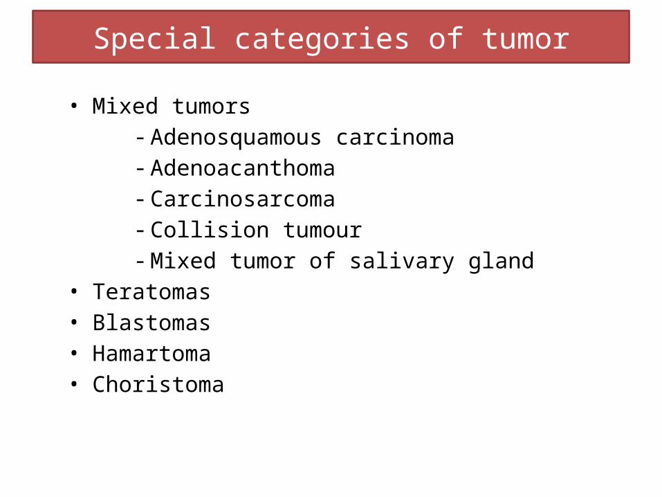
Special categories of tumor
• Mixed tumors- Adenosquamous carcinoma- Adenoacanthoma - Carcinosarcoma- Collision tumour- Mixed tumor of salivary gland
• Teratomas• Blastomas• Hamartoma• Choristoma

CANCER INCIDENCE

Cancer incidence
• 8.2 million people worldwide died from cancer in 2012.• 60% of world’s total new annual cases occur in Africa, Asia
and Central and South America.• 30% of cancers could be prevented.• More than 1 million new cases of cancer are diagnosed every
year in a population of 1·2 billion.• An estimated 600 000—700 000 deaths in India were caused
by cancer in 2012• Around 325,000 people were diagnosed with cancer in 2010
in the UK (approximately 890 people per day). More than 1 in 3 people in the UK will develop some form of cancer during their lifetime. It remains the most common cause of death (29%) followed by circulatory diseases(28%) such as heart disease and strokes.

PATHOGENESIS

Pathogenesis
• Biology of tumor growth- Malignant tumors can be divided into 4 phases:
Malignant change in target cell (Transformation)
Growth of the transformed cells
Local invasion
Distant metastasis

• Rates of growth
It is determined by 3 main factors:
Doubling time of tumor cells
Fraction of tumor cells that are in the replicative pool
Rate at which cells are shed & lost in the growing lesion.
Pathogenesis

Characteristics Benign MalignantDifferentiation/anaplasia Well differentiated;
structure may be typical tissue of origin
Some lack of differentiation with anaplasia; structure is often atypical
Rate of growth Usually progressive & slow; may come to a standstill or regress; mitotic figures are rare & normal
Erratic & may be slow to rapid; mitotic figures may be numerous & abnormal
Local invasion Usually cohesive & expansile well-demarcated masses that do not invade or infiltrate surrounding normal tissues
Local invasive, infiltering the surrounding normal tissues; sometimes may be seemingly cohesive & expansile
Metastasis absent Frequently present; the larger & more undifferentiated the primary, the more likely are metastasis
Pathogenesis

Pathways of spread-Dissemination of cancers may occur through one of three pathways-
• Seeding of body cavities & surfaces
• Lymphatic spread
• Hematogenous spread
Pathogenesis

Pathogenesis
cell cycle
Growth inhibitors(TGF-β, p53, others
Oncogenic viruses(HPV-E7 protein; SV40-T antigen
Growth factors(EGF. TGF-α, HGF, PDGF)

GRADING & STAGING

Grading & cancer staging
• These are the systems to determine the prognosis & choice of treatment after a malignant tumor is detected.
• Grading- cancers may be graded grossly & microscopically, based on histology- degree of anaplasia, & the rate of growth
• Grade I: well-differentiated (less than 25% anaplastic cells).• Grade II: Moderately- differentiated (25-50% anaplastic cells).• Grade III: Moderately- differentiated (50-75% anaplastic cells)• Grade IV: Poorly-differentiated or anaplastic (more than 75%
anaplastic cells).

• Stage 0- in situ cancer
• Stage 1- localised cancer
• Stage 2- local spread stage, usually includes spread to the nearest lymph nodes
• Stage 3- usually indicates more extensive lymph node involvement
• Stage 4- always indicates distant spread.
Grading & cancer staging

CAUSES OF CANCER

Causes of cancerAny cell in the body has the potential to mutate and become a cancer cell. Anything that can cause cancer is termed a “carcinogen”. Some of these include:• Genetics • Tobacco, smoking• Diet deficiencies• Racial & geographical factors• Physical Activity• Radiation carcinogenesis• Non- radiation Physical Carcinogenesis• Biologic Carcinogenesis- Parasites, Fungus, Bacteria• Viral Carcinogenesis (introducing foreign DNA into the cell)

Causes of cancer

DIAGNOSIS

Diagnosis of cancer
1. Histological Methods 2. Cytological Methods
- Exfoliative cytology - Fine needle aspiration cytology (FNAC)
3. Histochemistry & Cytochemistry4. Immunohistochemistry5. Electron Microscopy6. Tumor Markers (Biochemical Assays)- Alpha-foetoprotein
(AFP), Carcino-embryonic antigen (CEA)7. Modern Aids in Tumour Diagnosis- flow cytometry, In situ
hybridisation, Molecular diagnostic techniques, DNA microarray analysis of tumors

GENERAL APPROACHES TO CANCER TREATMENT

General approaches to cancer therapy
ChemotherapyImmunotherapy
Monoclonal antibodies, cytokines, therapeutic vaccines, the bacterium
bacillus Calmet-Guérin, cancer-killing viruses, gene therapy, and
adoptive T-cell transfer.
Targeted Therapies
TransplantationBone Marrow Transplantation and Peripheral Blood Stem Cell Transplantation
Radiation Therapy
Other Treatment MethodsAngiogenesis Inhibitors
Cancer VaccinesCryosurgery
Photodynamic Therapy

General approaches to cancer therapy• Kill or remove malignant cells: - Cytotoxic drugs - Surgery - Irradiation - Targeted cytotoxic agents (e.g. antibody-linked toxins or radioactive agents)• Inactivate components of oncogene signaling pathway: - Inhibitors of growth factor receptors (e.g. receptor tyrosine kinases) - Inhibitors of adapter proteins (e.g. Ras), cytoplasmic kinases, cyclins, cyclin-dependent kinases, etc. - Antisense oligonucleotides - Inhibitors of antiapoptotic factors or stimulators of pro apoptotic factors
-

General approaches to cancer therapy
• Restore function of tumor suppressor genes: - Gene therapy• Employ tissue-specific proliferation inhibitors: - oestrogens, antioestrogens, androgens, antiandrogens,
glucorticoids, gonadotophin releasing hormone analogues.• Inhibit tumor growth, invasion, metastasis: - Inhibitors of angiogenesis - Matrix metalloproteinase inhibitors• Enhance host immune response: - Cytokine-based therapies - Gene therapy- based approaches - Cell- based approaches (e.g. antitumor T cells).• Reverse drug resistance: - Inhibitors of multidrug resistance transport.

General approaches to cancer therapy

CANCER CHEMOTHERAPY

Cancer chemotherapy
Drugs acting directly on cells (cytotoxic drugs)
1. Alkylating agents- forms covalent bonds with DNA & thus impede replication.Mechlorethamine, Busulfan
2. Antimetabolites- Block or subvert one or more of the metabolic pathways involved in DNA synthesis.Methotrexate, 6-Mercaptopurine (6-MP)
3. Vinca alkaloids- specifically affect microtubule functionVincristine, Vinblastine

Cancer chemotherapy
4. Taxanes- Paclitaxel, Docetaxel
5. Epipodophyllotoxin- Etoposide
6. Camptothecin analogues- Topotecan
7. Antibiotics- Prevent mammalian cell division Actinomycin D, Doxorubicin
8. Miscellaneous- Hydroxyurea, Procabazine

Cancer chemotherapy
Drugs altering hormonal milieu- Suppress hormone secretion or antagonize hormone action.
– Glucocorticoids- Prednisolone– Estrogens- Fosfestrol– Selective estrogen receptor modulators- Tamoxifen– Selective estrogen receptor down regulators- Fulvestrant– Aromatase inhibitors- Letrozole– Antiandrogen- Flutamide– 5-α reductase inhibitor- Finasteride– GnRH analogues- Nfarelin– Progestins- Hydroxyprogesterone acetate

LIMITATIONS

Limitations of chemotherapy
Resistance Bone marrow suppression Lymphocytopenia GIT
- Stomatitis- diarrhoea- shedding of mucosa- haemorrhages
Nausea Vomiting
Skin- Alopecia- Dermatitis
Gonads- Oligozoospermia- impotence
Teratogenesis Secondary cancers Hyperuricaemia
- Gout- Urate stones

CANCER MODELS

Cancer In-vitro models
• In- vitro methods-
- Tetrazolium Salt Assay (Microculture Tetrazolium Test or MTT)- Sulphorhodamine B Assay- 3H-thymidine Uptake Assay- Fluorescence- Dye Exclusion Tests- Clonogenic Assays- Cell Counting Assay

MTT assay
• Developed by Mossman 1• Cell proliferation assay• For determination of cell growth rates• Testing drug action• Cytotoxic agents• Screening biologically active compounds.• Most rapid & large scale assay
Cancer In-vitro models

MTT assayPrinciple:
MTT ( 3-4,5-dimethylthiazol-2-yl)-2,5-diphenyltetrazolium(water soluble)
Conversion Insoluble formazen
Further solubilisation, using DMSO
Concentration determined by O.D at 570 nm
Cancer In-vitro models

Cancer In-vitro models
MERITS
• Less time consuming• More cost effective• Small quantities & large
number of compounds can be tested.
• Easy to manage• Can be cultured under
controlled environment (pH, temperature, humidity, O2 , CO2
DEMERITS
• Furnish false positive results & false negative results
• There is need for in-vivo biotransformation
• Pharmacokinetics can not be determined
• Difference lies in solid tumor geometry of in- vivo & in- vitro experiments.

Cancer In-vivo models
• In- vivo methods-
Chemically Induced Tumor Models
Models involving cell line /tumor pieces implantation

Chemically Induced Tumor Models-
- DMBA-induced Mouse Skin Papillomas
- N-methyl, N-nitrosourea (MNU)-induced Rat Mammary Gland Carcinogenesis
- DMBA-induced Rat Mammary Gland Carcinogenesis
- MNU-induced Tracheal Squamous Cell Carcinoma in Hamster.
Cancer In-vivo models

- N,N-Diethylnitrosamine (DEN)-induced Lung Adenocarcinoma in Hamster
- 1,2-Dimethylhydralazine (DMH)-induced Colorectal Adenocarcinoma in Rat & Mouse
- Azoxymethane (AOM)- induced Aberrant Crypt Foci in Rat
- N-Butyl-N-(4-hydroxybutyl)- nitrosamine (OH-BBN)-induced Bladder Carcinoma in Mouse
Cancer In-vivo models

Models involving cell line /tumor pieces implantation-
Specified number of cells of a particular cell line are inoculated into sensitive mouse strain.
Tumors can be developed rapidly as compared to chemical carcinogen-induced tumors
Time saving
Cancer In-vivo models

Methods involving cell line /tumor pieces implantation-
• Hollow-fiber technique
• Use of xenografts
• Nude mouse model
• Transgenic mouse model
• Newborn rat model
Cancer In-vivo models

Merits
• Detect host mediated activity
• Predictive
• Estimate therapeutic ratio
• Supports in-vitro results
Demerits
• Low sensitivity
• Costly
• Time consuming
• Large number of samples are difficult to manage
• Toxicological profile of test drug is needed.
Cancer In-vivo models
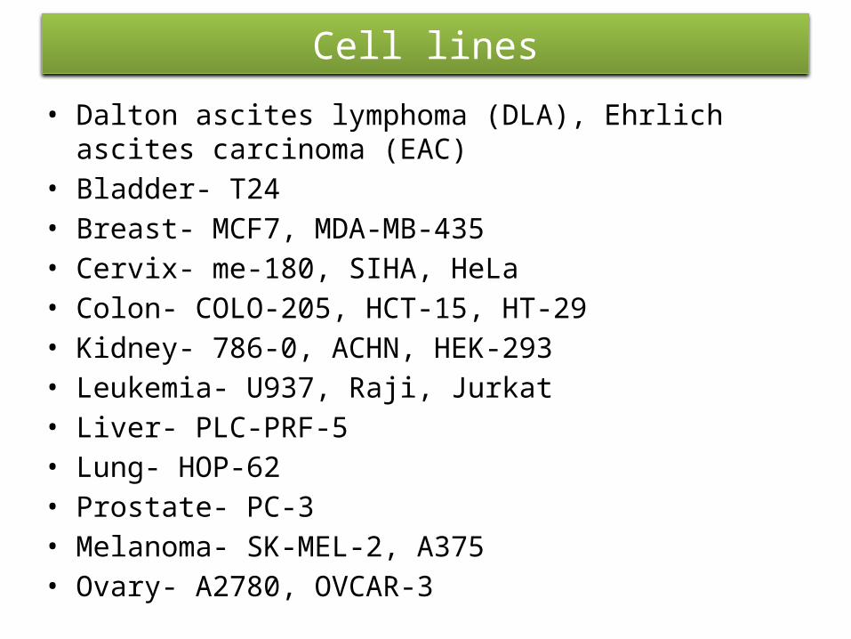
Cell lines
• Dalton ascites lymphoma (DLA), Ehrlich ascites carcinoma (EAC)
• Bladder- T24• Breast- MCF7, MDA-MB-435• Cervix- me-180, SIHA, HeLa• Colon- COLO-205, HCT-15, HT-29• Kidney- 786-0, ACHN, HEK-293• Leukemia- U937, Raji, Jurkat• Liver- PLC-PRF-5• Lung- HOP-62• Prostate- PC-3• Melanoma- SK-MEL-2, A375• Ovary- A2780, OVCAR-3

Cancer research organizations
• WHO• Indian Cancer Society (ICS)• Cancer aid society• American Association for Cancer Research (AACR)• Union for International Cancer Control (UICC)• Indian association of cancer research• European cancer organisation (ECCO)• Cancer association of south africa (CANSA)

RECENT ADVANCES
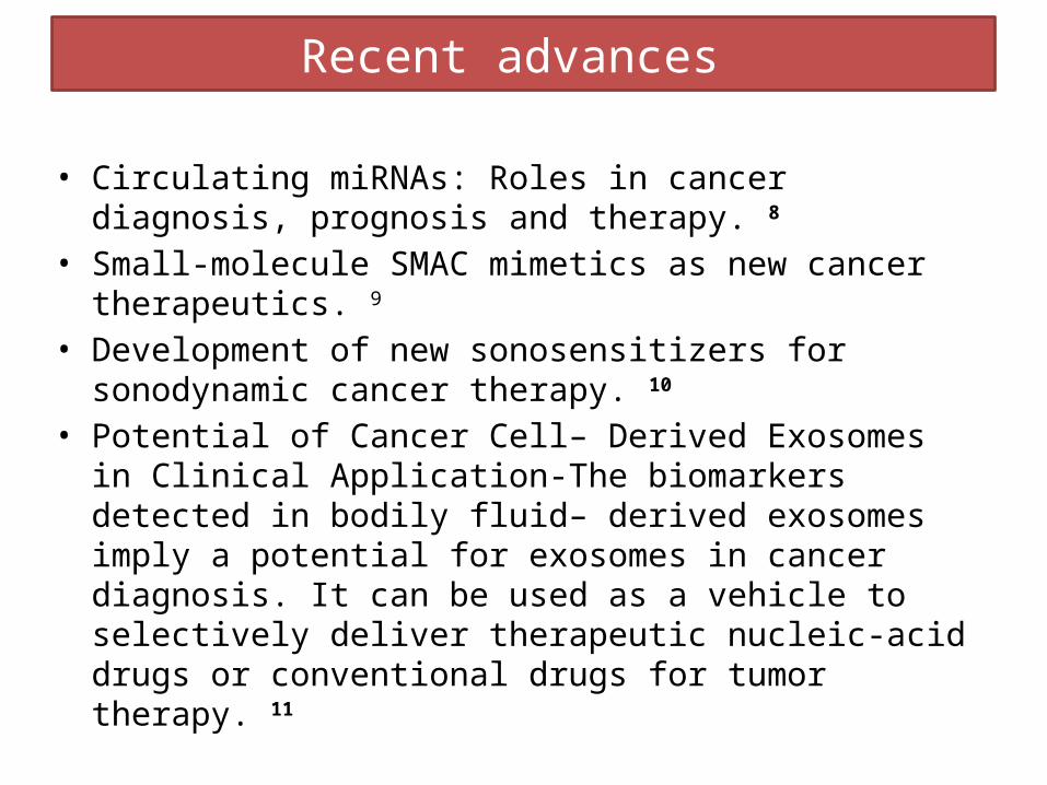
Recent advances
• Circulating miRNAs: Roles in cancer diagnosis, prognosis and therapy. 8
• Small-molecule SMAC mimetics as new cancer therapeutics. 9
• Development of new sonosensitizers for sonodynamic cancer therapy. 10
• Potential of Cancer Cell– Derived Exosomes in Clinical Application-The biomarkers detected in bodily fluid– derived exosomes imply a potential for exosomes in cancer diagnosis. It can be used as a vehicle to selectively deliver therapeutic nucleic-acid drugs or conventional drugs for tumor therapy. 11

Recent advances

Recent advances
• Cardiac dose sparing and avoidance techniques in breast cancer radiotherapy-
Maneuvers that displace the heart from the field such as coordinating the breathing cycle or through prone patient positioning
Technological advances such as intensity modulated radiation therapy (IMRT) or proton beam therapy (PBT)
Techniques that treat a smaller volume around the lumpectomy cavity such as accelerated partial breast irradiation (APBI), or intraoperative radiotherapy (IORT)
Akt and p53R2, partners that dictate the progression and invasiveness of cancer. 12

Recent advances
•Advanced nanovehicles for cancer management & delivery of drug–nucleic acid combinations for cancer treatment. 13
Figure: Schematics of the preparation process of the RBC membrane coated poly (lactic-co-glycolic) acid (PLGA) nanoparticles (NPs)

Recent advances
• Types of stem cell transplants for treating cancer-
Autologous stem cell transplantsTandem transplantsAllogeneic stem cell transplantsMini transplants (non-myeloablative transplants)Syngeneic stem cell transplants – for those with an
identical siblingHalf-matched transplants

Recent advances
• The p53-mediated cytotoxicity of photodynamic therapy of cancer. 14
• “Diamond” mammoplasty as a part of conservative management of breast cancer. 15
• Stereotactic radiotherapy (SABR) for the treatment of primary non-small cell lung cancer. 16

FUTURE TRENDS

Future trends• Clinical effectiveness of new drugs may improve if
developments in pharmacogenetics will be done.• Computer-assisted surgery with three-dimensional imaging
allow the tumour and anatomy of the organ to be clearly visible. Intra-operative diagnosis will also be more prevalent. Ultrasound scans used during surgery may show previously
undiscovered metastases, which can be dealt with immediately.
• Efforts to mark tumours with a radioactive isotope so that a Geiger counter passed over the body can detect distant metastases, or with fluorescent markers that glow in the dark.
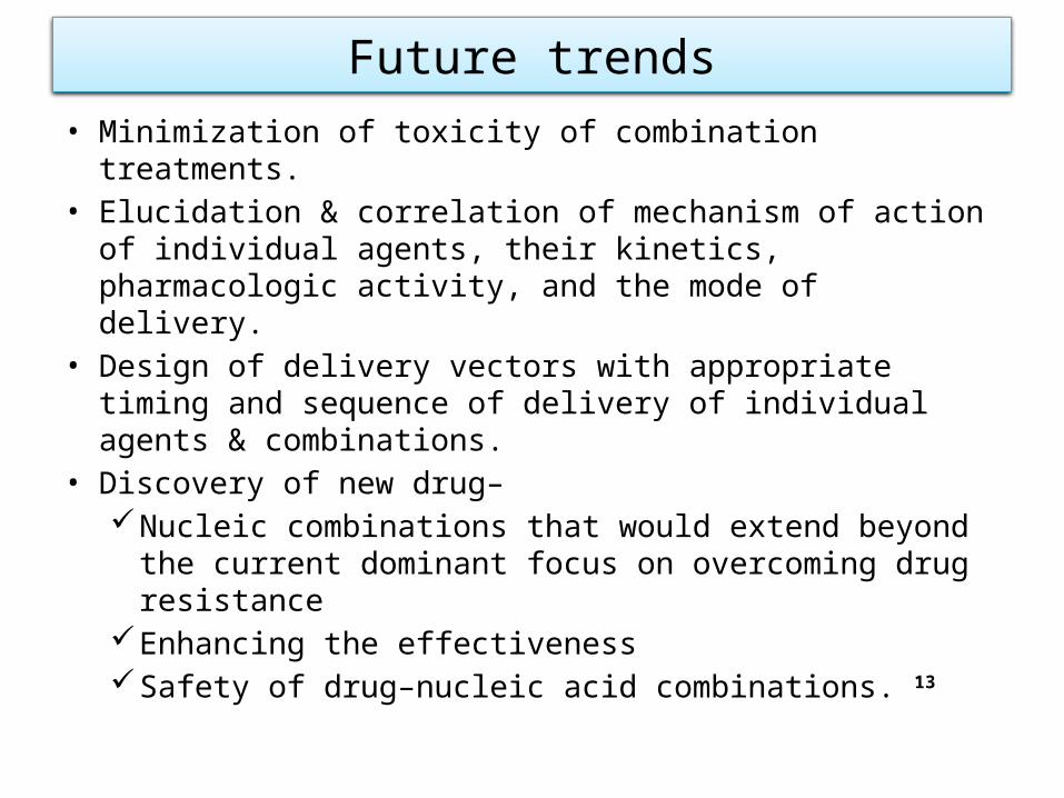
Future trends• Minimization of toxicity of combination treatments.• Elucidation & correlation of mechanism of action of individual
agents, their kinetics, pharmacologic activity, and the mode of delivery.
• Design of delivery vectors with appropriate timing and sequence of delivery of individual agents & combinations.
• Discovery of new drug–Nucleic combinations that would extend beyond the
current dominant focus on overcoming drug resistance Enhancing the effectivenessSafety of drug–nucleic acid combinations. 13

Future trends
• p53 is the main inducer of p53R2, in order to understand the precise role of p53R2 and how its levels are elevated in some cancers, it is suggested to study the ability of mutant p53 in induction of p53R2.
• On the other hand, since p21 has dual opposite roles in regulation of cell cycle, and p53R2 can up-regulate p21, it is better to focus future investigation on the role of p21 in cancer progression and its association/interactions with p53R2.
• The need to develop novel sonosensitizers with optimal properties for treating human cancers. 10

Conclusion

Conclusion
• Basic research in cancer biology has provided new targets for cancer drug development & has brought older targets into sharper focus, leading to new & novel approaches to cancer prevention & treatment.
• Conventional methods of drug screening are continuously being refined or replaced with newer methods & thereby accelerating the drug development process.
• Efforts are being done to achieve specific predefined locus of action.
• It is inevitable that in the years to come we will see high technology, high speed, high volume & information- intensive approaches to the identification of novel targets genes, proteins & drugs.
• However, the importance of basic research in the molecular biology & pharmacotherapy of cancer remains critical.

References

References
1. Tripathi KD. Essentials of Medical Pharmacology. New Delhi: Jaypee Brothers Medical Publishers (P) Ltd; 2013.
2. Rang HP, Dale MM, Ritter JM. Rang and Dale’s Pharmacology. London: Elsevier Churchill Livingstone; 2012.
3. Mohan H. Textbook of Pathology. New Delhi: Jaypee Brothers Medical Publishers; 2013.
4. Gupta SK. Drug Screening Methods. New Delhi: Jaypee Brothers Medical Publishers; 2009.
5. Kumar V, Abbas AK, Fausto N. Robbins and Cotron Pathologic Basis of disease. Philadelphia: Saunders ; 2008.
6. Alberts B, Johnson A, Lewis J, Molecular Biology of the cell. New York: Garland Science; 2008.

7. http/ www. WHO.int/mediacentre/ factsheets/ fs 297/ en/ index.html8. G. Cheng, Circulating miRNAs: Roles in cancer diagnosis,
prognosis and therapy, Adv. Drug Deliv. Rev. (2014), http://dx.doi.org/10.1016/j.addr.2014.09.001
9. Bai L, Smith DC , Wang S. Small-molecule SMAC mimetics as new cancer therapeutics: Pharmacology & Therapeutics.
2014; 144: 82–95.10. Chen H, Zhou X, Gao Y, Zheng B, Tang F and Huang J. Recent
progress in development of new sonosensitizers for sonodynamic cancer therapy: Drug Discovery Today REVIEWS - Elsevier 2014 Apr; 19.
References

11. Sun Y, Liu J. Potential of Cancer Cell–Derived Exosomes in Clinical Application: A Review of Recent Research Advances.
Clinical Therapeutics- 2014; 36. 12. Shah C, Badiyan S, Berry S, Khan AJ, Goyal S, Schulte K, Nanavati A, Lynch M,Vicini FA. Cardiac dose sparing and avoidance techniques in breast cancer radiotherapy: Review. Radiotherapy and Oncology. Elsevier. 2014; 112: 9–16.13. Burgo SD, Pedraz JL, Orive G. Advanced nanovehicles for cancer management. REVIEWS Drug Discovery Today: Review. Elsevier. 2014 Oct; 19.
References

14. Pankau JZ, Krachulec J, Grulkowski I, Bielawski KP, Selivanova G. The p53-mediated cytotoxicity of photodynamic therapy of cancer: Recent advances. Toxicology and Applied Pharmacology. Elsevier 2008; 232:487–97.
15. Husseina O, Khodaryb TE. Diamond mammoplasty as a part of conservative management of breast cancer: Description of a new technique. International Journal of Surgery Case Reports. Elsevier. 2012: 203–6.
16. Soldà F, Lodge M, Ashley S, Whitington A, Goldstraw P, Brada M. Stereotactic radiotherapy (SABR) for the treatment of primary non-small cell lung cancer; Systematic review and comparison with a surgical cohort. Radiotherapy and Oncology. Elsevier. 2013; 109: 1–7.
References

MUCH OBLIGED