cancer detection examination
TRANSCRIPT

8/6/2019 Cancer Detection Examination
http://slidepdf.com/reader/full/cancer-detection-examination 1/20

8/6/2019 Cancer Detection Examination
http://slidepdf.com/reader/full/cancer-detection-examination 2/20
� The Pap smear is done as part of a gynecologicalexam. You will lie on a table and place your feet instirrups to position your pelvis for examination. Thehealth care provider will insert an instrument(speculum) into your vagina and open it slightly tosee inside the vaginal canal.
� The health care provider will take a sample of cellsfrom the outside and just inside the opening of thecervix (cervical canal) by gently scraping the outside
of the cervix with a wooden or plastic spatula, theninserting a small brush that looks like a pipe cleaner into the canal.
� The cells are placed on a glass slide, or put in a bottlecontaining a preservative, and then sent to the labfor examination.

8/6/2019 Cancer Detection Examination
http://slidepdf.com/reader/full/cancer-detection-examination 3/20
Tell your health care provider if you:� Are taking any medications or birth control pills� Have had an abnormal Pap smear
� Might be pregnantWithin 24 hours of the test, avoid:� Douching� Having intercourse� Taking a tub bath� Using tamponsAvoid scheduling your Pap smear while you have your period (are menstruating), because blood and cellsfrom the uterus may affect the accuracy of the Papsmear. Empty your bladder just before the test.

8/6/2019 Cancer Detection Examination
http://slidepdf.com/reader/full/cancer-detection-examination 4/20
� The Pap smear can detect cancerous or
precancerous conditions of the cervix.
Most invasive cancers of the cervix canbe detected early if women have Pap
tests and pelvic examinations.

8/6/2019 Cancer Detection Examination
http://slidepdf.com/reader/full/cancer-detection-examination 5/20
� Class I. Normal
� Class II. Inflammation
� Class III. Mild to Moderate Dysplasia
� Class IV. Probably Malignant
� Class V. Possibly Malignant

8/6/2019 Cancer Detection Examination
http://slidepdf.com/reader/full/cancer-detection-examination 6/20
� Class I result requires follow ² up
examination every 1-3 years as
recommended by the physician.� Class II and III results may require repeat
Pap exam in 3 to 6 months as prescribed.
� Class IV and V results require biopsy asprescribed.

8/6/2019 Cancer Detection Examination
http://slidepdf.com/reader/full/cancer-detection-examination 7/20

8/6/2019 Cancer Detection Examination
http://slidepdf.com/reader/full/cancer-detection-examination 8/20
� A biopsy is the removal of a small piece of tissue for laboratory examination.
� It is most often done to examine tissue for disease.
What Abnormal Results Mean� An abnormal biopsy means that the tissue or cells
have an unusual structure, shape, size, or condition.� This may mean you have a disease, such as cancer,
but it depends on your biopsy.
Risks� Bleeding� Infection

8/6/2019 Cancer Detection Examination
http://slidepdf.com/reader/full/cancer-detection-examination 9/20
� Needle Biopsy ² done by aspiration of
tumor cells with needle and syringe.
� Excisional Biopsy ² done by removing theentire tumor. It is done when the tumor is
small.
� I
nc
isional or subtotal biopsy ² done bytaking only a part of the tumor. This is
done when the tumor is large.

8/6/2019 Cancer Detection Examination
http://slidepdf.com/reader/full/cancer-detection-examination 10/20

8/6/2019 Cancer Detection Examination
http://slidepdf.com/reader/full/cancer-detection-examination 11/20
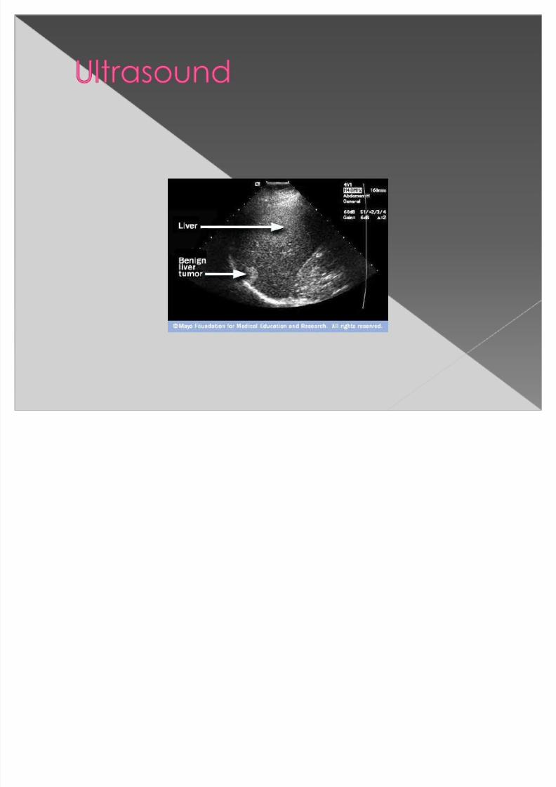
� Ultrasound involves the use of high-frequency soundwaves to create images of organs and systems withinthe body.
�
The test is done in the ultrasound or radiologydepartment. You will be lying down for theprocedure. A clear, water-based conducting gel isapplied to the skin over the area being examined tohelp with the transmission of the sound waves. Ahandheld probe called a transducer is moved over
the area being examined. You may be asked tochange position so that other areas can beexamined.
� The reason for the examination will depend on your symptoms.
8/6/2019 Cancer Detection Examination
http://slidepdf.com/reader/full/cancer-detection-examination 12/20

8/6/2019 Cancer Detection Examination
http://slidepdf.com/reader/full/cancer-detection-examination 13/20
� Magnetic resonance imaging (MRI) is anoninvasive method to take pictures of thebody.
� Unlike x-rays and computed tomographic(CT)scans, which use radiation, MRI usespowerful magnets and radio waves. Signalsfrom the magnetic field bounce off your body
and are sent to a computer, where they areturned into images. Different types of tissuessend back different signals.
� Single MRI images are called slices. The imagescan be stored on a computer or printed on
film. One exam produces dozens or sometimes

8/6/2019 Cancer Detection Examination
http://slidepdf.com/reader/full/cancer-detection-examination 14/20
Combining MRIs with other imaging methodscan often help the doctor make a moredefinitive diagnosis.
� MRI images taken after a special dye(contrast) is delivered into the body mayprovide additional information about theblood vessels.
�
An MRA, or magnetic resonanceangiogram, is a form of magneticresonance imaging, that creates three-dimensional pictures of blood vessels. It isoften used when traditional angiography
cannot be done.

8/6/2019 Cancer Detection Examination
http://slidepdf.com/reader/full/cancer-detection-examination 15/20
8/6/2019 Cancer Detection Examination
http://slidepdf.com/reader/full/cancer-detection-examination 16/20
� A computed tomography (CT) scan is animaging method that uses x-rays to createcross-sectional pictures of the body.
Why the Test is Performed� CT rapidly creates detailed pictures of the
body, including the brain, chest, spine, andabdomen. The test may be used to:
� Diagnose an infection� Guide a surgeon to the right area during a
biopsy� Identify masses and tumors, including cancer � Study blood vessels

8/6/2019 Cancer Detection Examination
http://slidepdf.com/reader/full/cancer-detection-examination 17/20

8/6/2019 Cancer Detection Examination
http://slidepdf.com/reader/full/cancer-detection-examination 18/20
Hematologic (CBC)
Hemoglobin
Hematocrit
Leukocytes
Platelets

8/6/2019 Cancer Detection Examination
http://slidepdf.com/reader/full/cancer-detection-examination 19/20
� Tumor markers are measurable biochemicals that areassociated with a malignancy. They are either producedby tumor cells (tumor-derived) or by the body in responseto tumor cells (tumor-associated). They are typically
substances that are released into the circulation and thusmeasured in the blood. There are a few exceptions to this,such as tissue-bound receptors that must be measured ina biopsy from the solid tumor or proteins that are secretedinto the urine.
Purpose
� Though tumor markers are rarely specific enough to beused alone to diagnose cancer, they do have a number of clinical uses. They can be used to stage cancer, toindicate a prognosis, to monitor treatment, or in follow-upto watch for cancer recurrence. Changes in some tumor markers have been sensitive enough to be used as targetsin clinical trials.

8/6/2019 Cancer Detection Examination
http://slidepdf.com/reader/full/cancer-detection-examination 20/20
� AFP ² (Alpha ² feto ² protein )
� CEA (Carcinoembryonic Antigen)
� HCG (Human Chorionic Gonadotropin)
� Prostatic Acid Phosphatase
� PSA (Prostatic Specific Antigen)