cancer microenvironment and therapeutic implications · attilio baronzio fellow department of...
TRANSCRIPT

Cancer Microenvironment and TherapeuticImplications

Gianfranco Baronzio · Giammaria Fiorentini ·Christopher R. CogleEditors
Cancer Microenvironmentand Therapeutic Implications
Tumor Pathophysiology Mechanismsand Therapeutic Strategies
123
Editors
Dr. Gianfranco BaronzioPoliclinico di MonzaDipto. Radioterapia eIpertermiaVia Amati, 11120052 MonzaItaly
Dr. Giammaria FiorentiniOspedale San GiuseppeServizio di OncologiaVia Giovanni Boccaccio, 1850053 EmpoliItaly
Dr. Christopher R. CogleUniversity of Florida1600 SW. Archer RoadP.O.Box 100278Gainesville FL 32610-0278USA
ISBN 978-1-4020-9575-7 e-ISBN 978-1-4020-9576-4
DOI 10.1007/978-1-4020-9576-4
Library of Congress Control Number: 2008942118
c© Springer Science+Business Media B.V. 2009No part of this work may be reproduced, stored in a retrieval system, or transmittedin any form or by any means, electronic, mechanical, photocopying, microfilming, recordingor otherwise, without written permission from the Publisher, with the exceptionof any material supplied specifically for the purpose of being enteredand executed on a computer system, for exclusive use by the purchaser of the work.
Printed on acid-free paper
9 8 7 6 5 4 3 2 1
springer.com

The book is dedicated to Pietro M Gullino,an uncommon friend and teacher

Contents
Part I Tumor Physiopathology and Microenvironment Genesis
1 Inflammation and Carcinogenesis: A Change in the Metabolic Process 3L. Schwartz, M. Israel and Icard Philippe
2 Tumor Microenvironment: Aspects of Stromal-ParenchymalInteraction . . . . . . . . . . . . . . . . . . . . . . . . . . . . . . . . . . . . . . . . . . . . . . . . . . . . 19Attila Zalatnai
3 Significance of Tumor Microenvironment on the Genesis of:Interstitial Fluid, Angiogenesis, Haemostatic/HaemorheologicAbnormalities. Pathogenesis and Therapeutic Aspects . . . . . . . . . . . . . 39Gianfranco Baronzio, Isabel Freitas and Kwan Hau
4 Cancer, Stem Cells and the Neoplastic Niche . . . . . . . . . . . . . . . . . . . . . . 63Christopher R. Cogle
Part II Tumor Microenvironment, Therapeutic Perspectives and Strategiesfor Normalization
5 Barriers to Drug Delivery in Cancer: Clinical Implications . . . . . . . . . 81Gianfranco Baronzio, Isabel Freitas, Attilio Baronzio, Miriam Baronzio,Elisabetta Crespi and Paolo Antonio Netti
6 Hypoxia, Hyperthermia, Chemotherapy: Interactionsand Opportunities . . . . . . . . . . . . . . . . . . . . . . . . . . . . . . . . . . . . . . . . . . . . . 105Giammaria Fiorentini, Maurizio Cantore, Francesco Montagnani,Alfonso Del Freo, Stefano Guadagni and Gianfranco Baronzio
vii

viii Contents
7 Effects of Molecularly Targeting Hypoxia in Oncology . . . . . . . . . . . . . 117Giammaria Fiorentini, Camillo Aliberti, Giorgio Benea,Francesco Montagnani, Andrea Mambrini, Pier Luigi Ballardini,Maurizio Cantore and Stefano Guadagni
8 Significance of the Tumour Microenvironmentin Radiotherapy . . . . . . . . . . . . . . . . . . . . . . . . . . . . . . . . . . . . . . . . . . . . . . . 137Michael R. Horsman and Dietmar W. Siemann
9 Effects of Tumor Microenvironment on Immunity and ConsequentClinical Considerations . . . . . . . . . . . . . . . . . . . . . . . . . . . . . . . . . . . . . . . . . 157Gianfranco Baronzio, Isabel Freitas, Giammaria Fiorentini, Anna RitaCrugnola, Dieter Hager, Dino Ceppodomo and Mikhail V. Kiselevsky
10 Effects of Tumor Microenvironment on Hyperthermia,Photodynamic and Nanotherapy . . . . . . . . . . . . . . . . . . . . . . . . . . . . . . . . . 181Gianfranco Baronzio, Attilio Baronzio, Elisabetta Crespi and IsabelFreitas
11 Targeting Tumour Vascularization from Bench to Bedside:Suggestions for Combination with Hyperthermia . . . . . . . . . . . . . . . . . . 203Girolamo Ranieri, Annamaria Catino, Vittorio Mattioli, Vito Fazio,Gennaro Gadaleta-Caldarola and Cosmo Damiano Gadaleta
12 New Indications for Established Drugs Able to Modify Tumour-HostInteractions . . . . . . . . . . . . . . . . . . . . . . . . . . . . . . . . . . . . . . . . . . . . . . . . . . . 221Annika Bundscherer and Christian Hafner
Index . . . . . . . . . . . . . . . . . . . . . . . . . . . . . . . . . . . . . . . . . . . . . . . . . . . . . . . . . . . . . 241

Contributors
Camillo Aliberti Department of Interventional Radiology, Delta Hospital,Azienda Sanitaria Locale, Ferrara, Italy
Pier Luigi Ballardini Department of Interventional Radiology, Delta Hospital,Azienda Sanitaria Locale, Ferrara, Italy
Attilio Baronzio Fellow Department of Pharmacology, University of Novara,28100 Novara, Italy, [email protected]
Gianfranco Baronzio Consultant Radiohyperthermia Dept, Via Amati 111,Policlinico di Monza, 20052 Monza, Italy, [email protected]
Miriam Baronzio Chemistry & Biology High School “Istituto Torno”, 20022Castano P, MI, Italy
Giorgio Benea Department of Interventional Radiology, Delta Hospital, AziendaSanitaria Locale, Ferrara, Italy
Annika Bundscherer Department of Dermatology, University of Regensburg,93042 Regensburg, Germany
Maurizio Cantore Department of Oncology, City Hospital, Carrara, Italy
Annamaria Catino Interventional Radiology Unit, National Cancer Institute,Giovanni Paolo II, Bari, Italy
Dino Ceppodomo Department of Laboratory Medicine, Policlinico di Monza, ViaAmati 111, 20052 Monza, [email protected]
Christopher R. Cogle University of Florida, 1600 SW Archer Road, P.O. Box100278, Gainesville, FL 32610-0278, USA, [email protected]
Elisabetta Crespi Fellow Department of Pharmacology, University of Novara,28100 Novara, Italy, [email protected]
Anna Rita Crugnola Fellow Department of Pharmacology, University of Novara,28100 Novara, Italy, [email protected]
Alfonso Del Freo Department of Oncology, City Hospital, Carrara, Italy
ix

x Contributors
Vito Fazio Department of Critical Area and Surgery, National Cancer InstituteGiovanni Paolo II, Bari, Italy
Giammaria Fiorentini Oncology Department, Nuovo ospedale San GiuseppeHospital, via Giovanni Boccaccio 18,50053, Empoli (Firenze), Italy,[email protected]
Isabel Freitas Department of Animal Biology and IGM-CNR Center forHistochemistry & Cytometry, University of Pavia, Pavia, Italy, [email protected]
Cosmo Gadaleta Director of Dept of Interventional Radiology, Tumor Institute“Giovanni Paolo II”, Bari, Italy, [email protected]
Cosmo Damiano Gadaleta Intrventional Radiology Unit, National CancerInstitute Giovanni Paolo II, Bari, Italy
Gennaro Gadaleta-Caldarola Intrventional Radiology Unit, National CancerInstitute Giovanni Paolo II, Bari, Italy
Stefano Guadagni Department of Surgical Sciences, University of L’Aquila,L’Aquila, Italy
Christian Hafner Department of Dermatology, University of Regensburg, 93042Regensburg, Germany, [email protected]
ED Hager BioMed-Klinik Bad Bergzabern, Tischberger Str. 5 – 8,76887 BadBergzabern, [email protected]
Kwan Hau Division of Hematology/Oncology, Northwestern University FeinbergSchool of Medicine, 303 East Chicago, Ave.Chicago, IL 60611, USA,[email protected]
Michael R. Horsman Department of Experimental Clinical Oncology, AarhusUniversity Hospital, Aarhus, Denmark, [email protected]
Maurice Israel Laboratoire de Neurochimie, CNRS Gif sur Yvette, France
Mikhail V. Kiselevsky NN Blokhin Russian Cancer Research Center RAMS,Moscow, Russia, [email protected]
Andrea Mambrini Medical Oncology Unit, Carrara, City Hospital, AziendaSanitaria Locale 1, Massa Carrara, Italy
Vittorio Mattioli Department of Critical Area and Surgery, National CancerInstitute Giovanni Paolo II, Bari, Italy
Francesco Montagnani Oncology Department, Nuovo ospedale San Giuseppe,via Giovanni Boccaccio 18, 50053, Empoli (Firenze.), Italy
Paolo Antonio Netti Department of Materials and Production Engineering,University of Naples “Federico II”, Piazzale Tecchio, 80 – 80125 Napoli, Italy,[email protected]

Contributors xi
Icard Philippe Service de chirurgie thoracique, CHU de Caen, Hopital Cote deNacre, Avenue de la Cote de Nacre, 14033 CAEN cedex 9, [email protected]
Girolamo Ranieri Intrventional Radiology Unit, National Cancer InstituteGiovanni Paolo II, Bari, Italy
L. Schwartz Service de Radiotherapie, Hopital De la Pitie-Salpetriere, 73Boulevard de L’hopital, 75013 Paris, France
Dietmar W. Siemann Department of Radiation Oncology, University of Florida,Shands Cancer Center, Gainesville, Florida, USA
Attila Zalatnai Semmelweis University, Faculty of Medicine, First Institute ofPathology and Experimental Cancer Research, H-1085 Budapest, Ulloi 26,Hungary

List of Abbreviations
BDMCs Bone marrow derived myeloid cellsCCL22 Chemokine ligand 22CD Cluster of differentiationDCs Dendritic cellsG-CSF Granulocyte colony stimulating factorGF-β Trasforming growth factor betaGM-CSF Granulocyte macrophage colony stimulating factorGAL-1 Galectin 1HIF-1 Hypoxia inducible factor 1HSP Heat shock proteinIFN-γ Interferon gammaIDO Indoleamine 2,3-dioxygenase immunoregulatory cellsIL-1 Interleukin1IL-2 Interleukin 2IL-4 Interleukin 4IL-6 Interleukin6IL-10 Interleukin 10IL-12 Interleukin 12IF Interstitial fluidIFP Interstitial fluid pressureLAK Lymphokine-activated killer cellMDSC Immature myeloid-derived suppressor cellsMHC Major histocompatibility complexNF-kB Nuclear factor kappa BNO Nitric oxidePDGF Platelet derived growth factorpHe Low extracellular pHROS Reactive oxygen speciesSDF-1 Stromal cell derived growth factor1TAA Tumor associated antigensTNF-α Tumor necrosis factor alphaTLR Toll like receptorsTreg Regulatory T cellsVEGF Vascular endothelial growth factor
xiii

Introduction
Revolutionary changes have swept through cancer diagnosis and treatment over thepast century. In the early 1900s the struggle was to diagnose. Then came the struggleto cut it out. Then the struggle to keep it away.
In the late 1900s scientists and physicians turned inward towards cancer cellmechanics and that is when the explosion of oncologic sciences erupted. A foun-tain of genes, transcription factors, signaling pathways, degradation fates, apoptosischains, receptor interactions and cell-cell contact points provide a wellspring ofopportunity to interfere with runaway cell growth and metastasis.
In the post-genomic era, cancer is a genetic disease.But somewhere in the rush of genetic reductionism, the cancer research commu-
nity has increasingly neglected the importance of the tumor microenvironment andthe interplay between cancer cells, dysplastic cells and normal cells.
It is in this realm that this book is written.The genesis of cancer is not as simple as a gene abnormality or even a set of gene
abnormalties. A permissive environment must allow unchecked cell division. Forcancer to initiate, the immune system must look the other way, nutrients and oxygenmust be served at the right time, temperature and concentration, and a hospitable bedof stroma and extracellular matrices must support burgeoning growth and spread.Cancer is a chance co-op with a panoply of factors traded between normal andmalignant.
Chief interactions in this co-op are exposed herein and implications for anti-cancer therapies are discussed. This book is not intended to serve as a simple review;rather, to spark new ideas and provocative questions so that better anti-neoplastictherapies are conceived and promulgated.
In this book we assembled a team of contributors who share amongst them seri-ous experiences at the laboratory bench and in the clinic. These physician-scientistshave dedicated themselves to the tension between the urgency for breakthroughsand the technical challenges of discovery. Their thoughts and perspectives on thestate of cancer biology, ecology and implications for treatment are gathered herein.We intend for this book to roadmap outstanding questions and potential answers forthe eradication of cancers.
xv

Part ITumor Physiopathology
and Microenvironment Genesis

Chapter 1Inflammation and Carcinogenesis: A Changein the Metabolic Process
L. Schwartz, M. Israel and Icard Philippe
Abstract Since the seminal work it is known that inflammation is a major riskfactor for cancer. Inflammation can be caused by agents as diverse as heat, cold,foreign body or chemicals. In every case, there is a protein leak from the damagedcapillaries. This results in increased oncotic pressure which is in turn responsiblefor the methylation of the PP2A phosphatase. The activation of PP2A results in thetranslocation of NF κB and the activation of several metabolic pathways.
Keywords Inflammation · Chronic inflammation · Carcinogenesis · Methylation ·PP2A phosphatase · NF-κB
Cancer is frequently associated with pre-existing inflammation and fibrosis.Between 60% and 90% of hepatocellular carcinoma occurs in patients with hep-atic macronodular cirrhosis (De Vita et al. 1993, Podolsky and Isselbacher 1994).Chronic liver disease of any type is a risk factor for liver cancer. The cancer may becaused by hepatitis B or C, alcoholic liver disease, antitrypsin deficiency,hemochro-matosis, and tyrosinemia. Its features result from hepatocyte necrosis, extensivefi-brosis, connective tissue deposition, vascular distortion and nodular regeneration ofthe remaining tissue parenchyma (Podolsky and Isselbacher 1994). Evidence fora cause-effect link between cirrhosis and hepatocarcinoma is lacking. The rela-tion may often be one of chance alone, since not all cirrhotics develop cancer.Nonetheless, diseases that cause cirrhosis also increase the risk of hepatocarcinoma(Podolsky and Isselbacher 1994). Furthermore, the more disorganized the liverbecomes, the higher the risk of hepatocarcinoma (Podolsky and Isselbacher 1994,Baffis et al. 1999).
Similarly, lung cancer is most common among patients suffering from any formof chronic lung disease (Ernster 1996, Maitre et al. 1996). History of chronic bron-chitis, emphysema, primary lung fibrosis, chronic lung infection and even lung
L. Schwartz (B)Service de Radiotherapie, Hopital De la Pitie-Salpetriere, 73 Boulevard de L’hopital,75013 Paris, Francee-mail: [email protected]
G. Baronzio et al. (eds.), Cancer Microenvironment and Therapeutic Implications,DOI 10.1007/978-1-4020-9576-4 1, C© Springer Science+Business Media B.V. 2009
3

4 L. Schwartz et al.
irradiation are associated with increased risk (Ernster 1996, Maitre et al. 1996).There is no evidence for a relation of bronchitis or emphysema with lung cancerthat could not be explained by independent links to exposure to tobacco smoke orother noxious agents. Nevertheless, the risk of lung cancer increases with the extentof disruption of normal lung architecture. For example, the risk of lung cancer ishigher among patients suffering from chronic bronchitis and severe impairmentof the lungs’carbon monoxide Breast cancer genesis also seems to be linked toarchitectural changes. A woman’s reproductive history is one of the most importantdeterminants of breast cancer risk. This is not a new notion. Ramaziani in 1700 firstshowed that breast cancer risk was higher among nuns (Haagensen 1986). Early inthe 1900s, investigations noted that nulliparity and a history of having never breast-fed an infant were risk factors. Modern epidemiological cohorts have confirmedthe increased risk for breast cancer after early puberty, late menarche, hormonalstimulation (Haagensen 1986).
In 1977, Ing reported a disproportionate increase of post menopausal breast can-cer in the left breast of Tanka women of Hong-Kong thought to have nursed onlywith the right breast (Ing et al. 1997). All these risk factors and others, like radiationto the developing breast, are related (causally or not) to change in architecture ofthe breast.
Breast cancer is rare among young women. It is before the menopause, when thearchitecture of the mammary gland starts to undergo fatty tissue involution, that theincidence of cancer rises (Haagensen 1986). With the completion of menopause,the breast changes, it becomes somewhat smaller and less dense. There is a decreasein the number and size of the ducts. These atrophic lobules are seen lying in adense fibrous matrix. Increase in the connective tissue is a prominent feature of thisaging process.
The rare cases of breast cancer in young women are often due to heredi-tary anomalies. The most studied gene is BRCA-1. BRCA-1 is a nuclear phos-phoprotein expressed in a broad spectrum of tissues during cell division. Theinheritance of a mutant BRCA1 allele dramatically increases a woman’s life-time risk for developing both breast and ovarian cancers. This increased riskmay be secondary to architectural changes. Analysis of a prophylactic subcuta-neous mastectomy after genetic counseling for either carrying the BRCA-1 gene orbelonging to a pedigree with familial breast cancer shows a different architecturalpattern. The BRCA-1 or related genes may have a functional role in the branchingpattern of the breast during lobular development, mainly in epithelial-stroma inter-action (Russo et al. 2001). BRCA-1 deficient mice display multiple malformations(Cressman et al.1999).
Cancers occurring during childhood (nephroblastoma, medulloblastoma, retino-blastoma or Li-Fraumeni syndrome) are also associated with tissue disorganiza-tion from embryonic remnants (De Vita et al. 1993, Pivnick et al. 1998, Boyleet al. 2000). Patients suffering from genetically-encoded hereditary tumors likeLi-Fraumeni syndrome and retinoblastoma have both mutated epithelial cells andfibroblasts with impaired growth, resulting in concomitant malformations (Boyleet al. 2000, Le Couter et al. 1998).

1 Inflammation and Carcinogenesis 5
Experimental Evidence that Chronic InflammationInduces Fibrosis and Cancer
Chemical Carcinogenesis
Exposure to chemical carcinogens is considered to cause most human cancer (DeVita et al. 1993). In animal carcinogenesis experiments, a first chemical (initiator)is responsible of an intense inflammatory reaction. A second chemical (promotor)is genotoxic. The word “genotoxic” has been created to replace the older previousterm: toxic. The effect of genotoxic compounds is not confined to the epithelial cell;they kill epithelial and stromal cells. Genotoxicity causes tissue disruption throughcell killing and replacement. For example, hepatocyte necrosis induced by geno-toxic compounds, which precedes hepatic carcinoma, is associated with substantialdamage to surviving hepatocytes, as well as extensive mesenchymal changes andloss of normal liver architecture (Ames and Gold 1990).
In vitro, however, carcinogens have not always successfully transformed normalhuman cells in culture (Mc Cormick et al. 1990). For these normal cells to be trans-formed, they often need to be immortalized by transfection with a cancer-associatedvirus prior to exposure to a carcinogen (Dipaolo et al. 1986).
Radiation-Induced Cancer
Ionizing radiation induces cancer in humans and animals (Rhim et al. 1993,Barcellos-Hoff and Ravani 2000). In vitro, the vast majority of attempts to achievetransformation of normal human cells into cancer cells have been unsuccessful(Barcellos-Hoff and Ravani 2000). In fact radiation induced carcinogenesis appearsto be a consequence of inflammation and fibrosis.
The female mammary gland is unique among all glands in that the epitheliumdevelops after the birth from a rudiment that can be easily removed at about threeweeks of age. Barcellos-Hoff irradiated the whole mammary gland of nurturingmice. After the irradiation, the epithelial cells are surgically removed and replacedwith transplanted normal mammary cells. The cancer arises from these normal non-irradiated epithelial cells. Tumor growth appears as a consequence of the changesin the irradiated stroma (Barcellos-Hoff and Ravani2000).
Physical Carcinogenesis
Chemicals and radiation induce inflammation and fibrosis. The question is whetherthese architectural changes and the fact that tissue disruption is a risk factor forneoplasia may be explained by chronically increased epithelial cell proliferationresulting in an increased rate of mutation (Moore and Tsuda 1998). The answer liesin the old literature on physical carcinogenesis. It has been documented that some

6 L. Schwartz et al.
foreign bodies induce cancer (Sonnenschein and Soto 1999, Brand 1982, Stantonand Wrench 1972, IARC 1999, Lipkin 1980). The carcinogenicity of foreign bodiesis linked to their shape. Cellulose membrane filters of specific shape, texture orsize generate sarcoma. Intense inflammation and proliferative fibrosis precede tumorformation. The combination of shape and size (about the width of a human cell) mayalso be critical (Lipkin 1980). The carcinogenicity of this chemically inert moleculeis also linked to particle shape and size.
The International Agency for Research on Cancer evaluated the carcinogeniceffect of surgical implants and other foreign bodies in humans (IARC 1999). Theevaluation resulted in a group 2B classification (possibly carcinogenic for humans)for polymeric implants prepared as thin smooth films, and implanted foreign bodiesconsisting of metallic cobalt, or nickel, and a particular alloy powder consisting of66–67% nickel, 13–16% chromium and 7% iron. The evaluation also resulted ina group 3 classification (not classifiable as to their carcinogenicity to humans) fororganic polymeric materials as a group, orthopaedic implants of complex compo-sition, cardiac pacemakers, silicone breast implants, dental materials and ceramicimplants.
Physical carcinogenesis may also be a “transforming” factor. In nude mice,the implantation of both colon adenoma cells and of a plastic plate are neces-sary for tumorigenic growth. Again, locally, there is intense inflammation (Okadaet al. 2000).
Inflammation Is a Metabolic Disease
Of the cardinal symptoms of inflammation: rubor, calor, dolor and tumor, we shallparticularly consider a cellular aspect covered by the word tumor. Indeed, inflamma-tion, like cancer, triggers the mitosis of a variety of cells, and common mechanismsmay have to be controlled in order to remain within physiological limits. We shallfirst recall a few basic facts.
Inflammation is a natural response of the organism to an aggression, which can betraumatic, bacterial or viral. The defense involves first non-specific (innate) mech-anisms, and second specific immunological mechanisms. The innate mechanismscover the complement cascade, which leads to the activation of proteases able todestroy cells, the complement found in the serum, responds to an antigen-antibodycomplex, or might be directly activated by bacterial antigens. The innate defenseis also associated to the release by the liver, of a variety of proteins the Hagemanfactor for example. The latter, induces a variety of events, first the coagulation cas-cade (thrombine-fibrinogen-fibrine); then the induction of the fibrinolytic-plasminecascade; finally, the activation of the so called kallikrein-kinin system, which willhave major actions on vascular permeability, leading to infiltration of tissues andpain, and to the release of lipid mediators of inflammation, eicosanoids-leucotrienes.The liver releases many other proteins (C reactive protein) the latter, binds phos-pholipids from bacteria, controlling the recruitment of macrophages, it also pro-motes the activation of the complement. The increase of proteins in the serum

1 Inflammation and Carcinogenesis 7
increases the sedimentation velocity of red blood cells, measuring the intensity ofinflammation.
An essential player of the innate line of defense is the mast cell; it will releasehistamine, causing vasodilatation, and TNF α (tumor necrosis factor); it will produceand release Interleukins (IL1) and generate lipid mediators from arachidonic acid:leukotrienes, prostaglandins. The mast cell also releases a platelet activation factor(PAF), which promotes the release of serotonin from platelets. The activation of themast cell may result from an injury, or from an allergen-IgE recognition. Endothelialcells neutrophils and macrophages, which are attracted on the site by chemotacticfactors (e.g., RANTES), form this first line of defense. There is a vasodilatation,the site is red and hot (histamine-serotonin), the vascular permeability increases(prostaglandin), swelling gives pain since nerves are compressed, and bradykinineis involved. As for IL1 and TNF α, they act on the hypothalamic center control-ling fever. They cytokines also trigger the release of prostaglandin (E2), whichactivates cAMP dependent processes and catabolism, providing substrates to thesite of inflammation. The patient experiences cachexia, anorexia, fatigue, and fever.This innate line of defense already explains three of the major symptoms of inflam-mation: rubor, calor, and dolor. But what about cell multiplication and “tumor”?We shall see that both lines of defense trigger the increased mitosis leading to theaccumulation of neutrophils and macrophages.
The mitogenic effect involves signaling processes mediated by tyrosine phos-phorylations, that have common features with tyrosine kinase receptors such as theinsulin receptor. If we consider for example the Toll/ILl-1 receptors, or the TLR4receptors that respond respectively to interleukin 1 (IL1) or to bacterial lipopolysac-charides, they both have a tyrosine kinase, intracellular domain. Downstream ofthe tyrosine kinase signal, and with the help of an adapter protein (MyD88), theIL-1R kinase (IRAK) gets activated, which leads after several steps, to a phospho-rylation and to proteolysis of Iκb. This dissociates Iκb from its partener NF-κb.The latter can then move to the nucleus and transcribes proteins controlling cellmitosis. Nitric oxide released by macrophages may nitrosylate Iκb and dissociate itfrom NF-κb.
We shall now discuss mitogenic effects associated to the specific defense line.When a virus infects a cell for example, it presents on its surface the antigen, viralproteins for example, associated to the major histocompatibility complex of class 1(MHC1). The complex will be recognized by a T cell lymphocyte of the cytotoxictype. The T cell receptor recognizes not only MHC1 as a self-identification device,but also motifes of the antigen. The cytotoxic T cell receptor will signal via its CD3components, and via an essential protein (CD8) that a correct recognition of boththe antigen and MHC1 took place, which activates again a tyrosine kinase (P56lck).In the case of cytotoxic T cells, the tyrosine kinase signal induces synthesis andsecretion of enzymes perforating the contaminated cell, which is eliminated. Thesituation is different for helper T cells, they recognize antigens presented by themajor histocompatibility complex of class 2 (MHC2) that appear on the surfaceof macrophages or B lymphocytes. The helper T cell receptor recognizes not onlyself-MHC2, but also the presented antigen. The helper T cell receptor will signal via

8 L. Schwartz et al.
its CD3 component, but also via an essential protein of helper T cells (CD4), thatthe recognition of the antigen-MHC2 complex took place, which activates againthe P56lck kinase. A variable menu of lymphokines are consequently produced,triggering a massive multiplication of immunologically competent cells. In all casesinterleukin 2 (IL-2) autoactivates the multiplication of the helper T cell. In the caseof an antigen presented by a macrophage, the helper T cell and the macrophagesecrete a cocktail of lymphokines: Il-6, TNFα, INFα, and colony stimulating factorsfor granulocyte and macrophages (G-CSF and GM-CSF). In the case of an antigenpresented to helpers T cells by a B lymphocyte, which binds circulating antigens,the helper T cell secretes IL-4 and IL-6, which induce proliferation of the antigenpresenting B lymphocyte. The selected population will proliferate and convert intoa plasmocyte secreting antibodies in the plasma, which neutralize circulating anti-gens. For macrophage presentation, a cocktail of the same lymphokines inducestheir proliferation.
After this brief survey of immunological defense mechanisms, the point we wantto make in relation to cancer is that these lymphokines will act on receptors thatactivate cellular proliferation via similar MAP kinase signaling pathways. In spiteof many common features, there are different modalities to consider. In responseto growth factors, the receptor is autophosphorylated on tyrosines, which bind theSH2 domains of adapter proteins. Then, RAS-GTP steps activate the Raf serine-threonine kinase, follows the mixed MEK kinase, which finally activates ERK.The nuclear translocation of ERK will allow the phosphorylation of transcriptionfactors, regulating the expression of genes involved in mitosis. Two other MAPkinase pathways have been identified, they are particularly activated in responseto inflammatory cytokines; in these pathways, JNK or P38 replaces ERK. At thereceptor level, there are also differences, particularly when the receptor itself hasno kinase activity. In this case it recruits a tyrosine kinase JAK, or src. In the caseof JAK, there is a short cut, which activates STAT, the latter being translocated tothe nucleus.
An interesting modality is related to the CD45 phosphatase that serves as receptorin lymphocytes, it will then modulate a downstream tyrosine kinase step, promotingmitosis. It is probable that scaffold proteins are involved for selecting among thedifferent MAP kinase pathways.
After having recalled these basic features of inflammation, we shall discuss somepossible links with cancer.
The proliferation of immunologically competent cells, the multiplication ofembryonic tissues, or the proliferation of tumor cells seems to require the activationof a signaling mechanism, which indeed, depend on MAP kinases. Their activationfirst depends on a tyrosine kinase reaction, mediated by specific enzymes, or resultfrom auto-phosphorylations, which can be induced by the binding of a ligand, or anantigen, to the tyrosine kinase receptor.
In cancer formation, oncogenes often results in a perturbation of normal signalingpathways. Similar perturbations can also occur in inflammation, when prolifer-ating cells involved in immunologic defense escape from homeostatic controls.Indeed, if factors that trigger the proliferation of cells related to inflammation,

1 Inflammation and Carcinogenesis 9
interleukins for example, are continually secreted because inflammation persists,the enhanced tyrosine kinase signals may result in uncontrolled proliferation oftissues and then oncogenic transformation. We know that tyrosine kinase receptorsignals are not only triggers for the MAP kinase cascade, but that they also activatePI3 kinase, this is the case for the insulin receptor for example. Receptors coupledto G proteins, activated by transmitters released by inflammatory cells, may alsoactivate PI3 kinase, leading in both cases to the conversion of phosphatidyl inositol4,5, bisphosphate (PIP2) into phosphatidyl inositol 3,4,5 (PIP3). We know fromthe work of Hokin and Hokin (Hokin and Hokin 1953) that transmitters such asacetylcholine (ACh) acting on muscarinic receptors, elicit hydrolysis of phospho-inositides. Other transmitters released during inflammation (serotonin or histamine)may also activate such “Gq coupled” receptors, increasing phosphoinositides viathe stimulation of a phospholipase C. If PI3kinase is activated as well, inositol1,3,4,5 (IP4) is formed, if not, inositol 1,4,5 (IP3) is released. In general, Gq cou-pled receptors activate phospholipase Cβ while tyrosine kinase receptors activatephospholipase Cγ. The released inositides mobilize calcium, thereby helping theexocytotic incorporation of the glucose transporter. Another action of phospholipids,like diacyl glycerol (DAG), activates protein kinase C, which mimics the tumoralaction of phorbol esters, probably via a direct activation of ERK. The inflamma-tory transmitters also activate a phospholipase A2, releasing arachidonic acid. Thelatter, lead via cyclooxygenases (COX1, COX2) to prostaglandins and thrombox-anes or to leukotrienes via lipoxygenases. The preventive effect of non-steroidalanti-inflammatory drugs in colon cancers likely depend on the inhibition of COX2,decreasing inflammation. But above all, we should recall that PTEN phosphatasehydrolyses phosphoinositides, decreasing their effects. Hence, if this phosphataseis down regulated, as it is observed in cancer, the PI3 kinase pathway and relatedlipidic mediators of inflammation will exaggerate their effects. If PI3 kinase sig-naling is upregulated, PTEN control is lost, resulting in increased frequency ofmitosis. This may not necessarily lead to cancer, unless some other perturbationtakes place at the level of the MAP kinase pathway, which controls mitosis. Thesynthesis of cell cycle proteins or tumor suppressor proteins is partly controlled bythis pathway through transcription factors activated by ERK, JUNE, or P38, aftertheir translocation in the nucleus.
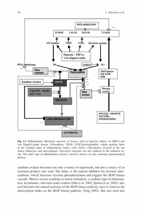
So what might perturb the MAP kinase pathway in inflammation, and whyshould this favor the transformation of cells? Fig. 1.1 answers in part the ques-tions raised. The tyrosine phosphorylation step is essential. In order to limit themitogenic effects of the MAP kinase pathway, we have a natural inhibitory systemperforming tyrosine nitrosylation of proteins. This inhibitory system competes withphosphorylation of these tyrosines when the pathway is activated. Normally, nitro-sylation is operated by peroxynitrite, formed when NO meets superoxides, whichare generated by NADH oxidase, or if respiration does not generate enough elec-trons to fully reduce oxygen. If an aggression takes place, hypoxia induces viaHIF1/Von Hippel-Lindau factor: NO synthase, glycolytic enzymes, carbonic anhy-drase, VEGF, COX2, leading to inflammation; except that cell mitosis is still limitedby the tyrosine nitrosylation mechanism. Then, purine catabolism gets activated,
10 L. Schwartz et al.
Fig. 1.1 Inflammation. Metabolic stressors of tissues, such as hypoxia, induce via HIF1α andVon Hippel-Lindau factors: NOsynthase, VEGF, COX2-prostaglandins, which explains threeof the cardinal signs of inflammation (rubor, calor, dolor). Chemokines secreted at the siteattract leukocytes and macrophages. Glycolytic enzymes are also induced in the inflamed tis-sue. The other sign of inflammation (tumor), involves mitosis of cells ensuring immunologicaldefense
xanthine oxidase becomes not only a source of superoxide, but also a source of anessential product: uric acid. The latter, is the natural inhibitor for tyrosine nitro-sylations, which increases tyrosine phosphorylation and triggers the MAP kinasecascade. Mitosis occurs resulting in tumor formation, a cardinal sign of inflamma-tion. In primates, who lack urate oxidase (Oda et al. 2002, Spitsin et al. 2002), uricacid becomes the natural activator of the MAP kinase pathway since it removes thenitrosylation brake on the MAP kinase pathway (Teng 2002). But uric acid also

1 Inflammation and Carcinogenesis 11
inhibits formation of nitrocatechols and nitroindols, which normally inhibit methy-lases (Perez and Avila 1999, Huotari et al. 2001). Hence, uric acid helps methylationof PP2A and its activation, thereby counteracting the MAP kinase trigger of mitosisand limits its tumorigenic effects.
In other works we have discussed the role of nitrocatechols and nitroindols inrelation to neurologic diseases, and compared them to endogenous neuroleptics(Israel 2004). The levels of these molecules decrease when uric acid increases. Innormal conditions, uric acid controls this defense mechanism in primates; it is stilloperational when an excess of peroxynitrite or peroxycarbonate is formed, such asafter inhaling NO or CO2 with cigarette smoke. But in this case, peroxynitrite con-verts urate into triurate, which inhibits tyrosine nitration, and thus pushes the MAPkinase pathway. Triurate has deleterious actions on lipid peroxidation (Robinsonet al. 2004) and cannot inhibit formation of nitrocatechols as well as urate. Nitro-catechols increase, and the methylase is more inhibited, leading to PP2A becomingpoorly methylated and inactive towards given substrates. We have seen that thissituation favors mitosis. In sum, toxin exposure generates triurates which lead topoorly activated PP2A, which then fails to counteract the MAP kinase activation ofmitosis.
A protection against the effects of triurate on lipid peroxidation and on PP2Acould come from ascorbate (Frei 1991) or vitamine E, but primates do not synthesizethese vitamins. Other mammals, such as rodents, can synthesize ascorbate. Rodentsuse ascorbate, as a single protection mechanism against lipid peroxidation and tyro-sine nitrosylation. Probably, this explains why cigarette smoke interferes with uratedefenses of primates, resulting in lung cancer, while rodents with different defensemechanisms, based on ascorbate, rather than urate, are more resistant to developinglung cancer after toxin exposure.
In this discussion we highlight major players in inflammation such as PP2Aand PTEN. Specifically, PTEN is down regulated in cancer because the gene ishypermethylated,while PP2A is inactivated because of a cytosolic hypomethylation.
Cancer as a Metabolic Disease: Back to Otto Warburg
To understand regulation of human cell division, a detour to the world of micro-biology is necessary, beginning with the work of Louis Pasteur himself. Pasteur(1822–1895) showed that the conversion from sugar to ethanol required livingorganisms, rather than a chemical catalyst, demonstrating that by decreasing theoxygen content in a yeast broth, the yeast cells could be made to divide, multiply,and ferment vigorously (the “Pasteur Effect”). In modern terms, this effect could bedescribed as an activation of anaerobic glycolysis to meet cellular ATP needs.
Otto Warburg developed the work of Pasteur on fermentation. During his life-time Warburg was generally regarded as the greatest biochemist of the 20th century.Warburg, Krebs and Meyerhoff showed that cancer cells were anaerobic in nature,more akin to fungus or bacteria than to normal mammalian cells.

12 L. Schwartz et al.
Fig. 1.2 A vicious circle leading to cancer 1. There are conditions related to food: methyl sources/methyl baits, vitamins, or genetic susceptibilities, that lead to a methylation deficit in the cytosol.2-This affects the activity of PP2A phosphatase, this enzyme has hundreds of variable isoformswhich may be affected by many different external agents: viral, physical or chemicals that afterthe different isoforms. The resulting effect is a hypomethylation in affected cells. The PP2Asubunits do not assemble. 3-The phosphatase looses its specificity for given protein targets, lead-ing to effects on proteins controlling mitosis, and also to a glycolytic metabolic “bottle neck”,because Pyruvate kinase remains in its inactive M2 phosphorylated form, rather than switchingto the active M4 tetramer. 4-there is a compensatory insulin signaling process, entering glucose,saturating mitochondria shuttles above the neck, Lactate dehydrogenase forms NAD+ and lac-tate (Warburg effect). Below the neck, Alanine transaminase (ALAT), the Malic enzyme, formpyruvate, but the lactate sink is deep. Hence, fatty acid catabolism has to form the necessary acetyl-coA, not provided by pyruvate dehydrogenase, oxaloacetate comes via an abnormal carboxylationof phosphoenolpyruvate by mitochondrial PEP carboxy kinase. 5-The activation of insulin–tyrosine kinase–MAP kinase signaling, activates PP1 phosphatase; 6-PP1 acts on the cell cycle

1 Inflammation and Carcinogenesis 13
As early as 1920, Warburg knew how to inject tumoral suspensions into the peri-toneum of mice, and how to measure their gas concentration. He understood thatcancer is a disease of cellular breathing and that cancers are often hypoxic.
He also understood that all cancer-producing substances (arsenic, tars, andcyanide) decrease cellular respiration. Cancer cells ferment even in presence ofoxygen. Either oxygen cannot reach the cell, or it cannot be utilized. In the 1920’s,Warburg had identified these two phases: first, hypoxia alters cell metabolism; sec-ond, if the cell survives these anomalies, the latter will produce cancer. Despitethe Nobel prizes discerned to Otto Warburg, Krebs and Meyerhoff their work haslargely been forgotten. The main reason for this ignorance of the work done at thebeginning of the past century lies in the multiple dramas of that period.
And what could be easier, when you understand cellular respiration than tochoke it? From the start of the First World War, German scientists working oncellular respiration lent their knowledge to the German war effort, with the result-ing devastation caused by yperite and other combat gases. Yet another poison wassynthesized: Zyklon B, used so “successfully” in the death camps of the SecondWorld War. Needless to say, this research on cellular respiration mechanisms –whatever it’s original merits – suffered greatly from the ensuing madness. Fur-thermore, Warburg was an eccentric genius who had many enemies. He proposeda theory of cancer dependent on glycolysis that was initially greeted enthusias-tically but was later widely ridiculed in academic circles (Nachmansohn 1979,Guillemin and Krasnow 1997). Although Warburg’s data were impeccable, otherscientists later claimed to find exceptions to Warburg’s rule. In time, Warburg’stheory became not just old-fashioned but anathema to a scientific establishment thatwas increasingly focused on viruses and aberrant genes as the source of cancer.
Since then, the Positron Emitting Tomography (PET) scan has revived Warburg’swork.
For long, the prodigious expense and ponderous size of the cyclotron (housed inits own building and managed via a network of intricate controls) and the cumber-some requirements of staff prohibited its clinical use. Over the years, refinementsmade it more practical, more manageable, and less complicated. The cyclotron wasdownsized. It could fit into a hospital.
←−−−−−−−−−−−−−−−−−−−−−−−−−−−−−−−−−−−−−−−−−−−−−−−−−−−−−−−−−−−−−−−−−−−−−Fig. 1.2 (continued) synergistically with the PP2A deficit, leading to the permanent activation ofmitosis. 7-MAP kinases affect lamine phosphorylation, the nuclear membrane becomes permeableto methyl donors such as S-adenosyl methionine (SAM), it is retained in the nucleus, cytosolicmethylase become less active, while the nuclear methylases are boosted. 8-There is an increaseof nuclear methylations, the hypermethylation of promoters will silence genes (PTEN), but alsodemethylase inhibitors; thus, other parts of the genome get hypomethylated, activating othergenes (hexokinase gene). 9-Consequently PTEN and HPM1 phosphatases are silenced, increasinginsulin – PI3 kinase – MAP kinase effects, while hexokinase and glucose influx increase, aggra-vating the effect of the PP2A failure, on the cell cycle. In sum there is a cytosolic hypomethylationand a nuclear hypermethylationhypomethylation process, which upsets the phosphatases and leadsto catastrophic changes of metabolism and mitosis, generating cancer

14 L. Schwartz et al.
Using a radioactive analog of glucose, PET scan examination has revivedWarburg’s work. Modern imaging confirms the increased uptake by cancer of largequantities of glucose.
Fermentation provides energy, though less efficiently than respiration (Guilleminand Krasnow 1997, Schwartz 2004) for, in the presence of sufficient oxygen, glu-cose is completely degraded into water and carbonic gas, hence the efficiency ofrespiratory metabolism. At lower concentrations of oxygen, by contrast, the glucoseis incompletely degraded and waste is produced, a portion of which is released intothe extracellular component. Some wastes such as ethanol produced by glycolysisare highly valuable.
Other “waste products” of anaerobic glycolysis remain within cells, causing cellmass to increase, and reacting to form aminoacids, lipids, glucids and nucleic acids(Schwartz 2004, Palmer 1985, Wang et al. 1995, Gerasimovskaya et al. 2002, Wadaet al. 2002). These aminoacids, in turn, form proteins, the lipids are transformedinto hormones; and the nucleic acid provides DNA and RNA. In this manner, anaer-obic glycolysis provides cells with all the requirements for mitosis, both physical(sufficient cell mass) and biochemical (suitable molecular species).
Modern biology has been hindered by the discoveries of hundreds of what havebeen identified and named or, rather, misnamed, “growth factors”. Originally, theconcept of “growth factors” was limited to polypeptides, but it now extends toinclude sugars and fats such as triglycerides. All such molecules have one thingin common: they deliver energy to the cell. There are, for example, no polypeptidic“growth factors” which cannot be transformed into energy. These so-called growthfactors can be utilized on the spot or exported as secretions to be used by distantcells, as in the case of steroid hormones (derivatives of cholesterol, that is, of fat).Some growth factors like insulin or insulin like growth factor (IGF1 and 2) furtherincrease the glucose uptake and metabolism.
Pharmacologists modify “growth factors” and synthesize false nutrients that can-not be transformed into available energy for the cell. As a result, cell metabolism isreduced and the cell may ultimately starve to death.
Once cancerous, the metabolism of a cancer cell remains glycolytic even in thepresence of oxygen (the Warburg Effect), with concurrent hypoxia responsible foran elevated incidence of mutations whose pattern is similar to that observed intumors (Reynolds et al. 2002). For example, an oncogene originally activated byhypoxia will result in the accumulation of p53 and an increased concentration ofthe Hypoxia Inducible Factor (HIF), thus mimicking a situation of reduced oxygenavailability (Chan et al. 2002). Hypoxia also results in the secretion of proteases.These may destroy the basement membrane and enable the cancer cells to invadethe surrounding normal tissue and spread to distant organs.
Most if not all of the properties of cancer can be explained by hypoxia and theresulting anaerobic glycolysis: carcinogenesis, cancer fractal growth, cell prolifer-ation, loss of cell differentiation, loss of cell polarity, metastasis, and resistance toconventional cancer

1 Inflammation and Carcinogenesis 15
Inflammation and Cancer Are Dysmethylation Syndroms
Recent work from Abdolhassani (Abolhassani et al. 2008) and his colleaguesdemonstrates that inflammation is a consequence of a dysmethylation syndrome.In short, they show that in every model of inflammation studied, the inflammatorycascade is a direct consequence of the methylation of the catalytic subunit of PP2A.
Similarly work by Israel (Israel and Schwartz 2006) and then Guenin (Gueninet al. 2008) demonstrates that tumor response is controlled by methylation of PP2Aadding further insight into the epigenetic regulation of cancer.
The cholinergic deficit and the increase of homocysteine are often found inAlzheimer’s disease. Another essential methylation is also deficient in this disease:the methylation of PP2A phosphatase. As previously stated, this phosphatase con-trols key enzymes of glycolysis, pyruvate kinase, but also the phosphorylation ofother proteins, Tau protein, controlling in this way, tubulin polymerization. ThePP2A deficit leads to hyperphosphorylated Tau and tangles. Homocysteine alsoreacts with serine, giving cysteine, a step requiring vitamin B6. Cysteines like glu-tathion control the folding of proteins, their proteolysis and are in this way involvedin the formation of plaques.
The same effect could take place in inflammation and tumors, and the PP2Ablock towards given proteins, presumably shuts down pyruvate kinase, leading to a“bottle neck” in glycolysis, and to the Warburg effect (Israel and Schwartz 2006).The PP2A methylation deficit also alters tubulin polymerization and perturbs thespindle. But in cancer, the possible SAM decrease, which impaired the cytoso-lic methylation of PP2A, seems to be associated to an excess of nuclear DNAmethylations, leading to the silencing of genes such as PTEN, and to changes acti-vating demethylases, which favor the expression of other genes such as hexokinase.The metabolic result, is a facilitation of PI3kinase signals linked to the insulin-tyrosine kinase pathway, which also gets enhanced. Consequently MAP kinases areactivated, and mitosis is triggered.
The activation of nuclear methylases, could be a consequence of inflammationor hypoxia acting via arachidonic inflammatory derivatives on these methylases,the nuclear membrane becomes permeable to SAM, while the cytosolic methy-lases are blocked in a non-specific configuration. We have abundantly discussedthe different external triggers leading to this metabolic situation in which cytoso-lic hypomethylations are associated to nuclear hypermethylations-hypomethylationshifts. In comparison and schematically, Alzheimer’s disease displays only thecytosolic methylation deficit, while tumors alter as well their nuclear methylationprograms.
Tumors also secrete proteases that disrupt the controls of differentiation, whichare also linked to the proteolysis of contact proteins. Mitosis and the development ofthe cancer mass is certainly most impressive, but the catastrophic metabolic situationin which the organism burns proteins and lipids for burning glucose is really terrible.Clinically, this can present as cancer cachexia.