cap-snatching mechanism in yeast l-a double-stranded … · cap-snatching mechanism in yeast l-a...
TRANSCRIPT
Cap-snatching mechanism in yeastL-A double-stranded RNA virusTsutomu Fujimura1 and Rosa Esteban
Instituto de Biología Funcional y Genómica, Consejo Superior de Investigaciones Científicas/Universidad de Salamanca, Edificio Departamental, Avenidadel Campo Charro s/n, Salamanca 37007, Spain
Edited* by Reed B. Wickner, National Institutes of Health, Bethesda, MD, and approved September 8, 2011 (received for review July 22, 2011)
The 5′ cap structure (m7GpppX-) is an essential feature of eukaryo-tic mRNA required for mRNA stability and efficient translation.Influenza virus furnishes its mRNA with this structure by a cap-snatching mechanism, in which the viral polymerase cleaves hostmRNA endonucleolytically 10–13 nucleotides from the 5′ end andutilizes the capped fragment as a primer to synthesize viral tran-scripts. Here we report a unique cap-snatching mechanism bywhich the yeast double-stranded RNA totivirus L-A furnishes itstranscript with a cap structure derived from mRNA. Unlike influen-za virus, L-A transfers only m7Gp from the cap donor to the 5′ endof the viral transcript, thus preserving the 5′ α- and β-phosphatesof the transcript in the triphosphate linkage of the final product.This in vitro capping reaction requires His154 of the coat proteinGag, a residue essential for decapping of host mRNA and knownto form m7Gp-His adduct. Furthermore, the synthesis of cappedviral transcripts in vivo and their expression were greatly compro-mised by the Arg154 mutation, indicating the involvement of Gagin the cap-snatching reaction. The overall reaction and the struc-ture around the catalytic site in Gag resemble those of guanylyl-transferase, a key enzyme of cellular mRNA capping, suggestingconvergent evolution. Given that Pol of L-A is confined insidethe virion and unable to access host mRNA in the cytoplasm, thestructural protein Gag rather than Pol catalyzing this uniquecap-snatching reaction exemplifies the versatility as well as theadaptability of eukaryotic RNA viruses.
transcription ∣ Saccharomyces cerevisiae ∣ killer toxin
Eukaryotic mRNA is capped at the 5′ end by a 7-methyl GMPmoiety via an inverted 5′-5′ triphosphate linkage (m7GpppX)
(1, 2). The cap structure is required for mRNA stability and effi-cient translation. Capping of the RNA 5′ end is accomplished incells and for most viruses in three sequential catalytic reactions(3, 4): removal of the 5′ γ-phosphate by RNA triphosphatase, ad-dition of GMP fromGTP by guanylyltransferase, and methylationof the added GMP by methyltransferase. Influenza virus, how-ever, employs a different mechanism (cap-snatching) to furnishits mRNA with the structure (5–7). The trimeric viral polymerasebinds the cap of host mRNA, cleaves the RNA endonucleolyti-cally 10–13 nucleotides downstream, and utilizes the capped frag-ment as a primer to synthesize its transcript. So far, only negativestranded RNA viruses and ambiviruses (the Orthomyxoviridae,Bunyaviridae, and Arenaviridae families) have been known to usethis strategy to furnish their mRNAs.
The totivirus L-A, which infects the yeast Saccharomyces cer-evisiae, has a nonsegmented dsRNA genome of 4.6 kb (8). Typicalof fungal viruses, L-A has no extracellular transmission pathway.This virus is transmitted vertically from mother to daughter cells,or horizontally through mating. The L-A genome contains twooverlapping genes, gag and pol, and the latter is expressed as aGag-Pol fusion protein by a −1 ribosomal frameshift (9, 10).The genome is packed inside of a 39-nm icosahedral capsid con-sisting of 60 asymmetric Gag dimers, in which one or two Gagmolecules are substituted by Gag-Pol. The N-terminal Pol regionis necessary for genome packaging, although Gag alone is suffi-cient to form morphologically normal capsids (11). M1, a satellite
RNA of L-A, has a dsRNA genome (1.6–1.8 kb) that encodes aprotein toxin and immunity (12) but no proteins necessary for itsown replication. It requires L-A for encapsidation andreplication. Thus M1 can be maintained in the cell without thehelper virus provided that L-A proteins are expressed from a vec-tor (13). Two decades ago Blanc et al. (14) found that the L-Acoat protein Gag covalently binds the cap structure of mRNA.The reaction was inhibited by the cap analogue m7GpppG butnot by the nonmethylated GpppG. The subsequent study re-vealed that m7Gp decapped from mRNA covalently attachedto His154 of Gag (15). Site-directed mutagenesis found His154essential for decapping of mRNA, but its mutation (Arg154)did not affect transcription, replication, and encapsidation ofviral RNA (15). L-A virions can synthesize positive strand tran-scripts in vitro in a conservative manner (16). Recently we foundthat L-A and M1 transcripts have diphosphate at their 5′ termini(5′-ppGAAAAAU . . . . . .. . . . . ; L-A and M1 share the same 7-ntsequences at the 5′ ends) (17). More strikingly, when transcrip-tion was primed with GTP or GMP, the transcripts again bore thediphosphate at the 5′ ends. In the latter case, the 5′ β-phosphatewas derived from the γ-phosphate of ATP present in the reaction.Therefore, L-A virus deliberately keeps its transcripts dipho-sphorylated at the 5′ ends.
We speculated that, if m7Gp derived from host mRNA wastransferred to the diphosphorylated 5′ end of the viral tran-script, it would produce an authentic cap structure in yeast. Wetested this hypothesis. Here we describe a unique cap-snatchingmechanism in the dsRNA virus L-A. L-A only transfers the m7Gpmoiety derived from mRNA to its transcript. We also demon-strate that this capping reaction is essential for efficient expres-sion of the viral transcript.
ResultsCap Transfer Reaction.A 5-nt cap donor, 32p-labeled at the γ-phos-phate of the triphosphate linkage, was incubated with L-A virionsin a transcription mixture containing the 4 NTPs. The full-lengthL-A transcript incorporated the radioactivity (Fig. 1A, lane 1).To further analyze the reaction, CTP was omitted and GTP wasreplaced by GDP. Previously we observed that GDP is a bettersubstrate to prime transcription than GTP (17). Because the firstC appears at position 17 in the L-A transcript, the virions synthe-size a 16-nt L-A fragment in the absence of CTP. L-A virions canalso incorporate a cap analogue (m7GpppG) as a primer and pro-duced a 17-nt transcript (17). In the presence of the cap donor,GDP-primed transcripts incorporated the label, which moved in apolyacrylamide gel slightly slower than the 16-nt transcript but asfast as a cap analogue (m7GpppG)-primed 17-nt transcript did(Fig. 1B, lanes 4–6). The omission of GDP abrogated the incor-
Author contributions: T.F. designed research; T.F. and R.E. performed research; T.F. and R.E.analyzed data; and T.F. and R.E. wrote the paper.
The authors declare no conflict of interest.
*This Direct Submission article had a prearranged editor.1To whom correspondence may be addressed. E-mail: [email protected].
This article contains supporting information online at www.pnas.org/lookup/suppl/doi:10.1073/pnas.1111900108/-/DCSupplemental.
www.pnas.org/cgi/doi/10.1073/pnas.1111900108 PNAS ∣ October 25, 2011 ∣ vol. 108 ∣ no. 43 ∣ 17667–17671
BIOCH
EMISTR
Y

poration, indicating that the reaction is transcription-dependentand that the cap donor (or part of it) was not used as a primer(Fig. 1B, lane 3). The labeled product was isolated from thegel and analyzed by TLC. Bacterial alkaline phosphatase (BAP)treatment did not release radioactivity (Fig. 1C, lane 7), indi-cating that the product was internally labeled. Nuclease S1treatment digested the RNA body but could not work on thetriphosphate linkage, thus releasing the labeled cap analogue(m7GpppG) (Fig. 1C, lane 5). Tobacco acid pyrophosphatase(TAP) treatment released labeled m7Gp (Fig. 1C, lane 6). Be-cause TAP can hydrolyze anhydrous bonds between α- andβ-, and also β- and γ-phosphates of the triphosphate linkage, it
indicates that the label was located at the γ-phosphate in thecapped product. These results demonstrate that, at least, them7Gp moiety of the cap donor was transferred to the 5′ end ofthe viral transcript and formed a new cap structure on it. Thereaction required a high concentration of PEG 4000 (Fig. S1)whereas a nonmethylated capped molecule made in the absenceof SAM did not function as cap donor (Fig. 1D, lane 4). PEG isknown to have bulk structure in aqueous solution that causescrowding of macromolecules in the solution and has successfullybeen used to reconstitute the transcriptase activity of L-A virus invitro (18). A 107-nt-capped molecule exhibited similar or betterdonor activity than the 5-nt donor (Fig. S2).
Preservation of the 5′ α- and β-Phosphates of the Viral Transcripts inthe Triphosphate Linkage During Cap Transfer. The gel mobility ofthe capped transcript (Fig. 1B) strongly suggests that only them7Gp moiety of the cap donor was transferred to the 5′ end ofthe viral transcript. Here we confirm this. We found that the capanalogue m7GpppG but not GpppG could be used as cap donorand produced the 17-nt-capped product (Fig. 2A). This allowedus to specifically label the 5′ end of the viral transcript (acceptor)and examine its fate during cap transfer. In the absence of CTP,
Fig. 1. Transfer of m7Gp from cap donor to the 5′ end of viral transcript.(A) Labeled 5-nt cap donor was incubated in a transcription reaction mixturewith L-A virions (lane 1) or M1 virions with WT (lane 2) or mutant (Arg154,lane 3) Gag protein and the full-sized transcripts were separated in an agar-ose gel. Ethidium bromide staining and autoradiogram of the gel are shown.λ, lambda-HindIII markers. (B) L-A virions were incubated with 5-nt cap donorin a transcription mixture from which CTP and GTP were omitted. Transcrip-tion was done in the presence of GDP (lane 4) or in its absence (lane 3), andthe products were analyzed on a 15% acrylamide gel. As mobility markers,GTP (lane 1), cap donor (lane 2), 16-mer (lane 5), and 17-mer primed withm7GpppG (lane 6) were run in parallel. The sequence of the cap donorand the position of 32P (indicated by the asterisk) are shown (Left). Underthe panel the 5′ end sequence of L-A transcript is shown. The first C appearsat position 17. (C) The labeled product (marked by the arrowhead in lane 4of B) was isolated from the gel, treated with the enzymes as indicated,and analyzed on polyethyleneimine-cellulose (PEI-cellulose) with 0.3 MðNH4Þ2SO4. Cap donor was also processed in parallel as control. The mobilityof nonlabeled nucleotides is indicated (Left). Deduced transfer reaction isshown under the panel along with TAP and S1 cleavage sites. (D) 7-methy-lated and nonmethylated cap donors were synthesized in the presence and inthe absence of S-adenosyl methionine (SAM), respectively, and incubatedwith L-A virions as described in the legend to B. The product (17-mer) wasseparated in a 8-M urea/15% acrylamide gel.
Fig. 2. Preservation of 5′ diphosphate of L-A transcript in the triphosphatelinkage of the capped product during cap snatching. (A) L-A virions were in-cubated in a CTP-omitted transcription mixture in the presence or absence ofmethylated or nonmethylated cap analogue (0.5 mM). Transcription wasprimed with [α-32P] GTP. BAP-treated or nontreated products were separatedin 8 M urea/15% acrylamide gel. The arrowheads indicate BAP-resistantcapped transcript (17-mer). (B) BAP-resistant 17-mer (capped) shown in Awas isolated from the gel, treated with the enzymes singularly or sequen-tially as indicated, and analyzed by TLC using 0.3 M ðNH4Þ2SO4 as solvent.Noncapped transcript (16-mer) was also processed in parallel as control.Deduced reaction scheme is drawn under the panel along with TAP andS1 cleavage sites. (C) L-A virions were incubated in a GTP- and CTP-omittedtranscription mixture in the presence or absence of m7GpppG (0.5 mM). Tran-scription was primed with GMP in the presence of [γ-32P] ATP. BAP-treated ornontreated products were separated in an 8 M urea/15% acrylamide gel. Thearrowheads indicate BAP-resistant capped transcripts (17-mer). (D) BAP-resis-tant 17-mer (capped) shown in C was isolated and processed as described inthe legend to Fig. 1C except that TLC was carried out with 1 M LiCl as solvent.Noncapped transcript (16-mer) was also processed in parallel as control. RPP:RNA 5′ polyphosphatase that hydrolyses β- and γ- phosphates at the 5′ ter-minus of RNA. Lane 8: Pi standard. Deduced cap transfer reaction with someenzymatic cleavage sites is drawn under the panel. The asterisks show thepositions of the 32P label.
17668 ∣ www.pnas.org/cgi/doi/10.1073/pnas.1111900108 Fujimura and Esteban
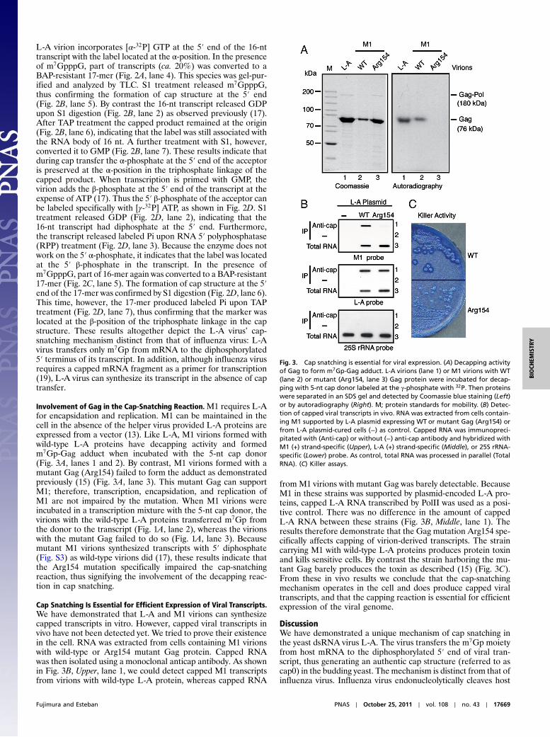
L-A virion incorporates [α-32P] GTP at the 5′ end of the 16-nttranscript with the label located at the α-position. In the presenceof m7GpppG, part of transcripts (ca. 20%) was converted to aBAP-resistant 17-mer (Fig. 2A, lane 4). This species was gel-pur-ified and analyzed by TLC. S1 treatment released m7GpppG,thus confirming the formation of cap structure at the 5′ end(Fig. 2B, lane 5). By contrast the 16-nt transcript released GDPupon S1 digestion (Fig. 2B, lane 2) as observed previously (17).After TAP treatment the capped product remained at the origin(Fig. 2B, lane 6), indicating that the label was still associated withthe RNA body of 16 nt. A further treatment with S1, however,converted it to GMP (Fig. 2B, lane 7). These results indicate thatduring cap transfer the α-phosphate at the 5′ end of the acceptoris preserved at the α-position in the triphosphate linkage of thecapped product. When transcription is primed with GMP, thevirion adds the β-phosphate at the 5′ end of the transcript at theexpense of ATP (17). Thus the 5′ β-phosphate of the acceptor canbe labeled specifically with [γ-32P] ATP, as shown in Fig. 2D. S1treatment released GDP (Fig. 2D, lane 2), indicating that the16-nt transcript had diphosphate at the 5′ end. Furthermore,the transcript released labeled Pi upon RNA 5′ polyphosphatase(RPP) treatment (Fig. 2D, lane 3). Because the enzyme does notwork on the 5′ α-phosphate, it indicates that the label was locatedat the 5′ β-phosphate in the transcript. In the presence ofm7GpppG, part of 16-mer again was converted to a BAP-resistant17-mer (Fig. 2C, lane 5). The formation of cap structure at the 5′end of the 17-mer was confirmed by S1 digestion (Fig. 2D, lane 6).This time, however, the 17-mer produced labeled Pi upon TAPtreatment (Fig. 2D, lane 7), thus confirming that the marker waslocated at the β-position of the triphosphate linkage in the capstructure. These results altogether depict the L-A virus’ cap-snatching mechanism distinct from that of influenza virus: L-Avirus transfers only m7Gp from mRNA to the diphosphorylated5′ terminus of its transcript. In addition, although influenza virusrequires a capped mRNA fragment as a primer for transcription(19), L-A virus can synthesize its transcript in the absence of captransfer.
Involvement of Gag in the Cap-Snatching Reaction.M1 requires L-Afor encapsidation and replication. M1 can be maintained in thecell in the absence of the helper virus provided L-A proteins areexpressed from a vector (13). Like L-A, M1 virions formed withwild-type L-A proteins have decapping activity and formedm7Gp-Gag adduct when incubated with the 5-nt cap donor(Fig. 3A, lanes 1 and 2). By contrast, M1 virions formed with amutant Gag (Arg154) failed to form the adduct as demonstratedpreviously (15) (Fig. 3A, lane 3). This mutant Gag can supportM1; therefore, transcription, encapsidation, and replication ofM1 are not impaired by the mutation. When M1 virions wereincubated in a transcription mixture with the 5-nt cap donor, thevirions with the wild-type L-A proteins transferred m7Gp fromthe donor to the transcript (Fig. 1A, lane 2), whereas the virionswith the mutant Gag failed to do so (Fig. 1A, lane 3). Becausemutant M1 virions synthesized transcripts with 5′ diphosphate(Fig. S3) as wild-type virions did (17), these results indicate thatthe Arg154 mutation specifically impaired the cap-snatchingreaction, thus signifying the involvement of the decapping reac-tion in cap snatching.
Cap Snatching Is Essential for Efficient Expression of Viral Transcripts.We have demonstrated that L-A and M1 virions can synthesizecapped transcripts in vitro. However, capped viral transcripts invivo have not been detected yet. We tried to prove their existencein the cell. RNA was extracted from cells containing M1 virionswith wild-type or Arg154 mutant Gag protein. Capped RNAwas then isolated using a monoclonal anticap antibody. As shownin Fig. 3B, Upper, lane 1, we could detect capped M1 transcriptsfrom virions with wild-type L-A protein, whereas capped RNA
from M1 virions with mutant Gag was barely detectable. BecauseM1 in these strains was supported by plasmid-encoded L-A pro-teins, capped L-A RNA transcribed by PolII was used as a posi-tive control. There was no difference in the amount of cappedL-A RNA between these strains (Fig. 3B, Middle, lane 1). Theresults therefore demonstrate that the Gag mutation Arg154 spe-cifically affects capping of virion-derived transcripts. The straincarrying M1 with wild-type L-A proteins produces protein toxinand kills sensitive cells. By contrast the strain harboring the mu-tant Gag barely produces the toxin as described (15) (Fig. 3C).From these in vivo results we conclude that the cap-snatchingmechanism operates in the cell and does produce capped viraltranscripts, and that the capping reaction is essential for efficientexpression of the viral genome.
DiscussionWe have demonstrated a unique mechanism of cap snatching inthe yeast dsRNA virus L-A. The virus transfers the m7Gp moietyfrom host mRNA to the diphosphorylated 5′ end of viral tran-script, thus generating an authentic cap structure (referred to ascap0) in the budding yeast. The mechanism is distinct from that ofinfluenza virus. Influenza virus endonucleolytically cleaves host
Fig. 3. Cap snatching is essential for viral expression. (A) Decapping activityof Gag to form m7Gp-Gag adduct. L-A virions (lane 1) or M1 virions with WT(lane 2) or mutant (Arg154, lane 3) Gag protein were incubated for decap-ping with 5-nt cap donor labeled at the γ-phosphate with 32P. Then proteinswere separated in an SDS gel and detected by Coomassie blue staining (Left)or by autoradiography (Right). M; protein standards for mobility. (B) Detec-tion of capped viral transcripts in vivo. RNA was extracted from cells contain-ing M1 supported by L-A plasmid expressing WT or mutant Gag (Arg154) orfrom L-A plasmid-cured cells (−) as control. Capped RNA was immunopreci-pitated with (Anti-cap) or without (−) anti-cap antibody and hybridized withM1 (+) strand-specific (Upper), L-A (+) strand-specific (Middle), or 25S rRNA-specific (Lower) probe. As control, total RNA was processed in parallel (TotalRNA). (C) Killer assays.
Fujimura and Esteban PNAS ∣ October 25, 2011 ∣ vol. 108 ∣ no. 43 ∣ 17669
BIOCH
EMISTR
Y
mRNA 10–13 nt downstream from the 5′ end and utilizes theresulting capped oligo fragment as a primer to synthesize its tran-script. The L-A cap-snatching mechanism is essential for efficientexpression of viral transcripts and, strikingly, His154 of Gag isinvolved in this process. In influenza virus the trimeric polymer-ase performs cap snatching. It is surprising to find a unique cap-snatching activity in the structural coat protein Gag. However,considering that the Pol domain of the Gag-Pol fusion proteinis confined inside the virion and unable to access mRNA in thecytoplasm, it is quite reasonable that Gag but not Pol performsthe reaction. Thus these findings exemplify the versatility andadaptability of eukaryotic RNA viruses.
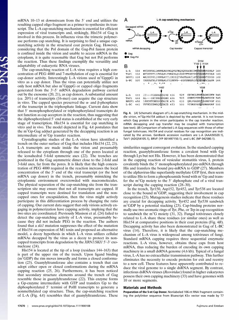
The cap-snatching reaction of L-A virus requires a high con-centration of PEG 4000 and 7-methylation of cap is essential forcap-donor activity. Interestingly L-A virions used m7GpppG invitro as a cap donor. Thus the virus can potentially utilize notonly host mRNA but also m7GpppG or capped oligo fragmentsgenerated from the 3′-5′ mRNA degradation pathway carriedout by the exosome (20, 21), as cap donors. A substantial amount(ca. 20%) of transcripts (16-mer) can acquire the cap structurein vitro. The capped species preserved the α- and β-phosphatesof the transcript in the triphosphate linkage. Current data showthat 5′ monophosphorylated or triphosphorylated transcripts donot function as cap acceptors in the reaction, thus suggesting thatthe diphosphorylated 5′ end status is established at the very earlystage of transcription. His154 is essential for cap snatching invitro and in vivo. The model depicted in Fig. 4A postulates thatthe m7Gp-Gag adduct generated by the decapping reaction is anintermediate of m7Gp transfer reaction.
Crystallographic studies of the L-A virion have identified atrench on the outer surface of Gag that includes His154 (22, 23).L-A transcripts are made inside the virion and presumablyreleased to the cytoplasm through one of the pores located atthe icosahedral 5-fold symmetric axes (22). The trenches arepositioned in the Gag asymmetric dimer close to the 2-fold and3-fold axes, far from the pores. It is likely that the high concen-tration of PEG 4000 required in the reaction increases the localconcentration of the 5′ end of the viral transcript (or the hostmRNA cap donor) in the trench, presumably mimicking thecytoplasmic environment overcrowded with macromolecules.The physical separation of the cap-snatching site from the tran-scription site may ensure that not all transcripts are capped. Ifcapped transcripts were destined for translation and the non-capped ones for encapsidation, then the virus might activelyparticipate in this differentiation process by changing the ratioof capping. Our current data suggest that only virions actively en-gaging in polymerization have capping activity, implying that thetwo sites are coordinated. Previously Masison et al. (24) failed todetect the cap-snatching activity of L-A virus, presumably be-cause they did not include PEG in the reaction. The authorsfound that a ski1 mutation suppresses the effect of the mutationof His154 on expression of M1 toxin and proposed an alternativemodel, a decoy hypothesis in which L-A virus utilizes cellularmRNAs decapped by the virus as a decoy to protect its non-capped transcripts from degradation by the XRN1/SKI1 5′-3′ exo-nuclease (24).
His154 is located at the tip of a loop (residues 144–163) thatis part of the upper rim of the trench. Upon ligand binding(m7GDP) the rim moves inwardly and forms a closed conforma-tion (23). Guanylyltransferase also contains a trench that canadopt either open or closed conformations during the mRNAcapping reaction (25, 26). Furthermore, it has been noticedthat secondary structure elements around the trench of Gagresemble those in guanylyltransferase (22). This enzyme formsa Gp-enzyme intermediate with GTP and transfers Gp to thediphosphorylated 5′ termini of PolII transcripts to generate anonmethylated cap structure. Thus, the cap-snatching reactionof L-A (Fig. 4A) resembles that of guanylyltransferase. These
similarities suggest convergent evolution. In the standard cappingreaction, guanylyltransferase forms a covalent bond with Gpthrough Lys but not His. However, it has been demonstrated thatin the capping reaction of vesicular stomatitis virus, L proteincovalently binds the 5′ monophosphorylated pre-mRNA throughHis and transfers the bound pre-mRNA to GDP (27). Membersof the alphavirus-like superfamily methylate GTP first, then seemto utilize His to form a phosphoamide bond with m7Gp and trans-fer the m7Gp moiety to the 5′ diphosphate end of a viral tran-script during the capping reaction (28–30).
In the trench, Tyr150, Asp152, Tyr452, and Tyr538 are locatedclose to the bound m7GDP, suggesting their involvement in caprecognition (23). Mutagenesis studies indicate that these residuesare crucial for decapping activity. Tyr452 and Tyr538 sandwichm7GDP by a potential stacking (23). Cap-binding proteins nor-mally use two aromatic rings of Tyr, Phe, or Trp in cap recognitionto sandwich the m7G moiety (31, 32). Fungal totiviruses closelyrelated to L-A share these residues (or similar ones) as well asHis154 at comparative positions in their coat proteins (Fig. 4B).Decapping activity has also been demonstrated in Gag of L-BCvirus (14). Therefore, it is likely that the cap-snatching me-chanism of L-A virus is widespread among totiviruses of fungi.Standard mRNA capping requires three sequential enzymaticreactions. L-A virus, however, obtains these caps from hostmRNA, thus reducing the burden of encoding its own cappingmachinery in a small dsRNA genome (4.6 kb). Typical of a fungalvirus, L-A has no extracellular transmission pathway. This furthereliminates the necessity to encode proteins for exit and reentryto a new cell. These features have apparently contributed to re-duce the viral genome to a single dsRNA segment. By contrast,infectious dsRNA viruses (Reoviridae) found in higher eukaryotespossess their own capping machinery (33) and have genomes with10 or more segments.
Materials and MethodsPreparation of the 5-nt Cap Donor. Nonlabeled 106-nt RNA fragment contain-ing the polylinker sequence from Bluescript KS+ vector was made by T7
Fig. 4. (A) Schematic diagram of L-A cap-snatching mechanism. In the mid-dle virion, m7Gp-His154 adduct is depicted by the asterisk. It is not knownwhich Gag protein in the virion participates in the cap transfer reaction.mRNA decapping and cap transfer may be coupled with transcription(see text). (B) Comparison of selected L-A Gag sequences with those of otherfungal totiviruses. His154 and crucial residues for cap recognition are indi-cated by the arrows. GenBank accession numbers are L-A (AAA50320.1),TaV1 (ADQ54105.1), BRVF (YP 001497150.1), and L-BC (NP 042580.1).
17670 ∣ www.pnas.org/cgi/doi/10.1073/pnas.1111900108 Fujimura and Esteban
runoff transcription. The cap structure was installed on it using [α-32P] GTPwith the aid of mScript mRNA production system (Epicentre) following theinstructions provided by the manufacturer. The 5-nt cap donor wasgenerated by digesting it with RNase A (14) and purified through a 15%acrylamide gel.
Cap Transfer Reaction. The standard cap transfer reaction contained 50 mMTris HCl pH 7.5, 5 mM MgCl2, 0.1 mM EDTA, 20 mM NaCl, 5 mM KCl, 0.5 mMeach of ATP, CTP, GTP, and UTP, 20% PEG 4000, and labeled 5-nt cap donor(2–5,000 cpm). The reaction was started by the addition of L-A or M1 virionsand kept at 30 °C for 1 h. Phenol-extracted products were analyzed on eitheragarose or acrylamide gels. TLC using PEI-cellulose was done as described(17). Decapping reaction was done as described (14).
Immunoprecipitation with Anticap Monoclonal Antibody. Freshly growing cells(109 cells) were harvested and broken by vortex mixing (15 s, 10 times) with
glass beads in a buffer containing 20 mM Tris HCl, pH 8.0 and 100 mM NaCl.After removing cell debris, RNA was extracted with phenol from the super-natant and precipitated with ethanol. RNA was incubated for 30 min at 4 °Cwith 30 μL of protein G-Sepharose (GE Healthcare) prebound to anti-m3G∕m7G-cap monoclonal antibody (Synaptic Systems). RNA bound to theSepharose was extracted, blotted to a nylon membrane, and hybridized withan L-A-, M1-, or 25S rRNA-specific probe as described previously (34). The L-Aand M1 probes recognize the positive strand sequences of L-A (nt 1,323–1,786) and M1 (nt 14–500), respectively. The 25S rRNA probe recognizes nt139–608 of 25S rRNA.
ACKNOWLEDGMENTS. We thank Reed B. Wickner (National Institutes ofHealth, Bethesda, MD) for providing the L-A-free killer strains. This workwas supported by Grants BFU2007–60057 and BFU2010–15768 from theSpanish Ministry of Education and Science.
1. Muthukrishnan S, Both GW, Furuichi Y, Shatkin AJ (1975) 5′-Terminal 7-methylguano-sine in eukaryotic mRNA is required for translation. Nature 255:33–37.
2. Furuichi Y, LaFiandra A, Shatkin AJ (1977) 5′-Terminal structure and mRNA stability.Nature 266:235–239.
3. Venkatesan S, Gershowitz A, Moss B (1980) Modification of the 5′ end of mRNA As-sociation of RNA triphosphatase with the RNA guanylyltransferase-RNA (guanine-7-)methyltransferase complex from vaccinia virus. J Biol Chem 255:903–908.
4. Shuman S (1995) Capping enzyme in eukaryotic mRNA synthesis. Prog Nucleic Acid ResMol Biol 50:101–129.
5. Plotch SJ, BouloyM, Ulmanen I, Krug RM (1981) A unique cap(m7GpppXm)-dependentinfluenza virion endonuclease cleaves capped RNAs to generate the primers thatinitiate viral RNA transcription. Cell 23:847–858.
6. Guilligay D, et al. (2008) The structural basis for cap binding by influenza viruspolymerase subunit PB2. Nat Struct Mol Biol 15:500–506.
7. Boivin S, Cusack S, Ruigrok RW, Hart DJ (2010) Influenza A virus polymerase: Structuralinsights into replication and host adaptation mechanisms. J Biol Chem285:28411–28417.
8. Wickner RB (2007) Fields Virology, eds DM Knipe and PM Howley (Lippincott Williansand Wilkins, Philadelphia), 5th Ed,, pp 737–768.
9. Icho T,Wickner RB (1989) The double-stranded RNA genome of yeast virus L-A encodesits own putative RNA polymerase by fusing two open reading frames. J Biol Chem264:6716–6723.
10. Dinman JD, Icho T, Wickner RB (1991) A-1 ribosomal frameshift in a double-strandedRNA virus of yeast forms a gag-pol fusion protein. Proc Natl Acad Sci USA 88:174–178.
11. Fujimura T, Ribas JC, Makhov AM, Wickner RB (1992) Pol of gag-pol fusion proteinrequired for encapsidation of viral RNA of yeast L-A virus. Nature 359:746–749.
12. Bostian KA, et al. (1984) Sequence of the preprotoxin dsRNA gene of type I killer yeast:Multiple processing events produce a two-component toxin. Cell 36:741–751.
13. Wickner RB, Icho T, Fujimura T, Widner WR (1991) Expression of yeast L-A double-stranded RNA virus proteins produces derepressed replication: A ski- phenocopy. J Vir-ol 65:155–161.
14. Blanc A, Goyer C, Sonenberg N (1992) The coat protein of the yeast double-strandedRNA virus L-A attaches covalently to the cap structure of eukaryotic mRNA. Mol CellBiol 12:3390–3398.
15. Blanc A, Ribas JC, Wickner RB, Sonenberg N (1994) His-154 is involved in the linkage ofthe Saccharomyces cerevisiae L-A double-stranded RNA virus Gag protein to the Capstructure of mRNAs and is essential for M1 satellite virus expression. Mol Cell Biol14:2664–2674.
16. Fujimura T, Esteban R, Wickner RB (1986) In vitro L-A double-stranded RNA synthesisin virus-like particles from Saccharomyces cerevisiae. Proc Natl Acad Sci USA83:4433–4437.
17. Fujimura T, Esteban R (2010) Yeast double-stranded RNA virus L-A deliberately synthe-sizes RNA transcripts with 5′-diphosphate. J Biol Chem 285:22911–22918.
18. Fujimura T, Wickner RB (1989) Reconstitution of template-dependent in vitro tran-scriptase activity of a yeast double-stranded RNA virus. J Biol Chem 264:10872–10877.
19. Yuan P, et al. (2009) Crystal structure of an avian influenza polymerase PA(N) reveals anendonuclease active site. Nature 458:909–913.
20. Mitchell P, Petfalski E, Shevchenko A, MannM, Tollervey D (1997) The exosome: A con-served eukaryotic RNA processing complex containing multiple 30 → 50 exoribonu-cleases. Cell 91:457–466.
21. Schaeffer D, Clark A, Klauer AA, Tsanova B, van Hoof A (2011) Functions of the cyto-plasmic exosome. Adv Exp Med Biol 702:79–90.
22. Naitow H, Tang J, Canady M, Wickner RB, Johnson JE (2002) L-A virus at 3.4 A resolu-tion reveals particle architecture and mRNA decapping mechanism. Nat Struct Biol9:725–728.
23. Tang J, et al. (2005) The structural basis of recognition and removal of cellularmRNA 7-methyl G ‘caps’ by a viral cased protein: A unique viral response to hostdefense. J Mol Recognit 18:158–168.
24. Masison DC, et al. (1995) Decoying the cap-mRNA degradation system by a double-stranded RNA virus and poly(A)- mRNA surveillance by a yeast antiviral system. MolCell Biol 15:2763–2771.
25. HÅkansson K, Doherty AJ, Shuman S, Wigley DB (1997) X-ray crystallography revealsa large conformational change during guanyl transfer by mRNA capping enzymes.Cell 89:545–553.
26. Chu C, et al. (2011) Structure of the guanylyltransferase domain of human mRNAcapping enzyme. Proc Natl Acad Sci USA 108:10104–10108.
27. Ogino T, Yadav SP, Banerjee AK (2010) Histidine-mediated RNA transfer to GDP forunique mRNA capping by vesicular stomatitis virus RNA polymerase. Proc Natl AcadSci USA 107:3463–3468.
28. Ahola T, Laakkonen P, Vihinen H, Kääriäinen L (1997) Critical residues of Semliki Forestvirus RNA capping enzyme involved in methyltransferase and guanylyltransferase-likeactivities. J Virol 71:392–397.
29. Ahola T, Ahlquist P (1999) Putative RNA capping activities encoded by brome mosaicvirus: methylation and covalent binding of guanylate by replicase protein 1a. J Virol73:10061–10069.
30. Huang YL, Hsu YH, Han YT, MengM (2005) mRNA guanylation catalyzed by the S-ade-nosylmethionine-dependent guanylyltransferase of bamboo mosaic virus. J Biol Chem280:13153–13162.
31. Marcotrigiano J, Gingras AC, Sonenberg N, Burley SK (1997) Cocrystal structure ofthe messenger RNA 5′ cap-binding protein (eIF4E) bound to 7-methyl-GDP. Cell89:951–961.
32. Fechter P, Brownlee GG (2005) Recognition ofmRNA cap structures by viral and cellularproteins. J Gen Virol 86:1239–1249.
33. Luongo CL, Reinisch KM, Harrison SC, Nibert ML (2000) Identification of the guanylyl-transferase region and active site in reovirus mRNA capping protein lambda2. J BiolChem 275:2804–2810.
34. Fujimura T, Esteban R (2007) Interactions of the RNA polymerase with the viralgenome at the 5′- and 3′-ends contribute to 20S RNA narnavirus persistence in yeast.J Biol Chem 282:19011–19019.
Fujimura and Esteban PNAS ∣ October 25, 2011 ∣ vol. 108 ∣ no. 43 ∣ 17671
BIOCH
EMISTR
Y