capn5 mutation in hereditary uveitis: the r243l mutation increases
TRANSCRIPT

OR I G INA L ART I C L E
CAPN5 mutation in hereditary uveitis: the R243Lmutation increases calpain catalytic activity andtriggers intraocular inflammation in a mouse modelKatherine J.Wert1,2, Alexander G. Bassuk5,Wen-HsuanWu1, Lokesh Gakhar6,7,Diana Coglan8,9, MaryAnn Mahajan8,9, Shu Wu5, Jing Yang7,8, Chyuan-ShengLin3, Stephen H. Tsang1,2,4,* and Vinit B. Mahajan8,9,*1Barbara and Donald Jonas Laboratory of Stem Cells and Regenerative Medicine and Bernard and Shirlee BrownGlaucoma Laboratory, Edward S. Harkness Eye Institute, 2Institute of HumanNutrition, College of Physicians andSurgeons, 3Herbert Irving Comprehensive Cancer Center, 4Department of Pathology and Cell Biology, College ofPhysicians and Surgeons, Columbia University, New York, NY, USA, 5Department of Pediatrics and Neurology,6Department of Biochemistry, 7Protein Crystallography Facility, 8Omics Laboratory and 9Department ofOphthalmology and Visual Sciences, University of Iowa, Iowa City, IA, USA
*Towhom correspondence should be addressed at: Department of Ophthalmology and Visual Sciences, the University of Iowa, 200 Hawkins Drive, Iowa City,IA 52242, USA. Tel: +1 3194675151; Email:[email protected] (V.B.M.); Edward S. Harkness Eye Institute, Columbia University, NewYork, NY 10032, USA.Tel: +1 2123421189; Email: [email protected] (S.H.T.)
AbstractAsingle aminoacidmutationnear the active site of theCAPN5proteasewas linked to the inherited blindingdisorder, autosomaldominant neovascular inflammatory vitreoretinopathy (ADNIV, OMIM #193235). In homology modeling with other calpains,this R243L CAPN5 mutation was situated in a mobile loop that gates substrate access to the calcium-regulated active site. Inin vitro activity assays, the mutation increased calpain protease activity and made it far more active at low concentrations ofcalcium. To test whether the disease allele could yield an animal model of ADNIV, we created transgenic mice expressinghuman (h) CAPN5R243L only in the retina. The resulting hCAPN5R243L transgenic mice developed a phenotype consistent withhuman uveitis and ADNIV, at the clinical, histological andmolecular levels. The fundus of hCAPN5R243L mice showed enhancedautofluorescence (AF) and pigment changes indicative of reactive retinal pigment epithelial cells and photoreceptordegeneration. Electroretinography showed mutant mouse eyes had a selective loss of the b-wave indicating an inner-retinasignalingdefect. Histological analysis ofmutantmouse eyes showedprotein extravasation fromdilated vessels into the anteriorchamber and vitreous, vitreous inflammation, vitreous and retinal fibrosis and retinal degeneration. Analysis of geneexpression changes in the hCAPN5R243L mouse retina showed upregulation of several markers, including members of theToll-like receptor pathway, chemokines and cytokines, indicative of both an innate and adaptive immune response. Sincemanyforms of uveitis share phenotypic characteristics of ADNIV, thismouse offers amodelwith therapeutic testing utility for ADNIVand uveitis patients.
Received: March 1, 2015. Revised and Accepted: May 18, 2015
© The Author 2015. Published by Oxford University Press. All rights reserved. For Permissions, please email: [email protected]
Human Molecular Genetics, 2015, Vol. 24, No. 16 4584–4598
doi: 10.1093/hmg/ddv189Advance Access Publication Date: 20 May 2015Original Article
4584
Downloaded from https://academic.oup.com/hmg/article-abstract/24/16/4584/745418by gueston 29 March 2018

IntroductionThe molecular basis of uveitis (intraocular inflammation) ispoorly understood (1–7). Until recently, primary (non-syndromic)uveitis was not linked to any causative gene, since families withinherited uveitis are rare.We ascertained two large kindredswithan inherited uveitis; autosomal dominant neovascular inflam-matory vitreoretinopathy (ADNIV, OMIM #193235). ADNIV pa-tients’ eyes develop a severe autoinflammatory uveitis andretinal degeneration with vitreous inflammation, vitreous andretinal fibrosis, cataract, cystoid macular edema and retinal neo-vascularization. Our studies of human autopsy eyes indicatedthat ADNIV uveitis is primarily driven by cell-mediated immun-ity (8,9). Proteomic profiling of vitreous biopsies revealed thatinterleukin-6 (IL-6) was elevated in ADNIV, suggesting a stimu-lated inflammatory or autoimmune response (10). Nevertheless,like many other uveitis patients, ADNIV patients respond poorlyto conventional immunosuppressive therapies and incompletelyto corticosteroids (11).
At different disease stages, ADNIVmimics severe, progressiveuveitis, retinitis pigmentosa, proliferative diabetic retinopathyand proliferative vitreoretinopathy (8,9,11–16), a set of eye dis-eases that account for a large fraction of visual morbidity andblindness (17). Discoveries about the causes of ADNIV mighthave therapeutic implications for a wide variety of ocular dis-eases. To find causative genes for ADNIV, two ADNIV kindredswere analyzed via whole exome sequencing, and each harboreda uniquemissensemutation in the coding region of CAPN5, a cal-cium-activated cysteine protease expressed in photoreceptorcells (18,19). We recently identified a third ADNIV family (20),and all three mutations lie in the catalytic domain of CAPN5(21,22). Our homology modeling to calpains generated a three-dimensional structure for calpain-5 (23–26), which showed allmutations were in a calcium-sensitive, flexible loop that gatessubstrate access to the active site (20,25,27). However, the effectof the mutation on calpain activity was not known until it wastested in this study.
Despite CAPN5 expression in multiple tissues, ADNIV patientsonly develop disease in the eyes. It is not clear whether disease isdriven by the immune system or by the retina. Retina-specificCAPN5 vitreoretinopathy was tested in a mouse model, using len-tiviral transduction to express one of the ADNIV human CAPN5mutant cDNAs, hCAPN5R243L (16). Lentiviral vectors were injectedinto the sub-retinal space of perinatal mice. Retinal expressionof the disease allele was sufficient to recapitulate the majorfeatures of the ADNIV phenotype: loss of the electroretinogram(ERG) b-wave, photoreceptor degeneration and inflammatorygene expression (16). Despite these changes, the inflammatoryphenotypewas limited compared with the human disease pheno-type, likely because the lentivirus deliverymethod, although rapid,has a number of shortcomings. First, effects are usually localizednear the site of injection and the number of transduced cells islow (typically 20–30% of cells). Thus it was not surprising that thelentivirus model caused a less severe inflammatory response (16),a limited loss of ERG function, and death in a minor fraction ofphotoreceptors and cells lining the inner nuclear layer of the eye.In addition, with lentiviral transduction, the mutation was nottransmitted through generations, so the system offers a limitedutility for testing therapeutics. Given the encouraging resultsof ERG b-wave reduction, photoreceptor degeneration and inflam-matory gene expression with the hCAPN5R243L lentiviral studies,we were optimistic that a transgenic mouse expressing thehCAPN5R243L transgene in the retina would provide a better modelof the ADNIV disease.
ResultsThe ADNIV DII gating-loop mutation increases CAPN5catalytic activity
All calpains have a proteolytic core with two subdomains(domains IIa and IIb) that contain four flexible loops that undergosignificant conformational changes upon binding calcium inCAPN1 (Fig. 1A), shifting the enzyme into an active form(26,28,29). Although areas of domain II (DII) are highly conserved,CONSURF analysis (data not shown) and previous studies indi-cate that the mobile loops are among the more variable featuresnear calpain active sites (23), which may give the calpain familyitswide range of calcium sensitivity and substrate specificity. Thefour flexible loops are highlighted in the sequence alignmentcomparing CAPN5 to CAPN1 (Fig. 1B). In both ADNIV cohorts,the second flexible loop in DIIb contains a CAPN5 pointmutation.This loop-2 has been implicated in gating substrate access tothe catalytic groove (23,26). Calcium binds to other loops withinthe proteolytic core (green spheres), eliciting a conformationalchange in the DIIb gating loop-2 that opens the catalytic grooveand activates the enzyme (23,26,28,29).
Based on structural modeling (13), our hypothesis was that aCAPN5 gain-of-function might arise if the ADNIV mutations dis-rupt the DII gating loop-2 and increase substrate access to thecatalytic groove. We aimed to recreate the disease scenario inmice, but first needed to confirm the gain-of-function phenotypefor a DIIb gating loop-2 mutation. Testing calpain enzymatic ac-tivity in vitro, however, is not straightforward, because purifiedCAPN5 is inherently unstable (unpublished observation), andits substrates are not known (so no in vitro activity assay hasbeen developed). To circumvent these problems, we designed aCAPN1/5 hybrid, in which the DIIb gating loop-2 from humanCAPN5 (amino acids KAVTAADMEARLACG) replaced the corre-sponding sequence in the proteolytic core of a rat mini-CAPN1(NISDIRDLEAITFKN; Fig. 1C). This hybrid mini-calpain strategyhas beenused previously to purify and test the enzymatic activityof other calpains (23) and takes advantage of a robust purificationmethod and established activity assay for rat mini-CAPN1.
Mini-CAPN1/5 hybrids were created in three maltose-bindingprotein (MBP)-tagged forms: one containing the wild-type (WT)CAPN5DIIb gating loop-2, a form expected to be catalytically active;another, carrying a p.C81S mutation at the catalytic triad, whichwas expected to be inactive; and a third carrying the disease-causing mutation (p.R243L), which we hypothesized would showa gain-of-function. Introduction of the CAPN5 gating loop did notaffect cleavage activity. The CAPN1/5 WT hybrid (positive control)robustly cleaved a fluorescent, peptide substrate (AC-LLY-AFC;Fig. 2A and B) recognized by mini-CAPN1. Adding a peptide sub-strate inhibitor abolished all cleavage activity (Fig. 2A). In addition,the negative-control p.C81S mutant, with its defective catalytictriad, showed no activity (Fig. 2B). These controls demonstratedthat the mini-CAPN1/5 hybrid with the DIIb gating loop-2 fromWT CAPN5 displays characteristic CAPN1 activity.
Next, the hybrid containing the p.R243L mutant DIIb gatingloop-2 was compared with the WT hybrid. The disease mutanthad significantly increased calpain catalytic activity at all timepoints and calcium concentrations tested compared with the WT(Fig. 3). This suggests that the gain-of-function mutation relievesCAPN5 of strict calcium regulation. Indeed, the mutant hybridcleaved almost as much substrate in the presence of 0.1 m cal-cium (Fig. 3B) as did the WT hybrid at 100 m calcium (Fig. 3D).These results support the assertion that the DIIb gating loop-2 con-trols calpain catalytic activity, and mutations in that loop can
Human Molecular Genetics, 2015, Vol. 24, No. 16 | 4585
Downloaded from https://academic.oup.com/hmg/article-abstract/24/16/4584/745418by gueston 29 March 2018

Figure 1. Generation of a recombinantmini-CAPN1/5 hybrid with loop 2 of proteolytic domain IIb (DIIb) swapped from CAPN5 into the homologous region inmini-CAPN1.
Themini-calpain systemallows the catalytic activity of domain II to be isolated and assayed, since full-length calpains are generally not stable. (A) Cartoon representation
of the rat mini-CAPN5 structure with calcium bound (green spheres), highlighting the active site residues (yellow sticks), the four flexible loops (pink) and the ADNIV
mutations (red dotted circle). (B) Sequence alignment of domain II of rat mini-CAPN1 and human mini-CAPN5 highlighting the flexible loops (pink box). (C) Schematic
representation showing proteins used in our modified mini-calpain system. Mini-CAPN1, a hybrid mini-CAPN1/5 with loop 2 of mini-CAPN1 swapped out with that of
mini-CAPN5 and a hybrid mini-CAPN1/5 harboring the p.R243L ADNIV mutation.
4586 | Human Molecular Genetics, 2015, Vol. 24, No. 16
Downloaded from https://academic.oup.com/hmg/article-abstract/24/16/4584/745418by gueston 29 March 2018

increase enzymatic catalysis.Moreover, these experiments stronglysuggest that the effect of the ADNIVmutation is to increase CAPN5catalytic activity, an ideaconsistentwith thedominant inheritance,
high penetrance and disease severity observed in mutant CAPN5vitreoretinopathy patients. This insight was next supported byphysiologic testing of the CAPN5R243L mutation in vivo.
Figure 2. A mini-CAPN1/5 hybrid is active in in vitro assays. A mini-CAPN1/5 is sensitive to a calpain inhibitor as well as mutation of the catalytic cysteine residue (C81S)
that renders all calpains enzymatically inactive. (A) Time course of substrate peptide (AC-LLY-AFC) shows catalytic activity of CAPN1/5 hybrid with (green) and without
(blue) a calpain peptide inhibitor (Z-LLY-AFC). (B) Catalytic activity of CAPN1/5 hybrid bearing amutation of the catalytic cysteine residue (C81S; green) renders it inactive.
RFU, relative fluorescent units.
Figure 3. R243LADNIVmutation increases catalytic activity of a recombinantmini-CAPN1/5 hybrid. The R243Lmutation renders themini-CAPN1/5 hybrid over 300%more
sensitive to calcium (activity is a ratio normalized to mini-CAPN1; control is the reaction without calpain). (A) A time-course comparison of the CAPN1/5 hybrid and a
hybrid bearing the R243L mutation, showing catalysis of a standard peptide substrate at 0.01 m calcium, (B) 0.10 m calcium, (C) 1.0 m calcium and (D) 100 m
calcium. RFU, relative fluorescent units.
Human Molecular Genetics, 2015, Vol. 24, No. 16 | 4587
Downloaded from https://academic.oup.com/hmg/article-abstract/24/16/4584/745418by gueston 29 March 2018
A transgenic mouse model expresses ADNIV-associatedhCAPN5R243L in the retina
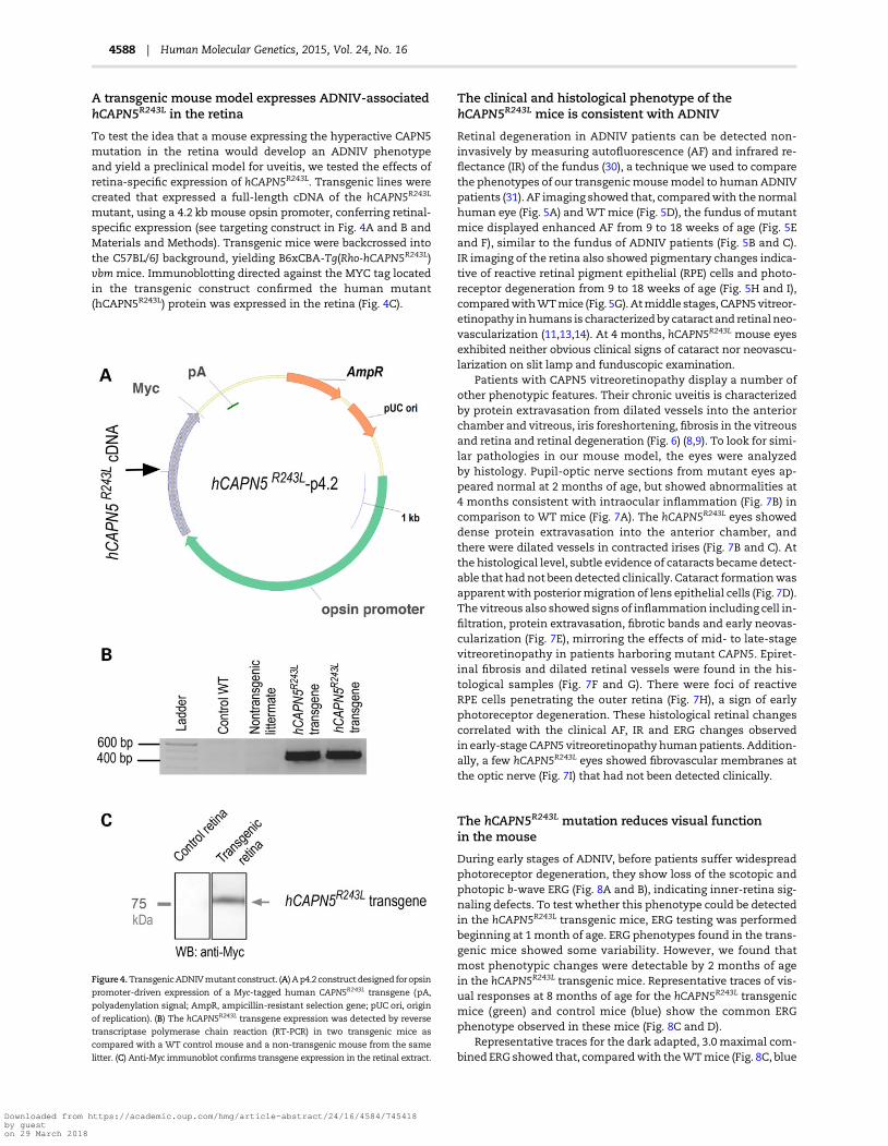
To test the idea that a mouse expressing the hyperactive CAPN5mutation in the retina would develop an ADNIV phenotypeand yield a preclinical model for uveitis, we tested the effects ofretina-specific expression of hCAPN5R243L. Transgenic lines werecreated that expressed a full-length cDNA of the hCAPN5R243L
mutant, using a 4.2 kb mouse opsin promoter, conferring retinal-specific expression (see targeting construct in Fig. 4A and B andMaterials and Methods). Transgenic mice were backcrossed intothe C57BL/6J background, yielding B6xCBA-Tg(Rho-hCAPN5R243L)vbm mice. Immunoblotting directed against the MYC tag locatedin the transgenic construct confirmed the human mutant(hCAPN5R243L) protein was expressed in the retina (Fig. 4C).
The clinical and histological phenotype of thehCAPN5R243L mice is consistent with ADNIV
Retinal degeneration in ADNIV patients can be detected non-invasively by measuring autofluorescence (AF) and infrared re-flectance (IR) of the fundus (30), a technique we used to comparethe phenotypes of our transgenicmousemodel to human ADNIVpatients (31). AF imaging showed that, comparedwith the normalhuman eye (Fig. 5A) andWTmice (Fig. 5D), the fundus of mutantmice displayed enhanced AF from 9 to 18 weeks of age (Fig. 5Eand F), similar to the fundus of ADNIV patients (Fig. 5B and C).IR imaging of the retina also showed pigmentary changes indica-tive of reactive retinal pigment epithelial (RPE) cells and photo-receptor degeneration from 9 to 18 weeks of age (Fig. 5H and I),comparedwithWTmice (Fig. 5G). Atmiddle stages, CAPN5vitreor-etinopathy inhumans is characterized by cataract and retinal neo-vascularization (11,13,14). At 4 months, hCAPN5R243L mouse eyesexhibited neither obvious clinical signs of cataract nor neovascu-larization on slit lamp and funduscopic examination.
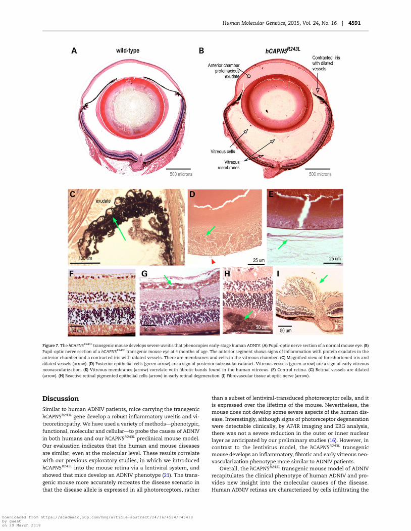
Patients with CAPN5 vitreoretinopathy display a number ofother phenotypic features. Their chronic uveitis is characterizedby protein extravasation from dilated vessels into the anteriorchamber and vitreous, iris foreshortening, fibrosis in the vitreousand retina and retinal degeneration (Fig. 6) (8,9). To look for simi-lar pathologies in our mouse model, the eyes were analyzedby histology. Pupil-optic nerve sections from mutant eyes ap-peared normal at 2 months of age, but showed abnormalities at4 months consistent with intraocular inflammation (Fig. 7B) incomparison to WT mice (Fig. 7A). The hCAPN5R243L eyes showeddense protein extravasation into the anterior chamber, andthere were dilated vessels in contracted irises (Fig. 7B and C). Atthe histological level, subtle evidence of cataracts became detect-able thathadnot beendetected clinically. Cataract formationwasapparentwith posteriormigration of lens epithelial cells (Fig. 7D).The vitreous also showed signs of inflammation including cell in-filtration, protein extravasation, fibrotic bands and early neovas-cularization (Fig. 7E), mirroring the effects of mid- to late-stagevitreoretinopathy in patients harboring mutant CAPN5. Epiret-inal fibrosis and dilated retinal vessels were found in the his-tological samples (Fig. 7F and G). There were foci of reactiveRPE cells penetrating the outer retina (Fig. 7H), a sign of earlyphotoreceptor degeneration. These histological retinal changescorrelated with the clinical AF, IR and ERG changes observedin early-stageCAPN5 vitreoretinopathyhumanpatients. Addition-ally, a few hCAPN5R243L eyes showed fibrovascular membranes atthe optic nerve (Fig. 7I) that had not been detected clinically.
The hCAPN5R243L mutation reduces visual functionin the mouse
During early stages of ADNIV, before patients suffer widespreadphotoreceptor degeneration, they show loss of the scotopic andphotopic b-wave ERG (Fig. 8A and B), indicating inner-retina sig-naling defects. To test whether this phenotype could be detectedin the hCAPN5R243L transgenic mice, ERG testing was performedbeginning at 1month of age. ERG phenotypes found in the trans-genic mice showed some variability. However, we found thatmost phenotypic changes were detectable by 2 months of agein the hCAPN5R243L transgenic mice. Representative traces of vis-ual responses at 8 months of age for the hCAPN5R243L transgenicmice (green) and control mice (blue) show the common ERGphenotype observed in these mice (Fig. 8C and D).
Representative traces for the dark adapted, 3.0 maximal com-bined ERG showed that, comparedwith theWTmice (Fig. 8C, blue
Figure 4.TransgenicADNIVmutant construct. (A) Ap4.2 construct designed foropsin
promoter-driven expression of a Myc-tagged human CAPN5R243L transgene (pA,
polyadenylation signal; AmpR, ampicillin-resistant selection gene; pUC ori, origin
of replication). (B) The hCAPN5R243L transgene expression was detected by reverse
transcriptase polymerase chain reaction (RT-PCR) in two transgenic mice as
compared with a WT control mouse and a non-transgenic mouse from the same
litter. (C) Anti-Myc immunoblot confirms transgene expression in the retinal extract.
4588 | Human Molecular Genetics, 2015, Vol. 24, No. 16
Downloaded from https://academic.oup.com/hmg/article-abstract/24/16/4584/745418by gueston 29 March 2018

trace), the hCAPN5R243L transgenicmice generated b-waveswith asmaller maximal amplitude (Fig. 8C, green trace). In contrast, thea-wave was unchanged between the control and hCAPN5R243L
transgenic mice, similar to human ADNIV patients (Fig. 8A).Under photopic conditions, the maximal b-wave amplitude waslower in the hCAPN5R243L transgenic mice (Fig. 8D, green trace)compared with control mice (Fig. 8D, blue trace), and similar tothe b-wave reduction found in human ADNIV patients (Fig. 8B).This suggests hCAPN5R243L transgenicmice lose cone-specific vis-ual function. In a prior study, we reported a cone and inner-retinacell loss-of-function with lentiviral delivery of the single p.R243Lmutation in CAPN5within the mouse eye (16). Here, we see simi-lar effects, which additionally correlate with the ERG phenotypefound in ADNIV (32,33), before the appearance of widespreadphotoreceptor degeneration.
hCAPN5R243L transgenic mice display biomarkersof uveitis
We previously found lentiviral induction of hCAPN5R243L inmouse photoreceptor cells elevated expression of IL-6 mRNAin the mouse retina. IL-6 is a cytokine associated with uveitisand fibrosis (16) and ADNIV patients display high levels of
the IL-6 cytokine in their vitreous (10). To determine if the trans-genic hCAPN5R243Lmice showed elevated IL-6, we performed im-munohistochemistry for this profibrotic cytokine. Elevated IL-6was not seen in retinal cells, but the vitreous contained veryhigh levels of the cytokine in regions clustered near the parsplana and ciliary body (Fig. 9). This anatomic location is a keysite where ADNIV patients develop fibrosis despite corticoster-oid implantation (15), and the profibrotic properties of IL-6 andits localization may offer some explanation of this diseasephenotype.
CAPN5 is expressed in the photoreceptor cell and its nucleus(13), and it can influence transcription of genes that triggerinflammation (18,21,34). We previously found that lentiviraltransduction of the mutant CAPN5 alters inflammatory gene ex-pression, but gene expression could only be surveyed after thevirus was transduced and was found to correlate to late stagesof human ADNIV disease. This current transgenic hCAPN5R243L
mouse model allowed us to survey retinal inflammatory geneexpression at the earliest stages of disease. Thus, we used quan-titative PCR (qPCR) arrays to survey mouse retinal mRNA for 84inflammatory transcripts, at 1.5 months, before all the signs ofinflammatory retinal degeneration were clinically and histologi-cally detectable. We identified 11 genes that were significantly
Figure 5.AF and IR imaging of human andmouseWTand hCAPN5R243L retinas reveals early retinal degeneration. (A) The normal humanAF fundus image. (B) Mid- to late-
stage ADNIV eyes show increased AF that may be associated with photoreceptor or RPE cell dysfunction. (C) IR imaging of an ADNIV patient shows scattered pigment
accumulation (arrows). Image quality is limited by cataract and vitreous membranes. (D) AF image of a control WT mouse at 18 weeks of age. (E) AF imaging of
hCAPN5R243L mice at 9 weeks of age shows increased AF compared with the control. (F) Further autofluorescent increase is observed by 18 weeks of age, as seen in
human ADNIV eyes. (G) IR imaging of a control WT mouse fundus. (H) IR imaging of a hCAPN5R243L mouse fundus at 9 weeks of age, and (I) by 18 weeks of age
scattered, melanin-like pigment is accumulating in the hCAPN5R243L mouse fundus (arrow).
Human Molecular Genetics, 2015, Vol. 24, No. 16 | 4589
Downloaded from https://academic.oup.com/hmg/article-abstract/24/16/4584/745418by gueston 29 March 2018

upregulated compared with control mouse retinas (Fig. 10). Six ofthe 11 genes represented a novel Toll-like receptor (TLR) pathway(Tlr6, Tlr9, Il1a, Itgb2, Ltb and Myd88), components of which arepart of the innate immune system that have been implicated inmouse models of autoimmune uveitis (35). Other upregulatedgenes included chemokine (C-C motif ) ligands (Ccl2, Ccl7 andCcl11), signals shown to be upregulated in the RPE cells thathave come into contact with activated T-cells. The T-cell marker,Cd40, and the innate immunity gene, Crp, were also upregulatedin diseased eyes. Elevation of these transcripts might suggest
stimulated immune cells infiltrating from both the innate andadaptive arms of the immune system. Alternatively, retinal cellsthemselvesmight overexpress some of thesemolecules, since ex-pression of the TLR pathway has been shown inneurons aswell ascells of the immune system (36). These early changes in gene ex-pression implicate several potential drug targets. Four genes weredownregulated, including Fos, Bcl6, Il1rap and Nr3c1 (Fig. 10). Inter-estingly, Nr3c1 is a glucocorticoid receptor, and its lower expres-sion might explain why ADNIV patients have a limited clinicalresponse to glucocorticoid therapy (18,21,34).
Figure 6. Human ADNIV pathology. (A) Histology reveals a foreshortened, fibrotic iris with dilated vessels and exudate in the anterior chamber (visualized by Masson’s
trichrome stain). (B) A cataractous lens with lens epithelial cells (arrow) located along the posterior capsule (visualized by Masson’s trichrome stain). (C). RPE cells (arrow)
infiltrating the degenerating retina (visualized by H&E stain). (D) Vitreous containing fibrous membranes (arrow) and filled with exudate (visualized by H&E stain).
(E) Epiretinal membrane (arrow). (F) Fibrovascular membrane at the optic nerve (arrow). Scale bar = 250 μm.
4590 | Human Molecular Genetics, 2015, Vol. 24, No. 16
Downloaded from https://academic.oup.com/hmg/article-abstract/24/16/4584/745418by gueston 29 March 2018
DiscussionSimilar to human ADNIV patients, mice carrying the transgenichCAPN5R243L gene develop a robust inflammatory uveitis and vi-treoretinopathy.We have used a variety ofmethods—phenotypic,functional, molecular and cellular—to probe the causes of ADNIVin both humans and our hCAPN5R243L preclinical mouse model.Our evaluation indicates that the human and mouse diseasesare similar, even at the molecular level. These results correlatewith our previous exploratory studies, in which we introducedhCAPN5R243L into the mouse retina via a lentiviral system, andshowed that mice develop an ADNIV phenotype (21). The trans-genic mouse more accurately recreates the disease scenario inthat the disease allele is expressed in all photoreceptors, rather
than a subset of lentiviral-transduced photoreceptor cells, and itis expressed over the lifetime of the mouse. Nevertheless, themouse does not develop some severe aspects of the human dis-ease. Interestingly, although signs of photoreceptor degenerationwere detectable clinically, by AF/IR imaging and ERG analysis,there was not a severe reduction in the outer or inner nuclearlayer as anticipated by our preliminary studies (16). However, incontrast to the lentivirus model, the hCAPN5R243L transgenicmouse develops an inflammatory, fibrotic and early vitreous neo-vascularization phenotype more similar to ADNIV patients.
Overall, the hCAPN5R243L transgenic mouse model of ADNIVrecapitulates the clinical phenotype of human ADNIV and pro-vides new insight into the molecular causes of the disease.Human ADNIV retinas are characterized by cells infiltrating the
Figure 7. The hCAPN5R243L transgenic mouse develops severe uveitis that phenocopies early-stage human ADNIV. (A) Pupil-optic nerve section of a normalmouse eye. (B)Pupil-optic nerve section of a hCAPN5R243L transgenic mouse eye at 4 months of age. The anterior segment shows signs of inflammation with protein exudates in the
anterior chamber and a contracted iris with dilated vessels. There are membranes and cells in the vitreous chamber. (C) Magnified view of foreshortened iris and
dilated vessels (arrow). (D) Posterior epithelial cells (green arrow) are a sign of posterior subcasular cataract. Vitreous vessels (green arrow) are a sign of early vitreous
neovascularization. (E) Vitreous membranes (arrow) correlate with fibrotic bands found in the human vitreous. (F) Control retina. (G) Retinal vessels are dilated
(arrow). (H) Reactive retinal pigmented epithelial cells (arrow) in early retinal degeneration. (I) Fibrovascular tissue at optic nerve (arrow).
Human Molecular Genetics, 2015, Vol. 24, No. 16 | 4591
Downloaded from https://academic.oup.com/hmg/article-abstract/24/16/4584/745418by gueston 29 March 2018

vitreous (8,9); similarly, the hCAPN5R243Lmouse eyes showed cellspopulating the vitreous. In addition, our screen for early biomar-kers revealed ADNIV production of specific molecules, such as
TLR pathways, and these might be targeted therapeutically.Together, these findings suggest a disease model where molecu-lar factors in the retina are sufficient to trigger uveitis (Fig. 11).
Figure 8. Electroretinography shows hCAPN5R243L mice have a specific loss of the inner retinal cells and cone cell function. (A) ERG tracing of ADNIV patient shows
reduction of b-wave relative to the a-wave (14). The scotopic b-wave was 83% of normal (normal scotopic b-wave was 450 ± 100 μV in this system). (B) The photopic ERG
b-wave was ∼32.5% of normal (339 ± 85 μV). The calibration mark before stimulus shows 100 μV. (C) A representative ERG test on an 8 month-old hCAPN5R243L transgenic
mouse (green) compared with a control mouse (blue) displays the frequently observed functional phenotype. hCAPN5R243L transgenic mice have a reduced scotopic
maximal b-wave amplitude compared with control mice, with the a-wave remaining unchanged. (D) Photopic, cone-specific ERGs showed a reduction in the b-wave
maximal amplitude in the hCAPN5R243L transgenic mice compared with controls. Such a pattern correlates to ADNIV patients.
Figure 9. IL-6 is upregulated in the hCAPN5R243L transgenic mouse ciliary body. (A) Control IgG antibody. (B) No IL-6 expression in 4-month-oldWT vitreous, as assayed by
immunohistochemistry. (C) Vitreous IL-6 expression is upregulated in the hCAPN5R243L mouse anterior vitreous. CB, ciliary body; Vit, vitreous; R, retina. Scale bar = 25 μm.
4592 | Human Molecular Genetics, 2015, Vol. 24, No. 16
Downloaded from https://academic.oup.com/hmg/article-abstract/24/16/4584/745418by gueston 29 March 2018

Some of the biomarkers detected have been linked to othernon-inherited forms of uveitis and retinal degenerations (37–39), and hence we expect this transgenic ADNIV mouse modelto have utility for other types of human uveitis. Indeed, thehCAPN5R243L mouse offers distinctions over current animal mod-els of autoimmune uveitis, which are typically generated by ecto-pically expressing proteins in the retina (e.g. hen egg lysoszymeand beta-galactosidase) or injecting antigens and adjuvants(7,33,40). These models did not develop a constellation of path-ologies or naturally develop disease phenotypes without extramanipulation.
Furthermore, we provide evidence of the first example of anoveractive calpain causing a dominant, inherited disease. Onlyone other Mendelian disease has been shown so far to be causedby a mutant calpain: mutations in CAPN3 cause limb-musclegirdle dystrophy type 2A. In this case, however, the disease-associated mutation causes a calpain-3 loss-of-function (41).Excess calpain activity is associated with non-inherited patholo-gies, such as Alzheimer’s disease (calpain 1) and myocardial in-farction (calpains 1, 2 and 4) (18–21). Similarly, our biochemicalstudies demonstrate that the hCAPN5R243L mutation increasedcalpain activity, such that the mutant protease activity is not
Figure 10. The hCAPN5R243L transgenic retina shows early changes in inflammatory gene expression before any clinical or histological signs of disease. (A) Volcano plot
displays genes in the hCAPN5R243L retinas at post-natal day (P) 45 that had statistically significant expression changes that were greater than 2-fold (P < 0.05), when
compared with P45 control retinas. Four genes (Fos, Bcl6, IL1rap and Nr3c1) were downregulated (blue dots, upper left). (B) Schematic representation shows that several
members of the TLR pathways were among the upregulated inflammatory genes.
Figure 11. An integrated ADNIV disease model. (A) Histology of the CAPN5R243L uveitis model. (B) The CAPN5R243L disease allele transgene is expressed in retina
photoreceptors. Because of the high calcium levels in the retina for phototransduction, CAPN5 mutations in the retina are particularly sensitive. TLR signaling and
cyotokine secretion trigger retinal and iris vessel hyperpermeability and a cell-mediated autoinflammatory reaction that includes vitreous inflammation and fibrosis.
Human Molecular Genetics, 2015, Vol. 24, No. 16 | 4593
Downloaded from https://academic.oup.com/hmg/article-abstract/24/16/4584/745418by gueston 29 March 2018

only relieved from strict calcium regulation, it is also three ordersof magnitude more sensitive to calcium (Fig. 3). Given the majorchanges in the protein’s response to calcium, it is interesting thatthe hCAPN5R243L mutation is not in a calcium binding loop, butinstead, in loop 2, which likely gates access of substrates to thecalpain active site (Fig. 1). Interestingly, we have identified athird family with ADNIV (42), and linked their disease to anotherpoint mutation in CAPN5, also in the sequence encoding the DIIbmobile gating loop.
Among the calpains, CAPN5might be particularly susceptibleto mutational effects. The calcium residues in the catalytic do-main provide one layer of calcium dependence, but most cal-pains in higher vertebrates have evolved a second regulatorylayer by acquiring a calmodulin-like C-terminal domain (34). Incontrast, CAPN5 is a ‘non-classical’ calpain that most closely re-sembles Tra-3, a calpain ortholog conserved in flies and wormsthat regulates neuronal degeneration and sex determination. Inaddition, CAPN5 cannot form heterodimers with the regulatorysubunit CAPNS1, whichmight remove yet another layer of poten-tial regulation (18,26). Thus, CAPN5 might rely more heavily oncalcium binding in the catalytic domain to control proteolyticactivity than other members of the calpain family.
Since CAPN5 is preferentially expressed in the CNS (43), and isgenetically linkedwith retinal disease (13), generalized calpain orCAPN5-specific inhibition is a promising strategy through whichto develop future uveitis and retinal disease therapy. Moreover,oral delivery of these inhibitors to the retinamaymake thempar-ticularly effective for treating an eye disorder linked to overactiveCAPN5 (44). In a Caenorhabditis elegans genetic model, reducingTra-3 rescued genetic lesions that cause necrotic (inflammatory)neuronal degeneration (34). Similarly, inhibiting calpain with asmall-molecule inhibitor (SNJ-1945) rescued neuronal cell deathinmodels of traumatic brain injury (which elicit necrotic neuron-al degeneration) (45). The molecule also blocked experimentalautoimmune encephalitis (46), a condition that strongly parallelsuveitis. Calpain inhibitors additionally rescued retinal degener-ation in several other models, including models of light-inducedretinal toxicity (47,48), retinal hypoxia (49), retinitis pigmentosa(50–52), optic neuritis (53,54), diabetic retinopathy (55) and re-tinal angiogenesis (49,56). Since the inhibitors are not isoform-specific, and RNA sequencing showed expression of at leastseven different calpains in the retina (unpublished observation),the main limit of prior studies was that they could not pinpointthe critical calpain isoform. In contrast, our studies clearly linkretinal dysfunction to an overactive CAPN5 protease.
CAPN5 expression is highest in the nervous systembut our pa-tients do not present with any other diseases, including neuro-logical disease. ADNIV eye disease is the only manifestation ofthemutation. Retinal-specific diseasemight arise for several rea-sons. First, the extremely high levels of calcium in the retinacould make it more susceptible to a CAPN5 that is activated atvery low levels of calcium. Alternatively, the retina could be af-fected by the overactive CAPN5 because some other regulatorymechanism that is redundant in other tissues is lacking in theretina, allowing the ectopic activation of CAPN5 proteolysis.
A third reason for retina-specific diseasemight lie in an alteredCAPN5 target specificity. Thenatural calpain targets have beendif-ficult to determine, since there is no consensus cleavage site se-quence. The calpain family has multiple members and howtheir precise targetsmight vary is not currently known. In general,calpains have been shown to cleave a wide variety of exposedpeptide surfaces. Nevertheless, the structure and sequence ofthe calpain proteolytic core revealed that some level of sequencespecificity is likely found at the mobile loops of its substrate
binding site. Moreover, the loop 2 is among the more variable fea-tures among calpains, with each member having a distinctivelength and sequence (23). This variability has led others to hy-pothesize that themobile loops are good candidates for conferringsubstrate specificity (23). Since the three ADNIVmutationswe un-coveredeachaffect the sameDIIbmobile gating loop, theymaynotonly be relieving calcium regulation and boosting catalytic activitybut also changing CAPN5 targets, such that a retinal-specific pro-tein is pathologically cleaved, rendering the ADNIV phenotype.
To discriminate between these possibilities it will be advanta-geous to compare the specificity and sensitivity of proteolytic tar-gets for WT CAPN5 and mutant CAPN5R243L. Understanding howchanges in CAPN5 activity cause ADNIV should reveal informa-tion about the causes of other diseases linked to aberrant calpainactivity. For example, the true targets of calpain and how theyare affected by excess proteolysis is currently not clear. Withthis new hCAPN5R243L transgenicmousemodel in hand, however,it might be possible to begin to probe the substrate specificity ofcalpain proteases.
Materials and MethodsCAPN5 sequence and structure analysis
Using MODELLER 9.14 (57), a homology model of human mini-CAPN5 was generated as previously described (20), using thecrystal structure of human mini-CAPN9 as a template, whichwas the closest match in the Protein Database (PDB). An analysisof the conserved residues in the catalytic domain across variousmembers of the calpain family was performed using the CON-SURF server (58–61). The CONSURF scores were mapped to theB-factor column of mini-CAPN1 (PDB ID 2ARY) and visualizedusing PyMOL (http://www.pymol.org).
CAPN5 cloning, purification and activity assay
Sequences encoding the calpain-1/5 catalytic domain and theinactive calpain-1/5-C81S catalytic domain were cloned intopMal-c5x (New England Biolabs, Ipswich, MA) with a c-terminalHis tag. Mini-calpains were expressed as MBP-fusion proteins inBL21(DE3) E. coli and purified on an amylose resin (New EnglandBiolabs). At different calcium concentrations, purified calpaincatalytic activitywasmeasuredby cleavage of a peptide substrate(AC-LLY-AFC) and quantification on a fluorometric plate reader(Tecan Infinite M200 Pro, Männedorf, Switzerland) using a cal-pain activity assay and peptide inhibitor (Z-LLY-AFC) accordingto the manufacturer’s instructions (Abcam, ab65308, Cambridge,MA).
Human ADNIV case
The collection of data used in this study was approved by the In-stitutional ReviewBoard forHuman Subjects Research at theUni-versity of Iowa, was compliant with the Health InsurancePortability and Accountability Act and adhered to the tenets ofthe Declaration of Helsinki. Clinical examination and testingwere performed as previously described (16). Stereoscopic colorfundus images and AF images were obtained using a TopconTRC 50DX camera (Topcon, Pyramus, NJ). Optical coherence tom-ography imaging was obtained from the spectral-domain Heidel-berg HRA2 Spectralis, version 1.6.1 (Heidelberg Engineering Inc,Vista, CA). A full-field ERG was performed according to inter-national standards. Briefly, the eyes were dilated and darkadapted for 30 min. ERGs were recorded simultaneously fromboth eyes using Burian-Allen bipolar contact lens electrodes.
4594 | Human Molecular Genetics, 2015, Vol. 24, No. 16
Downloaded from https://academic.oup.com/hmg/article-abstract/24/16/4584/745418by gueston 29 March 2018

Evoked waveforms, a 100 μV calibration pulse, and a stimulusartifact were recorded on Polaroid film.
Generation of mutant mouse lines
Transgenicmicewere created on a CBA × B6 background (JacksonLaboratory, Bar Harbor, ME). B6CBA-Tg(Rho-hCAPN5R243L)vbmmice were maintained in the Columbia University Pathogen-free Eye Institute Annex Animal Care Services Facility under a12/12-h light/dark cycle. The line was verified to be negative formutations in rd1, Gnat2 and rd8 (not shown). The InstitutionalAnimal Care and Use Committee (IACUC) approved all experi-ments. Mice were used in accordance with the Statement forthe Use of Animals in Ophthalmic and Vision Research of theAssociation for Research in Vision and Ophthalmology, as wellas the Policy for the Use of Animals in Neuroscience Researchof the Society for Neuroscience.
Human CAPN5 cDNA was obtained from Origene (Catalogue# RG202045). DNA constructs for the expression of hCAPN5contained 4.2 kilobases of the mouse opsin promoter to driveretina-specific expression, the complete open reading frame ofthe human CAPN5 cDNA and the polyadenylation signal of themouse protamine gene. The R243L and p.C81S mutations wereintroduced by a standard PCR-based site-specific mutagenesisstrategy (16). The entire CAPN5 cDNA coding region in the trans-genic construct was sequenced to confirm introduction ofthe point mutation and no other inadvertent changes. Vectorsequences were excised using SalI and AscI.
Oocytes were obtained from superovulated B6xCBA F1 fe-males mated with B6xCBA F1 males. Control and R243L con-structs were injected into the male pronuclei of oocytes under adepression slide chamber. Microinjected oocytes were culturedovernight in M16 (Specialty Media, Phillipsburg, NJ) and trans-ferred into the oviducts of 0.5-day post coitum pseudopregnantB6xCBA F1 females. The resulting transgenic mice were thenbackcrossed, initially into the C57BL/6J background.
Transgenic mice were identified by analyzing genomic DNAisolated from tail tips. Tissue was homogenized, digested exten-sively with proteinase K and extracted with phenol. PCR primerstargeted sequences originating from exons 12 and 13 of humanCAPN5. PCR amplification with the forward primer <5′-CCAGGTGCCGGCCGTTTAAACC-3′ > and the reverse primer <5′-CCAGTGTGAGGGAGACAAAGTC-3′> amplified a 405-bp fragment from thecassette containing the human CAPN5R243L cDNA that had inte-grated in the mouse genome. A control fragment of 524 bp wasamplified using the forward primer <5′- CCTGGGCTAGTCATAGCACGATACCACTCTC-3′> and the reverse primer <5′- TGGTGGTGATGGTGGGGGTTCGATAAGCC-3′>. PCR amplification was per-formed as follows: 1 cycle at 94°C for 5 min; 35 cycles at 94°C for30 s and 55°C for 30 s one cycle at 72°C for 7 min.
AF and IR imaging
For the mice, AF fundus imaging was obtained with the Spectra-lis scanning laser confocal ophthalmoscope (OCT-SLO Spectra-lis 2; Heidelberg Engineering, Heidelberg, Germany). Pupils weredilated using topical 2.5% phenylephrine hydrochloride and 1%tropicamide (Akorn Inc., Lakeforest, IL, USA). Mice were an-esthetized by intraperitoneal injection of 0.1 ml/10 g bodyweight of anesthesia [1 ml ketamine 100 mg/ml (Ketaset III,Fort Dodge, IA, USA) and 0.1 ml xylazine 20 mg/ml (Lloyd La-boratories, Shenandoah, IA, USA) in 8.9 ml phosphate-bufferedsaline (PBS)]. Body temperature was maintained at 37°C usinga heating pad during the procedure. AF imaging was obtained
at 488 nm absorption and 495 nm emission using a 55° lens. IRimaging was performed as previously described (62). Imageswere taken of the central retina, with the optic nerve locatedin the center of the image.
ERGs
Mice were dark adapted overnight, manipulations were con-ducted under dim red light illumination and recordings weremade using Espion ERG Diagnosys equipment (Diagnosys LLL,Littleton, MA, USA), as previously described (16,62).
Histochemical analyses
Whole mouse eyes were enucleated at 2-months (12 eyes) and4-months (14 eyes) of age, fixed, sectioned and stained withhematoxylin and eosin (H&E) as previously described (63).Human eyes were sectioned and stained with H&E or Masson’strichrome as previously described (9).
Immunofluorescence
Sections were deparaffinized and rehydrated with dH2O. Antigenretrieval was performed using 10 m citrate buffer (pH 6.0) in a650-watt microwave. Slides were microwaved for 5 min, cooled5 min, microwaved 5 min, cooled for 30 min and then rinsed inPBS 3 × 3 min. Sections were permeabilized using 0.1% Triton-X-100 in PBS (pH 7.4) for 10 min, followed by 3 × 3 min rinses inPBS. Sections were blocked in 5% normal goat sera (G-9023,Sigma) for 2 h. Primary antibody was incubated using a 1:200 di-lution (final 370 µg/ml) rabbit anti-IL6 (ab6672, Abcam) or normalrabbit IgG as a control overnight at 4°C. Sections were rinsed 3 ×3 min in PBS and then incubated with 1.2 µg/ml goat anti-rabbitIgG-biotin conjugated antibody (111-066-047, Jackson Immuno-Research) for 30 min at RT. Sections were rinsed and incubatedwith streptavidin-Alexa 647 fluorescent conjugate (S11223, Invi-trogen) diluted 1:500 in blocking solution for 1 h and then rinsedagain. Slides were mounted with VectaShield containing DAPI(H-1200, Vector Laboratories). Images were collected using aZeiss 710 confocal microscope with 405 and 633 nm excitationfor nuclei and anti-calpain5, respectively.
Real-time PCR array analysis
Mouse retinas were isolated as previously described (64). RNAwas extracted from the retina as described above and reversetranscribed using the RT2 First Strand Kit according to manufac-turer’s instructions (SABiosciences/Qiagen, Valencia, CA). MousecDNAwas added to SABiosciences RT2 qPCRmastermix and thenadded to the 96wells ofmouse Inflammation and AutoimmunityRT2 Profiler PCR Array System (Product # PAMM-077Z-A; SABio-sciences/Qiagen). Quantitative PCR was performed using theApplied Biosystems Model 7000 sequence detection system(Applied Biosystems Inc., Foster City, CA), and Applied Biosys-tems analysis software SDS 2.3 was used to determine Ct values.Significant gene expression changes were based on fold changesgreater than 2 and P-values <0.05.
Authors’ ContributionsK.J.W., S.H.T. and V.B.M. designed research; K.J.W., J.Y., W.-H.W.,C.-S.L., V.B.M., L.G. and S.H.T. performed research; K.J.W., S.H.T.,S.W., A.G.B., D.C., L.G. and V.B.M. analyzed research; K.J.W., A.G.B.,S.H.T., D.C., L.G. and V.B.M. wrote the article.
Human Molecular Genetics, 2015, Vol. 24, No. 16 | 4595
Downloaded from https://academic.oup.com/hmg/article-abstract/24/16/4584/745418by gueston 29 March 2018

AcknowledgementsMelinda Smits and Jessica Skeie provided technical assistance.
Conflict of Interest statement. None declared.
FundingV.B.M. is supported by the National Institute of Health(K08EY020530, R01EY024665, R01EY025225 and Doris Duke Char-itable Foundation, Grant #2013103) and Research to PreventBlindness NewYork, NY, USA. S.H.T. is supported by the NationalInstitute of Health Core (5P30EY019007), National Cancer Insti-tute Core (5P30CA013696), the National Institute of Health(R01EY018213) and unrestricted funds from Research to PreventBlindness, New York, NY, USA. S.H.T. is a member of theRD-CURE Consortium and is supported by Tistou and CharlotteKerstan Foundation, the Research to Prevent BlindnessPhysician-Scientist Award, the Schneeweiss Stem Cell Fund,New York State (N09G-302 and N13G-275), and the FoundationFighting Blindness New York Regional Research Center Grant(C-NY05-0705-0312), the Joel Hoffman Fund, Gale and RichardSiegel Stem Cell Fund, Charles Culpeper Scholarship, LaszloBito and Olivia Carino Foundation, Irma T. Hirschl CharitableTrust, Bernard and Anne Spitzer Stem Cell Fund, Professor Ger-trude Rothschild Stem Cell Foundation and Gebroe Family Foun-dation. K.J.W. was supported by the National Institute of Health(5T32EY013933, 5T32DK007647-20) and is supported by theNational Cancer Institute (F32CA196065).
References1. Rose, C.D., Doyle, T.M., McIlvain-Simpson, G., Coffman, J.E.,
Rosenbaum, J.T., Davey,M.P. andMartin, T.M. (2005) Blau syn-drome mutation of CARD15/NOD2 in sporadic early onsetgranulomatous arthritis. J. Rheumatol., 32, 373–375.
2. Caspi, R.R., Silver, P.B., Luger, D., Tang, J., Cortes, L.M.,Pennesi, G., Mattapallil, M.J. and Chan, C.C. (2008) Mousemodels of experimental autoimmune uveitis. OphthalmicRes., 40, 169–174.
3. Luger, D. and Caspi, R.R. (2008) New perspectives on effectormechanisms in uveitis. Semin. Immunopathol., 30, 135–143.
4. Caspi, R.R. (2010) A look at autoimmunity and inflammationin the eye. J. Clin. Invest., 120, 3073–3083.
5. Zenewicz, L.A., Abraham, C., Flavell, R.A. and Cho, J.H.(2010) Unraveling the genetics of autoimmunity. Cell, 140,791–797.
6. Zhou, R., Horai, R., Mattapallil, M.J. and Caspi, R.R. (2011) Anew look at immune privilege of the eye: dual role forthe vision-related molecule retinoic acid. J. Immunol., 187,4170–4177.
7. Forrester, J.V., Klaska, I.P., Yu, T. and Kuffova, L. (2013) Uveitisin mouse and man. Int. Rev. Immunol., 32, 76–96.
8. Mahajan, V.B., Vallone, J.G., Lin, J.H., Mullins, R.F., Ko, A.C.,Folk, J.C. and Stone, E.M. (2010) T-cell infiltration in auto-somal dominant neovascular inflammatory vitreoretinopa-thy. Mol. Vis., 16, 1034–1040.
9. Mahajan, V.B. and Lin, J.H. (2013) Lymphocyte infiltration inCAPN5 autosomal dominant neovascular inflammatoryvitreoretinopathy. Clin. Ophthalmol., 7, 1339–1345.
10. Mahajan, V.B. and Skeie, J.M. (2013) Personalized proteomicsfor inflammatory retinal disease therapy [abstract]. HUPO2013 Annual World Congress, Yokohama, Japan.
11. Tlucek, P.S., Folk, J.C., Orien, J.A., Stone, E.M. andMahajan, V.B.(2012) Inhibition of neovascularization but not fibrosis withthe fluocinolone acetonide implant in autosomal dominantneovascular inflammatory vitreoretinopathy. Arch. Ophthal-mol., 130, 1395–1401.
12. Azuma, M. and Shearer, T.R. (2008) The role of calcium-acti-vated protease calpain in experimental retinal pathology.Surv. Ophthalmol., 53, 150–163.
13. Mahajan, V.B., Skeie, J.M., Bassuk, A.G., Fingert, J.H., Braun, T.A.,Daggett, H.T., Folk, J.C., Sheffield, V.C. and Stone, E.M. (2012)Calpain-5 mutations cause autoimmune uveitis, retinalneovascularization, and photoreceptor degeneration. PLoSGenet., 8, e1003001.
14. Rowell, H.A., Bassuk, A.G. and Mahajan, V.B. (2012) Monozy-gotic twinswith CAPN5 autosomal dominant neovascular in-flammatory vitreoretinopathy.Clin. Ophthalmol., 6, 2037–2044.
15. Tlucek, P.S., Folk, J.C., Sobol, W.M. and Mahajan, V.B. (2013)Surgical management of fibrotic encapsulation of the fluoci-nolone acetonide implant in CAPN5-associated proliferativevitreoretinopathy. Clin. Ophthalmol., 7, 1093–1098.
16. Wert, K.J., Skeie, J.M., Bassuk, A.G., Olivier, A.K., Tsang, S.H.and Mahajan, V.B. (2014) Functional validation of a humanCAPN5 exome variant by lentiviral transduction into mouseretina. Hum. Mol. Genet., 23, 2665–2677.
17. Pascolini, D. and Mariotti, S.P. (2012) Global estimates ofvisual impairment: 2010. Br. J. Ophthalmol., 96, 614–618.
18. Sorimachi, H., Ishiura, S. and Suzuki, K. (1997) Structure andphysiological function of calpains. Biochem. J., 328(Pt 3),721–732.
19. Croall, D.E. and Ersfeld, K. (2007) The calpains: modular de-signs and functional diversity. Genome Biol., 8, 218.
20. Bassuk, A.G., Yeh, S.,Wu, S., Martin, D.F., Tsang, S.H., Gakhar,L. and Mahajan, V.B. (2015) Structural modeling of a novelCAPN5 mutation that causes uveitis and neovascular retinaldetachment. PLoS One, 10, e0122352.
21. Goll, D.E., Thompson, V.F., Li, H., Wei, W. and Cong, J. (2003)The calpain system. Physiol. Rev., 83, 731–801.
22. Suzuki, K., Hata, S., Kawabata, Y. and Sorimachi, H. (2004)Structure, activation, and biology of calpain. Diabetes, 53(Suppl. 1), S12–S18.
23. Moldoveanu, T., Campbell, R.L., Cuerrier, D. and Davies, P.L.(2004) Crystal structures of calpain-E64 and -leupeptin inhibi-tor complexes reveal mobile loops gating the active site.J. Mol. Biol., 343, 1313–1326.
24. Davis, T.L.,Walker, J.R., Finerty, P.J. Jr,Mackenzie, F., Newman,E.M. and Dhe-Paganon, S. (2007) The crystal structures ofhuman calpains 1 and 9 imply diverse mechanisms of actionand auto-inhibition. J. Mol. Biol., 366, 216–229.
25. Hanna, R.A., Campbell, R.L. and Davies, P.L. (2008) Calcium-bound structure of calpain and its mechanism of inhibitionby calpastatin. Nature, 456, 409–412.
26. Campbell, R.L. and Davies, P.L. (2012) Structure–function rela-tionships in calpains. Biochem. J., 447, 335–351.
27. Cuerrier, D., Moldoveanu, T., Inoue, J., Davies, P.L. and Camp-bell, R.L. (2006) Calpain inhibition by alpha-ketoamide andcyclic hemiacetal inhibitors revealed by X-ray crystallog-raphy. Biochemistry, 45, 7446–7452.
28. Moldoveanu, T., Hosfield, C.M., Lim, D., Elce, J.S., Jia, Z. andDavies, P.L. (2002) ACa(2+) switch aligns the active site of cal-pain. Cell, 108, 649–660.
29. Moldoveanu, T., Hosfield, C.M., Lim, D., Jia, Z. and Davies, P.L.(2003) Calpain silencing by a reversible intrinsic mechanism.Nat. Struct. Biol., 10, 371–378.
4596 | Human Molecular Genetics, 2015, Vol. 24, No. 16
Downloaded from https://academic.oup.com/hmg/article-abstract/24/16/4584/745418by gueston 29 March 2018

30. Marsiglia, M., Lee, W., Mahajan, V.B., Zernant, J., Delori, F.C.,Tsang, S.H. and Sparrow, J.R. (2015) Quantitative autofluores-cence as a clinical tool for expedited differential diagnosis ofretinal degeneration. JAMA Ophthalmol., 133, 219–220.
31. Wang, N.K., Fine, H.F., Chang, S., Chou, C.L., Cella, W., Tosi, J.,Lin, C.S., Nagasaki, T. and Tsang, S.H. (2009) Cellular origin offundus autofluorescence in patients and mice with a defect-ive NR2E3 gene. Br. J. Ophthalmol., 93, 1234–1240.
32. Bennett, S.R., Folk, J.C., Kimura, A.E., Russell, S.R., Stone, E.M.and Raphtis, E.M. (1990) Autosomal dominant neovascularinflammatory vitreoretinopathy. Ophthalmology, 97, 1125–1135; discussion 1135–1136.
33. Stone, E.M., Kimura, A.E., Folk, J.C., Bennett, S.R., Nichols, B.E., Streb, L.M. and Sheffield, V.C. (1992) Genetic linkageof autosomal dominant neovascular inflammatory vit-reoretinopathy to chromosome 11q13. Hum. Mol. Genet., 1,685–689.
34. Syntichaki, P., Xu, K., Driscoll, M. and Tavernarakis, N. (2002)Specific aspartyl and calpain proteases are required for neu-rodegeneration in C. elegans. Nature, 419, 939–944.
35. Fang, J., Fang, D., Silver, P.B., Wen, F., Li, B., Ren, X., Lin, Q.,Caspi, R.R. and Su, S.B. (2010) The role of TLR2, TRL3, TRL4,and TRL9 signaling in the pathogenesis of autoimmune dis-ease in a retinal autoimmunity model. Invest. Ophthalmol. Vis.Sci., 51, 3092–3099.
36. Lafon, M., Megret, F., Lafage, M. and Prehaud, C. (2006) The in-nate immune facet of brain: human neurons express TLR-3and sense viral dsRNA. J. Mol. Neurosci., 29, 185–194.
37. Horai, R. and Caspi, R.R. (2011) Cytokines in autoimmuneuveitis. J. Interferon Cytokine Res., 31, 733–744.
38. Chinnery, H.R., McLenachan, S., Binz, N., Sun, Y., Forrester, J.V.,Degli-Esposti, M.A., Pearlman, E. and McMenamin, P.G. (2012)TLR9 ligand CpG-ODN applied to the injured mouse corneaelicits retinal inflammation. Am. J. Pathol., 180, 209–220.
39. Tarallo, V., Hirano, Y., Gelfand, B.D., Dridi, S., Kerur, N., Kim,Y., Cho, W.G., Kaneko, H., Fowler, B.J., Bogdanovich, S. et al.(2012) DICER1 loss and Alu RNA induce age-related maculardegeneration via the NLRP3 inflammasome and MyD88.Cell, 149, 847–859.
40. Gasparin, F., Takahashi, B.S., Scolari, M.R., Gasparin, F.,Pedral, L.S. and Damico, F.M. (2012) Experimental models ofautoimmune inflammatory ocular diseases. Arq. Bras. Oftal-mol., 75, 143–147.
41. Jia, Z., Petrounevitch, V., Wong, A., Moldoveanu, T., Davies,P.L., Elce, J.S. and Beckmann, J.S. (2001) Mutations in calpain3 associated with limb girdle muscular dystrophy: analysisby molecular modeling and by mutation in m-calpain.Biophys. J., 80, 2590–2596.
42. Bassuk, A.G., Yeh, S.,Wu, S., Martin, D.F., Tsang, S.H., Gakhar,L. and Mahajan, V.B. (2015) Structural modeling of a novelCAPN5 mutation that causes uveitis and neovascular retinaldetachment. PLoS One, 10, e0122352.
43. Singh, R., Brewer, M.K., Mashburn, C.B., Lou, D., Bondada, V.,Graham, B. and Geddes, J.W. (2014) Calpain 5 is highly ex-pressed in the central nervous system (CNS), carries dual nu-clear localization signals, and is associated with nuclearpromyelocytic leukemia protein bodies. J. Biol. Chem., 289,19383–19394.
44. Shirasaki, Y., Yamaguchi, M. and Miyashita, H. (2006) Retinalpenetration of calpain inhibitors in rats after oral administra-tion. J. Ocul. Pharmacol. Ther., 22, 417–424.
45. Bains, M., Cebak, J.E., Gilmer, L.K., Barnes, C.C., Thompson,S.N., Geddes, J.W. and Hall, E.D. (2013) Pharmacological ana-lysis of the cortical neuronal cytoskeletal protective efficacy
of the calpain inhibitor SNJ-1945 in a mouse traumaticbrain injury model. J. Neurochem., 125, 125–132.
46. Trager, N., Smith, A., Wallace Iv, G., Azuma, M., Inoue, J.,Beeson, C., Haque, A. and Banik, N.L. (2014) Effects of anovel orally administered calpain inhibitor SNJ-1945 onimmunomodulation and neurodegeneration in a murinemodel of multiple sclerosis. J. Neurochem., 130, 268–279.
47. Kanan, Y., Moiseyev, G., Agarwal, N., Ma, J.X. and Al-Ubaidi,M.R. (2007) Light induces programmed cell death by activat-ing multiple independent proteases in a cone photoreceptorcell line. Invest. Ophthalmol. Vis. Sci., 48, 40–51.
48. Imai, S., Shimazawa,M., Nakanishi, T., Tsuruma, K. andHara,H. (2010) Calpain inhibitor protects cells against light-induced retinal degeneration. J. Pharmacol. Exp. Ther., 335,645–652.
49. Hoang, M.V., Smith, L.E. and Senger, D.R. (2011) Calpain inhi-bitors reduce retinal hypoxia in ischemic retinopathy by im-proving neovascular architecture and functional perfusion.Biochim. Biophys. Acta, 1812, 549–557.
50. Shimazawa, M., Suemori, S., Inokuchi, Y., Matsunaga, N.,Nakajima, Y., Oka, T., Yamamoto, T. and Hara, H. (2010) Anovel calpain inhibitor, ((1S)-1-((((1S)-1-benzyl-3-cyclopropy-lamino-2,3-di-oxopropyl)amino)carbonyl)-3-methylbutyl)carbamic acid 5-methoxy-3-oxapentyl ester (SNJ-1945),reduces murine retinal cell death in vitro and in vivo.J. Pharmacol. Exp. Ther., 332, 380–387.
51. Ozaki, T., Nakazawa, M., Yamashita, T., Sorimachi, H., Hata,S., Tomita, H., Isago, H., Baba, A. and Ishiguro, S. (2012) Intra-vitreal injection or topical eye-drop application of a mu-calpain C2L domain peptide protects against photoreceptorcell death in Royal College of Surgeons’ rats, a model of retin-itis pigmentosa. Biochim. Biophys. Acta, 1822, 1783–1795.
52. Ozaki, T., Ishiguro, S., Hirano, S., Baba, A., Yamashita, T.,Tomita, H. and Nakazawa, M. (2013) Inhibitory peptide ofmitochondrial mu-calpain protects against photoreceptordegeneration in rhodopsin transgenic S334ter and P23Hrats. PLoS One, 8, e71650.
53. Smith, A.W., Das, A., Guyton, M.K., Ray, S.K., Rohrer, B. andBanik, N.L. (2011) Calpain inhibition attenuates apoptosis ofretinal ganglion cells in acute optic neuritis. Invest. Ophthal-mol. Vis. Sci., 52, 4935–4941.
54. Das, A., Guyton,M.K., Smith, A.,Wallace, G.t., McDowell, M.L.,Matzelle, D.D., Ray, S.K. and Banik, N.L. (2013) Calpain inhibi-tor attenuated optic nerve damage in acute optic neuritis inrats. J. Neurochem., 124, 133–146.
55. Shanab, A.Y., Nakazawa, T., Ryu, M., Tanaka, Y., Himori, N.,Taguchi, K., Yasuda, M., Watanabe, R., Takano, J., Saido, T.et al. (2012) Metabolic stress response implicated in diabeticretinopathy: the role of calpain, and the therapeutic impactof calpain inhibitor. Neurobiol. Dis., 48, 556–567.
56. Ma, H., Tochigi, A., Shearer, T.R. andAzuma,M. (2009) Calpaininhibitor SNJ-1945 attenuates events prior to angiogenesis incultured human retinal endothelial cells. J. Ocul. Pharmacol.Ther., 25, 409–414.
57. Eswar, N., Webb, B., Marti-Renom, M.A., Madhusudhan, M.S.,Eramian, D., Shen, M.Y., Pieper, U. and Sali, A. (2006) Com-parative protein structure modeling using Modeller. Curr.Protoc. Bioinformatics, doi: 10.1002/0471250953.bi0506s15.
58. Glaser, F., Pupko, T., Paz, I., Bell, R.E., Bechor-Shental, D.,Martz, E. and Ben-Tal, N. (2003) ConSurf: identification offunctional regions in proteins by surface-mapping of phylo-genetic information. Bioinformatics, 19, 163–164.
59. Landau, M., Mayrose, I., Rosenberg, Y., Glaser, F., Martz, E.,Pupko, T. and Ben-Tal, N. (2005) ConSurf 2005: the projection
Human Molecular Genetics, 2015, Vol. 24, No. 16 | 4597
Downloaded from https://academic.oup.com/hmg/article-abstract/24/16/4584/745418by gueston 29 March 2018

of evolutionary conservation scores of residues on proteinstructures. Nucleic Acids Res., 33, W299–W302.
60. Ashkenazy, H., Erez, E., Martz, E., Pupko, T. and Ben-Tal, N.(2010) ConSurf 2010: calculating evolutionary conservationin sequence and structure of proteins and nucleic acids.Nucleic Acids Res., 38, W529–W533.
61. Celniker, G., Nimrod, G., Ashkenazy, H., Glaser, F., Martz, E.,Mayrose, I., Pupko, T. and Ben-Tal, N. (2013) ConSurf: usingevolutionary data to raise testable hypotheses about proteinfunction. Isr. J. Chem., 53, 199–206.
62. Wert, K.J., Davis, R.J., Sancho-Pelluz, J., Nishina, P.M. andTsang, S.H. (2013) Gene therapy provides long-term visualfunction in a pre-clinical model of retinitis pigmentosa.Hum. Mol. Genet., 22, 558–567.
63. Mahajan, V.B., Skeie, J.M., Assefnia, A.H., Mahajan, M. andTsang, S.H. (2011) Mouse eye enucleation for remote high-throughput phenotyping. J. Vis. Exp., 57, doi: 10.3791/3184.
64. Skeie, J.M., Tsang, S.H. and Mahajan, V.B. (2011) Eviscerationof mouse vitreous and retina for proteomic analyses. J. Vis.Exp., 50, e2795. doi: 2710.3791/2795.
4598 | Human Molecular Genetics, 2015, Vol. 24, No. 16
Downloaded from https://academic.oup.com/hmg/article-abstract/24/16/4584/745418by gueston 29 March 2018