capsular tension rings - phaco...
TRANSCRIPT
115
Cataract surgery in an eye with absent or weak zonules presents unique challenges for the anterior segment surgeon. With the introduction of the capsular tension ring (CTR), and its several modified versions, the ability to perform safe cataract extraction with the implantation of a stable and well-centered intraocular lens (IOL) within the capsular bag has increased sub-stantially. In order to use CTRs safely and effectively, the modern cataract surgeon must have a thorough understanding of their design, indications, methods of insertion, and limitations.
Although there were descriptions of endocapsular ring devices made in the 1980s (initially intended to decrease capsular fibrosis), it was not until 1991 that the idea of the use of an endocapsular device solely for the purpose of maintaining a circular bag was pub-lished.1 In 1993, Leger and Witschel introduced the first iteration of the modern open-ringed polymeth-ylmethacrylate (PMMA) CTR and demonstrated its placement in a human eye during cataract surgery.2
Multiple studies and variations have followed, but the simple design and concept behind this revolutionary device has remained.
Description of the DeviceThe CTR is a PMMA open-ring device with blunt-
tipped eyelets at either end (Figure 14-1). The CTR is designed to be implanted into the capsular bag and left permanently in place. CTRs work by imparting a cen-trifugal force to the equator of the capsular bag. This force is equalized throughout the entire zonular-cap-sular apparatus, thereby transmitting the tension from intact and normal zonules to those areas of zonule laxity or absence. By increasing overall bag stability, the risk of intraoperative complication is reduced.3,4 In addition, the tension imparted to the entire bag with a CTR decreases postoperative capsular contrac-tion (phimosis) and posterior capsular opacification5,6
and improves IOL centration.7 CTRs appear to have no effect on the refractive results from cataract sur-gery.8 Whether or not a CTR will decrease the rate of late IOL/bag subluxation is still being evaluated and debated.9,10
In order to be most effective, the CTR should be larger in diameter than the capsular bag diameter and an appropriately sized ring should have its ends overlap slightly.11 Ultrasound biomicroscopy has shown that a correctly placed CTR lies between the IOL haptic and the ciliary body with no iris touch, and that its position is stable, safe, and consistent.12
Capsular Tension Rings
Patrick J. Riedel, MD, and Thomas W. Samuelson, MD
Chapter14

116 Chapter 14
Presently in the United States there are two Food and Drug Administration (FDA) approved CTRs. One is made by Morcher GmbH (Stuttgart, Germany) and the other by Ophtec (Groningen, The Netherlands). The Morcher ring (marketed in the United States as the Reform Ring and distributed exclusively by FCI Ophthalmics of Marshfield Hills, Mass) is available in three different sizes. The Type 14 CTR has an uncom-pressed diameter of 12.3 mm and can be compressed to a diameter of 10 mm. The Type 14C has an uncom-pressed diameter of 13 mm and can be compressed to 11 mm. The Type 14A has an uncompressed diameter of 14.5 mm with a compressible diameter of 12 mm. The Ophtec CTR is distributed by AMO (Santa Ana, CA) and marketed under the name StabilEyes. It is available in uncompressed diameters of 12 mm and 13 mm (compressed diameters of 10 mm and 11 mm respectively). Both the Reform Ring and the StabilEyes can be implanted using a universal injector made by Ophtec (Figure 14-2) or bimanually with forceps.
In order to get maximum zonular support for 360 degrees, the ends of a CTR should overlap slightly after being inserted. Since the CTR cannot be visual-ized once it is inserted, some surgeons advocate white-to-white measurements or axial eye length to predict the diameter of the capsular bag.13 There appears to be no disadvantage to having too large a CTR in an eye, so many surgeons opt for placing the largest available CTR in all cases (author’s preference).
inDicationsAlthough zonular laxity can be encountered in
any patient, there are common conditions that should be recognized preoperatively as having a higher risk for bag instability. Pseudoexfoliation is by far the most common of these conditions. Other conditions include uveitis, Marfan syndrome, homocystinuria, hyperma-ture cataracts, microspherophakia, iatrogenic or trau-matic zonular laxity, retinitis pigmentosa, myotonic dystrophy, and eyes that have previously undergone vitrectomy or filtering surgery.
Careful attention during the preoperative exami-nation can often identify mild iridodonesis or phaco-donesis. With the judicious use of a CTR, such cases often proceed without complication and result in a well-centered and stable capsular bag and IOL. Moderate or severe irido or phacodonesis (or frank lens subluxation) are signs of significant zonule com-promise and alternative methods of cataract removal and lens implantation should be considered. In cases of severe bag/lens instability, the capsular tension seg-ment (CTS) and the modified capsular tension ring (M-CTR) may be utilized. Descriptions and use of these devices will be discussed later in this chapter.
In cataract surgeries requiring a CTR, the device can be implanted at any point after the capsulorrhexis is made.14 To facilitate the remaining steps in the sur-gery, the CTR should be implanted as late as possible but as soon as necessary during a case with compro-mised zonules. A CTR should never be used if there is a tear in the anterior or posterior capsule, as the tear will almost certainly extend given the force placed on the capsular bag during insertion. Although there is no consensus, most surgeons believe that a CTR should not be utilized if there are more than 4 clock hours of zonulysis or more than mild diffuse zonular laxity.15 Generally, a standard CTR will have no beneficial effect in an eye with severe laxity or significant loss of zonules.
Figure 14-1. Standard CTR. A single-piece PMMA semicircular device with blunt tip eyelets. (Photo courtesy of Morcher GmbH, Stuttgart, Germany.)
Figure 14-2. Universal CTR injector. This device has a plunger-style mechanism for delivery of the ring into the capsular bag through a small cataract inci-sion. (Photo courtesy of Ophtec, Groningen, The Netherlands.)

Capsular Tension Rings 117
surgical issuesIdeally, zonular compromise is identified preopera-
tively, but may go unnoticed until surgery commences. During surgery, the first sign of compromised zonules usually occurs while performing the capsulorrhexis. Wrinkling of the capsular bag and quivering of the underlying lens during the creation of the rhexis are signs of zonular instability. Zonular weakness may also be present if a circular rhexis takes on an oval or ellip-soid shape after its completion. During phacoemulsifi-cation, zonular laxity can manifest as significant lens and bag movement, or difficulty in rotating the lens in the bag. While performing irrigation and aspiration (I/A), the surgeon should watch carefully for any evi-dence of the equator of the bag coming into view dur-ing cortex stripping. Effective cortex removal requires that the capsular bag be well supported to provide countertraction for cortical stripping. Therefore, if there is difficulty in stripping cortex it may be due to zonular weakness and a lack of countertraction. If I/A is not proceeding easily due to difficulty stripping cortex from an area of bag weakness, then the CTR should be placed at that time. As mentioned, the CTR can be placed at any time during cataract surgery once a con-tinuous curvilinear capsulorrhexis is made, but place-ment of the CTR with nucleus or significant cortex still remaining will make the removal of those structures much more difficult.14 If a CTR is required prior to the nucleus being removed, copious viscodissection under the anterior capsule and into the fornices of the bag should be attempted. This will make placement of the
CTR easier and produce less capture of lens and cortex material between the bag and the ring. If the CTR is placed after the nucleus is removed but prior to corti-cal aspiration, the surgeon needs to be prepared for a more tedious and time-consuming I/A as the cortical material is often trapped by the CTR against the bag.
insertion techniquesThere are several techniques described for CTR
insertion, but the authors have found the universal CTR injector (Ophtec) to be a simple and predictable means of CTR placement.
Step-by-Step Approach to Capsular Tension Ring Insertion Using the Universal CTR InjectorStep 1. Load the Injector. To use this device, the hook
on the extended arm of the injector captures one eyelet of the CTR and the CTR is with-drawn into the injector by releasing the spring-loaded plunger.
Step 2. Introduce the CTR Into the Capsular Bag. After adequate viscoelastic fill of the anterior chamber and capsular bag, the injector is in-troduced into the eye through the cataract incision. A slow and controlled depression of the plunger on the injector allows the CTR to slowly emerge (Figure 14-3). Initially, the lead-ing eyelet of the CTR can be seen entering the capsular bag but the remainder of the insertion maneuver is done with a somewhat blind ap-proach. In most cases, it is impossible to see the CTR as it makes its way circumferentially around the fornix of the bag.
Step 3. Disengage the CTR From the Injector. When the CTR is nearly implanted, the trailing eye-let, hooked on the tip of the plunger arm, will come into view. The trailing eyelet can usually be easily disengaged by a slight twisting motion of the injector, or by brushing the ring against the capsular edge. Occasionally a second in-strument is required to dislodge the trailing eyelet. Although this method of CTR insertion places some stress on the bag, it rarely will tear the capsule due to the smooth PMMA material and the ski-tip-like eyelet, which reduces snag-ging.
Some surgeons prefer to place the CTR bi-manually without the use of an injector, but to the beginning surgeon this method will seem less controlled and more difficult to master.
Figure 14-3. The appearance of a CTR as it emerges from, or is being drawn into, a CTR injector. (Photo courtesy of Ophtec, Groningen, The Netherlands.)

118 Chapter 14
Step-by-Step Approach to Manual Insertion of the Capsular Tension RingStep 1. Grasp the CTR. Non-toothed forceps are
used to grasp the CTR and direct the leading eyelet into the capsular bag.
Step 2. Introduce and Dial the CTR Into the Cap-sular Bag. The ring is then slowly pushed and dialed into the capsule. The trailing eyelet is placed into the bag with a Sinskey hook or a forceps. The manual technique is a bit more difficult since the CTR remains in its circular shape during insertion and requires a two-handed approach to not only dial the CTR into the bag, but also to dial it into the eye; an injector system eliminates this difficulty. One advantage of the bimanual technique is that it may reduce shearing and tangential forces placed on the bag and zonules during insertion by allowing the surgeon to direct the angle of the insertion more easily. In effect, the manual approach allows the surgeon to assist the CTR in curving around the bag circumference. A technique that requires no dialing of the CTR has also been recently described and is called the fishtail technique.16
More coMplex casesA standard CTR can be placed at anytime during
surgery after the capsulorrhexis is made, yet it pro-vides no advantage in cases of moderate to severe gen-eral zonular laxity or in cases with greater than 4 clock hours of zonule dialysis.15 For these cases, several new devices and techniques have been described.11
In cases of severe zonular inadequacy, support-ing the capsular bag to allow phacoemulsification and placement of an intracapsular IOL can be very challenging. Iris hooks have been employed to act as artificial zonules in such cases, but they cannot be left permanently in place.17 In addition, standard iris hooks may cause a tear in an otherwise intact capsular bag. Richard Mackool, MD, has invented a capsular support system using hooks that have a 2.5-mm hook return and an angle that allows more direct and level capsular tension. The reusable Mackool titanium hooks are available from Duckworth and Kent (Baldock, Hertfordshire, England), and similar disposable hooks are available from Impex Surgical (Staten Island, NY).
The need to create permanent artificial zonules led to the development of two revolutionary products.
Cionni conceived of the modified CTR (M-CTR or Cionni ring) in 199818 and FDA approval was granted in 2005. The M-CTR is essentially a CTR with the addition of suturing eyelets to allow the entire ring to be fixed permanently to the sclera with sutures (Figure 14-4). The suturing eyelets are positioned slightly anterior to the ring, allowing the ring to be placed into the capsular bag but allowing the eyelets to remain anterior to the capsule. This configura-tion allows suturing of the ring, via the eyelet, to the scleral wall without compromising the integrity of the capsular bag. Different models of the Cionni ring were produced to provide more versatility in placement of scleral fixation depending on the amount and area of zonular weakness. The M-CTR can have one eyelet to the left (model 1-L) or the right (model 1-R) or two eyelets 180 degrees apart (model 2-L) (Figure 14-5). A standard CTR cannot be used in cases with severe zonular instability, but the Cionni ring can be used in any case as long as the capsular bag is intact.
One downside of the Cionni ring is that its inser-tion can be difficult. Not only is the bag usually very loose when this ring has to be used, but the ring also has to be dialed into the bag while positioning and maintaining the suturing eyelets in front of the ante-rior capsule. This configuration can be challenging to
Figure 14-4. M-CTR (or Cionni ring) with a single suturing eyelet. Note that the suturing eyelet is slightly offset anteriorly to allow for suturing to the sclera without having to penetrate the capsular bag. (Photo courtesy of Morcher GmbH, Stuttgart, Germany.)

Capsular Tension Rings 119
achieve. In addition, the sutures intended to affix the ring to the sclera often have to be preplaced and can easily become tangled during the process of dialing the ring into the eye. Another product, the CTS, was designed to circumvent some of the difficulties inher-ent with the Cionni ring.
The CTS, invented by Ahmed in 2002, is essen-tially a small segment of a CTR with a suturing eyelet.15 This device requires no dialing maneuvers for insertion and it can be placed relatively simply in the bag supporting any area of zonular weakness or absence. Either an iris hook or a suture through the eyelet can then stabilize the CTS. Multiple CTSs can be placed in cases of severe zonular laxity or dialysis. Another advantage of this device is that it can be uti-
lized in cases with a non-curvilinear capsulorrhexis, or frank capsular tear (absolute contraindications for the use of a CTR or M-CTR). Additionally, since the CTS can be removed from the eye, it can be utilized anytime during surgery without concern that it will trap nuclear or cortical material. Three sizes are avail-able: 4.75 mm (model 6D), 5.00 mm (model 6E), and 5.5 mm (model 6C). The most commonly used is the 6D because its small size allows for easier intraocular manipulation. Presently, the CTR is not approved for use in the United States.
aDDitional ring DevicesExpanding on the basic design of the CTR, other
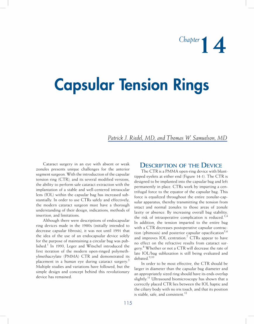
ring-like devices have also been developed. One of these is the artificial iris ring from Morcher and Ophtec. These PMMA rings are much like a CTR with segmented colored wing-like flanges that produce an artificial iris in cases of aniridia (Figure 14-6). In order to create a complete artificial iris, two rings need to be placed in the capsular bag and the wings offset slightly. Different models are available to produce dif-ferent artificial pupil diameters and to try to match iris color for a cosmetic result. In cases with only a sector of iris missing (coloboma or trauma), a ring with only one large wing-like flange has been developed (Figure 14-7). Although these rings are not yet FDA approved, they can be obtained and used on a compassionate care basis.
coMplicationsGenerally, the insertion of a CTR is straight-
forward with an immediate and beneficial effect. Complications can arise, however, either during sur-gery or in the postoperative period. Reports have been
Figure 14-5. M-CTR. The left image is of the M-CTR with a single sutur-ing eyelet to the right (model 1-R). The middle image is of the double eyelet version (model 2-L). The right image is of the M-CTR with a single suturing eyelet to the left (model 1-L). (Photo cour-tesy of Morcher GmbH, Stuttgart, Germany.)
Figure 14-6. The aniridia CTR. Usually two of these rings will be utilized and offset slightly to produce a complete artificial iris. (Photo courtesy of Morcher GmbH, Stuttgart, Germany.)
120 Chapter 14
published of CTRs being inadvertently inserted into the anterior chamber angle instead of the capsular bag and not identified until postoperatively.19 Insertion into the vitreous has also occurred either through an iatrogenic capsule tear from ring insertion or through an occult bag laceration.20 It is also possible for the ring to be placed in the ciliary sulcus. Great care must be taken to confirm that the capsular bag is intact before placing a CTR. If there is any question as to the status of the bag, the ring should not be inserted. Again, a CTS can be used in cases in which the bag integrity is questionable or already violated.
If a CTR is placed completely inside an intact capsular bag, and there is no inadvertent capsular tear after the ring is in position, there is little that could happen postoperatively except for possible subluxation of the entire IOL/CTR/capsular bag complex. In cases of progressive zonular weakening such as pseudoexfo-liation and Marfan syndrome, a significant subluxation of the entire complex has been described.21 This can occur years after the initial surgery. To prevent late subluxation, some surgeons advocate the placement of a CTR in all patients with a progressive zonular weak-ening condition such as pseudoexfoliation. However, no evidence yet exists that such a maneuver would reduce or prevent IOL/CTR/capsular bag subluxation. Cases of subluxation of the entire bag and its contents (including a CTR) have already been reported, so the widespread use of a standard CTR in such cases may have little impact on long-term results.9,10 In cases with obvious significant zonular absence or laxity, a device that can be suture fixated to the scleral wall (such as the M-CTR or a CTS) is more logically employed.
Several techniques have been described to reposi-tion the IOL/CTR/capsular bag complex when it has subluxed. The CTR can be directly sutured to the sclera, but this requires placing one end of the suture through the capsular bag (and under the CTR), there-by lacerating it.22 A CTS or M-CTR could be utilized to reposition the bag and suture it to the sclera without disruption of the capsular bag if the ring or segment could be appropriately positioned.
Removing a CTR from the vitreous space can be accomplished in several ways including directly removing the ring through a sclerostomy, cutting the ring into pieces and removing it, or removing it by capturing it and retracting it into a ring injector.23 Cases of ring dislocation or inadvertent insertion into the vitreous are often best handled by posterior seg-ment surgeons.
suMMaryCTRs increase the safety of anterior segment
surgery in the face of zonular weakness. A modern phacoemulsification surgeon must be adept at utilizing these devices to provide for a more stable capsular bag during and after cataract surgery. By mastering the use of these devices in cases of zonular instability, the rate of capsular tears, vitreous loss, IOL decentration, and capsular phimosis may be significantly reduced.
references1. Hara T, Yamada H. Equator ring for the maintenance of the
completely circular contour of the capsular bag equator after cataract removal. Ophthal Surg. 1991;22:358.
2. Leger U, Witschel BM, Lim SJ, et al. The capsular tension ring: a new device for complicated cataract surgery. Present-ed at: The Third American-International Congress on Cata-ract, Intraocular Lenses, and Refractive Surgery; Seattle, WA; May 11, 1993.
3. Bayraktar S, Alton T, Kucuksumer Y, et al. Capsular tension ring implantation after capsulorhexis in phacoemulsification of cataracts associated with pseudoexfoliation syndrome: in-traoperative complications and early post-operative findings. J Cataract Refract Surg. 2001;27:1620.
4. Gimbel HV, Sun R, Heston JP. Management of zonular di-alysis on phacoemulsification and IOL implantation using the capsular tension ring. Ophthal Surg Lasers. 1997;28:273.
5. D’Eliseo D, Pastena B, Longanesi L, Gristani F, Negrini V. Prevention of posterior capsule opacification using capsular tension ring for zonular defects in cataract surgery. Eur J Oph-thalmol. 2003;13(2):151.
Figure 14-7. The coloboma CTR. This ring can be implanted to cover a section of missing iris as in cases of coloboma or trauma. (Photo courtesy of Morcher GmbH, Stuttgart, Germany.)

Capsular Tension Rings 121
6. Kim JH, Kim H, Joo CK. The effect of capsular tension ring on posterior capsular opacity in cataract surgery. Korean J Ophthalmol. 2005;19(1):23.
7. Lee DH, Shin SC, Joo CK. Effect of the capsular tension ring on intraocular lens decentration and tilting after cataract sur-gery. J Cataract Refract Surg. 2002;28(5):843.
8. Boomer JA, Jackson DW. Effect of the Morcher capsular tension ring on refractive outcomes. J Cataract Refract Surg. 2006;32(7):1180.
9. Scherer M, Bertelmann E, Rieck P. Late spontaneous in-the-bag intraocular lens and capsular tension ring disloca-tion in pseudoexfoliation syndrome. J Cataract Refract Surg. 2006;32(4):672.
10. Oner FH, Kocak N, Saatci AO. Dislocation of the capsular bag with intraocular lens and capsular tension ring. J Cataract Refract Surg. 2006;32(10):1756.
11. Goldman JM, Karp CL. Adjunct devices for managing chal-lenging cases in cataract surgery: pupil expansion and stabili-zation of the capsular bag. Curr Opin Ophthalmol. 2007;18:44.
12. Boomer JA, Jackson DW. Anatomic evaluation of the Morcher capsular tension ring by ultrasound biomicroscopy. J Cataract Refract Surg. 2006;32(5):846.
13. Vass C, Menapace R, Schetterer K, et al. Prediction of pseu-dophakic capsular bag diameter based on biometric variables. J Cataract Refract Surg. 1999;25:1376.
14. Ahmed II, Cionni RJ, Kranemann C, Crandall AS. Optimal
timing of capsular tension ring implantation: Miyake-Apple video analysis. J Cataract Refract Surg. 2005;31(9):1809.
15. Hasanee K, Ahmed II. Capsular tension rings: update on endo-capsular support devices. Ophthalmol Clin N Am. 2006;19:508.
16. Anqunawela RI, Little B. Fishtail technique for capsular ten-sion ring insertion. J Cataract Refract Surg. 2007:33(5):767.
17. Lee V, Bloom P. Microhook capsular stabilization for phaco-emulsification in eyes with pseudoexfoliation-induced lens instability. J Cataract Refract Surg. 1999;25:1567.
18. Cionni,RJ, Osher RH. Management of profound zonular di-alysis or weakness with a new endocapsular ring designed for scleral fixation. J Cataract Refract Surg. 1998;24:1299.
19. Little BC, Richardson T, Morris S. Removal of the capsular tension ring from the anterior chamber angle. J Cataract Re-fract Surg. 2004;30(9):1832.
20. Levy J, Klemperer I, Lifshitz T. Posteriorly dislocated capsu-lar tension ring. Ophthal Surg Lasers Imaging. 2005;36(5):416.
21. Jehan FS, Mamalis N, Crandall AS. Spontaneous late disloca-tion of the intraocular lens within the capsular bag in pseudo-exfoliation patients. Ophthalmology. 2001;108(10):1727.
22. Ahmed II, Chen SH, Kranemann C, Wong DT. Surgical re-positioning of dislocated capsular tension rings. Ophthalmol-ogy. 2005;112(10):1725.
23. Ma PE, Kaur H, Petrovic V, Hay D. Technique for removal of a capsular tension ring from the vitreous. Ophthalmology. 2003;110(6):1142.
