capsule endoscopy
TRANSCRIPT
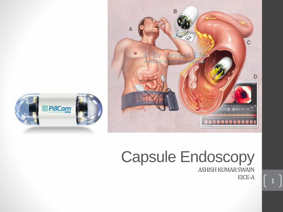
Capsule EndoscopyASHISH KUMAR SWAIN
EICE-A1
Gastrointestinal Bleeding
Is any type of bleeding that occurs in the gastrointestinal tract from
the pharynx to the rectum.
Treatments involves proton pump inhibitors, octeotride, and antibiotics
(Certain Cases).
There is also an emphasis on resuscitation by infusion of intravenous
fluids and blood transfusion.
Gastrointestinal Bleeding is usually discovered by Upper endoscopy,
Colonoscopy, or Capsule Endoscopy.2
Contents1. History of Endoscopy.
2. Introduction.
3. Understanding Capsule Endoscopy.
4. Description.
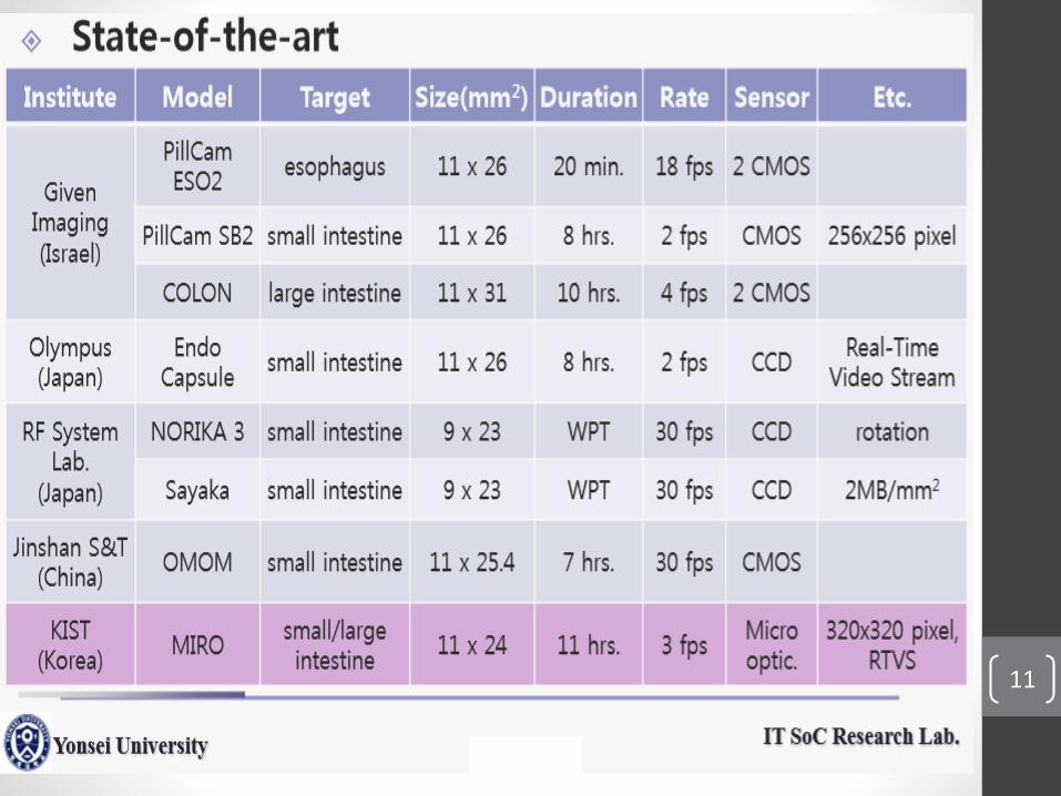
5. Some types Of Capsule Cameras.
6. Specifications.
7. Features.
8. Inside a capsule Cameras.
9. How Does It Work?
10. Endoscopy Procedure.
11. Images Obtained From Capsule.
12. Uses.
13. Advantages And Disadvantages.
14. Conclusion.
15. Work Cited.3
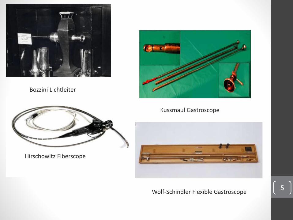
History Of EndoscopyThe first real endoscope that was developed was made by
Phillip Bozzini in 1805 to examine the urethra, the bladder and
vagina.
Adolf Kussmaul in 1868 used a straight rigid metal tube over a
flexible obturator to perform the first gastroscopy.
Building on the work of others, Rudolph Schindler constructed
the first practical gastroscope in 1932.
In 1957 Basil Hirschowitz developed his prototype fiberscope.
4
5
Bozzini Lichtleiter
Hirschowitz Fiberscope
Kussmaul Gastroscope
Wolf-Schindler Flexible Gastroscope
IntroductionThe capsule endoscopy procedure was invented by an Israel-based electro-optical engineer Gavriel Iddan, in order to detect gastro-intestinal bleed andother obscure problems that may even be life threatening.
• Aim of technology is to manufacture products at molecularlevel.
• Achieved by nanotechnology.
• One such product is capsule camera.
• A vitamin pill-sized, swallowable camera.
• Capture images of inside body..
• Study on captured images by the experts.
• Approved by EuropeanMedicines Agency .6
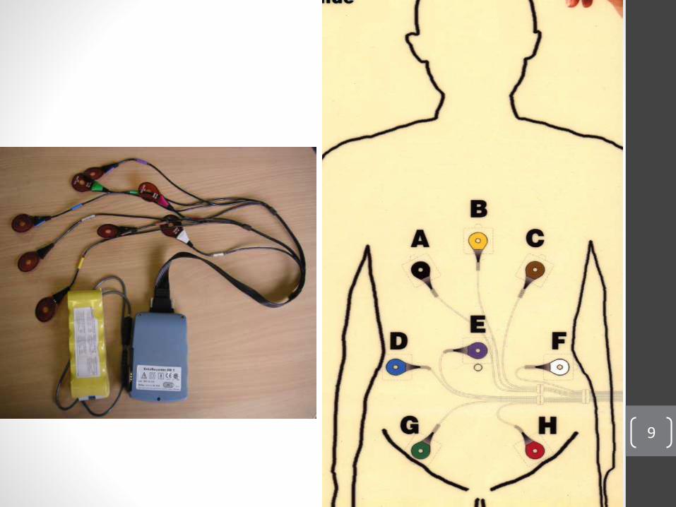
Understanding Capsule Endoscopy
What is Capsule Endoscopy?Capsule Endoscopy lets the doctor examine the lining of the middle part ofone’s gastrointestinal tract, which includes the three portions of the smallintestine (duodenum, jejunum, ileum). Doctor will give a pill sized videocamera for a patient to swallow. This camera has its own light source andtakes pictures of small intestine as it passes through. These pictures aresent to a small recording device patient have to wear on his body.
7
Why is Capsule Endoscopy Done?Capsule endoscopy helps doctor to evaluate the small intestine. The partof the bowel cannot be reached by traditional upper endoscopy or bycolonoscopy. The most common reason for doing capsule endoscopy is tosearch for a cause of bleeding from the small intestine. It may also beuseful for detecting polyps, inflammatory bowel disease (Crohn’sdisease), ulcers, and tumors of the small intestine.
Description
• The capsule (developed at university of Washington)consists of 7optical fibers.
• One for illumination and the rest six for collecting light.• Once swallowed, electric current flows through the pill
that causes the encased fibers to bounce back and forthsuch that its electronic eye would be able to scan the GItract.
• The tip will illuminate red, green and blue laser lighthelping in visuality,
• All this processing together combined will give us two-dimensional picture helping in diagnosis.
• The images can be retrieved from the recording deviceworn around patient's waist as a belt.
8
9
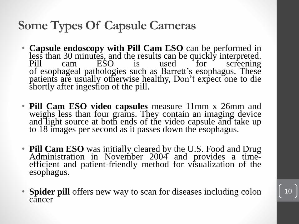
Some Types Of Capsule Cameras
• Capsule endoscopy with Pill Cam ESO can be performed inless than 30 minutes, and the results can be quickly interpreted.Pill cam ESO is used for screeningof esophageal pathologies such as Barrett’s esophagus. Thesepatients are usually otherwise healthy, Don’t expect one to dieshortly after ingestion of the pill.
• Pill Cam ESO video capsules measure 11mm x 26mm andweighs less than four grams. They contain an imaging deviceand light source at both ends of the video capsule and take upto 18 images per second as it passes down the esophagus.
• Pill Cam ESO was initially cleared by the U.S. Food and DrugAdministration in November 2004 and provides a time-efficient and patient-friendly method for visualization of theesophagus.
• Spider pill offers new way to scan for diseases including coloncancer
10
11
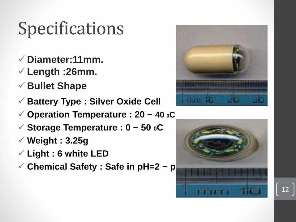
Specifications
Diameter:11mm.
Length :26mm.
Bullet Shape
Battery Type : Silver Oxide Cell
Operation Temperature : 20 ~ 40 0C
Storage Temperature : 0 ~ 50 0C
Weight : 3.25g
Light : 6 white LED
Chemical Safety : Safe in pH=2 ~ pH=8
12
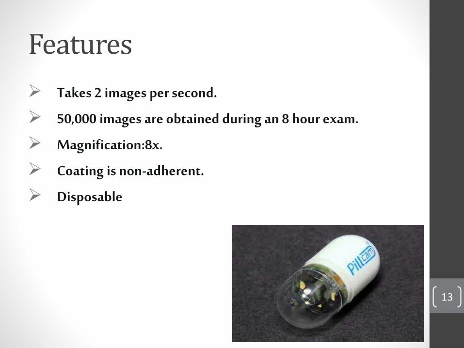
Features
Takes 2 images per second.
50,000 images are obtained during an 8 hour exam.
Magnification:8x.
Coating is non-adherent.
Disposable
13
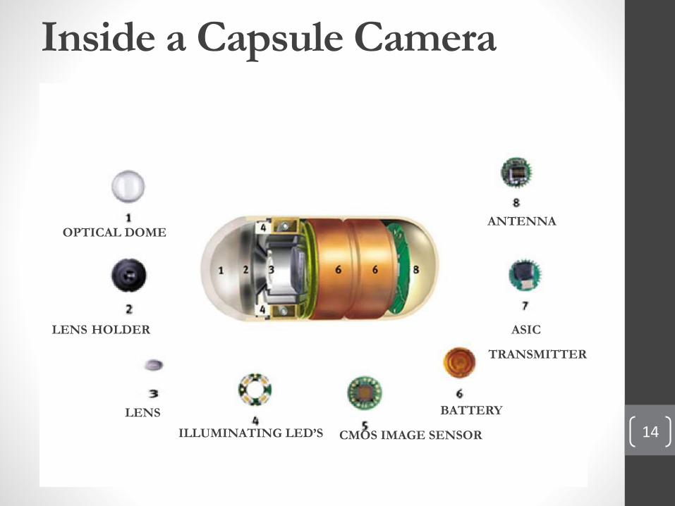
Inside a Capsule Camera
OPTICAL DOME
LENS HOLDER
LENS
ILLUMINATING LED’S CMOS IMAGE SENSOR
BATTERY
ASIC
TRANSMITTER
ANTENNA
14
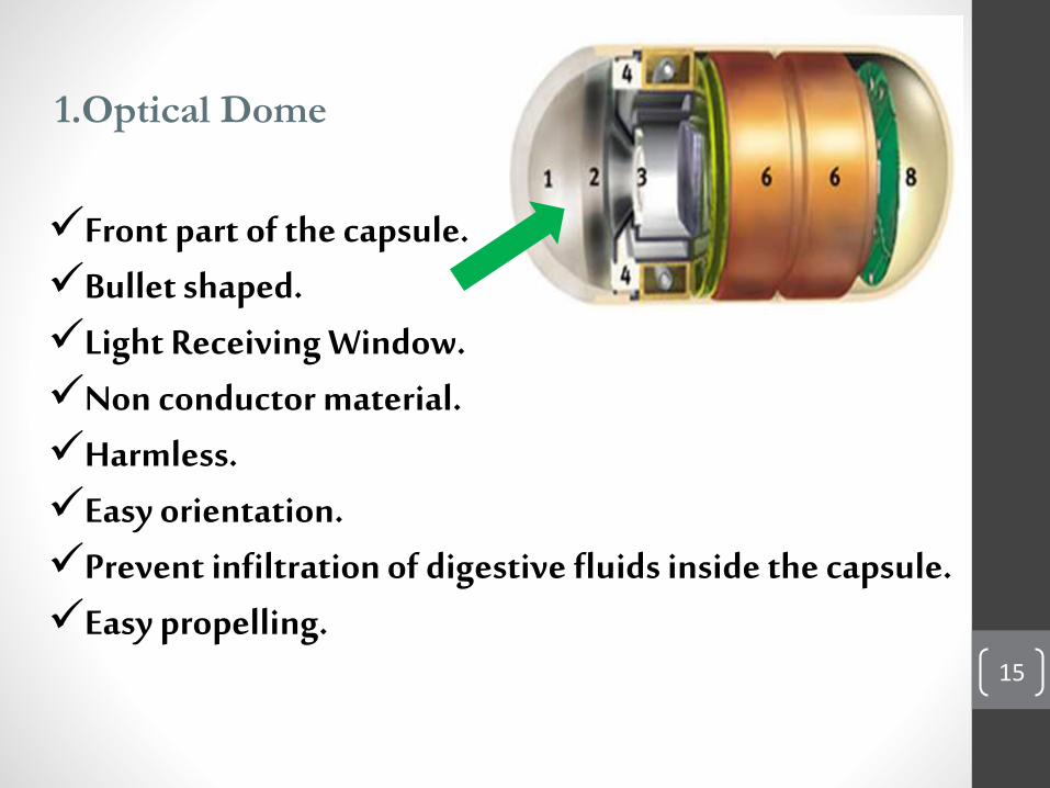
1.Optical Dome
Front part of the capsule.Bullet shaped.Light ReceivingWindow.Non conductor material.Harmless.Easy orientation.Prevent infiltration of digestive fluids inside the capsule.Easy propelling.
15
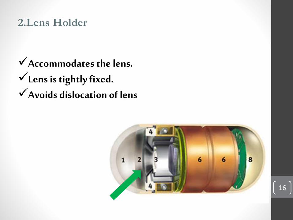
2.Lens Holder
Accommodates the lens.Lens is tightly fixed.Avoids dislocation of lens
16
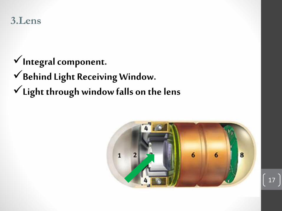
3.Lens
Integral component.Behind Light ReceivingWindow.Light through window falls on the lens
17
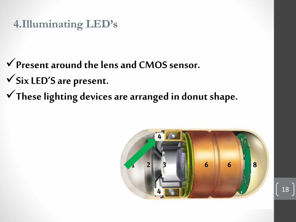
4.Illuminating LED’s
Present around the lens and CMOS sensor.Six LED’S are present.These lighting devices are arranged in donut shape.
18
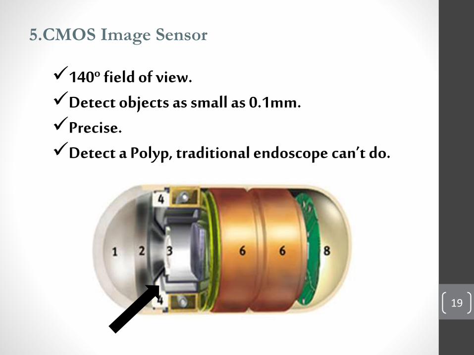
5.CMOS Image Sensor
140º field of view.Detect objects as small as 0.1mm.Precise.Detect a Polyp, traditional endoscope can’t do.
19
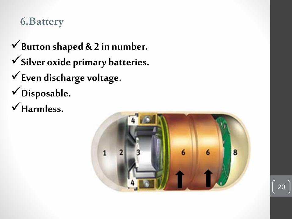
6.Battery
Button shaped & 2 in number.Silver oxide primary batteries.Even discharge voltage.Disposable.Harmless.
20
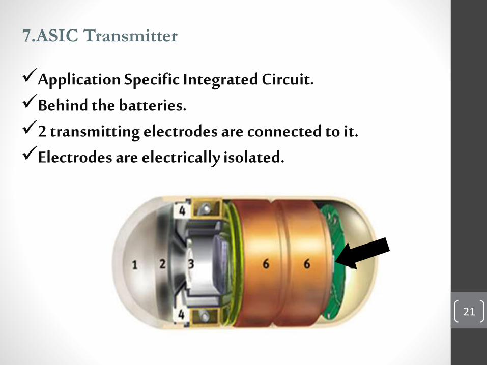
7.ASIC Transmitter
21
Application Specific Integrated Circuit.Behind the batteries.2 transmitting electrodes are connected to it.Electrodes are electrically isolated.
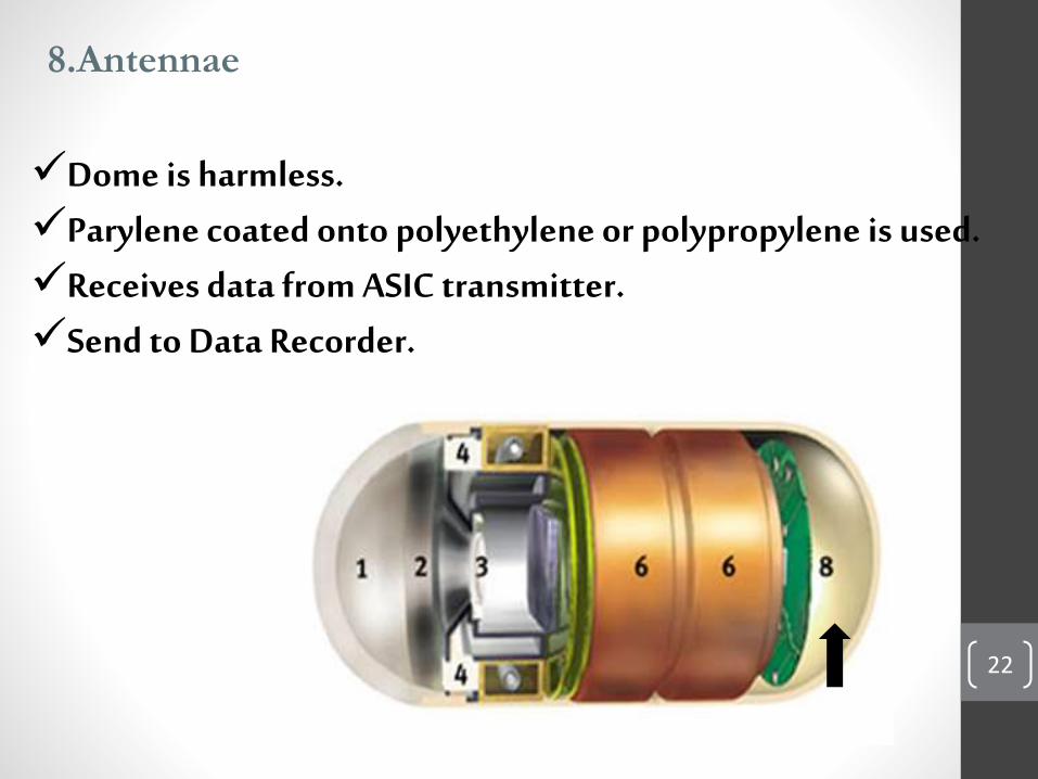
8.Antennae
Dome is harmless.Parylene coated onto polyethylene or polypropylene is used.Receives data from ASIC transmitter.Send to Data Recorder.
22
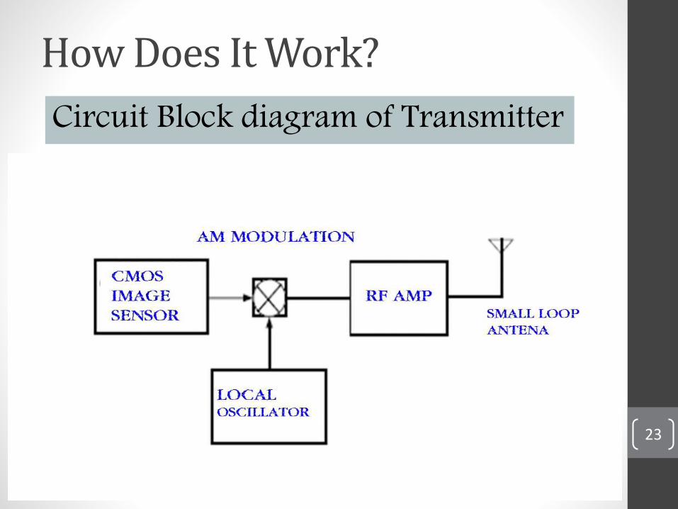
How Does It Work?
23
Circuit Block diagram of Transmitter
24
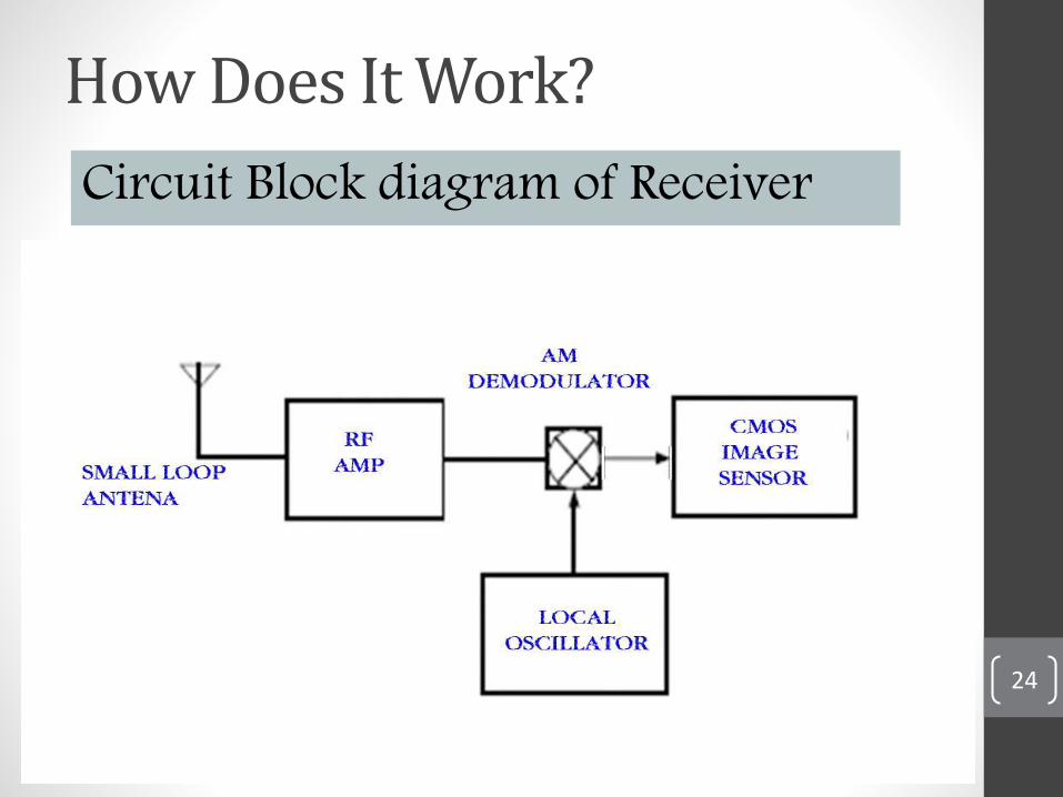
How Does It Work?
Circuit Block diagram of Receiver
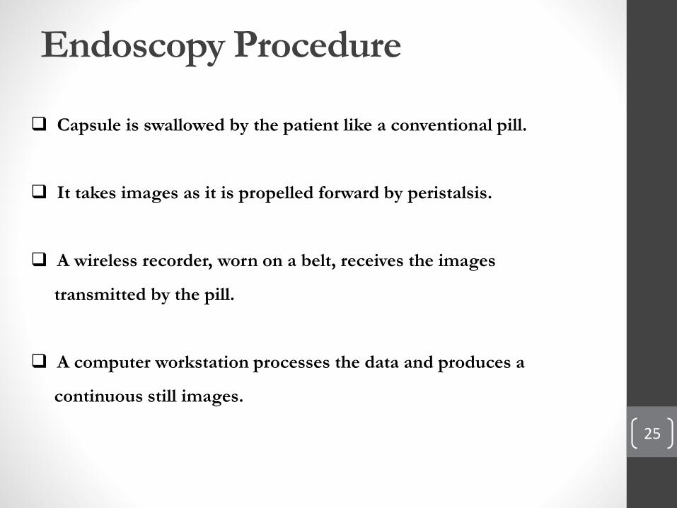
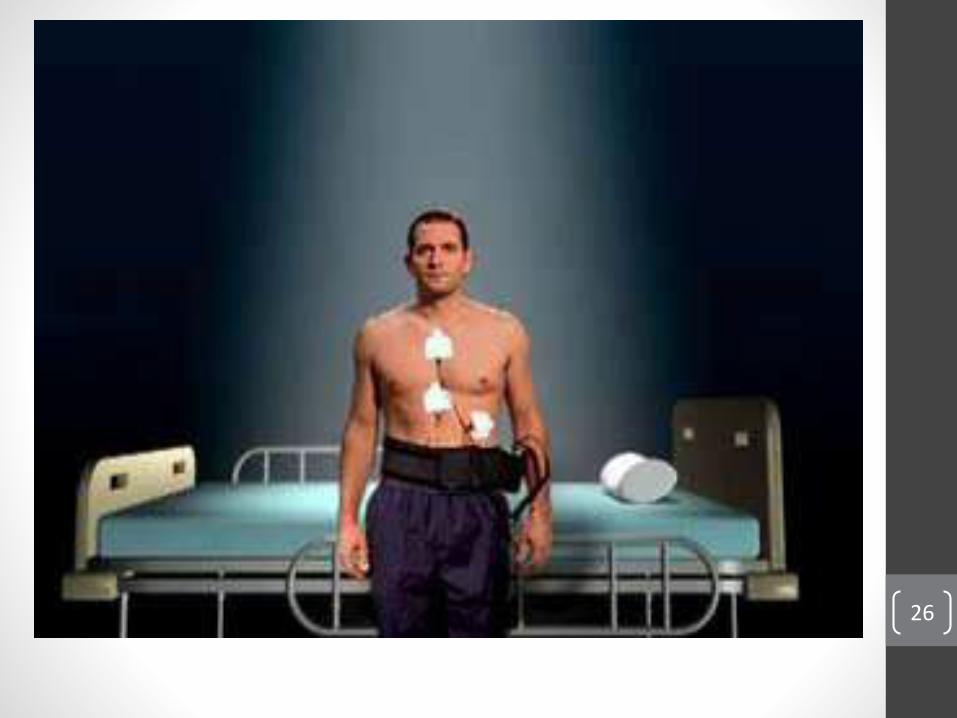
Endoscopy Procedure
25
Capsule is swallowed by the patient like a conventional pill.
It takes images as it is propelled forward by peristalsis.
A wireless recorder, worn on a belt, receives the images
transmitted by the pill.
A computer workstation processes the data and produces a
continuous still images.
26
27
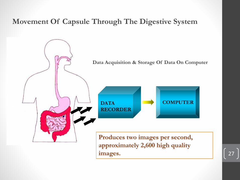
Movement Of Capsule Through The Digestive System
DATA
RECORDER
COMPUTER
Data Acquisition & Storage Of Data On Computer
Produces two images per second,
approximately 2,600 high quality
images.
28
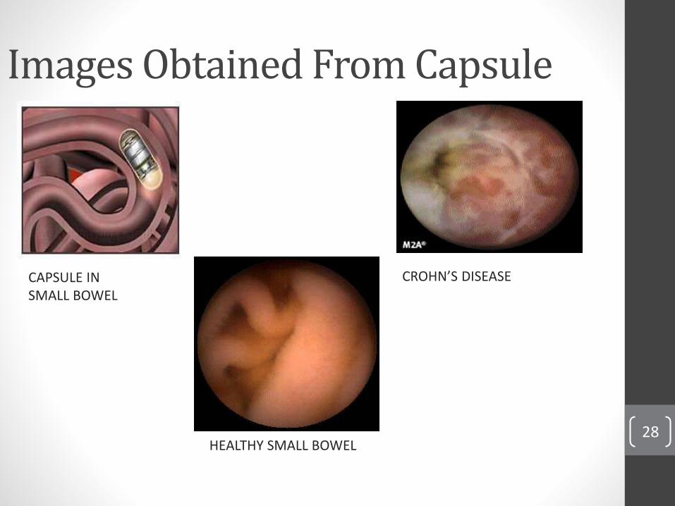
Images Obtained From Capsule
CROHN’S DISEASE
HEALTHY SMALL BOWEL
CAPSULE IN SMALL BOWEL
29
Uses
• Crohn's Disease.• Malabsorption Disorders.• Tumors of the small intestine & Vascular Disorders.• Medication Related To Small Bowel Injury• Ulcerative Colitis
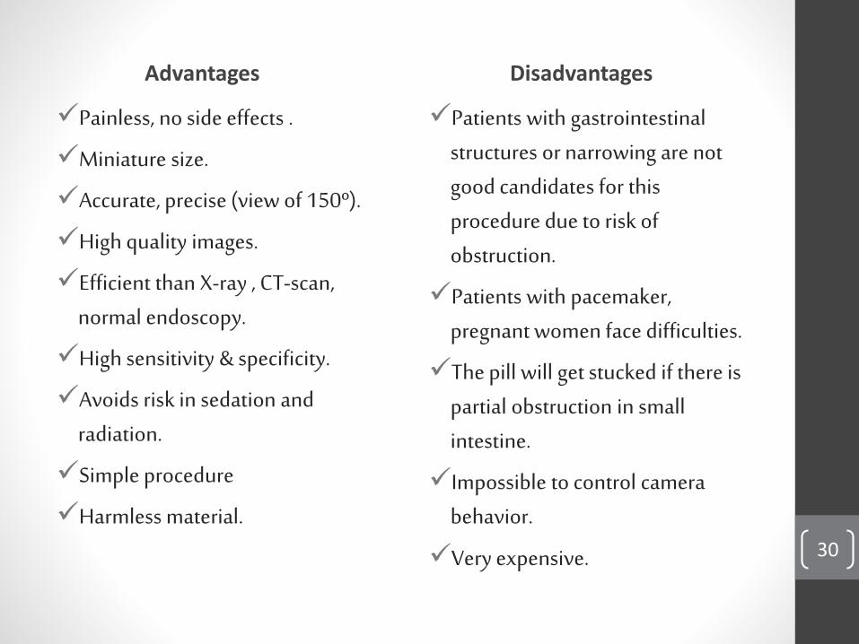
Advantages
Painless, no side effects .
Miniature size.
Accurate, precise (view of 150º).
High quality images.
Efficient than X-ray , CT-scan, normal endoscopy.
High sensitivity & specificity.
Avoids risk in sedation and radiation.
Simple procedure
Harmless material.
Disadvantages
Patients with gastrointestinal structures or narrowing are not good candidates for this procedure due to risk of obstruction.
Patients with pacemaker, pregnant women face difficulties.
The pill will get stucked if there is partial obstruction in small intestine.
Impossible to control camera behavior.
Very expensive. are not 30
Conclusion
31
The Given Endoscopy capsule is a pioneering concept for MedicalTechnology of the 21st century.
The endoscopy system is the first of its kind to be able to provide non-invasive imaging of the entire small intestine.
It has revolutionized the field of diagnostic imaging to a great extent andhas proved to be of great help to physicians all over the world.
32
Work Cited• Wikipedia: Capsure Endoscopy
<http://en.wikipedia.org/wiki/Capsule_endoscopy>.
• Lewis,Blair M.D,Swain,Paul, MD: write a paper. Capsule endoscopy in the evaluation of patients with suspected small intestinal bleeding:,2002
• Koichi, Soga: In vivo imaging of intestinal helminths by capsule endoscopy,2013
• Wikipedia:Gastro Intestinal Bleeding:http://en.wikipedia.org/wiki/Gastrointestinal_bleeding
• Engineering in Medicine and Biology Magazine,IEEE
(Volume: 24, Issue: 4 ) .
• http://www.capsuleendoscopy.org
• Sidhu, Reena, at al.Gastrointestinal capsule endoscopy:
from tertiary centres to primary "care". BMJ, March 4 2006.
332:528-531. doi:10.1136/bmj.332.7540.528

33

34
QUESTIONS