carbon nanomaterials based on graphene in (electro ... nanomaterials based on graphene in (electro-)...
TRANSCRIPT
Carbon Nanomaterials based on Graphene in (Electro-)
chemical Sensors: Characterization, Modification and
Application
Dissertation zur Erlangung des Doktorgrades der Naturwissenschaften
(Dr. rer. nat.)
der Fakultät Chemie und Pharmazie
der Universität Regensburg
Deutschland
vorgelegt von
Alexander Zöpfl
aus Ingolstadt
im Jahr 2015
II
Die vorgelegte Dissertation entstand in der Zeit von März 2012 bis Juni 2015 am Institut für
Analytische Chemie, Chemo- und Biosensorik der Universität Regensburg.
Die Arbeit wurde angeleitet von Prof. Dr. Frank-Michael Matysik.
Promotionsgesuch eingereicht am : 22. Juni 2015
Kolloquiumstermin: 20. Juli 2015
Prüfungsausschuss
Voritzender: Prof. Dr. Oliver Tepner
Erstgutachter: Prof. Dr. Frank-Michael Matysik
Zweitgutachter: Prof. Dr. Otto S. Wolfbeis
Drittprüfer: PD Dr. habil. Richard Weihrich

III
Acknowledgements
I am very grateful to Prof. Dr. Frank-Michael Matysik for giving me the possibility to work
on this topic, the helpful comments, discussions and his support.
My very special thanks go to Dr. Thomas Hirsch for supervising me. Many thanks for the
great help, good advices, scientific discussions and all the other tremendous support during
this time. It is really a fantastic experience working (and visiting conferences) with you!
Furthermore, I thank all my colleges from the 4th
floor (especially Michael) and all the other
members of the Institute of Analytical Chemistry for the friendly working atmosphere, helpful
comments and support. Thanks for the superb time we spend during, but also after working
hours.
I also want to thank Dr. Günther Ruhl at Infineon for the opportunity to work on this topic,
the great support and discussions. Further, I thank all the pleasant colleagues I met in this
company for their support. My special thanks go to Infineon Technologies AG for the
financial support.
I am very grateful to the people from the Department of Physics (especially Masoumeh
Sisakthi, Prof. Dr. Christoph Strunk) for the great collaboration. In this connection, I thank the
DFG Research Training Group GRK 1570 for the financial support enabling additional
conference visits.
Finally, I want to thank my family for their never-ending support. You are the best!
IV
Table of Contents
CURRICULUM VITAE VI
PUBLICATIONS AND PATENTS VII
PRESENTATIONS IX
DECLARATION OF COLLABORATIONS X
ABBREVIATIONS XII
1 INTRODUCTION AND OBJECTIVES 1
2 THE AUTHOR’S OWN PUBLICATIONS AND PATENTS 5
3 BACKGROUND 9
3.1 Preparation Methods and Transfer Techniques of Graphene 9
3.2 Graphene as Sensitive Material in Gas Detection Applications 14
Gas Sensing Fundamentals 14 3.2.1
Graphene in Gas Sensing Applications 16 3.2.2
Conclusion 19 3.2.3
3.3 Graphene in Biosensor Application 20
Biosensors for Glucose 20 3.3.1
Biosensors for Nucleic Acids 30 3.3.2
Biosensors for Proteins 35 3.3.3
Biosensors for other Biologically Relevant Analytes 42 3.3.4
Conclusion 45 3.3.5
4 EXPERIMENTAL 47
4.1 Materials and Instrumentations 47
4.2 Preparation of Different Graphene Materials 48
4.3 Modification of Graphene Materials 49
4.4 Fabrication of Graphene Electrodes 51
Sensor Preparation for Gas Sensing Applications 51 4.4.1
Preparation of Graphene Electrodes for Electrochemical Comparison 52 4.4.2
4.5 Gas Measurements 53
4.6 Electrochemical Characterization and Measurements 55
V
5 RESULTS AND DISCUSSION 56
5.1 Signal Enhancement in Amperometric Peroxide Detection by Using
Graphene Materials with Low Number of Defects 56
Characterization of Graphene Materials with Raman Spectroscopy 56 5.1.1
Electrochemical Characterization of Different Graphene Electrode Materials 58 5.1.2
Direct Amperometric Detection of H2O2 61 5.1.3
Conclusion 65 5.1.4
5.2 Reduced Graphene Oxide and Graphene Composite Materials for
Improved Gas Sensing at Low Temperature 66
Characterization of Reduced Graphene Oxide and Composite Materials 66 5.2.1
Gas Sensor Response 70 5.2.2
Principal Component Analysis for Pattern Recognition of Different Gases 75 5.2.3
Conclusion 76 5.2.4
5.3 Graphene Gas Sensor for the Detection of Carbon Dioxide at Room
Temperature 77
Formation and Characterization of CuO Modified Reduced Graphene Oxide 79 5.3.1
Detection of Carbon Dioxide 83 5.3.2
Cross Sensitivity 87 5.3.3
Comparison to a Commercial Sensor 89 5.3.4
Conclusion 91 5.3.5
6 SUMMARY 92
7 ZUSAMMENFASSUNG 95
8 REFERENCES 98
EIDESSTATTLICHE ERKLÄRUNG 127
VI
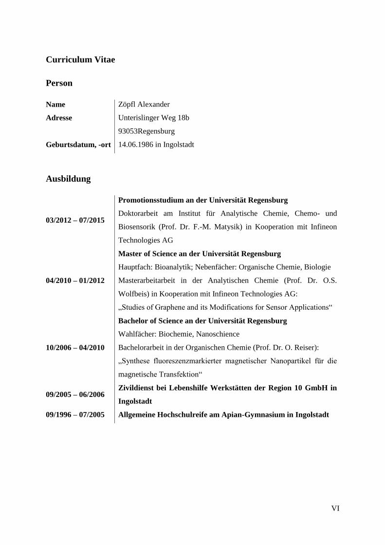
Curriculum Vitae
Person
Name Zöpfl Alexander
Adresse Unterislinger Weg 18b
93053Regensburg
Geburtsdatum, -ort 14.06.1986 in Ingolstadt
Ausbildung
03/2012 – 07/2015
Promotionsstudium an der Universität Regensburg
Doktorarbeit am Institut für Analytische Chemie, Chemo- und
Biosensorik (Prof. Dr. F.-M. Matysik) in Kooperation mit Infineon
Technologies AG
04/2010 – 01/2012
Master of Science an der Universität Regensburg
Hauptfach: Bioanalytik; Nebenfächer: Organische Chemie, Biologie
Masterarbeitarbeit in der Analytischen Chemie (Prof. Dr. O.S.
Wolfbeis) in Kooperation mit Infineon Technologies AG:
„Studies of Graphene and its Modifications for Sensor Applications“
10/2006 – 04/2010
Bachelor of Science an der Universität Regensburg
Wahlfächer: Biochemie, Nanoschience
Bachelorarbeit in der Organischen Chemie (Prof. Dr. O. Reiser):
„Synthese fluoreszenzmarkierter magnetischer Nanopartikel für die
magnetische Transfektion“
09/2005 – 06/2006 Zivildienst bei Lebenshilfe Werkstätten der Region 10 GmbH in
Ingolstadt
09/1996 – 07/2005 Allgemeine Hochschulreife am Apian-Gymnasium in Ingolstadt
VII
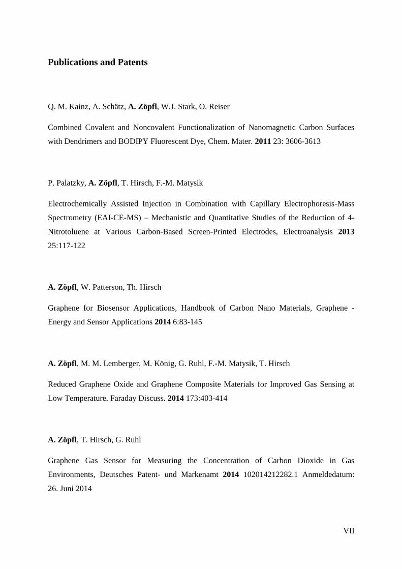
Publications and Patents
Q. M. Kainz, A. Schätz, A. Zöpfl, W.J. Stark, O. Reiser
Combined Covalent and Noncovalent Functionalization of Nanomagnetic Carbon Surfaces
with Dendrimers and BODIPY Fluorescent Dye, Chem. Mater. 2011 23: 3606-3613
P. Palatzky, A. Zöpfl, T. Hirsch, F.-M. Matysik
Electrochemically Assisted Injection in Combination with Capillary Electrophoresis-Mass
Spectrometry (EAI-CE-MS) – Mechanistic and Quantitative Studies of the Reduction of 4-
Nitrotoluene at Various Carbon-Based Screen-Printed Electrodes, Electroanalysis 2013
25:117-122
A. Zöpfl, W. Patterson, Th. Hirsch
Graphene for Biosensor Applications, Handbook of Carbon Nano Materials, Graphene -
Energy and Sensor Applications 2014 6:83-145
A. Zöpfl, M. M. Lemberger, M. König, G. Ruhl, F.-M. Matysik, T. Hirsch
Reduced Graphene Oxide and Graphene Composite Materials for Improved Gas Sensing at
Low Temperature, Faraday Discuss. 2014 173:403-414
A. Zöpfl, T. Hirsch, G. Ruhl
Graphene Gas Sensor for Measuring the Concentration of Carbon Dioxide in Gas
Environments, Deutsches Patent- und Markenamt 2014 102014212282.1 Anmeldedatum:
26. Juni 2014

VIII
C. Fenzl, C. Genslein, A. Zöpfl, A.J. Baeumner, T. Hirsch
A photonic crystal based sensing scheme for acetylcholine and acetylcholinesterase inhibitors,
J. Mater. Chem. B 2015 3:2089-2095
A. Zöpfl, M. Sisakthi, J. Eroms, F.-M. Matysik, C. Strunk, T. Hirsch
Signal Enhancement in Amperometric Peroxide Detection by Using Graphene Materials with
Low Number of Defects, submitted to Microchimica Acta

IX
Presentations
04/2012 Poster presentation at Graphene 2012 in Bussels, Belgium „The
Fluorescence Properties of Graphene Oxide”
06/2012 Oral presentation at Infineon Innovation Week in Regensburg, Germany
“Studies of Graphene and its Modifications for Gas Sensor Applications”
02/2013 Oral presentation at 7. Interdisziplinäres Doktorandenseminar in Berlin,
Germany „Carbon Nanomaterials based on Graphene in Electrochemical
Gas Sensors”
03/2013 Poster presentation at ANAKON in Essen, Germany “Studies of Graphene
and its Modifications for Gas Sensor Application”
04/2013 Poster presentation at International Graphene Workshop of RTG 1510 in
Regensburg, Germany “Chemically Derived Graphene for the Detection of
NO2”
04/2013 Poster presentation at Graphene 2013 in Bilbao, Spain “Comparison of
different Graphene Materials and their Electrochemical Application”
03/2014 Oral presentation at Mikrosystemtechnik Symposium in Landshut,
Germany „Chemisch hergestelltes Graphen als funktionalisierbares
Sensormaterial zur Detektion von NO2“
05/2014 Poster presentation at Graphene 2014 in Toulouse, France „Comparison of
Different Graphene Materials in Amperometric Sensors“
07/2014 Demonstration device and poster presentation at Infineon Innovation Week
in Regensburg, Germany “Gas Sensitive Chemiresistors Based on
Chemically Reduced Graphene Oxide”
09/2014 Oral presentation at Faraday Discussion 173 in London, England
“Reduced graphene oxide and graphene composite materials for improved
gas sensing at low temperature”
11/2014 Poster presentation at Infineon Innovation Week in Munich, Germany
“Demonstration of Graphene in Possible Fields of Application”
03/2015 Poster presentation at ANAKON in Graz, Graz “Graphene Nanocomposites
for Gas Sensing at Ambient Temperature”
03/2015 Poster presentation at MRS Spring Meeting 2015 in San Francisco, USA
“Graphene Modified Electrodes for Enzymatic Biosensing”
X
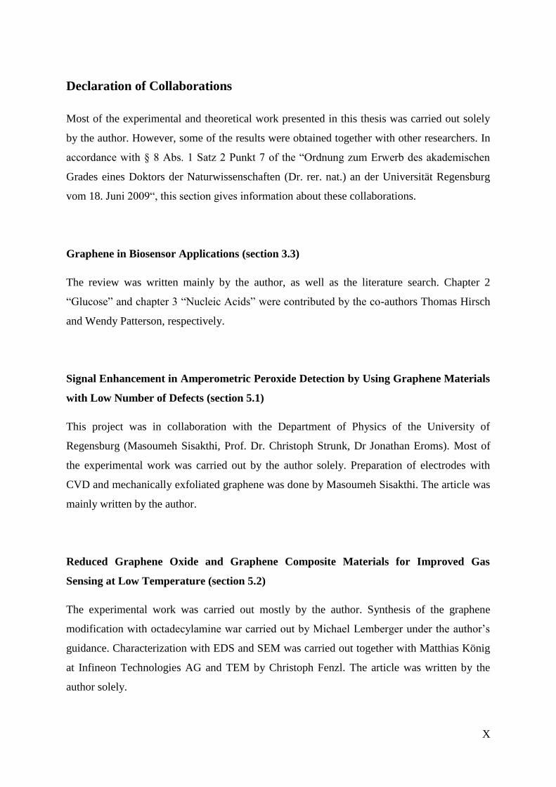
Declaration of Collaborations
Most of the experimental and theoretical work presented in this thesis was carried out solely
by the author. However, some of the results were obtained together with other researchers. In
accordance with § 8 Abs. 1 Satz 2 Punkt 7 of the “Ordnung zum Erwerb des akademischen
Grades eines Doktors der Naturwissenschaften (Dr. rer. nat.) an der Universität Regensburg
vom 18. Juni 2009“, this section gives information about these collaborations.
Graphene in Biosensor Applications (section 3.3)
The review was written mainly by the author, as well as the literature search. Chapter 2
“Glucose” and chapter 3 “Nucleic Acids” were contributed by the co-authors Thomas Hirsch
and Wendy Patterson, respectively.
Signal Enhancement in Amperometric Peroxide Detection by Using Graphene Materials
with Low Number of Defects (section 5.1)
This project was in collaboration with the Department of Physics of the University of
Regensburg (Masoumeh Sisakthi, Prof. Dr. Christoph Strunk, Dr Jonathan Eroms). Most of
the experimental work was carried out by the author solely. Preparation of electrodes with
CVD and mechanically exfoliated graphene was done by Masoumeh Sisakthi. The article was
mainly written by the author.
Reduced Graphene Oxide and Graphene Composite Materials for Improved Gas
Sensing at Low Temperature (section 5.2)
The experimental work was carried out mostly by the author. Synthesis of the graphene
modification with octadecylamine war carried out by Michael Lemberger under the author’s
guidance. Characterization with EDS and SEM was carried out together with Matthias König
at Infineon Technologies AG and TEM by Christoph Fenzl. The article was written by the
author solely.

XI
Graphene Gas Sensor for Measuring the Concentration of Carbon Dioxide in Gas
Environments (section 5.3)
The experimental work was carried by the author solely. Characterization with EDS and SEM
was carried out together with Matthias König at Infineon Technologies AG.
XII
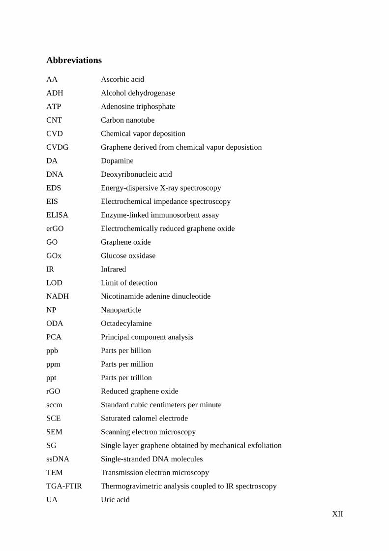
Abbreviations
AA Ascorbic acid
ADH Alcohol dehydrogenase
ATP Adenosine triphosphate
CNT Carbon nanotube
CVD Chemical vapor deposition
CVDG Graphene derived from chemical vapor deposistion
DA Dopamine
DNA Deoxyribonucleic acid
EDS Energy-dispersive X-ray spectroscopy
EIS Electrochemical impedance spectroscopy
ELISA Enzyme-linked immunosorbent assay
erGO Electrochemically reduced graphene oxide
GO Graphene oxide
GOx Glucose oxsidase
IR Infrared
LOD Limit of detection
NADH Nicotinamide adenine dinucleotide
NP Nanoparticle
ODA Octadecylamine
PCA Principal component analysis
ppb Parts per billion
ppm Parts per million
ppt Parts per trillion
rGO Reduced graphene oxide
sccm Standard cubic centimeters per minute
SCE Saturated calomel electrode
SEM Scanning electron microscopy
SG Single layer graphene obtained by mechanical exfoliation
ssDNA Single-stranded DNA molecules
TEM Transmission electron microscopy
TGA-FTIR Thermogravimetric analysis coupled to IR spectroscopy
UA Uric acid
1
1 Introduction and Objectives
Pioneering research by K. Novoselov and A. Geim of the University of Manchester led to the
isolation and characterization of the single atom thick layer of the carbon nanomaterial
graphene in 2004 [1]. It can be described as a monolayer of sp2 bound carbon atoms arranged
in a honeycomb lattice. In 2010, the two scientists also received the Nobel Prize in physics for
their innovative and groundbreaking studies. Starting from this, it has become one of the most
influential materials in science and technology in the last decade.
This is reinforced by the European Commission’s choice of graphene as a future emerging
technology. Research in Europe involving graphene will be funded in one of the first so-called
Flagship initiatives with 1,000 million euros over the next ten years [2]. The scientific and
technological roadmap of the graphene flagship describes graphene as a platform with many
beneficial properties. The two-dimensional material comprises extraordinary strength
(Young’s modulus of 1050 GPa) [3], but also high flexibility, having unique electrical (charge
carrier mobility at room temperature of up to 250,000 cm2V
-1s
-1) [4] and optical (~98%
transparency) [5] properties. Further exceptional characteristics are its high thermal
conductivity of 5000 Wm-1
K-1
and high surface to volume ratio of 2630 m2g
-1 [6].
First applications, which can be transferred from academic research to industrial products
were proposed for 2015. Transistors were predicted to lead the commercial success of the
graphene story, followed by spin valves, flexible displays, radio frequency identification tags,
ultra-light batteries, solar cells, ultrafast lasers, composite materials, and prostheses. Up to
now the nanomaterial can be found in only a few products like graphene based conductive
inks and a portable lightweight flexible power source from Vorbeck® [7], stronger and lighter
tennis rackets from HEAD [8], bicycle race wheels comprising graphene enhanced composite
materials from Vittoria [9] or graphene enhanced cycling helmets from Catlike [10]. This
gives a short impression about the current market situation and shows that graphene still has
not reached the market completely in the way it was proposed. However, many prototypes
have been demonstrated, e.g. a touch panels from Samsung [11] or further portable
supercapacitors from Zap&Go [12], and are expected to hit the market soon. Sensors and
biosensors based on graphene have been proposed by the Flagship initiative by 2018. For the
preliminary phase, a ramp up of 30 months with a total budget of 75 million euros is planned
2
by the European Flagship, where about 3-5% will be spent on the development of sensor
technologies. Not only in Europe, but also in Asia (mainly Singapore) and in the United
States, graphene is proclaimed as an important key technology. Therefore, the question arises:
Will graphene be smart enough to replace some of the materials currently used in sensor
technology? [partially adapted from P1]
Graphene holds great promise as tunable sensor material in miniaturized chemiresistive gas
sensing applications [13]. Simple and reliable monitoring of gas concentration is important in
everyday-life. In industrial processes hazardous gases need to be controlled to guarantee
safety. Controlling air quality can save energy in automated air conditioning. But also the
detection of environmental pollution (like NOx) is of great interest [14]. Up to now, solid-state
gas sensors based on metal oxide chemiresistors are well established in the field of chemical
sensors and widely used in detecting gases [15, 16]. They are operated at high temperature
[17], which consumes excessive energy and limits their long-term stability, thus leading to the
development of new gas sensor concepts which overcome these drawbacks. Similar to solid
state gas sensors, gas adsorption on graphene leads to a change in its electrical resistance.
Therefore, any kind of interaction between graphene sheets and adsorbates, influencing the
electronic structure of graphene, leads to an altered charge carrier concentration or
respectively electrical conductance of the material already at low operating temperature [18,
19]. One aim of this work was to study graphene in terms of gas adsorption and its potential
for gas sensor applications. [partially adapted from P2]
Since its discovery, it was suggested as highly sensitive material, allowing even the detection
of single molecules [20]. The ability to work as receptor and transducer, combined with the
high surface-to volume ratio results in fast and sensitive sensor responses. As a two-
dimensional material, all atoms can be considered as surface atoms providing a maximum of
surface, making it a very promising building block for sensor applications. Nevertheless,
experimentally measured surface areas (270-1500 m2g
-1) [21, 22] are remarkable but a little
lower than the theoretically predicted 2630 m2g
-1 [6]. Many different preparation methods for
graphene are known so far, but only a few of them are applicable in terms of sensor
preparation (chapter 3.1). Chemically derived graphene obtained by reduction of graphene
oxide (GO) [23, 24] is inexpensive, the synthesis is easily scalable and was therefore chosen
for sensor preparation. The electrical properties are not as outstanding as the ones of pristine
graphene due to a more defective structure [18, 25, 26]. However, it is also described that
graphene with a more defective structure shows an improved adsorption of gas molecules.
3
[27] Furthermore, reduced graphene oxide (rGO) can be dispersed in aqueous solutions,
simplifying the transfer to a substrate by e.g. spraying, printing and casting methods [28, 29].
[partially adapted from P2]
This work was motivated by the question how graphene can be modified to get a selective
receptor for a certain type of analyte, especially for discriminating individual gases.
Application of rGO via spin coating onto pre-structured microelectrodes comprising an
interdigital structure was optimized and resulted in consistent layers of reproducible quality in
terms of the electrical properties. The electrical resistance of such modified electrodes was
measured in the presence of various gases diluted in synthetic air (NO2, CH4, and H2) at
moderate temperatures (85 °C). Chemical modifications were applied by insertion of
functional groups and by doping with metals and metal oxides. The resulting materials were
characterized and tested for their gas sensing behavior. It was demonstrated that a
combination of functionalization of rGO lead to sensors with different characteristics, suitable
to detect an individual by pattern recognition. Hereby, data analysis based on principal
component analysis (PCA) can help to classify the overall detection pattern and was therefore
applied. [partially adapted from P2]
Proving the feasibility of this sensor concept, another goal was to develop graphene based
sensors suitable for the room temperature detection of CO2. Since rGO itself provides bad
sensing performance towards the analyte gas, different graphene composite materials were
screened. Decoration with metal oxide nanoparticles (NPs) turned out to be an effective
strategy to improve the sensitivity towards CO2. Corresponding wet chemical and
electrochemical preparation methods were investigated for optimization of the sensors
response. Potential fields of application can be demand-controlled ventilation in buildings,
which is primarily based on CO2 detection and basically reflects the presence of persons in a
room [30, 31]. This can help to reduce power consumption but also can be used in terms of
security issues.
Another broad field of research is the use of graphene as electrode material, particularly in
biosensing applications. It has also been reported that carbon nanomaterials comprise
electrocatalytic effects in electrochemical detection systems [32]. Therefore numerous studies
deal with graphene as electrode material in (bio)sensing applications [33], but often these
materials are not exactly defined in their chemical structure, shape, size, or number of
layers [34]. To date, most graphene employed in electrochemical analysis is chemically
synthesized via oxidative methods with subsequent reduction, introducing a lot of defects and
4
heteroatoms [23, 35]. These materials still incorporate oxygen containing groups, which
tremendously influences the electrochemical properties. Graphene derived from other
methods like chemical vapor deposition (CVD) [36, 37] contain also structural defects or
impurities generated by its transfer from the metallic support to an insulating substrate. Many
other methods have been developed so far which allow the preparation of graphene of various
sizes, shapes and quality [38]. The methods most commonly used can be classified by
mechanical [1] or chemical exfoliation [39]. All of these methods are advantageous in some
ways, and except the mechanical exfoliation they allow to produce large quantities of
graphene. The defects in the carbon nanomaterials do not only negatively influence the
conductance [40] but also offer the possibility of functionalization with e.g. biomolecules
and/or metal and metal oxide nanoparticles. This step is mandatory to introduce selectivity to
the system with the perspective of creating sensor platforms in great variability. Tailoring the
size and morphology of the graphene in combination with other nanomaterials, results in
composite materials with enhanced sensing performance [41]. Hydrogen peroxide is one of
the most studied analytes in the development of amperometric detection systems based on
graphene materials. For example, an enzymatic system using horseradish peroxidase
immobilized onto graphene-based electrodes led to a limit of detection (LOD) of 0.1 μM [42].
Up to now there are still drawbacks assigned to this approach like complex immobilization
protocols, low temporal stability, and massive influence of pH, temperature, or humidity on
the enzyme activity. Therefore, great efforts have been paid for developing of non-enzymatic
peroxide detection systems, using noble metals [43, 44], metal oxides and sulfides [45], and
carbon nanomaterials composites [46]. These materials have been chosen for its enhanced
electron transfer rates and for catalytic activity, resulting in higher sensitivities. Lin et al. [47]
recently presented an electrochemical detection of H2O2 based on a carbon nanotube MoS2
composite, with an outstanding LOD of 5 nM. Nevertheless the role of the individual
materials within these composites is not fully understood yet. Another goal of this work was
to investigate the influence of the choice of the graphene material itself on the electrochemical
properties in direct detection of H2O2. Different prepared graphene materials where compared
in their ability to build an electrochemical biosensor. [adapted from P4]
5
2 The Author’s own Publications and Patents
Parts in this thesis were adapted from the author’s publications or patent. The adapted text
parts are indicated with [P1] - [P4]. The abstracts of the original publications are listed in this
section.
[P1]
Graphene for Biosensor Applications
A. Zöpfl, W. Patterson, T. Hirsch
in Handbook of Carbon Nano Materials, Graphene - Energy and Sensor Applications
2014 6:83-145
Abstract
During the last decade there have been numerous studies on biosensors employing graphene
materials. In this chapter, the latest approaches reported on most relevant analytes such as
glucose, nucleic acids, proteins, H2O2, lipids, pesticides, ions and even whole cells and viruses
are summarized.
6
[P2]
Reduced Graphene Oxide and Graphene Composite Materials for Improved Gas
Sensing at Low Temperature
A. Zöpfl, M. M. Lemberger, M. König, G. Ruhl, F.-M. Matysik, T. Hirsch
in Faraday Discussions 2014 173:403-414
Abstract
Reduced graphene oxide (rGO) was investigated as a material for chemiresistive gas sensors.
The carbon nanomaterial was transferred onto silicon wafer with interdigital gold electrodes.
Spin coating turned out to be the most reliable transfer technique, resulting in consistent rGO
layers of reproducible quality. Fast changes in the electrical resistance at a low operating
temperature of 85 °C could be detected for the gases NO2, CH4 and H2. Especially upon
adsorption of NO2 the high signal changes allowed a minimum detection of 0.3 ppm
(S/N = 3). To overcome the poor selectivity, rGO was chemically functionalized with
octadecylamine, or modified by doping with metal nanoparticles such as Pd and Pt, and also
metal oxides such as MnO2, and TiO2. The different response patterns for six different
materials allow discriminating all test gases by pattern recognition based on principal
component analysis.

7
[P3]
Graphene Gas Sensor for Measuring the Concentration of Carbon Dioxide in Gas
Environments
A. Zöpfl, T. Hirsch, G. Ruhl
Deutsches Patent- und Markenamt 2014 102014212282.1 Anmeldedatum:26. Juni 2014
Summary of the Invention
In one aspect the invention provides a gas sensor for measuring a concentration of carbon
dioxide in a gas environment, the gas sensor comprising: a graphene layer having a side
facing towards the gas environment; an electrode layer comprising a plurality of electrodes
electrically connected to the graphene layer; and a chalcogenide layer covering at least a part
of the side facing towards the gas environment of the graphene layer.
Of all materials known, graphene has the largest specific surface area (2630 m2/g) and
changes its electrical conductance as a function of adsorbed gas molecules. Since graphene is
a p semiconductor under ambient conditions, the adsorption of electron donators (e.g. NH3)
reduces its electrical conductance, the adsorption of electron acceptors (e.g. NO2) increases its
electrical conductance. The amount of the change in electrical conductance correlates with the
concentration of gas molecules and returns to the initial value once the gas molecules desorb.
This change in electrical conductance may be measured by means of 4 electrodes structure. If
the sheet resistance is significantly higher than the contact resistance toward the electrodes, it
is also possible to use a 2 electrodes configuration.
The invention uses graphene, functionalized with chalcogenide, as the active sensor material.
Chalcogenide is a chemical compound consisting of at least one chalcogen anion and at least
one more electropositive element. The term chalcogenide refers in particular to sulfides,
selenides, tellurides, and to oxides.
The functionalization improve adsorption of the desired types of gas, namely carbon dioxide,
e.g. by chemically selective bonds, as well as possibly the extent of the charge transfer.
Surprisingly it has been found that a class of materials that can be advantageously used for
binding CO2 is the class of chalcogenides.
8
[P4]
Signal Enhancement in Amperometric Peroxide Detection by Using Graphene
Materials with Low Number of Defects
A. Zöpfl†, M. Sisakthi
†, J. Eroms, F.-M. Matysik, C. Strunk, T. Hirsch
submitted to Microchimica Acta
† These authors contributed equally to this work.
Abstract
Two-dimensional carbon nanomaterials ranging from single-layer graphene to defective
structures such as chemically reduced graphene oxide were studied with respect to their use in
electrodes and sensors. Their electrochemical properties and utility in terms of fabrication of
sensing devices are compared. Specifically, the electrodes have been applied to reductive
amperometric determination of hydrogen peroxide. Low-defect graphene (SG) was obtained
through mechanical exfoliation of natural graphite, while higher-defect graphenes were
produced by chemical vapor deposition (CVDG) and by chemical oxidation of graphite and
subsequent reduction (rGO). The carbonaceous materials were mainly characterized by
Raman microscopy. The electrochemical behavior of the modified electrodes deposited on a
carbon disk electrode were investigated by chronocoulometry, cyclic voltammetry,
electrochemical impedance spectroscopy and amperometry. It is shown that the quality of the
graphenes has an enormous impact on the amperometric performance. The use of carbon
materials with many defects (like rGO) does not result in a significant improvement in signal
compared to a plain carbon disc electrode. The sensitivity is 173 mA·M-1
·cm-2
in case of
using CVDG which is about 50 times better than that of a plain carbon disc electrode and
about 7 times better than that of rGO. The limit of detection for hydrogen peroxide is 15.1 μM
(at a working potential of -0.3 V vs SCE) for CVDG. It is concluded that the application of
two-dimensional carbon nanomaterials offers large perspectives in amperometric detection
systems due to electrocatalytic effects that result in highly sensitive detection.
9
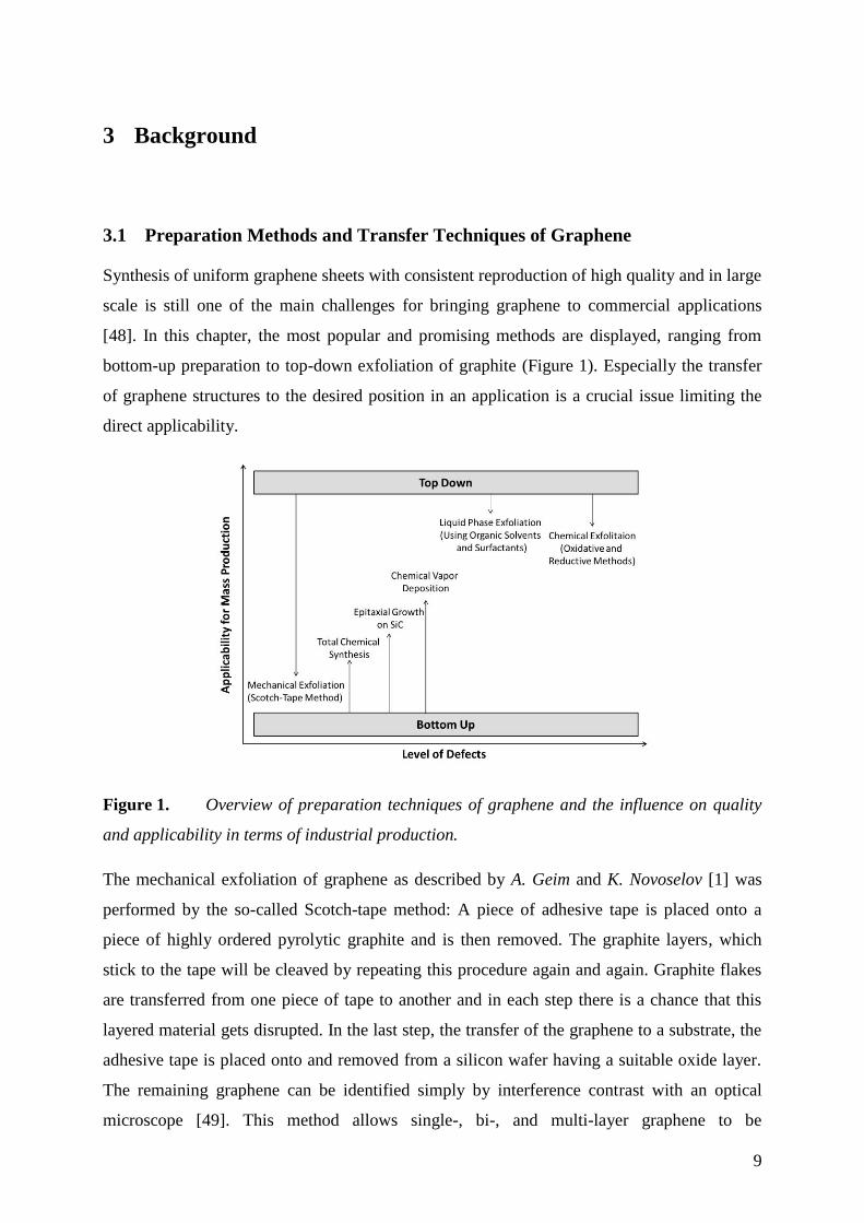
3 Background
3.1 Preparation Methods and Transfer Techniques of Graphene
Synthesis of uniform graphene sheets with consistent reproduction of high quality and in large
scale is still one of the main challenges for bringing graphene to commercial applications
[48]. In this chapter, the most popular and promising methods are displayed, ranging from
bottom-up preparation to top-down exfoliation of graphite (Figure 1). Especially the transfer
of graphene structures to the desired position in an application is a crucial issue limiting the
direct applicability.
Figure 1. Overview of preparation techniques of graphene and the influence on quality
and applicability in terms of industrial production.
The mechanical exfoliation of graphene as described by A. Geim and K. Novoselov [1] was
performed by the so-called Scotch-tape method: A piece of adhesive tape is placed onto a
piece of highly ordered pyrolytic graphite and is then removed. The graphite layers, which
stick to the tape will be cleaved by repeating this procedure again and again. Graphite flakes
are transferred from one piece of tape to another and in each step there is a chance that this
layered material gets disrupted. In the last step, the transfer of the graphene to a substrate, the
adhesive tape is placed onto and removed from a silicon wafer having a suitable oxide layer.
The remaining graphene can be identified simply by interference contrast with an optical
microscope [49]. This method allows single-, bi-, and multi-layer graphene to be
10
distinguished and gives access to the study of the physical properties of this unique carbon
nanomaterial. Graphene obtained from this technique is of high quality and has a well ordered
crystal structure [50]. For practical application, this synthesis is too laborious and
inconvenient, since individual flakes of a size of few micrometers only can be produced. Two
identical graphene flakes can never be obtained due to the imprecisely handling of the tape
and diversity of the starting material. Therefore, nearly a dozen methods have been developed
so far which allow the preparation of graphene of various dimensions, shapes and
quality [38]. [partially adapted from P1]
Another method for the preparation of graphene is the epitaxial growth on SiC [51]. Single
crystal SiC is annealed at 1500 °C evaporating Si atoms from the surface resulting in a
formation of graphene on the SiC substrate. The crystallinity of these samples is very good,
but this technique is also accompanied with several drawbacks. The starting material is very
expensive and the transfer of the graphene is very difficult because of the interaction between
graphene and substrate. Devices fabricated from this graphene are usually made directly on
the substrate [52, 53].
One of the most promising approaches is the growth via CVD [36], utilizing the catalytic
graphitization of hydrocarbon gases at a metal surface, namely Cu, Co and Ni. Here, a carbon
feedstock decomposes at high temperatures of ~1000 °C and rearranges to form sp2-carbon
species [54]. The solubility of carbon in the metal is hereby of major importance. Copper
provides only a low carbon solubility of ~0.03% and carbon atoms are only dissolved at the
surface leading to the preferential growth of single layer graphene [55, 56]. This is a self-
limiting process, since once the metal is covered with graphene the decomposition of the
hydrocarbon precursor is terminated. With this technique 30-inch graphene films were
realized in 2010 [57] and also prototype touch screens made out of this material have been
presented in 2014 [58]. Despite the significant progress in this field, there are still some issues
remaining for commercialization. Graphene grown on a metallic substrate has to be
transferred for further use, usually involving wet chemical etching of the metal and stamping
methods with a polymer support. This leads to contamination, but also to mechanical
distortions and damage to the film and thereby has an impact on the performance in
applications like touch panels [11]. Although the feasibility of such graphene applications has
been shown, the material cannot yet compete with widely established materials like indium tin
oxide and further improvements are needed.
11
The most popular concept for graphene production is the chemical exfoliation of graphite.
Oxidation of graphite with strong oxidizing agents and under harsh conditions leads to a
cleavage of the single carbon layers and single layers of graphene oxide (GO) are formed.
Here, the Hummers method is most often used involving stirring and/or sonication of graphite
in a solution of sulphuric acid and potassium permanganate [23]. The obtained product is
stable in suspension and can be further reduced chemically [59], thermally [60] or
electrochemically [61] forming rGO. Nevertheless, this material comprises a very defective
and ill-defined structure and its electrical properties are not as outstanding as the ones of
defect-free graphene. The oxidation leads to the insertion of hetero atoms like oxygen and
also many topographic defects [62]. Still, these features are not always considered as a
drawback. Functional groups like hydroxyl, epoxy and carboxyl groups are introduced, which
enables further modification of this material. The method has the main advantages of being
rapid, scalable and can be performed with basic requirements of equipment, but also the
resulting product is obtained in suspension and simplifies further use. Different transfer
techniques can be applied to generate graphene films and patterns, namely drop casting, spin
coating, spray coating [63], Langmuir-Blodgett assembly [64] or inkjet printing [65].
Other methods for the liquid phase exfoliation of graphite have been studied and hold great
promise for large-scale production [66]. A successful exfoliation requires the weakening of
the van der Waals attraction between the carbon layers in graphite. The interfacial tension
between liquid and solid should be low, hindering the exfoliated graphene sheets to
agglomerate again. Organic solvents like N,N-dimethylformamide, N-methyl-2-pyrrolidone,
or ortho-dichlorobenzene [67] and surfactants like pyrene derivatives [68] or sodium cholate
[69] are suitable for this propose. Exfoliation is achieved during sonication or by introducing
shearing forces. Recently, it has been shown that graphene can be prepared even with an
ordinary kitchen blender in large scale, using this concept [70]. Still one major drawback is
the residue of surfactant or organic solvent, which has to be eliminated after a transfer.
An interesting approach of a total bottom-up synthesis of graphene was presented by the
group of K. Müllen [71, 72]. Polymerization of rationally designed monomers and solution
mediated or surface assisted cyclodehydrogenation led to the formation of graphene
nanoribbons of precise length, width, edge structure and degree of heteroatom doping.
However, the solubility of the precursors is the main limiting factor and therefore the final
graphene sheets are only small in dimension (length x width) ranging from 50 x 50 nm,
12
500 x 1.1 nm or 30 x 2 nm. Up to now, yields of this synthesis are rather low, disqualifying
this method for industrial application.
All of these methods are advantageous in some ways, and most of them are able to produce
large quantities of graphene. However, the resulting material does not always consist of well-
defined, two-dimensional nanocrystals of unique chemical composition. Oftentimes, even
within the same production technique, an ill-defined material will result and can have slightly
different properties from batch to batch. The ideal graphene is characterized by a quasi-
infinite size. This cannot be achieved by any preparation method. Graphene is comprised of
only a surface, having no bulk phase, but there is always a border. At these edges the carbon
atoms are chemically bound to hydrogen, hydroxyl, carboxyl and other impurities, influencing
the characteristics of the material. So, as for every nanomaterial, the properties depend
strongly on the dimensions of the material. The smaller an ideal single flake, the higher the
ratio of carbon atoms located at the border to the ones within the two-dimensional crystal, and
the more such a material will differ in its physical properties from pristine graphene.
Furthermore, the structure itself can contain chemical functionalities or impurities, introduced
by the synthesis or the transfer of this material. For rGO for example, elemental analysis
showed that oxygen remains in this material. This has a tremendous influence on the behavior
and the properties of rGO, which are no longer the same as found for graphene prepared by
the Scotch-tape method. Additionally, many of the graphene materials synthesized have not
been adequately classified and so the term “graphene” is often used for materials that differ
significantly in chemical structure, shape, size, number of layers and, therefore, in their
properties. This problem was previously addressed [73], but remains unresolved. The term
graphene is often used without giving an exact characterization of the material. An urgent
need for all carbon nanomaterials from graphene to graphite is to come to a consensus to
describe this manifold of materials. Furthermore, standard characterization methods are
required whereby any researcher can immediately evaluate the quality of the material, in
terms of the number of layers, chemical doping, crystallite size, or presence of impurities.
Practically most of this information can be obtained from Raman spectroscopy studies [74].
Therefore, it should be a mandatory first step for all works regarding graphene synthesis to
present a Raman spectrum. [adapted from P1]
Despite all the advantages and drawbacks of different methods for preparing two-dimensional
carbon nanomaterials, the final application of the material will always direct to the method of
choice. Main issues are scaling, transfer, and reproducibility of the methods. Further possible
13
modifications of the material have also to be taken into account. The broad variety of
graphene materials provides a versatile toolbox and therefore can be applied to many fields of
application ranging from electrical devices like supercapacitors and solar cells to textiles and
clothing; or ranging from flexible products like electronics to stiff lightweight applications
like wind turbine blades or even cycling helmets; or large area applications like conductive
paint and heating elements to nanoapplications in chemical sensors and electronic devices.
14
3.2 Graphene as Sensitive Material in Gas Detection Applications
Gas Sensing Fundamentals 3.2.1
Reliable detection of gaseous analytes is essential for many aspects of our everyday life.
Applications are ranging from air quality control in cars or buildings for controlled ventilation
[75–77], over quality control in food industry [78] to security and safety issues [79, 80].
Recognition of harmful, toxic and hazardous gases will improve quality of life. Gas sensors
used so far can be principally divided in three big groups, namely spectroscopic methods,
optical detection and electrochemical solid-state gas sensors. Spectroscopic methods are based
on the direct analysis the vibrational spectrum or molecular mass of a target analyte enabling
a quantitative detection with high precision. Optical sensors usually based on the adsorption
measurements (particularly infrared adsorption) provide a characteristic fingerprint. Whereas
both of these concepts are expensive and require a certain instrumental effort, sensing based
on changes of the electrical properties of solid state devices can be miniaturized easily and
therefore have a broader scope of application. In this context the analyte interacts with an
active sensor material transducing an electrical signal that is most often potential, electrical
conductance or capacitance. Depending on the measurement type, such sensors can be
classified as potentiometric, amperometric or conductometric.
Especially conductometric or resistive type sensors hold great promise for the development of
miniaturized chemical sensors. The change in electrical resistance due to the interaction of
analyte with the sensor surface leads to changes in charge mobility or doping level of the
material. Many metal oxides show response in their electrical conductance and are suitable for
this propose [16]. Semiconductors like ZnO and SnO2 are widely applied in this technology as
thin-film or bulk resistor [81]. However, low electrical conductance and slow desorption
characteristics limit the operation temperatures of these sensors to 250-600 °C [17], having
negative effects on long term stability and power consumption [82]. A large surface area is
highly desirable, to absorb as much target analyte as possible on the surface, giving a stronger
response and better sensitivity. For all demands like fast response, large surface area and low
operation temperature, graphene offers a possible solution. Another key issue and major
challenge in the development of new sensitive materials is the cross sensitivity to other
species or external influences like temperature or humidity. Gas sensors often respond to a
broad variety of analytes limiting their potential fields of application. Multisensors, which
consist of an array of different sensors, can be a solution for improving the selectivity. Each
sensor element contributes to a dynamic response pattern helping to distinguish between

15
certain gases or even quantify concentrations in gas mixtures. Such sensor systems produce
higher dimensional datasets, which have to be processed with multivariate statistics for
evaluation. Here, PCA offers a tool to reduce the dimensions of the dataset into a manageable
form enabling a simple readout.
16
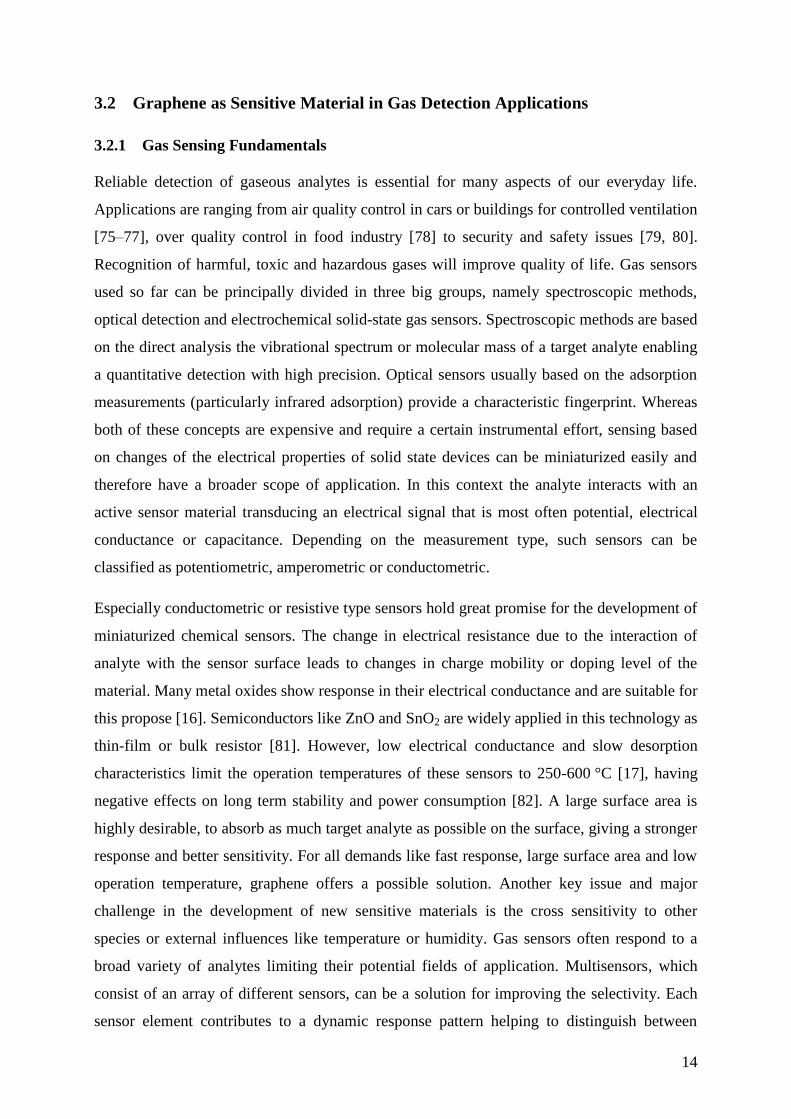
Graphene in Gas Sensing Applications 3.2.2
Carbon based nanomaterials, are promising candidates for electrochemical gas detection at
room temperature due to their intrinsic electrical properties, which are highly sensitive to
changes in the chemical environment [83, 84]. Extensive research has been carried out on
carbon nanotubes (CNTs) based chemical sensors [85]. A higher sensitivity compared to
CNTs, but also better opportunities for device integration and low cost, make graphene a
serious candidate for gas sensing devices in a commercial scenario [86].
The first graphene based gas sensor was presented by Schedin et al. [20] The device
comprised a field effect transistor (FET) made of mechanically exfoliated graphene tailored
and contacted by electron-beam lithography. Exposure to target gases like NO2, H2O, CO, and
NH3 led to a response which was attributed exclusively to gas adsorption and LODs in the
ppb range, comparable to exiting technologies, were observed. In a complex Hall bar setup it
was possible to display even the event of adsorption and desorption of a single NO2 molecule.
Nevertheless, the experimental conditions were far from reality, keeping the sensor under
vacuum, applying only one gas species and recovering the sensor material at 150 °C. But they
demonstrated the great potential of this material in gas sensing applications. The proof of
concept inspired many other researchers to further investigate this behavior theoretically [27,
87, 88] and experimentally [89–101]. An electrochemical detection based on a resistive setup
is here the most reasonable approach due to simplicity and applicability. The sensor concept is
similar to metal oxide solid-state chemiresistors and was also used in the frame of this work
(Figure 2). Graphene shows p-type semiconducting behavior, meaning that electron holes are
the dominant species involved in the charge transport [84]. Therefore, adsorbates on the
surface acting as electron donors lead to an increase of electrical resistance, whereas electron
acceptors result in a decrease, respectively.
17
Figure 2 Scheme of the sensor principle of chemiresistor gas sensors based on
graphene. [adapted from P2]
Comparing the graphene materials investigated as sensor material, most studies are performed
with graphene of low quality like rGO. Less works deal with materials of higher quality
derived from more efficient preparation methods compared to mechanical exfoliation. The use
of free-standing epitaxial grown graphene enabled the detection of NO2 in the sub-ppm
range [98]. But a recovery of the sensor material was only observed after heating to 150 °C.
Same observations were made by Yavari et al. presenting a chemiresistor based on CVD
grown graphene [99]. Unfortunately, the sensor needed a treatment of 200 °C and vacuum to
recover. Detection of NO2 concentrations in the sub-ppt regime at room temperature was
shown by Chen et al. [100], using a CVD chemiresistor, with additional in situ cleaning of the
sensing material with UV light illumination. However, whereas most of the graphene
applications benefit from a pristine graphene structure, this is not necessarily required in
chemical sensors. Target gas molecules may not easily adsorb and desorb at the defect-free
graphene surface [102]. Introduction of defects can be a possible solution and has been shown
to improve sensitivity compared to pristine graphene [103–105]. The incorporation of hetero
atoms within the graphene structure through doping with boron, nitrogen or sulfur showed to
enhance the adsorption of various gas molecules [87, 106, 107]. Chung et al. [108] reported
on the improved detection of NO2 using ozone treated graphene sheets compared to pristine
graphene. Theoretical studies underline these observations and the enhanced sensitivity was
attributed to the surface active defect sites like hydroxyl, epoxy and carbonyl groups [109,
110]. These features are already apparent in GO and rGO. Chemically exfoliation of graphene
is the easiest way to obtain the material and the resulting aqueous suspensions are rather
simple to process. This may be also a reason why rGO is utilized as sensor material in most
18
studies. Numerous studies showed the highly sensitive detection of various gaseous analytes
like NO2, CO, NH3, H2, CH4, H2O, formaldehyde, ethanol and ipropanol in the sub-ppm
range, applying rGO into a chemiresistor setup [18, 89, 91, 111–117]. However, the low
electrical conductance caused by the defects within the sp2 structure by oxygen containing
groups has to be considered. Robinson et al. [18] found that sensitivity and noise level are
affected by the level of reduction from GO to rGO using hydrazine vapor. An increase of
response and recovery time with increasing level of oxidation was observed. They also found
that the noise level greatly decreased with increasing film thickness accompanied with an
increase of electrical conductance, but also sensitivity decreased. In another study, Prezioso et
al. [118] showed that good electrical properties are not necessarily needed. A LOD of 20 ppb
for NO2 was observed applying highly oxidized GO in their device. The high sensitivity was
attributed to the increased number of oxygen functional groups serving as active adsorption
sites. Here, a tradeoff has to be found. Optimization of the defect density is required to
balance sensitivity, level of noise, response times and recovery rates of rGO-based sensors.
Nearly all of the devices presented in literature suffer from poor selectivity. Unprocessed
graphene materials are sensitive to nearly any kind of surface adsorbate, which alters its
electronic structure. Functionalization of the graphene material was shown to be the best way
to achieve improved sensor performance and was also a goal of this work. Introduction of
defects and hetero atoms [104, 119–121] or functional groups [122], decoration with metal,
[123–127] and metal oxide nanoparticles [93, 128–132], and polymer composite materials
[133–136] were shown to improve sensitivity to target analytes. The two major possibilities
are on the one hand, to enhance the gas adsorption process offering specific binding sites and
on the other hand it reduces nonspecific binding and therefore improves selectivity for a
certain analyte.
19
Conclusion 3.2.3
Graphene based gas sensors display superior sensitivity, reversibility and lower detection
limits compared to conventional metal oxide chemiresistors. They can be operated at room
temperature still showing remarkable high and fast responses upon gas adsorption and
therefore the carbon nanomaterial is seen as the sensor material for next generation gas
sensors. However, despite all advantages some critical issues have still to be addressed. Main
problem is the poor selectivity since a variety of gas molecules absorbed to surface of
graphene lead to similar changes in electrical conductance. Here, chemical functionalization
and also composite materials can provide a solution. Up to now, it has not been shown that the
material can be tailored to superior selectivity for a single analyte. But the altered sensor
response in combination with the concept of a sensor array can enable pattern recognition of
single gases in complex mixtures. Another issue in terms of commercialization of such
sensors is the reproducible preparation of devices, which is rarely addressed in literature. The
right choice of graphene preparation, followed by specific modification of the material is
mandatory. Graphene obtained by chemical and liquid exfoliation or CVD methods combined
with a defined introduction of defects is supposed to be most suitable for this purpose.
20
3.3 Graphene in Biosensor Application
During the last decade there have been numerous studies on biosensors employing graphene
materials. The following chapter summarizes approaches reported on most relevant analytes
such as glucose, nucleic acids, proteins, H2O2, lipids, pesticides, ions and even whole cells
and viruses. For modern biosensor applications, chemically exfoliated graphene is the most
frequently used type. It comprises many defects and high polydispersity, yet still has plenty of
interesting properties. The ability of GO being dispersible in aqueous solutions enables easy
modification with biomolecules via standard immobilization techniques. If the flakes are
small, the material shows weak fluorescence, which can be excited over nearly the entire
visible range. After a reduction step, the previously highly distorted aromatic system will be
recovered, and the resulting material has been shown to exhibit good electrical conductance,
as well as promising quenching abilities. The reduced chemically derived graphene (rGO) is
also dispersible in water, but does not form stable solutions. It has a high tendency to
aggregate due to excellent π-stacking properties. Nevertheless, chemically derived graphene
as well as GO can be processed in solution and therefore transferred to any sensor surface by
dip coating, drop casting or spin coating. As the material in solution is very inhomogeneous,
this is also the case for a sensor layer produced from such a solution. Furthermore, the desired
one atom thick layer will not result, but rather, most likely, few-layered graphene will be
deposited.
Biosensors for Glucose 3.3.1
Sensors for glucose have been now studied for more than 50 years since Clark and Lyons first
proposed the initial concept of enzyme-modified electrodes in 1962 [137]. One reason is that
diabetes continues to be an increasing worldwide public health problem. Sensors with good
precision and accuracy are already commercially available. Such sensors consume only
100 µL of whole blood for the measurement, and results are displayed within a very short
time. However, there are still many tasks and challenges for further research in glucose
biosensors, for example, the electrical contact between the redox center of the enzyme and the
electrode surface could be greatly improved [138]. Here, the electrical conductance of
graphene could be utilized. Another demanding need in the fabrication of enzymatic
electrochemical sensors is the immobilization of the biomolecule onto the electrode. In
principle one can entrap the enzyme in a thin polymer film or covalently attach it to the
electrode surface. For this first strategy, enzyme leaching can become an issue and long-term
21
stability will be critical to monitor. The second approach is often accompanied by changes in
the conformation of the biomolecule and therefore, limitations in the enzyme activity are
observed. Furthermore, good electrical communication between the active site of the enzyme
and the electrode is mandatory for achieving high sensitivity. The ultimate compromise
between the choice of materials and immobilization strategies, which results in the best
biosensor for glucose detection, has yet to be established. Thus, research in the field of
glucose biosensors, which can achieve high sensitivity, long-term stability, and excellent
selectivity is still a hot topic. Carbon nanomaterials, and especially graphene, have become
more prevalent in this research due to its good electrical transport properties, high surface to
volume ratio, as well as numerous possibilities to attach biomolecules.
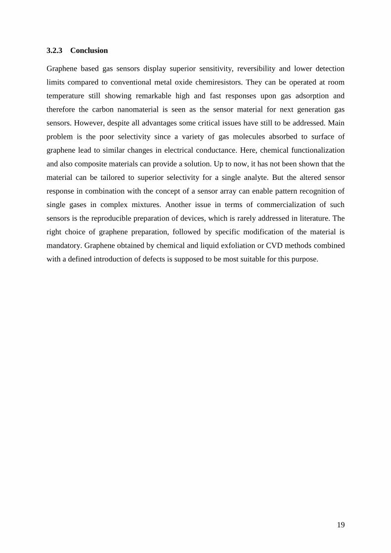
In amperometric glucose sensing, there are mainly two prominent detection strategies: First,
those that measure the direct electron transfer from the enzyme to the electrode during
oxidation of glucose. Second, those techniques, which catalyze the oxidation of the hydrogen
peroxide produced by the enzyme in the presence of glucose (Figure 3). Both strategies are
purported to enhance the performance by including graphene materials in the sensor design.
Figure 3 Scheme of enzymatic detection of glucose.
In the case of monitoring the direct electron transfer from the enzyme glucose oxidase (GOx)
to the electrode, electrodes where the enzyme is entrapped in conducting polymers have been
commonly used. However, by replacing the polymer with graphene, a mediator is no longer
required. Direct electron transfer has been observed thus far on electrochemically reduced
graphene (erGO) on polylysine [139], mixtures of 3-aminopropyltriethoxysilane and graphene
[140], a hybrid material of multiwalled carbon nanotubes and erGO [141], mesoporous
materials in combination with erGO [142], ionic liquid modified GO [143–145], bimetallic
Au/Pd NP decorated erGO [146], and on CdS quantum dot modified graphene [147].
22
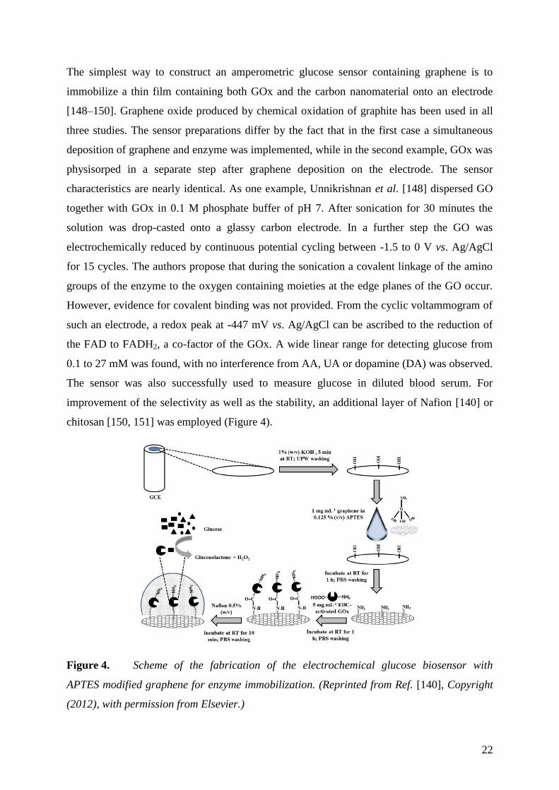
The simplest way to construct an amperometric glucose sensor containing graphene is to
immobilize a thin film containing both GOx and the carbon nanomaterial onto an electrode
[148–150]. Graphene oxide produced by chemical oxidation of graphite has been used in all
three studies. The sensor preparations differ by the fact that in the first case a simultaneous
deposition of graphene and enzyme was implemented, while in the second example, GOx was
physisorped in a separate step after graphene deposition on the electrode. The sensor
characteristics are nearly identical. As one example, Unnikrishnan et al. [148] dispersed GO
together with GOx in 0.1 M phosphate buffer of pH 7. After sonication for 30 minutes the
solution was drop-casted onto a glassy carbon electrode. In a further step the GO was
electrochemically reduced by continuous potential cycling between -1.5 to 0 V vs. Ag/AgCl
for 15 cycles. The authors propose that during the sonication a covalent linkage of the amino
groups of the enzyme to the oxygen containing moieties at the edge planes of the GO occur.
However, evidence for covalent binding was not provided. From the cyclic voltammogram of
such an electrode, a redox peak at -447 mV vs. Ag/AgCl can be ascribed to the reduction of
the FAD to FADH2, a co-factor of the GOx. A wide linear range for detecting glucose from
0.1 to 27 mM was found, with no interference from AA, UA or dopamine (DA) was observed.
The sensor was also successfully used to measure glucose in diluted blood serum. For
improvement of the selectivity as well as the stability, an additional layer of Nafion [140] or
chitosan [150, 151] was employed (Figure 4).
Figure 4. Scheme of the fabrication of the electrochemical glucose biosensor with
APTES modified graphene for enzyme immobilization. (Reprinted from Ref. [140], Copyright
(2012), with permission from Elsevier.)
23
Mesoporous materials such as ZrO2 have been used as a support material for amperometric
glucose sensors [142]. The high surface area, large pore volume, narrow pore size
distribution, as well as the tunable pore size, together with a modest electrical conductance,
make this material an attractive host for enzyme immobilization. The sensor was fabricated by
electrochemical reduction of GO on a glassy carbon electrode followed by electrodeposition
of a thin chitosan film. This polymer layer provides positively charged amino groups, which
helps to attach the mesoporous ZrO2 on top by drop casting. Following the deposition of the
ZrO2 layer, GOx was loaded onto the electrode. Direct electron transfer could be observed
when glucose was added. A high Michaelis-Menten constant of 28.01 mM indicated
limitations in mass transport, which results in a slow response time in the minutes regime. The
sensor was operated at 400 mV vs. Ag/AgCl in phosphate buffer of pH 7.4. The detection
limit for glucose was reported as 46 µM with a linear range from 0.2 to 1.6 mM. There was no
study of interference and no real sample analysis presented. Nevertheless, the high sensitivity
of 7.6 µA·mM-1
·cm-2
is remarkable. A comparison to the same sensor scheme without an
erGO-layer resulted only in poor redox peaks, therefore one can conclude that the carbon
nanomaterial is essential in providing the enhanced direct electron transfer. The simple
electrostatic adsorption of the enzyme suffers from long-term stability. Therefore, rGO was
modified with ionic liquids possessing positively charged groups [143]. The better adhesion
of the biomolecule to the carbon nanomaterial was proposed to be due to the ionic interaction.
In case of 1-(3-aminopropyl)-3-methylimidazolium bromide, the sensor covers a linear range
from 2 to 16 mM [143]. For the same sensor design with 1-vinyl-3-butylimidazolium bromide
as the ionic liquid a linear range from 0.8 to 20 mM was obtained [144] and with 1-butyl-3-
methylimidazolium hexafluorophosphate the linear range was reported between 2 and 20 mM
[145]. Such modifications using ionic liquids are not outstanding in their performance when
compared with other sensors, which use direct electrochemistry for transduction.
Direct electron transfer in amperometric enzyme sensors, with and without the use of
graphene, is limited due to the same reasons. The redox center is buried inside the structure of
the protein, which hinders the transfer of the electrons to the electrode. Therefore, electron
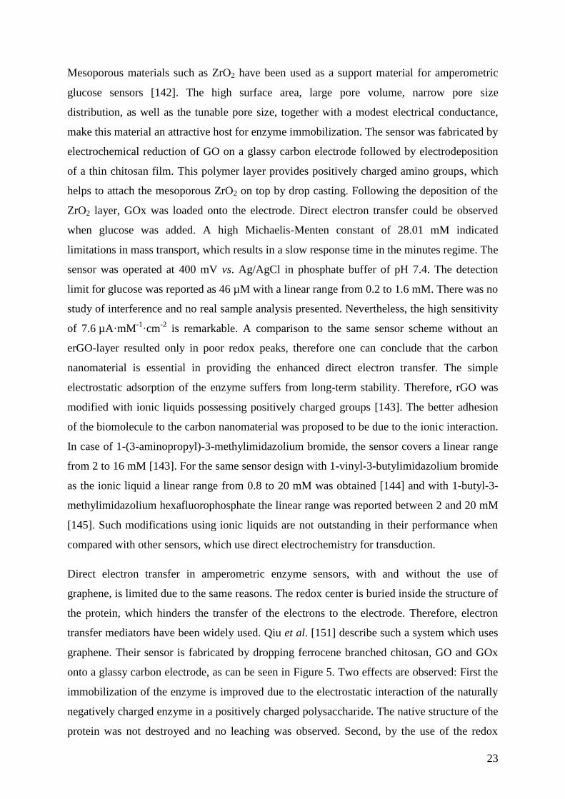
transfer mediators have been widely used. Qiu et al. [151] describe such a system which uses
graphene. Their sensor is fabricated by dropping ferrocene branched chitosan, GO and GOx
onto a glassy carbon electrode, as can be seen in Figure 5. Two effects are observed: First the
immobilization of the enzyme is improved due to the electrostatic interaction of the naturally
negatively charged enzyme in a positively charged polysaccharide. The native structure of the
protein was not destroyed and no leaching was observed. Second, by the use of the redox
24
mediator ferrocene, the sensitivity could be enhanced to 10 µA·mM-1
·cm-2
. A Michaelis-
Menten constant of 2.1 mM was calculated. The capability of this biosensor was demonstrated
in measuring plasma glucose level. The response displayed a linear glucose concentration
range from 0.02 to 6.78 mM with a LOD of 7.6 µM. By comparison of the resulting current
densities this sensor shows 2.7 times higher signal when GO is used. The working potential of
the electrode is lowered to 300 mV vs. Ag/AgCl, which increases the selectivity. No
interference of AA or UA was found.
Figure 5. Sensor design of a glassy carbon electrode modified by ferrocene modified
Chitosan (CS-Fc), GO and GOx. The sensor exhibits excellent sensitivity and a low working
potential of 300 mV vs. Ag/AgCl. (Reprinted from Ref. [151], Copyright (2011), with
permission from Elsevier.)
A novel platform for the in situ chemical immobilization of GOx was developed by Chen et
al. [152] Citric acid, which was applied to the Au NPs, was used as a capping and reducing
agent. The carboxyl groups of the citric acid, along with the amino groups of the GOx, form a
peptide bond when immobilization takes place. After drop casting the composite material onto
a glassy carbon electrode, glucose could be detected within the range of 0.1 to 10 mM, with
an LOD of 35 µM. An enhancement was found when electrochemically reduced GO
decorated with bimetallic nanoparticles of 1:1 Au:Pd was used. Extreme high sensitivity of
267 µA mM-1
cm-2
was reported. The enzyme was simply physically adsorbed on top of the
composite material and covered by a thin film of Nafion [146]. A detection limit of 6.9 µM
with linearity up to 3.5 mM was found.
25
Amperometric Glucose Biosensors with Detection of H2O2
The glucose level often is indirectly monitored using amperometric biosensors by measuring
the current in the oxidation of H2O2 produced during the enzymatic reaction of GOx with
glucose. In the case of a glassy carbon electrode coated by a thin polymer film of chitosan
incorporating rGO and GOx, the oxidation occurs at 780 mV vs. Ag/AgCl [153]. To enhance
the kinetics and to minimize high over potentials, metal and metal oxide nanoparticles are
often introduced. Low working potentials are desired to diminish the risk of interference by
other electroactive substances such as ascorbate, ureate, acetaminophen, etc., which are often
present in complex matrices. The introduction of additional nanomaterials for catalytic
purposes is favorable for large surface area materials such as graphene. Furthermore, a
support with high electrical conductance is needed to attach the nanoparticles without
agglomeration. For this reason graphene materials are of great interest, as they promote fast
electron transfer kinetics and allow simple preparation strategies with respect to decoration by
metal nanoparticles.
Composite materials consisting of Pd NPs attached to graphene are very popular in the design
of amperometric glucose sensors [154, 155]. Synthesis can be performed in one step by
simultaneous reduction of PdCl2 and GO in an 85% hydrazine solution [154]. The resulting
nanoparticles are attached to the carbon nanomaterial. With this method agglomeration of the
rGO is prevented. The electrode is modified by drop casting the nano-hybrid material. For
such electrodes an increase in the LOD to 1.0 µM for the oxidation of hydrogen peroxide at
500 mV vs. Ag/AgCl was found. For an identical sensor but without Pd NPs, the detection
limit was reported at 2.3 µM. To fabricate an amperometric glucose sensor further
immobilization of the enzyme is needed. This was performed by a well-known chemical
coupling procedure via glutaraldehyde, which results in a peptide bond between GOx and
rGO. Additional blocking is accomplished in the same way by the use of bovine serum
albumin (BSA). An alternative strategy is the functionalization of GO with chitosan and
reduction of GO with hydrazine in a solution containing PdCl2 [155]. A homogeneous
distribution of Pd NPs within the rGO-chitosan is found. This composite material was drop
cast onto a glassy carbon electrode. In a last step the GOx was chemically coupled to the
electrode via glutaraldehyde chemistry. Such a sensor shows best glucose detection properties
at a working potential of 700 mV vs. SCE with linearity in current response in the
concentration range from 1 to 10 mM. The LOD was reported to be 0.2 µM for glucose. It
was found that AA as well as UA interfered with the detection of glucose at such a high
26
electrode potential. Therefore, a suggestion was made to operate this sensor at -50 mV.
Although this change in working potential increased the selectivity, the sensitivity of the
system decreased by 93%.
A similar sensor concept was adapted by Claussen et al. [156] using Pt NPs to catalyze the
oxidation of hydrogen peroxide. In contrast to other reports, they do not use a glassy carbon
or a metal electrode; they used graphene on a SiO2 wafer as the electrode material. The
graphene was produced by CVD and consists, on average, of 12 layers. It was functionalized
by an oxygen plasma treatment and Pt NPs were electro-deposited on the edges. The GOx
was encased in a conductive polymer, poly(3,4-ethylenedioxythiophene). This glucose sensor
exhibits a broad linear range from 0.01 to 50 mM, with a detection limit of 0.3 µM. The
advantage of this complicated technology to produce such a sensor is to have the ability to
create a sensor array by structuring the graphene by plasma treatment.
When platinum is used as a metal coordination polymer (which allows an effective adsorption
of GOx), the LOD for glucose decreases to 5 nM [157]. Graphene oxide, modified with
poly(N-vinyl-2-pyrrolidone) and reduced by hydrazine was suspended together with p-
phenylenediamine and H2PtCl4 before being drop cast onto the electrode. The electrode
potential was adjusted to -300 mV vs. SCE and the sensing behavior was not influenced by
common interfering substances such as DA, AA or UA. Another approach describes graphene
decorated with platinum-gold alloy NANOPARTICLEs, synthesized by simultaneous
reduction of Pt- and Au-salts in the presence of GO [158]. As expected, such an electrode is
capable of detection of glucose. An LOD of 1 µM is reported. The drawback of this study is
the high working potential of 700 mV vs. Ag/AgCl, which limits the selectivity of this system.
Composite materials in electro-analytical sensors are most often prepared by simple drop
casting of a relatively complex sensor cocktail. There has been little work done in the
optimization and characterization of the resulting layer. By this simple technique in sensor
fabrication it is very easy to fabricate hybrid materials consisting of different nanomaterials.
Gu et al. [159] describe a system consisting of a composite layer of rGO with Au NPs
(G-AuNP), together with quantum dots (CdSe-CdS), Au NPs and GOx within a Nafion
membrane, drop casted onto a gold electrode. The electrocatalytic oxidation scheme is
depicted in Figure 6. This sensor exhibits a low working potential of -200 mV vs. Ag/AgCl,
which could increase the selectivity. One drawback of such systems may be the elaborate
synthesis of the nanomaterials required to produce high quality materials in a reproducible
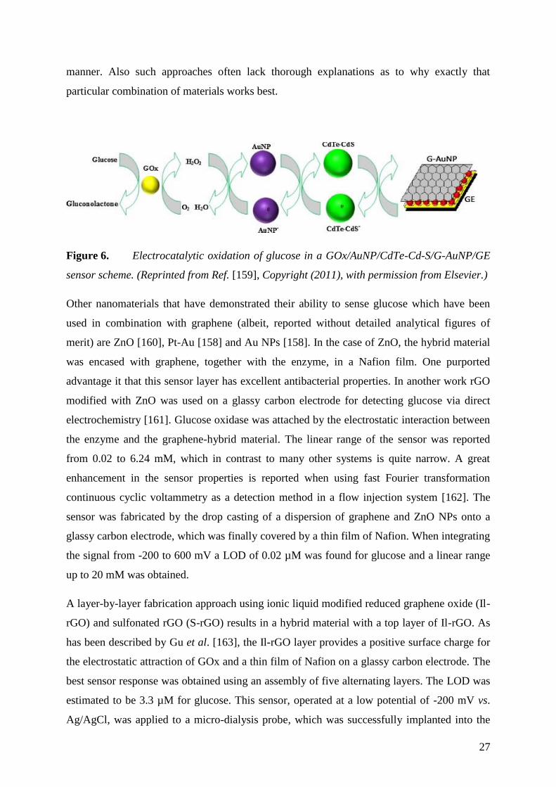
27
manner. Also such approaches often lack thorough explanations as to why exactly that
particular combination of materials works best.
Figure 6. Electrocatalytic oxidation of glucose in a GOx/AuNP/CdTe-Cd-S/G-AuNP/GE
sensor scheme. (Reprinted from Ref. [159], Copyright (2011), with permission from Elsevier.)
Other nanomaterials that have demonstrated their ability to sense glucose which have been
used in combination with graphene (albeit, reported without detailed analytical figures of
merit) are ZnO [160], Pt-Au [158] and Au NPs [158]. In the case of ZnO, the hybrid material
was encased with graphene, together with the enzyme, in a Nafion film. One purported
advantage it that this sensor layer has excellent antibacterial properties. In another work rGO
modified with ZnO was used on a glassy carbon electrode for detecting glucose via direct
electrochemistry [161]. Glucose oxidase was attached by the electrostatic interaction between
the enzyme and the graphene-hybrid material. The linear range of the sensor was reported
from 0.02 to 6.24 mM, which in contrast to many other systems is quite narrow. A great
enhancement in the sensor properties is reported when using fast Fourier transformation
continuous cyclic voltammetry as a detection method in a flow injection system [162]. The
sensor was fabricated by the drop casting of a dispersion of graphene and ZnO NPs onto a
glassy carbon electrode, which was finally covered by a thin film of Nafion. When integrating
the signal from -200 to 600 mV a LOD of 0.02 µM was found for glucose and a linear range
up to 20 mM was obtained.
A layer-by-layer fabrication approach using ionic liquid modified reduced graphene oxide (Il-
rGO) and sulfonated rGO (S-rGO) results in a hybrid material with a top layer of Il-rGO. As
has been described by Gu et al. [163], the Il-rGO layer provides a positive surface charge for
the electrostatic attraction of GOx and a thin film of Nafion on a glassy carbon electrode. The
best sensor response was obtained using an assembly of five alternating layers. The LOD was
estimated to be 3.3 µM for glucose. This sensor, operated at a low potential of -200 mV vs.
Ag/AgCl, was applied to a micro-dialysis probe, which was successfully implanted into the
28
striatum of a rat. A glucose concentration of 0.38 mM was monitored online. After an
injection of insulin into the striatum of the rat, a decrease in the glucose level could be
observed for about 25 minutes.
In contrast, by covalent attachment of GOx via glutaraldehyde coupling, a sensor based on
ERGO modified with an ionic liquid exhibits a greater linear range (0.005 to 10 mM) with an
LOD of 1 µM [164]. This illustrates the fact that it is not always clear which kind of
immobilization works best. Although these sensors were both operated under the same
conditions and exhibit the same selectivity, the chemically immobilized enzyme sensor [164]
exhibits a sensitivity that is three times higher than the sensor where the enzyme was
immobilized by ionic interaction [163].
Alwarappan et al. [165] report a unique strategy for enhanced glucose biosensing. Here they
develop a glassy carbon electrode which is electrochemically modified by polypyrrole that
acts as a host material for the immobilization of GOx which is chemically functionalized by
graphene. Unfortunately the covalent modification of the enzyme with the graphene was not
described in a convincing way. There is no real characterization of the formation of a specific
bond between graphene and GOx. The protocol involves simply mixing rGO with the enzyme
in phosphate buffer and vortexing for 50 s. Further purification of this mixture from unreacted
enzyme as well as excess graphene was also not described. Also the immobilization protocol
of the graphene-enzyme-conjugate to the polypyrrole is completely absent. Therefore, one can
only say that graphene and GOx was applied to a polypyrrole modified electrode.
Nevertheless, this system is reported to detect glucose with linearity in the range of 2 to
40 µM with an LOD of 3 µM.
Most studies of biosensors used for the detection of glucose use chemically derived graphene
[141–155, 157, 158, 161, 163–170], with various strategies for the reduction of the GO.
Electrochemical reduction has the advantage of convenience; electrode deposition can be
performed simultaneously with the reduction step. In contrast, dispersed GO allows easy
modification of the carbon nanomaterial with molecules, nanoparticles or biomolecules. To
recover the high electrical conductance, the functionalized GO is reduced afterwards. Many
studies lack proper characterization of the carbon nanomaterial and the final composite
material. Therefore, it is often not clear which parameter contributes most to the enhanced
sensor characteristics. To date, there has been little work on the investigation of the influence
of the doping of the graphene itself. Wang et al. [170] present a study where they use
nitrogen-doped graphene in electrochemical biosensing. Nitrogen-doping could be a potential
29
strategy to regulate the electronic properties in carbon nanomaterials. By treating a chemically
derived graphene with nitrogen plasma, it was possible to control the content of nitrogen
atoms of the graphene from 0.11 to 1.35%. This was achieved by mixing rGO with chitosan
and drop casting this mixture onto a glassy carbon electrode. The electrodes where then
exposed to a nitrogen plasma for a specified time. Finally, GOx was physisorbed onto the
modified electrodes. A comparison of undoped with N-doped graphene shows that there is an
enhancement in the resulting peak current. This sensor could be used either to monitor the
direct electron transfer from GOx or to measure the oxidation of hydrogen peroxide. The later
technique was performed at 150 mV vs. Ag/AgCl to achieve an LOD of 10 µM and a linear
range of 0.1 to 1.1 mM for glucose. Uric acid (UA) as well as ascorbic acid (AA) showed no
significant interference.
In summary, much research has been made into the use of graphene in enzymatic
electrochemical glucose sensing. Regarding hybrid materials, direct electrochemistry
involving metallic nanoparticles for the oxidation of hydrogen peroxide was well studied.
However, one can conclude that similar trends were found for the monitoring of glucose
sensors with and without the use of graphene. It is interesting to note that in most published
works the outstanding properties of graphene are discussed to be beneficial for the biosensor
design. Alwarappan et al. [171] systematically investigated the influence of the graphene
material itself on the immobilization of GOx and the resulting electrochemical sensor
properties. It is reported that there is almost no difference in the amount of immobilized
enzyme as well as in the resulting current density for single layer graphene, few layer
graphene and multilayer graphene. This is surprising due to the fact that it is well known that
single layer graphene is a semi-metallic conductor, whereas the multi layered material acts as
a metallic conductor [172]. Therefore, one can conclude that for glucose biosensing
applications, it is less important which kind of graphene material is used. The greater
challenge is to find a proper way to immobilize the enzyme or to design a hybrid material,
which can really improve the sensing properties in terms of better selectivity and higher
sensitivity. To date, hybrid materials with Pt NPs perform best in terms of sensitivity and the
indirect detection of glucose by oxidation of hydrogen peroxide is superior to the direct
electrochemical detection.
30
Biosensors for Nucleic Acids 3.3.2
Detection, monitoring and analysis of DNA play an important role in human health aiding in
the diagnosis of diseases and genetic mutations, fast detection of biological warfare agents
and in the development of improved pharmaceuticals. Thus, the development of DNA
detection methods with increased sensitivity and accuracy is highly desirable and is
anticipated to play an ever increasing role in the future of medicine.
A typical DNA biosensor system consists of a transducer surface with an immobilized single-
stranded DNA (ssDNA) probe. Upon binding with the complementary single stranded nucleic
acid sequence, hybridization occurs and the pair is detected. The hybridization event is most
typically measured optically, electrochemically or by mass sensitive detection. Historically,
the greatest strides in DNA detection using biosensors have been made with the
implementation of new or enhanced sensor materials. Such enhancers, or additives, have been
critical to the advancement of this field. Various carbon nanostructures (such as carbon
nanotubes) have been widely implemented in DNA biosensors. Among them, graphene has
recently received particularly strong attention. This section will focus on the developments of
graphene-based biosensors, which employ a variety of detection schemes, sensor additives
and types of graphene for the detection of oligonucleotides.
Graphene-based DNA biosensors show great promise when applied to diagnosis of genetic
diseases and environmental monitoring. The interaction between DNA molecules and
graphitic materials has been studied theoretically and experimentally [173–176]. It is
understood that the binding between graphene and nucleotides is dominated by the non-
electrostatic -stacking interaction [174, 175, 177–179], which explains how probe DNA is
immobilized on graphene. Primarily, fluorescence and electrochemical methods are used as
the sensor detection method, with other optical and electrical methods becoming more
prevalent. It has been well demonstrated that the hybridization of complementary strands can
be monitored with graphene-based sensors.
DNA hybridization can be detected using optical techniques such as fluorescence,
chemiluminescence, surface plasmon resonance or interferometry. Fluorescence quenching is
the most common and least complicated detection method used for DNA-sensors employing
graphene [180–189]. A single strand of nucleic acids has a natural affinity to the aromatic
system of graphene via π-stacking of its nucleobases. In this system, fluorescently labeled
ssDNA adsorbs onto the surface of the graphene. The interaction between the dye molecule
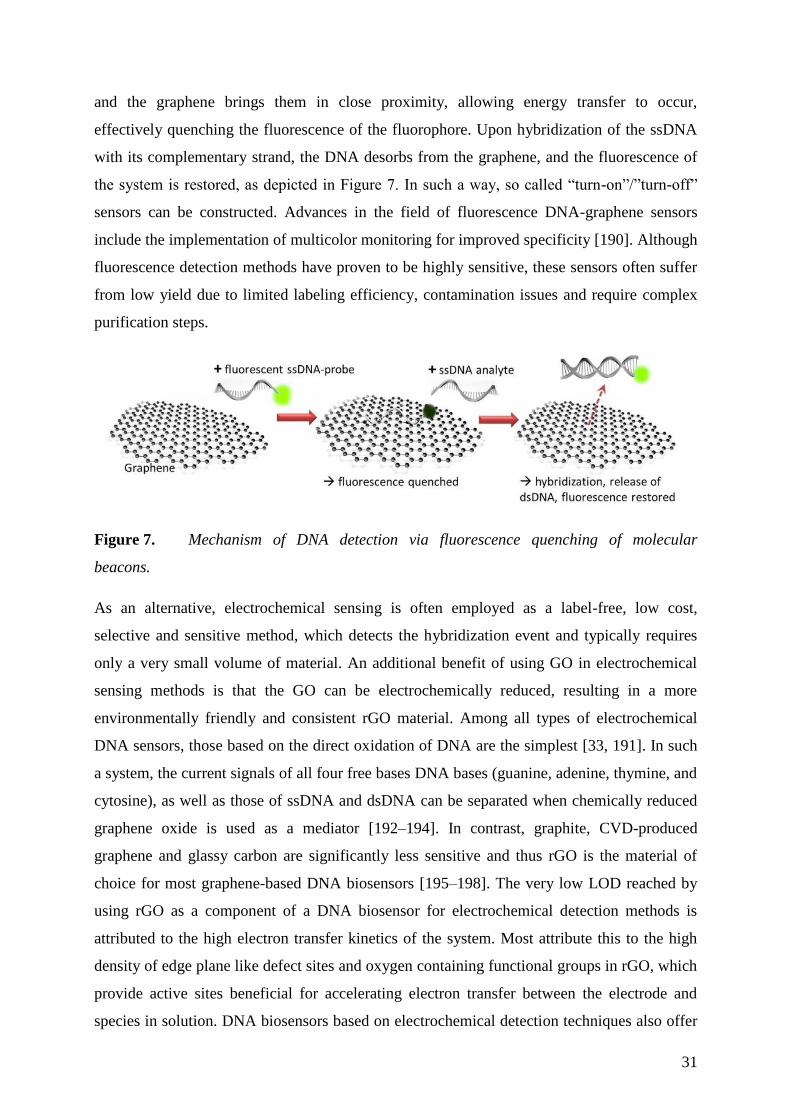
31
and the graphene brings them in close proximity, allowing energy transfer to occur,
effectively quenching the fluorescence of the fluorophore. Upon hybridization of the ssDNA
with its complementary strand, the DNA desorbs from the graphene, and the fluorescence of
the system is restored, as depicted in Figure 7. In such a way, so called “turn-on”/”turn-off”
sensors can be constructed. Advances in the field of fluorescence DNA-graphene sensors
include the implementation of multicolor monitoring for improved specificity [190]. Although
fluorescence detection methods have proven to be highly sensitive, these sensors often suffer
from low yield due to limited labeling efficiency, contamination issues and require complex
purification steps.
Figure 7. Mechanism of DNA detection via fluorescence quenching of molecular
beacons.
As an alternative, electrochemical sensing is often employed as a label-free, low cost,
selective and sensitive method, which detects the hybridization event and typically requires
only a very small volume of material. An additional benefit of using GO in electrochemical
sensing methods is that the GO can be electrochemically reduced, resulting in a more
environmentally friendly and consistent rGO material. Among all types of electrochemical
DNA sensors, those based on the direct oxidation of DNA are the simplest [33, 191]. In such
a system, the current signals of all four free bases DNA bases (guanine, adenine, thymine, and
cytosine), as well as those of ssDNA and dsDNA can be separated when chemically reduced
graphene oxide is used as a mediator [192–194]. In contrast, graphite, CVD-produced
graphene and glassy carbon are significantly less sensitive and thus rGO is the material of
choice for most graphene-based DNA biosensors [195–198]. The very low LOD reached by
using rGO as a component of a DNA biosensor for electrochemical detection methods is
attributed to the high electron transfer kinetics of the system. Most attribute this to the high
density of edge plane like defect sites and oxygen containing functional groups in rGO, which
provide active sites beneficial for accelerating electron transfer between the electrode and
species in solution. DNA biosensors based on electrochemical detection techniques also offer
32
the flexibility of various labels such as enzyme and metal complexes and various organic
compounds. Methylene blue is a common label used in DNA biosensors for electrochemical
detection. A further significant advantage of such a system is that label-free systems can also
be constructed when desired. Examples of such a system are detection based on a change in
capacitance or detection of the oxidation of guanine (the most redox-active nitrogenous base
in DNA).
Less widely implemented is the detection of DNA by direct electrical methods [199]. The
complementary DNA interacts with graphene and an n-doping (electronic doping) effect,
based on the graphene-nucleotide interaction, is observed and measurable. Another up and
coming technique, which has been applied to DNA biosensors is FETs [200, 201].
The immobilization of the probe DNA plays a key role in determining the final sensitivity of
the sensor [202]. In that regard, graphene materials have emerged to efficiently immobilize
DNA. With few exceptions [203–205], rGO or GO is used for graphene-based DNA
biosensors. However, as previously discussed, due to the lack of characterization of the
graphene, which is very often not included in most works involving graphene-based
biosensors, it is difficult to properly categorize and compare sensor systems. Most of the
recent studies of DNA-graphene-based sensors specifically involve chemically processed,
multilayer rGO. This material contains a high density of reactive edges providing functional
groups and adsorption sites.
It is evident when the literature is viewed as a whole while sorted by graphene type that rGO-
based DNA sensors perform best when tested using electrochemical detection methods. It also
appears that GO is perhaps a better choice for fluorescence based detection methods for DNA
biosensors. GO acts as a weak acid cation exchange resin due to its carboxylic groups located
at the basal plane (edge sites) of the GO [206]. A simple ion exchange, typically with a dye, to
form an energy/charge transfer complex for complexation with GO is a common explanation
for the mechanism [207, 208]. Additionally, GO is a highly suitable material for interaction
with biomolecules as the carboxylic groups render the material soluble in aqueous solutions.
The sensitivity, that is, the ability of the biosensor to detect low DNA concentrations, is
essential as is the selectivity and ability to detect a point mutation. With few exceptions, most
sensors based on graphene detect in the nM range. It appears that rGO is the preferred type of
graphene material, giving detection limits in the range of fM to aM, with graphene oxide also
33
giving excellent lower limits of detection in the pM to fM range. By comparison, CVD and
epitaxial graphene do not provide the low limits of detection offered by rGO or even GO.
In order to meet ever-increasing demands for the detection of trace amounts of DNA, new
strategies for enhanced detection and signal amplification are quickly evolving. Many past
reports were based on the use of single or duplex DNA signal amplification strategies,
although triplex sensing schemes are gaining more attention. The labels used in DNA
biosensors are used as a storage reservoir for electrical charge. Multiple labels can be
combined, often having different amplification mechanisms, to achieve excellent sensitivity.
In such strategies, enzyme schemes are often combined with inorganic nanoparticles such as
Au NPs. For example, in the case of one of the lowest reported LODs, Dong et al. [197] have
developed a triplex amplification strategy, which enabled them to achieve an LOD of 5 aM
with the combination of Au NPs, horseradish peroxidase (HRP) and functionalized carbon
nanoparticles. This sensor was highly selective to various single-base and three-base
mismatched sequences of DNA.
To further increase sensitivity, many DNA-graphene biosensor systems employ the use of
nanoparticles such as quantum dots [184, 209], upconverting nanoparticles [210] or metal
nanoparticles (most typically silver or gold) [186, 187, 193, 197, 199–201, 211–215], with
varying degrees of success for signal enhancement. Au NPs are considered to be
biocompatible and highly stable. However, inconsistent signal amplification using metal
nanoparticles remains a problem. They are often used in the immobilization layer to increase
the quantity of DNA, which is often functionalized with an amine group. The other benefit of
Au NPs in such a sensor system is the enhanced signal provided by the plasmon resonance
inherent in the properly engineered Au NPs. The adherence of such Au NPs effectively
magnifies the hybridized signal, and overall, aims to provide enhanced sensitivity and a lower
LOD. On a case-by-case basis, when sensors are developed with or without nanoparticle
enhancers, researchers do note an improvement, albeit typically not significant, in the sensor’s
performance. However, to date, only a few of the top performing sensors employ such sensor
additives, but rather contribute the enhanced signal to other material additives or improved
detection schemes.
Another area where graphene-based DNA biosensors have recently found moderate success is
in the detection of adenosine triphosphate (ATP) [183–185, 188, 210, 216–219], a nucleoside
triphosphate used in cells as a coenzyme. Most biosensors in this category use
carboxyfluorescein tagged ATP, adsorbed onto GO, which quenches the fluorescence. Upon
34
hybridization, the dye labeled ATP molecule is released from the GO, and fluorescence is
restored. The LODs usually lie in the nM range. Application of some of the advanced
techniques mentioned in this section could aid in the further reduction of the LOD for such
biosensors.
Recently, graphene-based DNA biosensors have found application for highly sensitive virus
detection [184, 189, 194, 220–224] by employing specific recognition DNA elements. SARS
[189], Simian [221], and Hepatitis B [184] viruses have been detected in the nM range.
Exceptional sensitivity has been obtained in the detection of HIV virus [220, 222–224], with
Hu et al. [222] reporting an LOD as low as 34 fM. It is worth noting that the authors also
tested the same sensor system with [222] and without [223] the addition of Au NPs and found
no discernible difference in the achieved LOD.
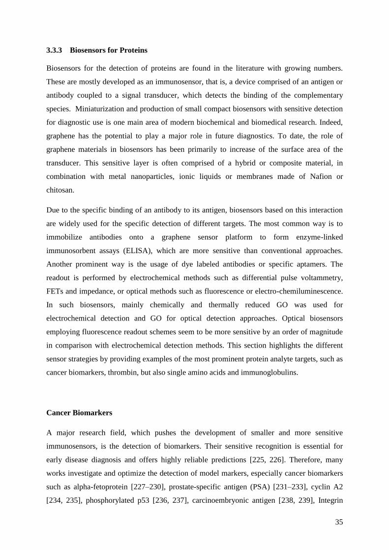
35
Biosensors for Proteins 3.3.3
Biosensors for the detection of proteins are found in the literature with growing numbers.
These are mostly developed as an immunosensor, that is, a device comprised of an antigen or
antibody coupled to a signal transducer, which detects the binding of the complementary
species. Miniaturization and production of small compact biosensors with sensitive detection
for diagnostic use is one main area of modern biochemical and biomedical research. Indeed,
graphene has the potential to play a major role in future diagnostics. To date, the role of
graphene materials in biosensors has been primarily to increase of the surface area of the
transducer. This sensitive layer is often comprised of a hybrid or composite material, in
combination with metal nanoparticles, ionic liquids or membranes made of Nafion or
chitosan.
Due to the specific binding of an antibody to its antigen, biosensors based on this interaction
are widely used for the specific detection of different targets. The most common way is to
immobilize antibodies onto a graphene sensor platform to form enzyme-linked
immunosorbent assays (ELISA), which are more sensitive than conventional approaches.
Another prominent way is the usage of dye labeled antibodies or specific aptamers. The
readout is performed by electrochemical methods such as differential pulse voltammetry,
FETs and impedance, or optical methods such as fluorescence or electro-chemiluminescence.
In such biosensors, mainly chemically and thermally reduced GO was used for
electrochemical detection and GO for optical detection approaches. Optical biosensors
employing fluorescence readout schemes seem to be more sensitive by an order of magnitude
in comparison with electrochemical detection methods. This section highlights the different
sensor strategies by providing examples of the most prominent protein analyte targets, such as
cancer biomarkers, thrombin, but also single amino acids and immunoglobulins.
Cancer Biomarkers
A major research field, which pushes the development of smaller and more sensitive
immunosensors, is the detection of biomarkers. Their sensitive recognition is essential for
early disease diagnosis and offers highly reliable predictions [225, 226]. Therefore, many
works investigate and optimize the detection of model markers, especially cancer biomarkers
such as alpha-fetoprotein [227–230], prostate-specific antigen (PSA) [231–233], cyclin A2
[234, 235], phosphorylated p53 [236, 237], carcinoembryonic antigen [238, 239], Integrin
36
avb3 [240], or progastrin releasing peptide [241]. Nearly all of the works combined a
sandwich assay with electrochemical readout of the coupled enzymatic reaction.
A very representative electrochemical immunosensor was prepared by Yang et al. [242]
investigating the detection of PSA, an important tumor marker for clinical diagnosis of
prostate cancer. In their work, thermally reduced GO was used in two ways (see Figure 8) to
promote the sensitivity to a LOD of 1 pg mL-1
. On the one hand, it was applied as an electrode
material for the immobilization of a primary antibody via --interactions of a linker
molecule. On the other hand, it was used as a support to build up a label consisting of a
secondary antibody, HRP and a mediator thionine. The biosensor was prepared by incubating
the electrode with PSA solutions of different concentrations, washing, incubating with the
label and washing again. The more PSA that was bound, the fewer labels that were attached to
the electrode, resulting in a lower amperometric response against fixed H2O2 concentrations.
This leads to a wide linear sensitivity of 0.002 - 10 ng·mL-1
. The large surface-to-volume ratio
(leading to a high enzyme loading of the label) and the good electrical conductance of
graphene are the main reasons for the improved sensitivity. To test interference and
applicability, the immunosensor was tested towards human immunoglobulin G, BSA, vitamin
C, glucose, and human serum samples. Satisfactory test results were obtained in comparison
with a commercial ELISA system.
Figure 8. Schematic representation of preparation (A) and mechanism (B) of the
graphene (GS) based immunosensor for PSA. (Reprinted from Ref. [242], Copyright (2010),
with permission from Elsevier.)
37
The main challenge in constructing good labels is to obtain a high loading of enzyme and
secondary antibody onto a support material. Therefore, Au NPs, but also several carbon
nanomaterials and their composites were demonstrated to be applicable.
Another promising amplification strategy for clinical screening of cancer biomarkers and
point-of-care diagnostics was developed by Du et al. two years before [243]. Here, the
primary alpha-fetoprotein antibody was attached to a screen-printed carbon electrode, which
modified with thermally reduced GO and chitosan. The label was comprised of carbon
nanospheres loaded with a secondary antibody and HRP. Using two carbon nanomaterials as a
support for the antibody coupling also resulted in a quite low LOD of 0.02 ng·mL-1
and a
linear sensitivity up to 6 ng·mL-1
. In contrast, measurements without graphene showed a
7-fold decrease in detection signal. With this sensor approach, graphene-based biosensors
became competitive with common ELISAs in terms of reproducibility and precision for the
investigation of human serum samples.
The detection of biomarkers is also possible via electrical conductance measurements, as was
demonstrated by Zhang et al. [244] Their biosensor was built-up like a common sandwich-
assay as previously mentioned, with rGO as support and HRP bound to a secondary antibody.
But instead of detecting only H2O2, AA was converted by the enzyme and led to a local pH
shift at the graphene surface, resulting in a change of the electrical conductance of the
graphene itself. Though PSA could be detected down to an incredible range of 4 fg·mL-1
to
0.4 pg·mL-1
, the sensor was highly unspecific. The electrical conductance of graphene is
highly sensitive to any molecule adsorbing onto its surface. Operating such a senor under real
conditions could cause some problems due to unspecific binding.
A solution was given here in a recent study of Kim et al. [231] by implementing a FET,
consisting of an electrically contacted layer of rGO, modified only with a PSA antibody. In
addition, they presented an efficient scheme for the formation of homogeneous sensor layers
due to the self-assembly of GO on a positively charged support with subsequent chemical
reduction. To prevent unspecific binding, the modified rGO as well as the Au electrodes used
for contacting were blocked. This enabled the specific binding of only the analyte with the
immobilized antibodies and therefore the detection of PSA in real blood samples in the range
of 0.1 - 10 ng·mL-1
.

38
Thrombin
In addition to the detection of cancer biomarkers, thrombin is an important analyte in
molecular biology in terms of regulation of tumor growth, metastasis and angiogenesis [245].
One of the most commonly used strategies for thrombin detection is based on aptamers, which
are selected single-stranded oligonucleotides, which bind with thrombin with excellent
specificity [246, 247]. The sensor principle is similar to the typical DNA detection scheme
using graphene and has been previously explained in Figure 7. The analyte in this case is a
peptide, which binds to the fluorescently labeled aptamer. The pair is then released from the
graphene, whereby fluorescence is restored. This was shown in the work of Chang et al.
[248], where by simply using rGO and a FAM-labeled aptamer, they were able to detect
thrombin down to an impressive LOD of 31.3 pM. Although the system led to good results in
serum samples, the linear range was only in the region of pM. One major criticism was the
poor long-term stability of the rGO suspensions. By simply applying the not reduced GO, it is
possible to detect thrombin in concentrations ranging from 40 pM to 10 nM [218] or even
higher [188, 249, 250].
There are also some approaches to detect thrombin electrochemically [251–254]. For
example, an ELISA-like biosensor has been constructed by replacing primary and secondary
antibody by the aptamer as was shown by Bai et al. [253] With their system, they were able to
improve the sensitivity to a LOD of 11 pM and a wide linear range up to 45 nM, which are
impressive results for electrochemical detection. Label free biosensors showed comparable
and even slightly better results [251, 252, 254]. For these aptasensors, an electrode was
modified with rGO, and a thrombin binding aptamer was immobilized on top. If the analyte
now binds on the surface of the electrode, an electron transfer barrier for redox probes is
formed and leads to a decrease in current with increasing thrombin concentrations. Here, one
outstanding work of Wang et al. [251] achieved a sensitivity range on the level of fM.
Readout via EIS without any redox probe was also possible [252]. The benefit of the graphene
in this sensor platform is explained by the greatly enhanced charge transfer of the carbon
nanomaterial. Therefore, one main reason for the outstanding sensitivity could be the linkage
of the aptamer to the electrode. In the work of Wang et al., this was accomplished via
covalent peptide bonding, whereas other studies only coupled the compounds via physical
adsorption onto rGO [252] or thiol-gold molecules [254] which resulted in poorer sensitivity.
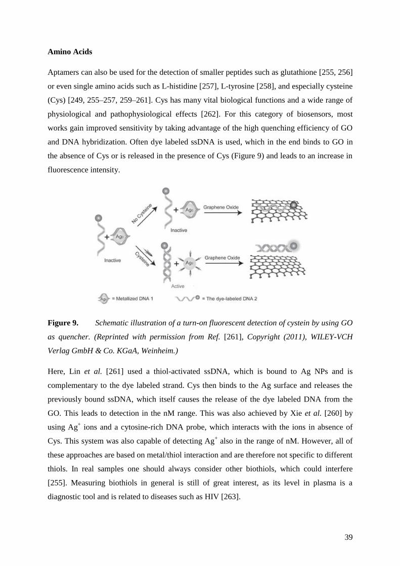
39
Amino Acids
Aptamers can also be used for the detection of smaller peptides such as glutathione [255, 256]
or even single amino acids such as L-histidine [257], L-tyrosine [258], and especially cysteine
(Cys) [249, 255–257, 259–261]. Cys has many vital biological functions and a wide range of
physiological and pathophysiological effects [262]. For this category of biosensors, most
works gain improved sensitivity by taking advantage of the high quenching efficiency of GO
and DNA hybridization. Often dye labeled ssDNA is used, which in the end binds to GO in
the absence of Cys or is released in the presence of Cys (Figure 9) and leads to an increase in
fluorescence intensity.
Figure 9. Schematic illustration of a turn-on fluorescent detection of cystein by using GO
as quencher. (Reprinted with permission from Ref. [261], Copyright (2011), WILEY-VCH
Verlag GmbH & Co. KGaA, Weinheim.)
Here, Lin et al. [261] used a thiol-activated ssDNA, which is bound to Ag NPs and is
complementary to the dye labeled strand. Cys then binds to the Ag surface and releases the
previously bound ssDNA, which itself causes the release of the dye labeled DNA from the
GO. This leads to detection in the nM range. This was also achieved by Xie et al. [260] by
using Ag+ ions and a cytosine-rich DNA probe, which interacts with the ions in absence of
Cys. This system was also capable of detecting Ag+ also in the range of nM. However, all of
these approaches are based on metal/thiol interaction and are therefore not specific to different
thiols. In real samples one should always consider other biothiols, which could interfere
[255]. Measuring biothiols in general is still of great interest, as its level in plasma is a
diagnostic tool and is related to diseases such as HIV [263].
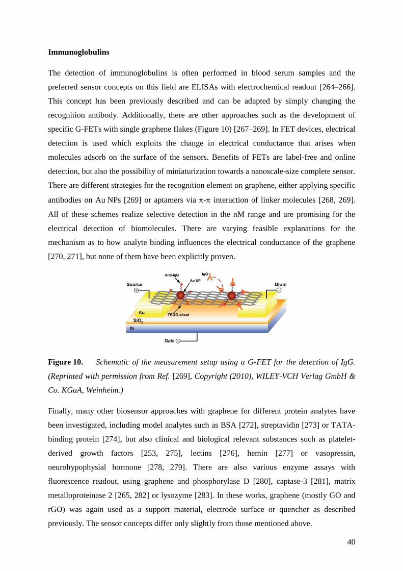
40
Immunoglobulins
The detection of immunoglobulins is often performed in blood serum samples and the
preferred sensor concepts on this field are ELISAs with electrochemical readout [264–266].
This concept has been previously described and can be adapted by simply changing the
recognition antibody. Additionally, there are other approaches such as the development of
specific G-FETs with single graphene flakes (Figure 10) [267–269]. In FET devices, electrical
detection is used which exploits the change in electrical conductance that arises when
molecules adsorb on the surface of the sensors. Benefits of FETs are label-free and online
detection, but also the possibility of miniaturization towards a nanoscale-size complete sensor.
There are different strategies for the recognition element on graphene, either applying specific
antibodies on Au NPs [269] or aptamers via - interaction of linker molecules [268, 269].
All of these schemes realize selective detection in the nM range and are promising for the
electrical detection of biomolecules. There are varying feasible explanations for the
mechanism as to how analyte binding influences the electrical conductance of the graphene
[270, 271], but none of them have been explicitly proven.
Figure 10. Schematic of the measurement setup using a G-FET for the detection of IgG.
(Reprinted with permission from Ref. [269], Copyright (2010), WILEY-VCH Verlag GmbH &
Co. KGaA, Weinheim.)
Finally, many other biosensor approaches with graphene for different protein analytes have
been investigated, including model analytes such as BSA [272], streptavidin [273] or TATA-
binding protein [274], but also clinical and biological relevant substances such as platelet-
derived growth factors [253, 275], lectins [276], hemin [277] or vasopressin,
neurohypophysial hormone [278, 279]. There are also various enzyme assays with
fluorescence readout, using graphene and phosphorylase D [280], captase-3 [281], matrix
metalloproteinase 2 [265, 282] or lysozyme [283]. In these works, graphene (mostly GO and
rGO) was again used as a support material, electrode surface or quencher as described
previously. The sensor concepts differ only slightly from those mentioned above.
41
This section illustrates the versatility in graphene application for protein analytics. By
immobilization of specific recognition elements such as enzymes, antibodies or aptamers in
combination with graphene, a broad spectrum of selective biosensors with impressive LODs
were constructed. Many of the sensor strategies could be adapted for other biomarkers, target
protein or other immuno targets.
However, explanations as to why certain combinations of materials result in better sensitivity
are lacking and descriptions of particular mechanisms with respect to the graphene are often
missing. Major advantages are improved sensitivity, less expensive production and smaller
sensor device, which could enable better point-of-care diagnostics and aid in early disease
recognition. However, to date, there is no commercial biosensor based on graphene, which
could potentially replace the older and more laborious techniques.
42
Biosensors for other Biologically Relevant Analytes 3.3.4
Aside from carbohydrates, nucleotides and proteins, there are several approaches to detect
smaller biomolecules such as lipids and biologically relevant substances such as pesticides,
alcohols single cations, anions and also whole cells and viruses.
Cholesterol is an important lipid and its accumulation in blood is related to hypertension,
arteriosclerosis, and heart disease [284, 285]. Conventional methods such as spectroscopic,
colorimetric, non-enzymatic and other electrochemical approaches suffer from poor
specificity and difficulty of analyte standardization [286, 287]. Using a graphene electrodes
modified with cholesterol oxidase enables electrochemical detection in the range of µM,
whereas an additional decoration with Pt NPs [288] or Au NPs [289, 290] leads to more
sensitivity. Further attachment of cholesterol esterase enables the detection of cholesterol
esters [288, 291]. But in principle the concept is analogous to the electrochemical enzymatic
detection of glucose, as described previously. Metal nanoparticles enhance the conversion of
H2O2 and their attachment to a graphene modified electrode surface leads to more sensitive
recognition. The readout of the H2O2 produced could also be realized via ECL with luminol,
additionally catalyzed by the iron ions of hemin, which was attached to the graphene [292].
This resulted in an LOD of 0.06 µM, which is two orders of magnitude lower compared to the
biosensors mentioned above.
Hydrogen peroxide is an important molecule in biological processes. It occurs as enzymatic
byproduct of oxidases or as substrate of peroxidases [293]. Furthermore it is an essential
mediator in food, pharmaceutical, or clinical analyses [33]. Therefore, also a lot of sensor
approaches, which are using graphene materials are described. The sensor principles are
almost the same as have been presented for the glucose sensors, except the usage of other
enzymes. Horseradish peroxidase is the most common enzyme to detect hydrogen peroxide
[294–307]. The enzyme is adsorbed or immobilized on electrodes surfaces together with GO
or rGO. The working potential could be decreased from -400 mV vs. SCE [296] by additional
incorporation of metallic nanoparticles [294, 298, 299, 305] to -200 mV vs. Ag/AgCl, which
makes these sensor more selective towards other electro active substances such as AA, UA or
DA. The selectivity could also be enhanced by applying perm-selective membranes, which
keep away potential interfering substances from the electrode surface. Nafion [302], chitosan
[306] or poly-lysine [297] have been used. Remarkable high sensitivity, with a LOD of 10 nM
is reported for a system comprising of rGO, HRP, Au NP and thionine [299]. The linear
change in the sensor signal ranges from 0.5·10-3
to 1.8 mM. Yet, most sensors reported so far
43
cannot compete with luminescent probes, which may have the additional benefit that they can
be used for the detection of hydrogen peroxide in cellular analysis [308].
The detection of alcohols is easily achievable by also simply changing the enzyme. The
enzymatic detection of ethanol with alcohol dehydrogenase (ADH) always involves the
sensitive recognition of the cofactor NADH. In conventional electrodes, the over-potential of
NADH is greater than 0.6 V (vs. Ag/AgCl) [309], which could lead to interference problems
in real samples [310]. When graphene is implemented as an electrode material, rGO is
typically used, as it enables oxidation of NADH at lower potentials and therefore leads to less
cross sensitivity. Many works investigated different sensor electrode additives such as Nafion,
[311] chitosan [312] or methylene green [313]. They yield good results, even for common
samples, such as wine or beer [312]. Simply casting ADH onto an rGO modified electrode
leads to sensitive detection in the range of µM, at a very low potential [314]. Like in other
sensor approaches, the question here arises, as to whether complex graphene composites are
even necessary at all, since they do not appear to drastically improve the sensitivity. By
applying a ruthenium (II) complex to the biosensor, the detection of NADH could be
performed again via ECL and was a slightly more sensitive than other approaches [315].
Other biological compounds of interest which have been investigated with graphene-based
biosensors are acetic acid, DA [316], UA [316, 317], glucosamine [318], acetylcholine [319],
oxygen [302, 320] and clenbuterol [321]. Since all of them use similar strategies that were
previously discussed, they will not be explained here again in detail. Again, rGO and GO are
used mainly as an electrode, substrate or label material due to their outstanding features.
Biosensors with remarkable sensitivity and selectivity were prepared by simple attachment of
enzymes, antibodies or aptamers.
In addition to small molecule detection, there are also approaches to detect even whole
bacteria or image cells with techniques previously. E.coli bacteria were detected by attaching
a specific antigen onto a CVD graphene FET and monitoring the change in electrical
conductance when the bacteria are bound to the sensor surface [322]. Although the detection
is not completely selective, and the sensing parameters are not suitable for a portable
biosensor, this could still find application in assays for drug screening. Sulfate reducing
bacteria were detected by a common assay with specific antibodies where GO enhanced the
potentiometric stripping analysis (PSA), but also the colorimetric detection [323]. GO sheets
were labeled with secondary antibodies, which bind to target bacteria previously attached to
the assay. In a further step, GO promotes the reduction of Ag(I) to Ag NPs, which are needed
44
for a further readout (Figure 11). In another study, they used an electrode covered with rGO
covalently modified with aptamers for the specific capture of different cancer cell lines [324].
Only upon cell adhesion, the redox signals of a mediator decreased due to the formation of an
inert barrier by the cells.
Figure 11. Schematic representation of a GO based immunoassay combined with silver
enhancement for the detection of sulphate-reducing bacteria (SBR). (Reprinted with
permission from Ref. [323], Copyright (2010), American Chemical Society.)
An optical readout was used for the detection of a rotavirus [325]. GO was used here as
transparent substrate material, which was further functionalized with antibodies via peptide
bonding, but also served as fluorescent dye. A secondary antibody was labeled with Au NPs
and through FRET the fluorescence of GO was quenched. With this setup they were able to
selectively detect the virus to an LOD of 10-5
pfu·mL-1
, comparable to common ELISA
technology [326].
In a different optical approach, GO was applied as quencher of dye-labeled aptamers, as
described previously in the DNA section. Pei et al. [272] were able to detect and distinguish
between different proteins and also various cell types with the appropriate aptamer receptor.
This again demonstrated the broad and easy adaptation of affinity-receptor based applications
for which GO can be used. The detection of cancer cells could be one interesting application
for this setup and has already been demonstrated in a microfluidic system [327].
45
Conclusion 3.3.5
The research discussed in this chapter shows that graphene has been extensively studied in
biosensors for the detection of glucose, DNA, various proteins, bacteria, and even whole cells.
Nevertheless, the advantages of including graphene in a biosensor scheme are not always
readily apparently. The lack of a consistent preparation method for large scale-synthesis of
well-defined graphene complicates the situation greatly. Most studies are based on rGO which
itself is an inhomogeneous material. The oxygen containing groups enable the adaption of
well-established immobilization techniques for biomolecules, receptors and linking
molecules. Working with this material and the severely lacking material characterization
make it difficult to compare results from one sensor concept. This is viewed as a severe
hindrance to the success of graphene-based biosensor development.
In order to exploit all of the extraordinary properties of graphene at once it is necessary to
produce high quality material on insulating, flexible substrates. The development of
ultrasensitive devices is challenging, but the impact of such technology can be great. Novel
concepts for surface nanostructuring based on semiconductor technology and for
nanopatterning using chemical approaches are needed. Graphene on its own has high
sensitivity but without any selectivity at all. One possibility to introduce selectivity is direct
chemical modification. Another possibility is to design composite materials together with
other nanomaterials.
For all of the optimistic and promising perceptions of graphene, three essential questions for
becoming a commercial success in chemo- and biosensors still remain: 1) Is there anything
beyond a good paper? 2) Will it be possible to produce more than one device? 3) Can it
compete with already existing products? As of now, these questions still are open. Yet, the
material cannot compete with existing biosensors, but the constant development and
improvement of the preparation and processing techniques can change the situation. One
exception seems to be the detection of oligonucleotides using graphene-based biosensors.
Electrochemical detection methods provide the best sensitivity when combined with rGO,
with typical detection limits in the range of fM to aM. Although GO also yields excellent
lower limits of detection in the pM to fM range, CVD and epitaxial graphene do not provide
the low limits of detection offered by rGO or even GO. Additionally, the immobilization of
DNA onto graphene is relatively easy and understood. With the advancement of sensor
additives such as polymers and nanoparticles, and improvements in detection schemes,

46
graphene can be a leading sensor material in DNA biosensors competitive with other
technologies.
The most significant strides will not be made by imitating or improving well-established
sensor technologies, but rather by focusing on exploiting the truly unique properties of carbon
nanomaterials and in finding new applications and markets. Some of these properties include
extremely high surface-to-volume-ratio of graphene, together with its transparency, high
electrical conductance, as well as its flexibility, which could lead to new miniaturized
biosensors and sensor arrays having high sensitivity, long term stability, multi-detection
capabilities and ultrafast response.
[Chapter 3.3 was mainly adapted from P1]
47
4 Experimental
4.1 Materials and Instrumentations
Unless otherwise stated, all chemicals were of analytical grade and purchased from Merck
(www.merck.de, Darmstadt, Germany) or Sigma-Aldrich (www.sigma-aldrich.de, Steinheim,
Germany) and used without further purification. Ultra-pure water (0.055 μS·cm-1
) was used in
all experiments. All gases and mixtures were purchased from Linde AG (http://www.linde-
gas.de, Pullach, Germany).
Characterization with Raman spectroscopy was performed on a Thermo Scientific DXR
Raman microscope with a 532 nm excitation laser (10 mW). Optical images were made with a
Leica DMRB microscope. Scanning electron microscopy (SEM) was performed with a Zeiss
Ultra 55 and on a JOEL JSM-6700F, as well as the energy-dispersive X-ray spectroscopy
(EDS), and thermogravimetric analysis coupled to IR spectroscopy (TGA-FTIR) with a
Netzsch Iris TG209 connected to a Bruker Equinox 55, all at Infineon Technologies AG
Regensburg. Transmission electron microscopy (TEM) was performed using a 120 kV Philips
CM12 microscope. Electrochemical characterizations and measurements were performed on a
CH Instrument electrochemical analyzer Model 602a, using a three-electrode system
consisted of a Pt wire as counter electrode and a saturated calomel electrode (SCE) as
reference electrode and graphene electrode as working electrode (chapter 4.4). [partially
adapted from P2 and P4]
48
4.2 Preparation of Different Graphene Materials
Single layer graphene (SG) was obtained through micromechanical exfoliation of graphite
flakes (NGS Naturgraphit GmbH, Leinburg, Germany) using an adhesive tape, and
transferred onto a Si substrate with a 300 nm thick SiO2 insulating layer. This kind of
graphene flakes is random in shape and size, with a typical size of about 50 μm2.
Graphene obtained by CVD (CVDG) was purchased on Si/SiO2 substrates
(www.graphenea.com, San Sebastian, Spain). Electron beam lithography and reactive ion
etching (O2/Ar) was used to remove excess graphene, forming rectangular graphene
electrodes of about 0.04 mm2 in size.
Reduced graphene oxide (rGO) was prepared by a slightly modified Hummers method [23]
with subsequent reduction [24]. Briefly, 1 g of China flake graphite (K. W. Thielmann & Cie
KG, Grolsheim, Germany) was mixed with 0.75 g NaNO3 in 75 mL conc. H2SO4 and 4.5 g
KMnO4 was added in small portions under vigorous stirring and cooling in an ice bath. The
mixture was sonicated for 3 hours and stirred for 3 days at room temperature. After the
addition of 75 mL of 5% H2SO4 the mixture was heated at 100 °C for 2 hours, followed by an
addition of 15 mL of 30% H2O2 and constant stirring for 1 hour at room temperature. For
purification, the produced GO was washed with the following solutions: four times in 3%
H2SO4 containing 0.5% H2O2; two times in 3% HCl; three times in water. Finally it was
dialyzed against 2 L water (14 kDa cut-off) for 10 days, changing the water three times. For
the chemical reduction of GO, 7 mL of a GO suspension (0.5 g·L-1
) were mixed with 31 μL of
32% NH3. After adding 5 μL of 98% hydrazine hydrate the reaction mixture was refluxed for
1 hour at 100 °C. The resulting black suspension was washed with water and isolated by
centrifugation. [partially adapted from P4]
49
4.3 Modification of Graphene Materials
Functionalization of Reduced Graphene Oxide with Pd and Pt Nanoparticles
Both approaches were adapted from a preparation of graphene-metal nanocomposites by Xu
et al. [328] and modified slightly. 5 mL of a GO suspension (1 mg∙mL-1
) were mixed with
10 mL ethylene glycol. Afterwards 0.5 mL of a 0.01 M solution of metal precursor (K2PtCl4,
respectively PdCl2) in water was added and the mixture was first stirred for 30 min at room
temperature and then 6 hours at 100 °C. During the heating in both cases the color changed to
black indicating that the GO was reduced by ethylene glycol. The resulting suspensions
containing the metal-doped carbon nanomaterials (rGO-Pd and rGO-Pt) were centrifuged and
washed three times with water. [adapted from P2]
Functionalization of Graphene Oxide with MnO2
A synthesis for GO-MnO2 nanocomposites was adapted [329]. 5.1 mg GO and 21 mg
MnCl2∙4H2O were dispersed in 5 mL ipropanol and sonicated for 30 min. The reaction
mixture was refluxed at 83 °C under vigorous stirring. Afterwards a solution of 12 mg
KMnO4 in 0.4 mL water was added and the slurry was refluxed at 83 °C for another 30 min,
before it was cooled to room temperature. The composite material was then centrifuged and
washed twice with water. The resulting brown product was dried at 60 °C over night. Thermal
reduction of GO was performed after the composite was applied to a microelectrode (chapter
4.4.1) heating it to 230°C for 30 s. [adapted from P2]
Functionalization of Reduced Graphene Oxide with TiO2
For the functionalization of carbon nanomaterials Li et al. [330] have reported the
modification of single-walled carbon nanotubes by TiO2. In analogous way rGO was modified
with TiO2 (rGO-TiO2): 1 mg of TiO2 with an average particle size of 1 μm was ground
thoroughly. Subsequently, the powder was dispersed in 2 mL water, sonicated for 1 hour and
1 mL of an aqueous rGO suspension (0.5 mg·mL-1
) was added. After another 1 hour
sonication a grey suspension was formed where the modified rGO flakes highly tended to
aggregate. [adapted from P2]
50
Functionalization of Graphene Oxide with Octadecylamine
To modify rGO with octadecylamine (ODA), a synthesis of surface functionalized graphene
by Wang et al. [331] was adapted. 5 mg GO were suspended in 5 mL dichlorobenzene. After
adding 50 mg ODA, the reaction mixture was sonicated for 24 hours at 80 °C. To get rid of
excess ODA and dichlorobenzene, the reaction mixture was treated with 40 mL ethanol
resulting in a black precipitate. The mixture was centrifuged and the resulting solid was
washed twice with ethanol and toluene. This washing step was repeated once more and the
product was dried at air. Thermal reduction of the product was performed after the composite
was applied to a microelectrode (chapter 4.4.1) heating it to 230°C for 30 s. [adapted from P2]
Functionalization of Reduced Graphene Oxide with CuO Nanoparticles
Deposition of CuO nanoparticles was achieved through a wet chemical procedure in a
water/ipropanol reaction system [332]. Different mass ratios of GO/CuO (1:2, 1:1, 1:0.2,
1:0.02, 1:0) were applied to alter the level of metal oxide doping within the nanocomposite. In
the following a typical reaction for e.g. a GO/CuO mass ratio of 1:2 is described: 5 mg of
freeze dried GO were dispersed in 5 mL ipropanol and sonicated for 30 min. After the
addition of 28 mg Cu(CH3COO)2 H2O and further 5 min of sonication, the mixture was
heated to reflux at 86 °C under vigorous stirring. The temperature was maintained for 30 min
and 0.5 mL water was rapidly introduced into the boiling reaction mixture. The reaction was
kept at 86 °C for another 30 min and cooled to room temperature. The product purified by
centrifugation, washing with EtOH and water and was dispersed in 5 mL water with 30 min
sonication. The resulting suspension was drop casted (1 µL) on the microelectrodes (chapter
4.4.1), dried at room temperature and heated to 230°C to thermally reduce the GO.
Microelectrodes modified with rGO (chapter 4.4.1) were used for the electrochemical
deposition of Cu nanoparticles with subsequent oxidation to CuO at 230 °C in air. The
deposition of metal nanoparticles on rGO was carried out from a solution of 10 mM CuSO4 in
0.5 M H2SO4. A short reduction pulse (10s) at -1.2 V vs SCE was applied. The electrode was
washed with water and heated to 230 °C for 30 s.
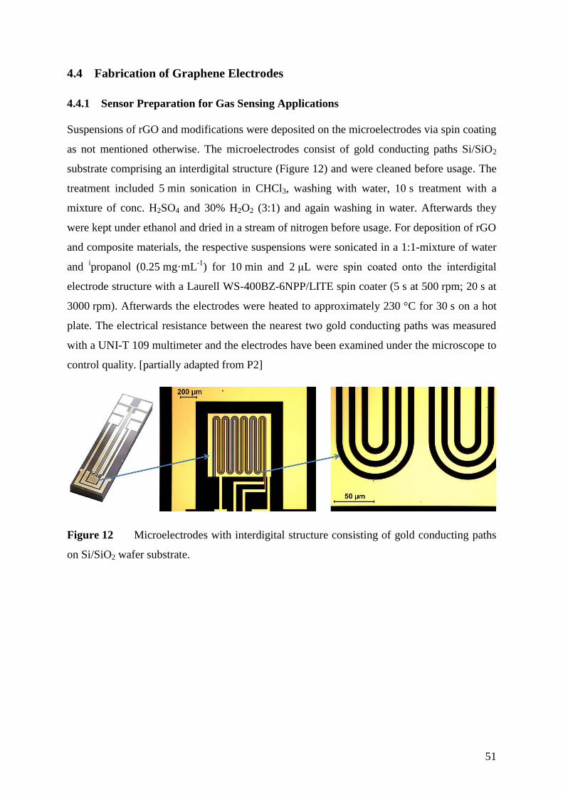
51
4.4 Fabrication of Graphene Electrodes
Sensor Preparation for Gas Sensing Applications 4.4.1
Suspensions of rGO and modifications were deposited on the microelectrodes via spin coating
as not mentioned otherwise. The microelectrodes consist of gold conducting paths Si/SiO2
substrate comprising an interdigital structure (Figure 12) and were cleaned before usage. The
treatment included 5 min sonication in CHCl3, washing with water, 10 s treatment with a
mixture of conc. H2SO4 and 30% H2O2 (3:1) and again washing in water. Afterwards they
were kept under ethanol and dried in a stream of nitrogen before usage. For deposition of rGO
and composite materials, the respective suspensions were sonicated in a 1:1-mixture of water
and ipropanol (0.25 mg·mL
-1) for 10 min and 2 μL were spin coated onto the interdigital
electrode structure with a Laurell WS-400BZ-6NPP/LITE spin coater (5 s at 500 rpm; 20 s at
3000 rpm). Afterwards the electrodes were heated to approximately 230 °C for 30 s on a hot
plate. The electrical resistance between the nearest two gold conducting paths was measured
with a UNI-T 109 multimeter and the electrodes have been examined under the microscope to
control quality. [partially adapted from P2]
Figure 12 Microelectrodes with interdigital structure consisting of gold conducting paths
on Si/SiO2 wafer substrate.
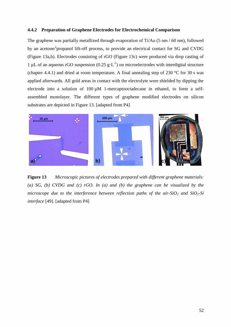
52
Preparation of Graphene Electrodes for Electrochemical Comparison 4.4.2
The graphene was partially metallized through evaporation of Ti/Au (5 nm / 60 nm), followed
by an acetone/ipropanol lift-off process, to provide an electrical contact for SG and CVDG
(Figure 13a,b). Electrodes consisting of rGO (Figure 13c) were produced via drop casting of
1 µL of an aqueous rGO suspension (0.25 g·L-1
) on microelectrodes with interdigital structure
(chapter 4.4.1) and dried at room temperature. A final annealing step of 230 °C for 30 s was
applied afterwards. All gold areas in contact with the electrolyte were shielded by dipping the
electrode into a solution of 100 µM 1-mercaptooctadecane in ethanol, to form a self-
assembled monolayer. The different types of graphene modified electrodes on silicon
substrates are depicted in Figure 13. [adapted from P4]
Figure 13 Microscopic pictures of electrodes prepared with different graphene materials:
(a) SG, (b) CVDG and (c) rGO. In (a) and (b) the graphene can be visualized by the
microscope due to the interference between reflection paths of the air-SiO2 and SiO2-Si
interface [49]. [adapted from P4]
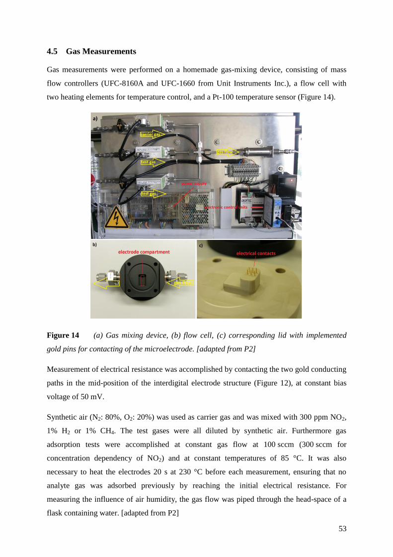
53
4.5 Gas Measurements
Gas measurements were performed on a homemade gas-mixing device, consisting of mass
flow controllers (UFC-8160A and UFC-1660 from Unit Instruments Inc.), a flow cell with
two heating elements for temperature control, and a Pt-100 temperature sensor (Figure 14).
Figure 14 (a) Gas mixing device, (b) flow cell, (c) corresponding lid with implemented
gold pins for contacting of the microelectrode. [adapted from P2]
Measurement of electrical resistance was accomplished by contacting the two gold conducting
paths in the mid-position of the interdigital electrode structure (Figure 12), at constant bias
voltage of 50 mV.
Synthetic air (N2: 80%, O2: 20%) was used as carrier gas and was mixed with 300 ppm NO2,
1% H2 or 1% CH4. The test gases were all diluted by synthetic air. Furthermore gas
adsorption tests were accomplished at constant gas flow at 100 sccm (300 sccm for
concentration dependency of NO2) and at constant temperatures of 85 °C. It was also
necessary to heat the electrodes 20 s at 230 °C before each measurement, ensuring that no
analyte gas was adsorbed previously by reaching the initial electrical resistance. For
measuring the influence of air humidity, the gas flow was piped through the head-space of a
flask containing water. [adapted from P2]
54
Measurements with CO2 were performed in analogous way, mixing 3000 ppm CO2 to a
constant flow (100 sccm) of synthetic air, in order to achieve concentrations ranging from 150
to 1500 ppm. The flow cell was held at a constant temperature of 25 or 60°C, respectively.
Cross sensitivity was tested in the presence of 100 ppm NO2, 3000 ppm H2, 3000 ppm CH4,
and 100 ppm CO. Temperature dependency was measured in synthetic air in the range from
25 to 85°C.
Additionally, microelectrodes modified with rGO-CuO (with a GO/CuO ratio of 1:2) were
compared to a commercial sensor device. A CDM4161-L00 CO2 Module from Figaro was
applied to the flow cell and sealed. The sensor was run with 4V power supply and the signal
output voltage was measured with an UNI-T UT109 multimeter. The output voltage measured
in mV is directly proportional to the concentration of CO2 in ppm. The sensor was calibrated
for 2 hours in air before measuring.
55
4.6 Electrochemical Characterization and Measurements
A custom build carbon disc electrode, comprising a carbon fiber composite (www.conrad.de,
Regensburg, Germany) with 2 mm diameter implemented in Teflon or the differently
prepared graphene materials (SG, CVDG, and rGO, chapter 4.4.2) assembled on a silicon
wafer substrate electrically contacted by gold leads were used as working electrode.
The modified electrodes and the carbon disc electrode were characterized with cyclic
voltammetry, chronocoulometry, electrochemical impedance spectroscopy (EIS) and
amperometry. For electrochemical experiments, the supporting electrolytes were 0.1 M KCl
or 10 mM phosphate buffer with 140 mM NaCl (pH 7.4). Before each measurement Argon
was bubbled through the solutions to remove dissolved oxygen. All experiments were
performed at room temperature. The amperometric response towards H2O2 was investigated
in a continuously stirred electrolyte solution (10 mM phosphate buffer, 140 mM NaCl,
pH 7.4) by doubling the concentration of peroxide with each addition. The studied analyte
concentration covers a range from 25 µM to 25.6 mM. [adapted from P4]
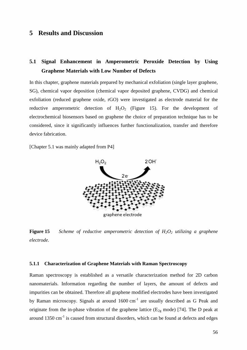
56
5 Results and Discussion
5.1 Signal Enhancement in Amperometric Peroxide Detection by Using
Graphene Materials with Low Number of Defects
In this chapter, graphene materials prepared by mechanical exfoliation (single layer graphene,
SG), chemical vapor deposition (chemical vapor deposited graphene, CVDG) and chemical
exfoliation (reduced graphene oxide, rGO) were investigated as electrode material for the
reductive amperometric detection of H2O2 (Figure 15). For the development of
electrochemical biosensors based on graphene the choice of preparation technique has to be
considered, since it significantly influences further functionalization, transfer and therefore
device fabrication.
[Chapter 5.1 was mainly adapted from P4]
Figure 15 Scheme of reductive amperometric detection of H2O2 utilizing a graphene
electrode.
Characterization of Graphene Materials with Raman Spectroscopy 5.1.1
Raman spectroscopy is established as a versatile characterization method for 2D carbon
nanomaterials. Information regarding the number of layers, the amount of defects and
impurities can be obtained. Therefore all graphene modified electrodes have been investigated
by Raman microscopy. Signals at around 1600 cm-1
are usually described as G Peak and
originate from the in-phase vibration of the graphene lattice (E2g mode) [74]. The D peak at
around 1350 cm-1
is caused from structural disorders, which can be found at defects and edges
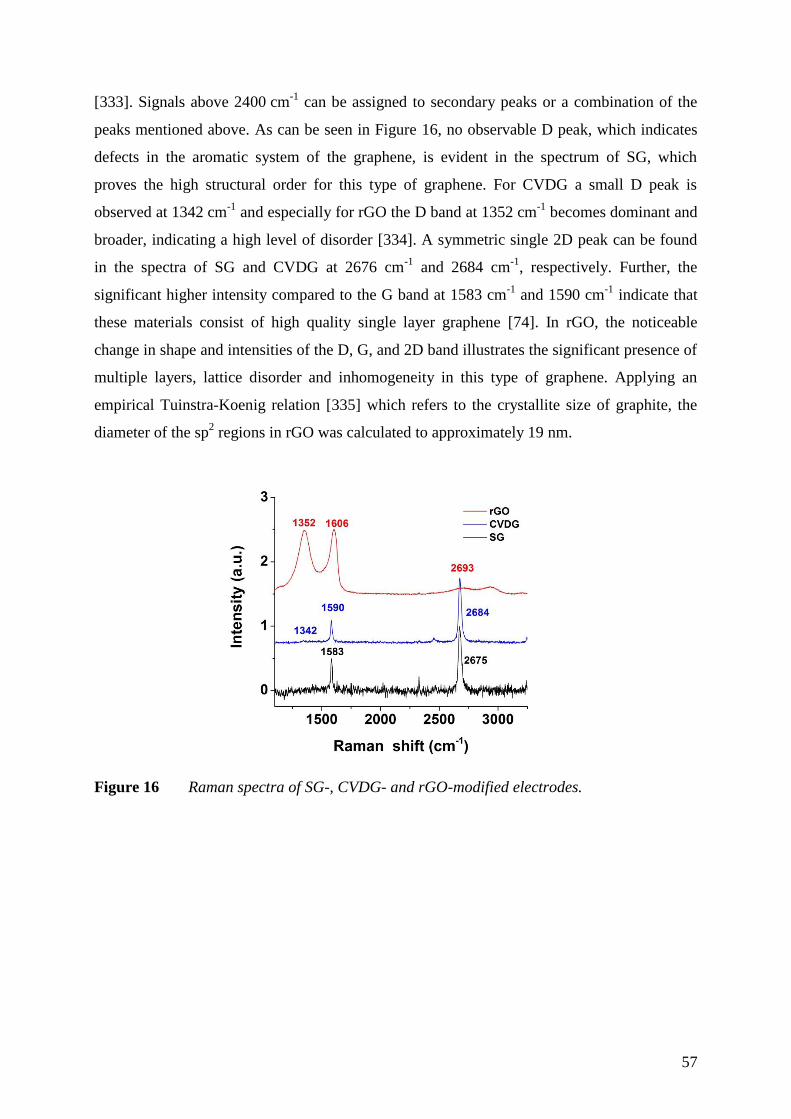
57
[333]. Signals above 2400 cm-1
can be assigned to secondary peaks or a combination of the
peaks mentioned above. As can be seen in Figure 16, no observable D peak, which indicates
defects in the aromatic system of the graphene, is evident in the spectrum of SG, which
proves the high structural order for this type of graphene. For CVDG a small D peak is
observed at 1342 cm-1
and especially for rGO the D band at 1352 cm-1
becomes dominant and
broader, indicating a high level of disorder [334]. A symmetric single 2D peak can be found
in the spectra of SG and CVDG at 2676 cm-1
and 2684 cm-1
, respectively. Further, the
significant higher intensity compared to the G band at 1583 cm-1
and 1590 cm-1
indicate that
these materials consist of high quality single layer graphene [74]. In rGO, the noticeable
change in shape and intensities of the D, G, and 2D band illustrates the significant presence of
multiple layers, lattice disorder and inhomogeneity in this type of graphene. Applying an
empirical Tuinstra-Koenig relation [335] which refers to the crystallite size of graphite, the
diameter of the sp2 regions in rGO was calculated to approximately 19 nm.
Figure 16 Raman spectra of SG-, CVDG- and rGO-modified electrodes.
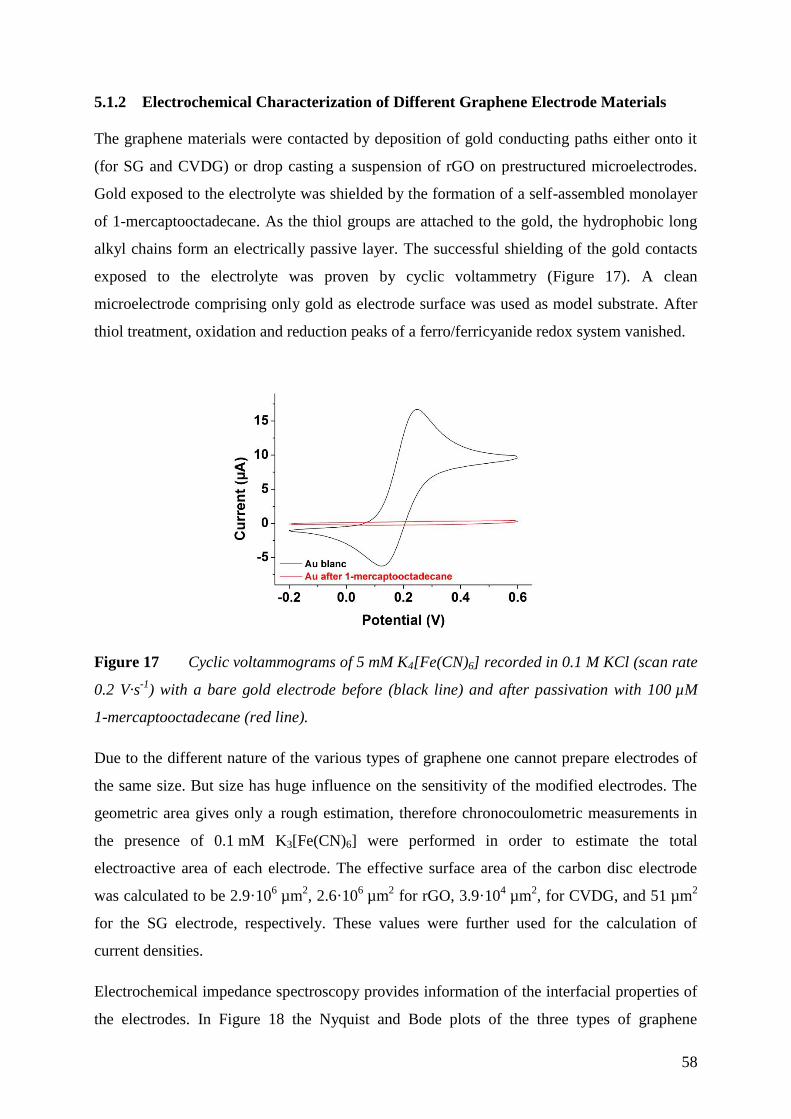
58
Electrochemical Characterization of Different Graphene Electrode Materials 5.1.2
The graphene materials were contacted by deposition of gold conducting paths either onto it
(for SG and CVDG) or drop casting a suspension of rGO on prestructured microelectrodes.
Gold exposed to the electrolyte was shielded by the formation of a self-assembled monolayer
of 1-mercaptooctadecane. As the thiol groups are attached to the gold, the hydrophobic long
alkyl chains form an electrically passive layer. The successful shielding of the gold contacts
exposed to the electrolyte was proven by cyclic voltammetry (Figure 17). A clean
microelectrode comprising only gold as electrode surface was used as model substrate. After
thiol treatment, oxidation and reduction peaks of a ferro/ferricyanide redox system vanished.
Figure 17 Cyclic voltammograms of 5 mM K4[Fe(CN)6] recorded in 0.1 M KCl (scan rate
0.2 V·s-1
) with a bare gold electrode before (black line) and after passivation with 100 µM
1-mercaptooctadecane (red line).
Due to the different nature of the various types of graphene one cannot prepare electrodes of
the same size. But size has huge influence on the sensitivity of the modified electrodes. The
geometric area gives only a rough estimation, therefore chronocoulometric measurements in
the presence of 0.1 mM K3[Fe(CN)6] were performed in order to estimate the total
electroactive area of each electrode. The effective surface area of the carbon disc electrode
was calculated to be 2.9·106 µm
2, 2.6·10
6 µm
2 for rGO, 3.9·10
4 µm
2, for CVDG, and 51 µm
2
for the SG electrode, respectively. These values were further used for the calculation of
current densities.
Electrochemical impedance spectroscopy provides information of the interfacial properties of
the electrodes. In Figure 18 the Nyquist and Bode plots of the three types of graphene

59
modified electrodes and the carbon disc electrode in presence of 5 mM K4[Fe(CN)6]
containing 0.1 M KCl at 0.2 V vs SCE are shown. Electrodes modified with SG, CVDG and
rGO cannot be fully described by a Randles equivalent circuit, consisting of the electrolyte
resistance Re in series with a parallel combination of double-layer capacitance (Cdl), an
impedance representing the charge transfer resistance (Rct) and the Warburg element (Zw)
taking diffusion into account. The results from the electrochemical impedance spectroscopy
do not take into account that there is a huge difference in the electrode surface area as can be
seen from the results of the chronocoulometric measurements. For high frequencies the
impedance (Z) for all systems is approaching the electrolyte resistance. For small frequencies
the charge-transfer resistance increases for the different electrodes in the order carbon disc,
rGO, CVDG to SG. The behavior of the CVDG can be explained by impurities adsorbed on
the graphene. These can be a consequence of the transfer process [36]. Here, a polymer layer
will deposited on top of the graphene layer; the metallic substrate where the graphene was
deposited is etched away so that the polymer modified graphene flake swims on top of the
solution. After transfer to the silicon wafer the polymer on top of the graphene is washed
away by organic solvents. By this process it is likely that polymer residues can remain. The
Nyquist plot in Figure 18a suggests that additional impedance for adsorbates need to be
applied to the equivalent circuit. For the rGO system, which consists of multilayers and
comprises many defects, it is expected that the impedance spectrum is not in accordance to
the simple model described by the Randles equivalent circuit. The SG modified electrode as
well as the carbon disc electrode show a diffusion process for the redox species at low
frequencies. The higher double-layer capacitance of SG modified electrodes in contrast to the
carbon disc electrode as can be seen from the imaginary part in the Nyquist plot (Z”) can be
attributed to the hydrophobic character of the aromatic system which is repelling the redox
species. After normalization of the total impedance Z to the electroactive surface area the
charge transfer resistance of carbon disc electrode is approximately 1.2·106 times higher than
for the SG modified electrode, for CVDG Rct per area is 1.7·103 times, for rGO 1.3·10
5 times
higher. This shows that all graphene types enhance the electron transfer of the
ferro/ferricyanide redox system compared to a plain carbon disc electrode. The differences
between the three different types are in accordance to the increase of the number of defects of
the two-dimensional carbon nanomaterial. The numbers given here are only rough estimations
not taking into account the influence of the border of the CVDG and SG. A detailed
investigation of different flake sizes will complete this picture.
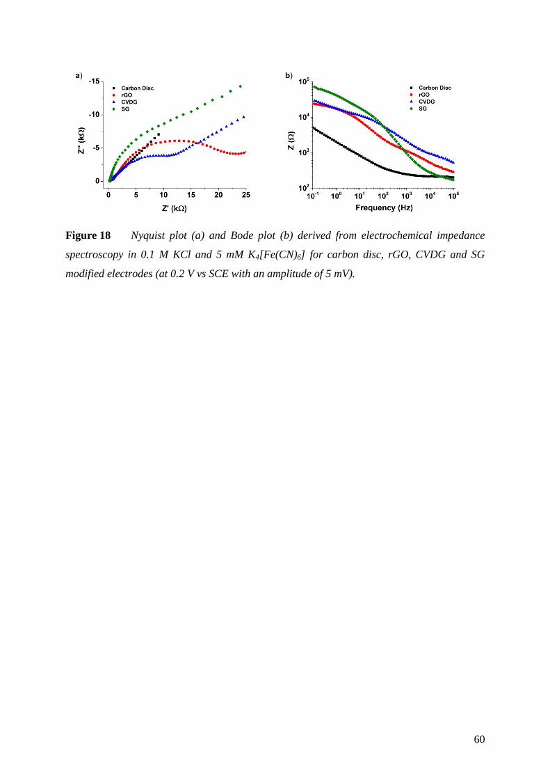
60
Figure 18 Nyquist plot (a) and Bode plot (b) derived from electrochemical impedance
spectroscopy in 0.1 M KCl and 5 mM K4[Fe(CN)6] for carbon disc, rGO, CVDG and SG
modified electrodes (at 0.2 V vs SCE with an amplitude of 5 mV).
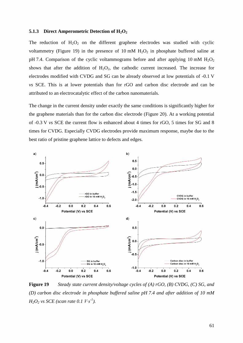
61
Direct Amperometric Detection of H2O2 5.1.3
The reduction of H2O2 on the different graphene electrodes was studied with cyclic
voltammetry (Figure 19) in the presence of 10 mM H2O2 in phosphate buffered saline at
pH 7.4. Comparison of the cyclic voltammograms before and after applying 10 mM H2O2
shows that after the addition of H2O2, the cathodic current increased. The increase for
electrodes modified with CVDG and SG can be already observed at low potentials of -0.1 V
vs SCE. This is at lower potentials than for rGO and carbon disc electrode and can be
attributed to an electrocatalytic effect of the carbon nanomaterials.
The change in the current density under exactly the same conditions is significantly higher for
the graphene materials than for the carbon disc electrode (Figure 20). At a working potential
of -0.3 V vs SCE the current flow is enhanced about 4 times for rGO, 5 times for SG and 8
times for CVDG. Especially CVDG electrodes provide maximum response, maybe due to the
best ratio of pristine graphene lattice to defects and edges.
Figure 19 Steady state current density/voltage cycles of (A) rGO, (B) CVDG, (C) SG, and
(D) carbon disc electrode in phosphate buffered saline pH 7.4 and after addition of 10 mM
H2O2 vs SCE (scan rate 0.1 V∙s-1
).
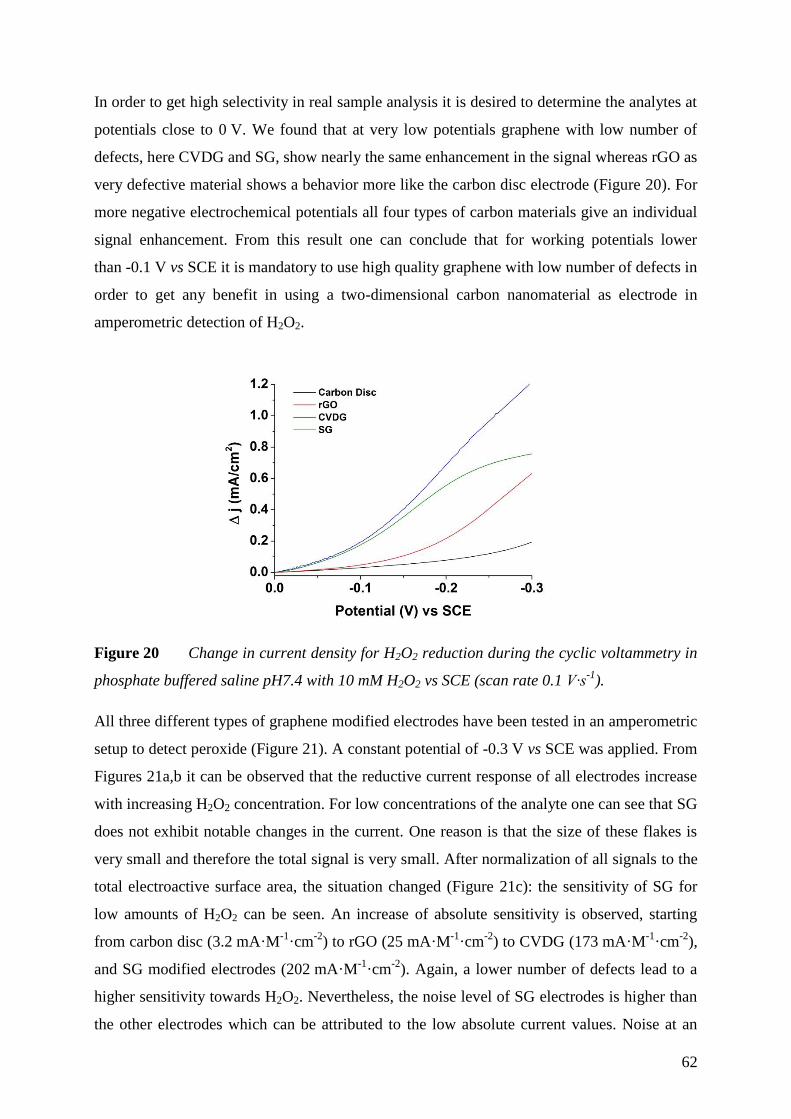
62
In order to get high selectivity in real sample analysis it is desired to determine the analytes at
potentials close to 0 V. We found that at very low potentials graphene with low number of
defects, here CVDG and SG, show nearly the same enhancement in the signal whereas rGO as
very defective material shows a behavior more like the carbon disc electrode (Figure 20). For
more negative electrochemical potentials all four types of carbon materials give an individual
signal enhancement. From this result one can conclude that for working potentials lower
than -0.1 V vs SCE it is mandatory to use high quality graphene with low number of defects in
order to get any benefit in using a two-dimensional carbon nanomaterial as electrode in
amperometric detection of H2O2.
Figure 20 Change in current density for H2O2 reduction during the cyclic voltammetry in
phosphate buffered saline pH7.4 with 10 mM H2O2 vs SCE (scan rate 0.1 V∙s-1
).
All three different types of graphene modified electrodes have been tested in an amperometric
setup to detect peroxide (Figure 21). A constant potential of -0.3 V vs SCE was applied. From
Figures 21a,b it can be observed that the reductive current response of all electrodes increase
with increasing H2O2 concentration. For low concentrations of the analyte one can see that SG
does not exhibit notable changes in the current. One reason is that the size of these flakes is
very small and therefore the total signal is very small. After normalization of all signals to the
total electroactive surface area, the situation changed (Figure 21c): the sensitivity of SG for
low amounts of H2O2 can be seen. An increase of absolute sensitivity is observed, starting
from carbon disc (3.2 mA·M-1
·cm-2
) to rGO (25 mA·M-1
·cm-2
) to CVDG (173 mA·M-1
·cm-2
),
and SG modified electrodes (202 mA·M-1
·cm-2
). Again, a lower number of defects lead to a
higher sensitivity towards H2O2. Nevertheless, the noise level of SG electrodes is higher than
the other electrodes which can be attributed to the low absolute current values. Noise at an
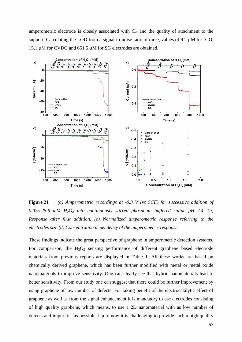
63
amperometric electrode is closely associated with Cdl and the quality of attachment to the
support. Calculating the LOD from a signal-to-noise ratio of three, values of 9.2 µM for rGO,
15.1 µM for CVDG and 651.5 µM for SG electrodes are obtained.
Figure 21 (a) Amperometric recordings at -0.3 V (vs SCE) for successive addition of
0.025-25.6 mM H2O2 into continuously stirred phosphate buffered saline pH 7.4. (b)
Response after first addition. (c) Normalized amperometric response referring to the
electrodes size.(d) Concentration dependency of the amperometric response.
These findings indicate the great perspective of graphene in amperometric detection systems.
For comparison, the H2O2 sensing performance of different graphene based electrode
materials from previous reports are displayed in Table 1. All these works are based on
chemically derived graphene, which has been further modified with metal or metal oxide
nanomaterials to improve sensitivity. One can clearly see that hybrid nanomaterials lead to
better sensitivity. From our study one can suggest that there could be further improvement by
using graphene of low number of defects. For taking benefit of the electrocatalytic effect of
graphene as well as from the signal enhancement it is mandatory to use electrodes consisting
of high quality graphene, which means, to use a 2D nanomaterial with as low number of
defects and impurities as possible. Up to now it is challenging to provide such a high quality
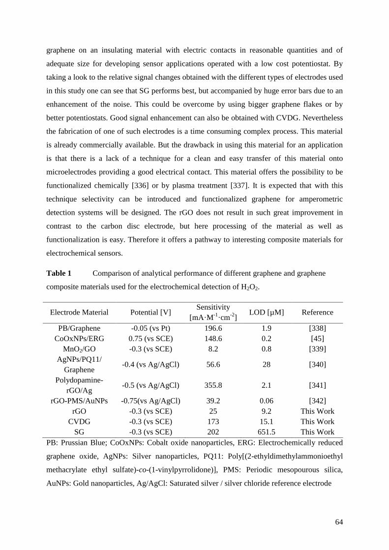
64
graphene on an insulating material with electric contacts in reasonable quantities and of
adequate size for developing sensor applications operated with a low cost potentiostat. By
taking a look to the relative signal changes obtained with the different types of electrodes used
in this study one can see that SG performs best, but accompanied by huge error bars due to an
enhancement of the noise. This could be overcome by using bigger graphene flakes or by
better potentiostats. Good signal enhancement can also be obtained with CVDG. Nevertheless
the fabrication of one of such electrodes is a time consuming complex process. This material
is already commercially available. But the drawback in using this material for an application
is that there is a lack of a technique for a clean and easy transfer of this material onto
microelectrodes providing a good electrical contact. This material offers the possibility to be
functionalized chemically [336] or by plasma treatment [337]. It is expected that with this
technique selectivity can be introduced and functionalized graphene for amperometric
detection systems will be designed. The rGO does not result in such great improvement in
contrast to the carbon disc electrode, but here processing of the material as well as
functionalization is easy. Therefore it offers a pathway to interesting composite materials for
electrochemical sensors.
Table 1 Comparison of analytical performance of different graphene and graphene
composite materials used for the electrochemical detection of H2O2.
Electrode Material Potential [V] Sensitivity
[mA·M-1
·cm-2
] LOD [µM] Reference
PB/Graphene -0.05 (vs Pt) 196.6 1.9 [338]
CoOxNPs/ERG 0.75 (vs SCE) 148.6 0.2 [45]
MnO2/GO -0.3 (vs SCE) 8.2 0.8 [339]
AgNPs/PQ11/
Graphene -0.4 (vs Ag/AgCl) 56.6 28 [340]
Polydopamine-
rGO/Ag -0.5 (vs Ag/AgCl) 355.8 2.1 [341]
rGO-PMS/AuNPs -0.75(vs Ag/AgCl) 39.2 0.06 [342]
rGO -0.3 (vs SCE) 25 9.2 This Work
CVDG -0.3 (vs SCE) 173 15.1 This Work
SG -0.3 (vs SCE) 202 651.5 This Work
PB: Prussian Blue; CoOxNPs: Cobalt oxide nanoparticles, ERG: Electrochemically reduced
graphene oxide, AgNPs: Silver nanoparticles, PQ11: Poly[(2-ethyldimethylammonioethyl
methacrylate ethyl sulfate)-co-(1-vinylpyrrolidone)], PMS: Periodic mesopourous silica,
AuNPs: Gold nanoparticles, Ag/AgCl: Saturated silver / silver chloride reference electrode
65
Conclusion 5.1.4
The non-enzymatic detection of H2O2 was studied using electrodes based on various types of
graphene, without utilizing any functionalization or modification steps. All three graphene
materials exhibit sensitivity to the catalytic reduction of H2O2 and are able to detect H2O2
concentrations in the µM range, showing fast amperometric response upon successive
additions of analyte. Moreover, it is clearly demonstrated that mechanically exfoliated
graphene as well as graphene prepared by CVD are promising candidates for sensor
applications, due to their efficiency in better detection of H2O2 with higher sensitivity
compared to rGO. However, due to the laborious process needed for production of exfoliated
graphene and its irregularity in shape, size and location, this type of graphene is more suitable
for theoretical study and proof-of-concept demonstration. In comparison, the CVDG suits
commercial purposes and mass production, since it offers uniform continuous films at
particular locations and with desired geometries, providing a superior candidate for
fabrication of high sensitivity biosensors and sensor arrays. Such carbon nanomaterials can
also be used as biosensors by further functionalization with enzymes.

66
5.2 Reduced Graphene Oxide and Graphene Composite Materials for Improved
Gas Sensing at Low Temperature
In this chapter, rGO was investigated as sensitive material for gas sensors in a chemiresitive
setup. Reduced graphene oxide was chosen due to features like solution processing, high
sensitivity and simple methods for functionalization owed to its defective structure. Covalent
attachment of functional groups or doping with metal and metal oxides can change the
electronic structure within the material, but may also increase the sensor surface or have a
catalytic effect on the gas adsorption.
[Chapter 5.2 was mainly adapted from P2]
Characterization of Reduced Graphene Oxide and Composite Materials 5.2.1
Oxidation of graphite with subsequent reduction leads to a material with inhomogeneous
shape and size of graphene sheets. The size of the graphene flakes ranging from 100 nm to
about 1 µm as it is indicated by SEM studies (Figure 22a). Characterization with Raman
spectroscopy also indicates the inhomogeneity of the material, showing a broad G and
D peaks and was discussed previously (chapter 5.1.1).
Regarding the thermogravimetric analysis (TGA) of GO and rGO (Figure 22b) a loss of mass
can be observed at temperatures of about 200 °C. In FTIR-TGA this effect can be ascribed to
a thermal reduction, where CO and CO2 is released [343]. Graphene oxide loses about 25% of
its mass, while rGO loses around 15% and shows that the reduction by hydrazine is
incomplete.
Figure 22 (a) SEM of rGO on a Si/SiO2 substrate and (b) thermogravimetric analysis of
GO and rGO.
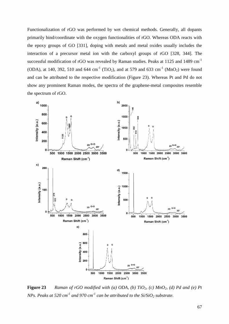
67
Functionalization of rGO was performed by wet chemical methods. Generally, all dopants
primarily bind/coordinate with the oxygen functionalities of rGO. Whereas ODA reacts with
the epoxy groups of GO [331], doping with metals and metal oxides usually includes the
interaction of a precursor metal ion with the carboxyl groups of rGO [328, 344]. The
successful modification of rGO was revealed by Raman studies. Peaks at 1125 and 1489 cm-1
(ODA), at 140, 392, 510 and 644 cm-1
(TiO2), and at 579 and 633 cm-1
(MnO2) were found
and can be attributed to the respective modification (Figure 23). Whereas Pt and Pd do not
show any prominent Raman modes, the spectra of the graphene-metal composites resemble
the spectrum of rGO.
Figure 23 Raman of rGO modified with (a) ODA, (b) TiO2, (c) MnO2, (d) Pd and (e) Pt
NPs. Peaks at 520 cm-1
and 970 cm-1
can be attributed to the Si/SiO2 substrate.
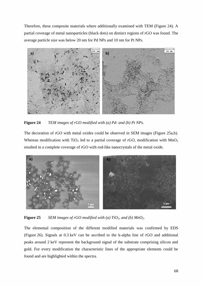
68
Therefore, these composite materials where additionally examined with TEM (Figure 24). A
partial coverage of metal nanoparticles (black dots) on distinct regions of rGO was found. The
average particle size was below 20 nm for Pd NPs and 10 nm for Pt NPs.
Figure 24 TEM images of rGO modified with (a) Pd- and (b) Pt NPs.
The decoration of rGO with metal oxides could be observed in SEM images (Figure 25a,b).
Whereas modification with TiO2 led to a partial coverage of rGO, modification with MnO2
resulted in a complete coverage of rGO with rod-like nanocrystals of the metal oxide.
Figure 25 SEM images of rGO modified with (a) TiO2, and (b) MnO2.
The elemental composition of the different modified materials was confirmed by EDS
(Figure 26). Signals at 0.3 keV can be ascribed to the k-alpha line of rGO and additional
peaks around 2 keV represent the background signal of the substrate comprising silicon and
gold. For every modification the characteristic lines of the appropriate elements could be
found and are highlighted within the spectra.
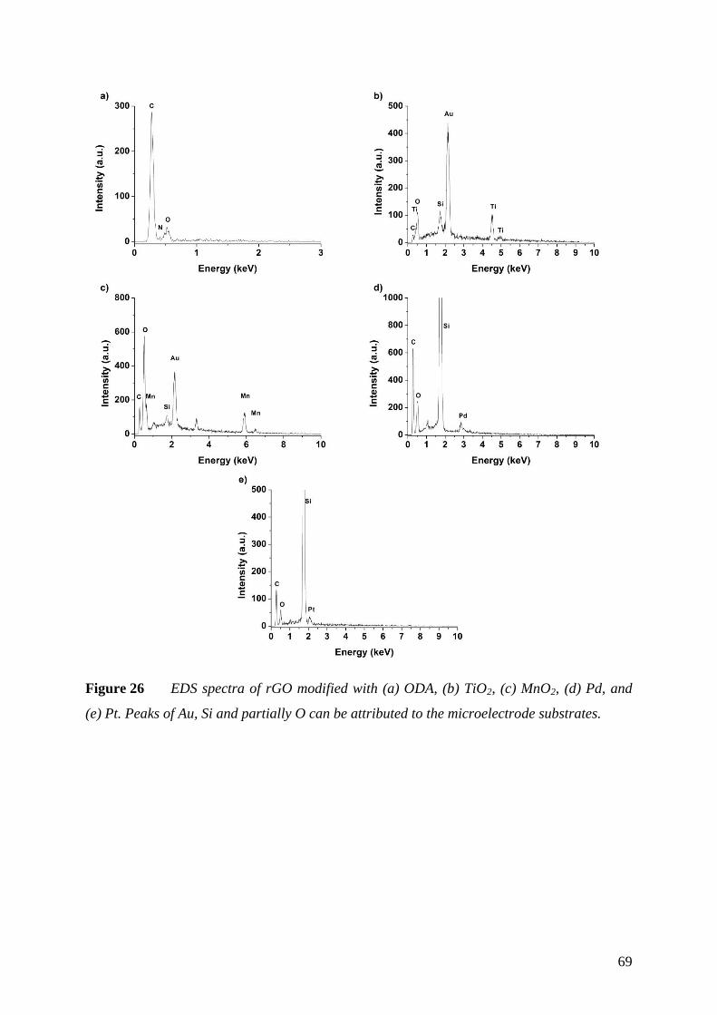
69
Figure 26 EDS spectra of rGO modified with (a) ODA, (b) TiO2, (c) MnO2, (d) Pd, and
(e) Pt. Peaks of Au, Si and partially O can be attributed to the microelectrode substrates.

70
Gas Sensor Response 5.2.2
The transfer of graphene materials is still a critical step in sensor preparation using this type
of carbon nanomaterial. Aqueous suspensions of rGO can be easily processed and the
interdigital electrode structure was covered with a thin layer of rGO via spin coating.
Parameters affecting spin coating as the concentration of rGO, the rotation speed and time as
well as the usage of certain additives, like ipropanol for improvement of the spreading of the
suspension, were optimized to obtain consistent layers with reproducible total electrical
resistance. Further, the rGO modified electrodes were heated to 230 °C to guarantee a thermal
reduction. During this step, the electrical resistance dropped from about 8.0 M ± 3.8 M to
180 k ± 78 k. The oxidation process of graphite introduces many defects to the sp2
structure which act like a barrier for the electron flux, resulting in better electrical
conductance at higher level of reduction.
To assess the effect of gas adsorption on the conductance of rGO, the electrical resistance of
coated microelectrodes was measured for different gases in various concentrations mixed in a
constant flow of synthetic air. An operating temperature of 85 °C was chosen to exclude the
influence of humidity on the sensor response (Figure 27c). Furthermore, heating to 85°C
improved the response time (< 1 min), and an increase in the signal change and the recovery
rate was found. Figure 27 displays the change of the relative resistance (R/R0) in the presence
of different gases, such as NO2, H2, and CH4. R0 is hereby the initial electrical resistance right
before the addition of a test gas.
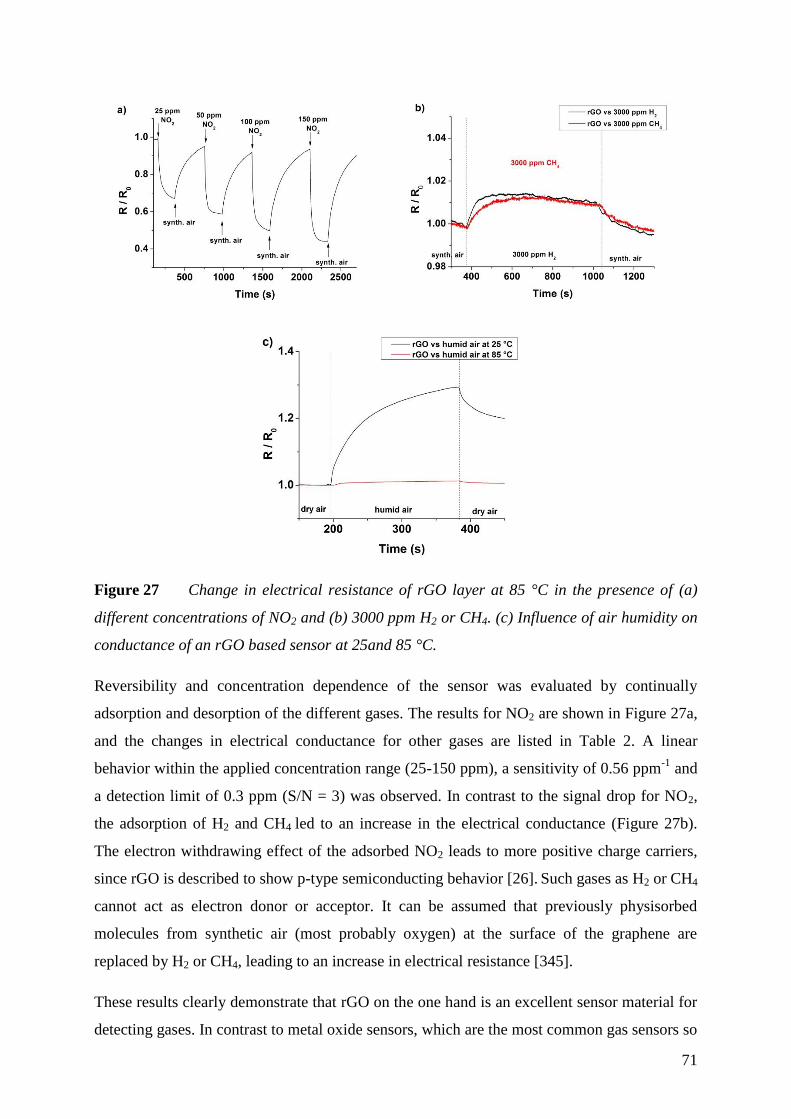
71
Figure 27 Change in electrical resistance of rGO layer at 85 °C in the presence of (a)
different concentrations of NO2 and (b) 3000 ppm H2 or CH4. (c) Influence of air humidity on
conductance of an rGO based sensor at 25and 85 °C.
Reversibility and concentration dependence of the sensor was evaluated by continually
adsorption and desorption of the different gases. The results for NO2 are shown in Figure 27a,
and the changes in electrical conductance for other gases are listed in Table 2. A linear
behavior within the applied concentration range (25-150 ppm), a sensitivity of 0.56 ppm-1
and
a detection limit of 0.3 ppm (S/N = 3) was observed. In contrast to the signal drop for NO2,
the adsorption of H2 and CH4 led to an increase in the electrical conductance (Figure 27b).
The electron withdrawing effect of the adsorbed NO2 leads to more positive charge carriers,
since rGO is described to show p-type semiconducting behavior [26]. Such gases as H2 or CH4
cannot act as electron donor or acceptor. It can be assumed that previously physisorbed
molecules from synthetic air (most probably oxygen) at the surface of the graphene are
replaced by H2 or CH4, leading to an increase in electrical resistance [345].
These results clearly demonstrate that rGO on the one hand is an excellent sensor material for
detecting gases. In contrast to metal oxide sensors, which are the most common gas sensors so
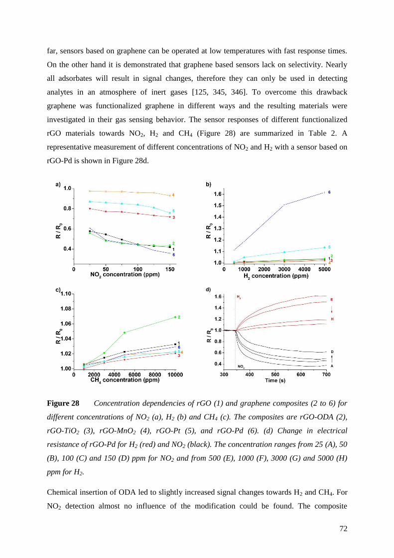
72
far, sensors based on graphene can be operated at low temperatures with fast response times.
On the other hand it is demonstrated that graphene based sensors lack on selectivity. Nearly
all adsorbates will result in signal changes, therefore they can only be used in detecting
analytes in an atmosphere of inert gases [125, 345, 346]. To overcome this drawback
graphene was functionalized graphene in different ways and the resulting materials were
investigated in their gas sensing behavior. The sensor responses of different functionalized
rGO materials towards NO2, H2 and CH4 (Figure 28) are summarized in Table 2. A
representative measurement of different concentrations of NO2 and H2 with a sensor based on
rGO-Pd is shown in Figure 28d.
Figure 28 Concentration dependencies of rGO (1) and graphene composites (2 to 6) for
different concentrations of NO2 (a), H2 (b) and CH4 (c). The composites are rGO-ODA (2),
rGO-TiO2 (3), rGO-MnO2 (4), rGO-Pt (5), and rGO-Pd (6). (d) Change in electrical
resistance of rGO-Pd for H2 (red) and NO2 (black). The concentration ranges from 25 (A), 50
(B), 100 (C) and 150 (D) ppm for NO2 and from 500 (E), 1000 (F), 3000 (G) and 5000 (H)
ppm for H2.
Chemical insertion of ODA led to slightly increased signal changes towards H2 and CH4. For
NO2 detection almost no influence of the modification could be found. The composite
73
material with TiO2 showed unaffected behavior towards H2 and CH4, but a lower response
towards NO2. This indicates that adsorption sites for NO2 may be blocked. Electrodes coated
with rGO which was doped with MnO2 showed almost no signal change to all of the utilized
gases. Therefore, this modification only could deal as a reference signal to compensate small
fluctuations in temperature. The most significant change in response was achieved by
chemical doping with Pd and Pt NPs. The resulting composite materials showed an increased
sensitivity towards H2 (Figure 28b). Same observations with comparable results have been
made with carbon nanotubes decorated with Pt and multilayered graphene nanoribbons doped
with Pd. Though, the composite material with rGO is easier to produce and to process. In
summary, all composites of rGO are rather simple to prepare and show different behavior in
their electrical conductance upon the presence of various gases.
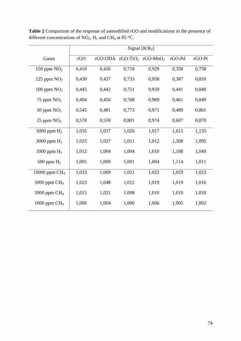
74
Table 2 Comparison of the response of unmodified rGO and modifications in the presence of
different concentrations of NO2, H2 and CH4 at 85 °C.
Signal [R/R0]
Gases rGO rGO-ODA rGO-TiO2 rGO-MnO2 rGO-Pd rGO-Pt
150 ppm NO2 0,419 0,436 0,718 0,929 0,358 0,758
125 ppm NO2 0,430 0,437 0,733 0,958 0,387 0,810
100 ppm NO2 0,445 0,442 0,751 0,959 0,441 0,840
75 ppm NO2 0,494 0,456 0,768 0,969 0,461 0,849
50 ppm NO2 0,545 0,481 0,773 0,971 0,489 0,861
25 ppm NO2 0,578 0,559 0,801 0,974 0,607 0,870
5000 ppm H2 1,035 1,037 1,026 1,017 1,615 1,135
3000 ppm H2 1,025 1,027 1,011 1,012 1,508 1,095
1000 ppm H2 1,012 1,004 1,004 1,010 1,188 1,049
500 ppm H2 1,005 1,000 1,001 1,004 1,114 1,011
10000 ppm CH4 1,033 1,069 1,021 1,022 1,029 1,023
5000 ppm CH4 1,023 1,048 1,012 1,019 1,019 1,016
3000 ppm CH4 1,015 1,021 1,008 1,010 1,010 1,010
1000 ppm CH4 1,006 1,004 1,000 1,006 1,005 1,002
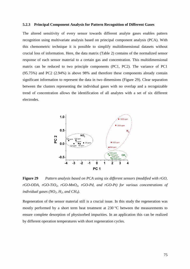
75
Principal Component Analysis for Pattern Recognition of Different Gases 5.2.3
The altered sensitivity of every sensor towards different analyte gases enables pattern
recognition using multivariate analysis based on principal component analysis (PCA). With
this chemometric technique it is possible to simplify multidimensional datasets without
crucial loss of information. Here, the data matrix (Table 2) contains of the normalized sensor
response of each sensor material to a certain gas and concentration. This multidimensional
matrix can be reduced to two principle components (PC1, PC2). The variance of PC1
(95.75%) and PC2 (2.94%) is above 98% and therefore these components already contain
significant information to represent the data in two dimensions (Figure 29). Clear separation
between the clusters representing the individual gases with no overlap and a recognizable
trend of concentration allows the identification of all analytes with a set of six different
electrodes.
Figure 29 Pattern analysis based on PCA using six different sensors (modified with rGO,
rGO-ODA, rGO-TiO2, rGO-MnO2, rGO-Pd, and rGO-Pt) for various concentrations of
individual gases (NO2, H2, and CH4).
Regeneration of the sensor material still is a crucial issue. In this study the regeneration was
mostly performed by a short term heat treatment at 230 °C between the measurements to
ensure complete desorption of physisorbed impurities. In an application this can be realized
by different operation temperatures with short regeneration cycles.
76
Conclusion 5.2.4
Reduced graphene oxide is an ideal sensor material for chemiresistive gas sensors, due to the
simple preparation and functionalization enabling an altered sensitivity. Compared to
commercial solid state gas sensors, such sensors have the advantage of being operated at quite
low temperatures of 85 °C to exclude the strong influences of humidity. It was demonstrated
that spin coating of rGO composites result in reproducible sensor behavior suitable for well-
established fabrication technologies. In a chemiresistor setup, unmodified rGO showed high
sensitivity towards NO2 in the ppm level at ambient conditions. The sensor was rather
unselective and showed also responses towards H2 and CH4. Upon different functionalization,
it was possible to achieve different sensor behavior for different gases. This can be used to
apply PCA and discriminate each individual gas. It is expected that this approach can be
extended to build up sensor arrays for detecting the concentration of many individual gases in
a complex matrix at low temperature.
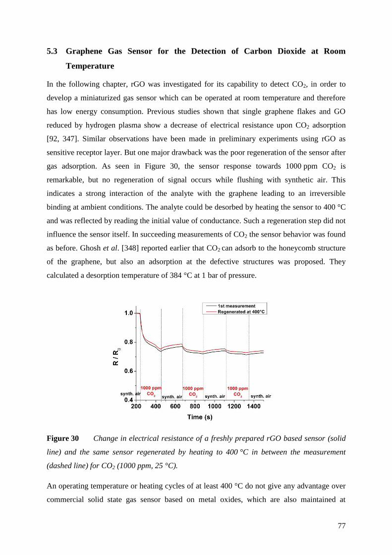
77
5.3 Graphene Gas Sensor for the Detection of Carbon Dioxide at Room
Temperature
In the following chapter, rGO was investigated for its capability to detect CO2, in order to
develop a miniaturized gas sensor which can be operated at room temperature and therefore
has low energy consumption. Previous studies shown that single graphene flakes and GO
reduced by hydrogen plasma show a decrease of electrical resistance upon CO2 adsorption
[92, 347]. Similar observations have been made in preliminary experiments using rGO as
sensitive receptor layer. But one major drawback was the poor regeneration of the sensor after
gas adsorption. As seen in Figure 30, the sensor response towards 1000 ppm CO2 is
remarkable, but no regeneration of signal occurs while flushing with synthetic air. This
indicates a strong interaction of the analyte with the graphene leading to an irreversible
binding at ambient conditions. The analyte could be desorbed by heating the sensor to 400 °C
and was reflected by reading the initial value of conductance. Such a regeneration step did not
influence the sensor itself. In succeeding measurements of CO2 the sensor behavior was found
as before. Ghosh et al. [348] reported earlier that CO2 can adsorb to the honeycomb structure
of the graphene, but also an adsorption at the defective structures was proposed. They
calculated a desorption temperature of 384 °C at 1 bar of pressure.
Figure 30 Change in electrical resistance of a freshly prepared rGO based sensor (solid
line) and the same sensor regenerated by heating to 400 °C in between the measurement
(dashed line) for CO2 (1000 ppm, 25 °C).
An operating temperature or heating cycles of at least 400 °C do not give any advantage over
commercial solid state gas sensor based on metal oxides, which are also maintained at
78
elevated temperatures above 250°C. It consumes too much energy, but also limits their long
term stability [17].
In the field of solid state gas sensors based on metal oxides, a tremendous amount of metal
oxides have been reported to show enhancement in sensitivity towards various gas analytes by
introducing new binding sites or catalytic effects [349–351]. In this context, different rGO
metal oxide composite nanomaterials had been synthesized and tested for their ability to
detect CO2 at room temperature. Modifications with different semiconductor nanoparticles,
like CuO, SnO2, Co3O4, TiO2, MnO2, NiO, and ZnO were screened. Only doping with CuO
showed promising first results in terms of high signal change, no drift of signal during gas
measurements, stability and especially regeneration of signal and was further investigated in
detail. The results are displayed in the following chapter.
79
Formation and Characterization of CuO Modified Reduced Graphene Oxide 5.3.1
A wet chemical approach for the decoration of graphene by oxide nanoparticles was adapted
[332]. A water/ipropanol system (1:10, v/v) turned out to result in CuO rod-like crystallites. It
is reported that the Cu2+
ions, which are formed in ipropanol, interact with the oxygen
functionalities of GO and nucleation and growth of CuO nanoparticles occurs directly at the
surface and edges of the GO sheets [332]. The addition of a small amount of water at a high
temperature of 86 °C leads to large amount of nuclei owing to the hydrolysis reaction of
Cu(CH3COO)2. The concentration of the copper precursor has been adjusted to achieve
different levels of metal oxide doping to assess its effect the sensitivity of the material
towards CO2. Subsequent heating to 230 °C at atmosphere, GO was thermally reduced to rGO
and the material became conductive.
The formation of CuO nanoparticles at the surface of GO could be observed with different
techniques. In SEM pictures, CuO nanoparticles of diameters ranging from 50 to 200 nm have
been found, decreasing in size using lower concentrations of copper precursor. Whereas a
mass ratio of 1:2 (GO:CuO) led to a complete coverage of the GO by CuO (Figure 31a), using
the same ratio of GO and CuO (1:1) resulted in a defined doping of the graphene material
(Figure 31a). Hereby, rGO flakes appear as darker areas and the lighter spots reflect the CuO
nanoparticles. Lower concentrations of CuO resulted in a composite material which does not
show any changes in electrical resistance when applying CO2.
An alternative route for the functionalization with CuO nanoparticles is the electrochemical
reduction of a copper precursor directly at the surface of rGO. This can be an option for later
industrial production of such devices and was shown here as a proof of concept. Applying a
reduction pulse of at -1.2 V versus SCE in a 10 mM solution of Cu2+
leads to a formation of
Cu nanoparticles on rGO. Whereas a longer pulse of 10 s result in a complete coverage of
rGO (Figure 31e), a shorter pulse led to a partial decoration of nanoparticles with a diameter
of around 200 nm (Figure 31f). Subsequent heating of the electrodes to 250°C at atmosphere
results in an oxidation of the deposited Cu to the thermodynamic more stable CuO.
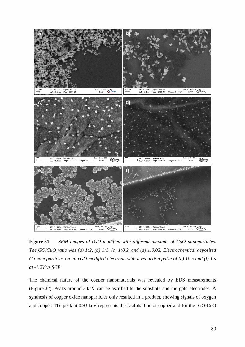
80
Figure 31 SEM images of rGO modified with different amounts of CuO nanoparticles.
The GO/CuO ratio was (a) 1:2, (b) 1:1, (c) 1:0.2, and (d) 1:0.02. Electrochemical deposited
Cu nanoparticles on an rGO modified electrode with a reduction pulse of (e) 10 s and (f) 1 s
at -1.2V vs SCE.
The chemical nature of the copper nanomaterials was revealed by EDS measurements
(Figure 32). Peaks around 2 keV can be ascribed to the substrate and the gold electrodes. A
synthesis of copper oxide nanoparticles only resulted in a product, showing signals of oxygen
and copper. The peak at 0.93 keV represents the L-alpha line of copper and for the rGO-CuO
a)
d)
b)
e)
c)
f)
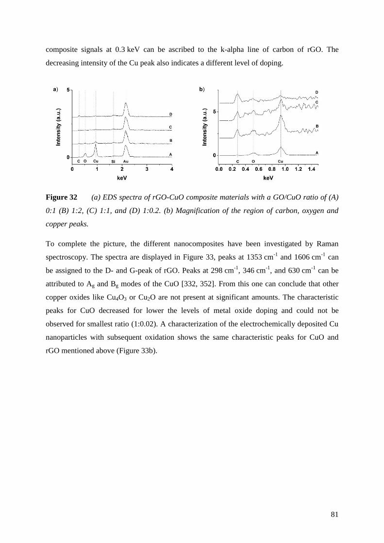
81
composite signals at 0.3 keV can be ascribed to the k-alpha line of carbon of rGO. The
decreasing intensity of the Cu peak also indicates a different level of doping.
Figure 32 (a) EDS spectra of rGO-CuO composite materials with a GO/CuO ratio of (A)
0:1 (B) 1:2, (C) 1:1, and (D) 1:0.2. (b) Magnification of the region of carbon, oxygen and
copper peaks.
To complete the picture, the different nanocomposites have been investigated by Raman
spectroscopy. The spectra are displayed in Figure 33, peaks at 1353 cm-1
and 1606 cm-1
can
be assigned to the D- and G-peak of rGO. Peaks at 298 cm-1
, 346 cm-1
, and 630 cm-1
can be
attributed to Ag and Bg modes of the CuO [332, 352]. From this one can conclude that other
copper oxides like Cu4O3 or Cu2O are not present at significant amounts. The characteristic
peaks for CuO decreased for lower the levels of metal oxide doping and could not be
observed for smallest ratio (1:0.02). A characterization of the electrochemically deposited Cu
nanoparticles with subsequent oxidation shows the same characteristic peaks for CuO and
rGO mentioned above (Figure 33b).

82
Figure 33 Raman spectra of (a) of rGO-CuO composite materials with a GO/CuO ratio
of (A) 0:1 (B) 1:2, (C) 1:1, (D) 1:0.2, (E) 1:0.02 and (b) electrochemical produced CuO
nanoparticles on rGO with a reduction pulse of (A) 10 s and (B) 1 s at -1.2V versus SCE.
Additional peaks at 520 and 980 cm-1
can be ascribed to the Si/SiO2 substrate.
83
Detection of Carbon Dioxide 5.3.2
Nanocomposites of rGO and CuO turned out to have a huge impact on the sensing behavior of
the material towards CO2. Whereas sensors based on rGO showed nearly no regeneration of
signal, combination with the metal oxide nanoparticle led to higher sensitivity and also to an
enhanced regeneration, even at room temperature (Figure 34a). The electrical resistance of
sensors modified with a GO/CuO ratio of 1:2 dropped by 40% while being exposed to 1000
ppm CO2. The response time changed from several minutes for rGO to less than 30 seconds
for the nanocomposite. The main benefit here is a nearly complete reversibility after flushing
200 seconds with synthetic air. Whereas a complete loading of rGO with metal oxide
nanoparticles, leads to an increased sensitivity, a decrease of the level of doping also results in
a decreasing sensitivity towards CO2 (Figure 34b). The electrical resistance dropped only by
7% and 1%, using a GO/CuO ratio of 1:1 and 1:0.2 respectively. A lower ratio of 1:0.02
resulted in a material comparable to rGO, with no signal changes in the presence of 1000 ppm
CO2. This shows the importance of the metal oxide in the gas sensing mechanism. Since
sensors with a GO/CuO ratio of 1:2 showed highest sensitivity towards CO2 and also stability,
further experiments were performed with this material referring to it as rGO-CuO.
Sensors with a layer comprising only CuO without involving any rGO show also a change in
electrical resistance upon exposure to 1000 ppm CO2 of about 15% (Figure 34c). Here, a
continuous drift of signal and no equilibration in a reasonable time are major drawbacks of
this system. One reason might be the mechanical instability of the sensor layer, consisting
only of metal particles and the use of rGO is mandatory for a stable sensor response.
Electrochemical preparation of the rGO-CuO composite resulted in a sensor material which
electrical resistance dropped by 6% in the presence of 1000 ppm CO2 with good regeneration
characteristics (Figure 34d). Though this approach did not yield in a sensor material with best
sensitivity, it reveals an alternative to the wet chemical synthesis and could be used in terms
industrial production.
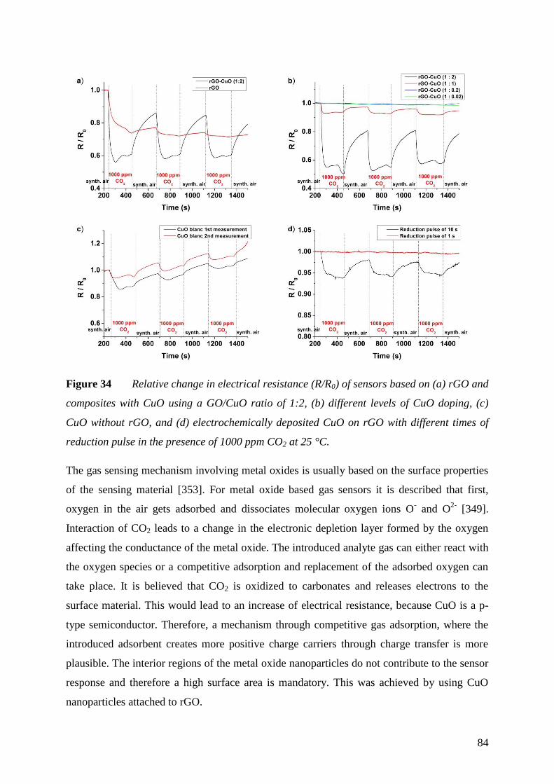
84
Figure 34 Relative change in electrical resistance (R/R0) of sensors based on (a) rGO and
composites with CuO using a GO/CuO ratio of 1:2, (b) different levels of CuO doping, (c)
CuO without rGO, and (d) electrochemically deposited CuO on rGO with different times of
reduction pulse in the presence of 1000 ppm CO2 at 25 °C.
The gas sensing mechanism involving metal oxides is usually based on the surface properties
of the sensing material [353]. For metal oxide based gas sensors it is described that first,
oxygen in the air gets adsorbed and dissociates molecular oxygen ions O- and O
2- [349].
Interaction of CO2 leads to a change in the electronic depletion layer formed by the oxygen
affecting the conductance of the metal oxide. The introduced analyte gas can either react with
the oxygen species or a competitive adsorption and replacement of the adsorbed oxygen can
take place. It is believed that CO2 is oxidized to carbonates and releases electrons to the
surface material. This would lead to an increase of electrical resistance, because CuO is a p-
type semiconductor. Therefore, a mechanism through competitive gas adsorption, where the
introduced adsorbent creates more positive charge carriers through charge transfer is more
plausible. The interior regions of the metal oxide nanoparticles do not contribute to the sensor
response and therefore a high surface area is mandatory. This was achieved by using CuO
nanoparticles attached to rGO.
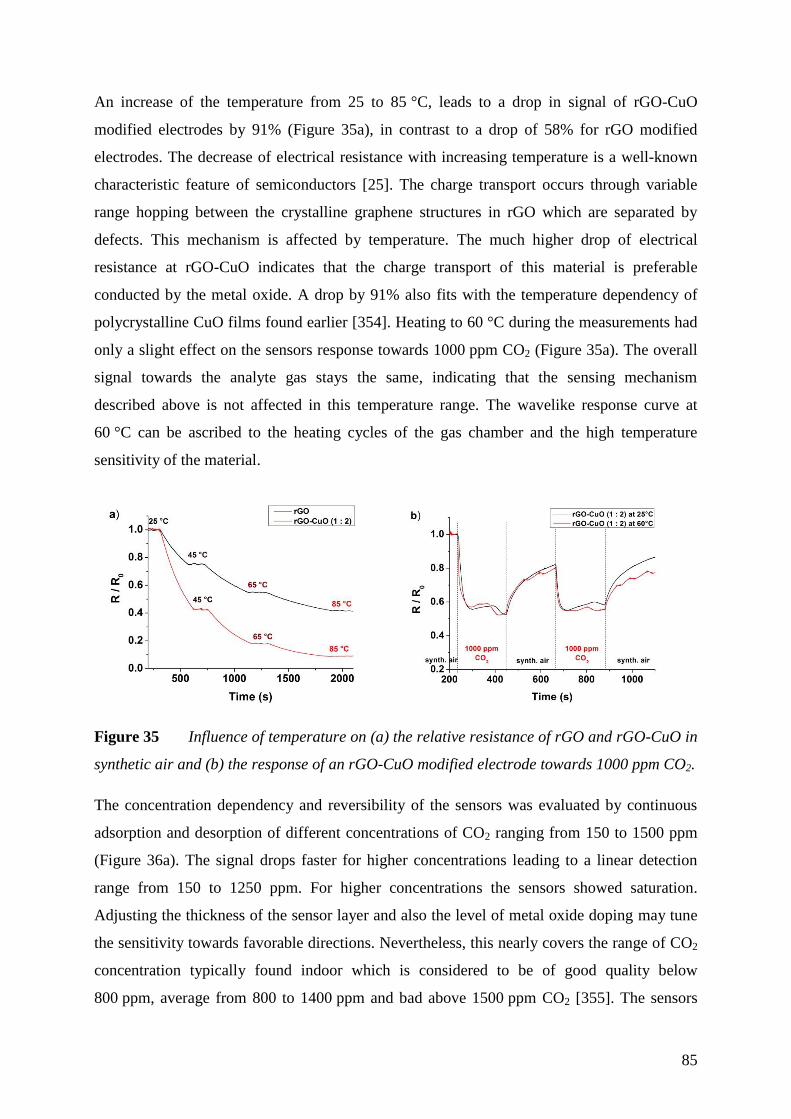
85
An increase of the temperature from 25 to 85 °C, leads to a drop in signal of rGO-CuO
modified electrodes by 91% (Figure 35a), in contrast to a drop of 58% for rGO modified
electrodes. The decrease of electrical resistance with increasing temperature is a well-known
characteristic feature of semiconductors [25]. The charge transport occurs through variable
range hopping between the crystalline graphene structures in rGO which are separated by
defects. This mechanism is affected by temperature. The much higher drop of electrical
resistance at rGO-CuO indicates that the charge transport of this material is preferable
conducted by the metal oxide. A drop by 91% also fits with the temperature dependency of
polycrystalline CuO films found earlier [354]. Heating to 60 °C during the measurements had
only a slight effect on the sensors response towards 1000 ppm CO2 (Figure 35a). The overall
signal towards the analyte gas stays the same, indicating that the sensing mechanism
described above is not affected in this temperature range. The wavelike response curve at
60 °C can be ascribed to the heating cycles of the gas chamber and the high temperature
sensitivity of the material.
Figure 35 Influence of temperature on (a) the relative resistance of rGO and rGO-CuO in
synthetic air and (b) the response of an rGO-CuO modified electrode towards 1000 ppm CO2.
The concentration dependency and reversibility of the sensors was evaluated by continuous
adsorption and desorption of different concentrations of CO2 ranging from 150 to 1500 ppm
(Figure 36a). The signal drops faster for higher concentrations leading to a linear detection
range from 150 to 1250 ppm. For higher concentrations the sensors showed saturation.
Adjusting the thickness of the sensor layer and also the level of metal oxide doping may tune
the sensitivity towards favorable directions. Nevertheless, this nearly covers the range of CO2
concentration typically found indoor which is considered to be of good quality below
800 ppm, average from 800 to 1400 ppm and bad above 1500 ppm CO2 [355]. The sensors
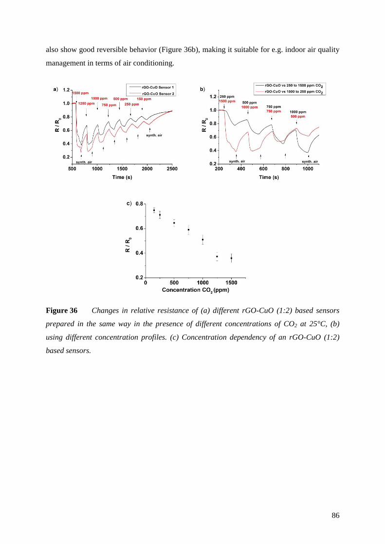
86
also show good reversible behavior (Figure 36b), making it suitable for e.g. indoor air quality
management in terms of air conditioning.
Figure 36 Changes in relative resistance of (a) different rGO-CuO (1:2) based sensors
prepared in the same way in the presence of different concentrations of CO2 at 25°C, (b)
using different concentration profiles. (c) Concentration dependency of an rGO-CuO (1:2)
based sensors.

87
Cross Sensitivity 5.3.3
Selectivity is a key issue in developing new sensor materials and concepts. Often sensors
show cross sensitivity to other gaseous analytes, disallowing the detection of single gases in a
gas mixture. A solution could be a sensor array consisting of different modified sensor
materials with altered sensitivities. The sensor response of the rGO-CuO composite is
characterized by high sensitivity for CO2, in contrast to rGO. Therefore, these type of sensors
were also tested against various analyte gases like NO2, CH4, H2 and CO (Figure 37a). The
sensor behavior is very similar to unmodified rGO. Oxidizing gases like NO2 transfer an
electron to the surface layer of CuO and therefore lower the concentration of charge carriers,
since CuO as well as rGO show p-type semiconductor behavior. Carbon monoxide can get
oxidized at metal oxides, but only at elevated temperatures [349]. The low signals for CO and
H2 are probably due to a competitive adsorption of the introduced gas molecules and
previously adsorbed oxygen. In the presence of CH4 no signal change occurs, assuming that
either there is no adsorption to the CuO surface or it no effect on the electrical resistance of
the sensor material.
Figure 37 (a) Response of sensors based on rGO-CuO composite with a GO/CuO ratio of
1:2 in the presence of 100 ppm NO2, 3000 ppm, CH4, 3000 ppm H2, and 100 ppm CO at 25°C.
(b) Plot of two principle components (scores plot, black) using a sensor array consisting of
unmodified rGO and rGO-CuO electrodes and the contribution to the signal of each sensor
(loadings plot, blue).
Whereas a single sensor based on rGO shows high changes in signal in the presence of NO2,
but cannot detect CO2, devices based on rGO-CuO are sensitive towards both gases. A
combination of both would capable to distinguish between these gases. Applying PCA, as
shown in the previous chapter (5.2.3), leads to a clear separation pattern of the applied gases

88
in the scores plot (Figure 37b). In the loading plot it is shown that the rGO-CuO sensor
mainly contributes to the CO2 detection, the rGO sensor is responsible for the NO2 signal and
both of them contribute to the detection of the other gases in nearly the same amount. The
resolution for test gases like CO, CH4 and H2 is rather low and other sensors with altered
sensitivity are needed for a clear separation of signals.
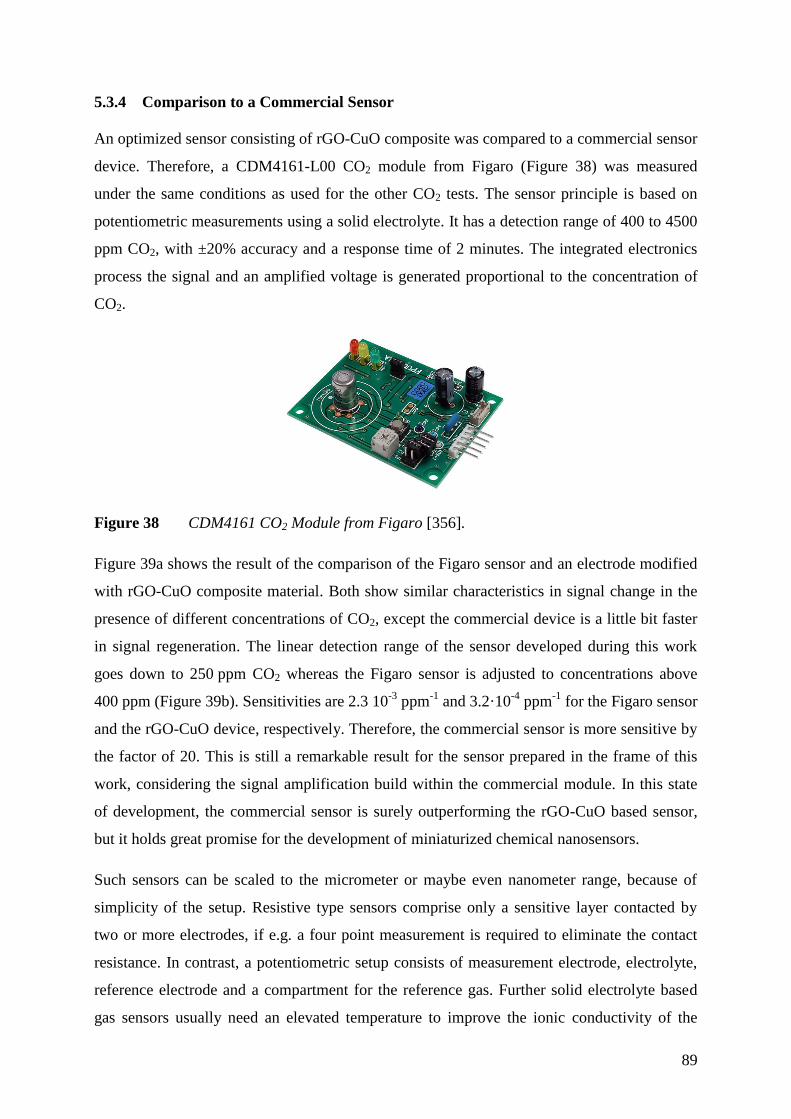
89
Comparison to a Commercial Sensor 5.3.4
An optimized sensor consisting of rGO-CuO composite was compared to a commercial sensor
device. Therefore, a CDM4161-L00 CO2 module from Figaro (Figure 38) was measured
under the same conditions as used for the other CO2 tests. The sensor principle is based on
potentiometric measurements using a solid electrolyte. It has a detection range of 400 to 4500
ppm CO2, with ±20% accuracy and a response time of 2 minutes. The integrated electronics
process the signal and an amplified voltage is generated proportional to the concentration of
CO2.
Figure 38 CDM4161 CO2 Module from Figaro [356].
Figure 39a shows the result of the comparison of the Figaro sensor and an electrode modified
with rGO-CuO composite material. Both show similar characteristics in signal change in the
presence of different concentrations of CO2, except the commercial device is a little bit faster
in signal regeneration. The linear detection range of the sensor developed during this work
goes down to 250 ppm CO2 whereas the Figaro sensor is adjusted to concentrations above
400 ppm (Figure 39b). Sensitivities are 2.3 10-3
ppm-1
and 3.2·10-4
ppm-1
for the Figaro sensor
and the rGO-CuO device, respectively. Therefore, the commercial sensor is more sensitive by
the factor of 20. This is still a remarkable result for the sensor prepared in the frame of this
work, considering the signal amplification build within the commercial module. In this state
of development, the commercial sensor is surely outperforming the rGO-CuO based sensor,
but it holds great promise for the development of miniaturized chemical nanosensors.
Such sensors can be scaled to the micrometer or maybe even nanometer range, because of
simplicity of the setup. Resistive type sensors comprise only a sensitive layer contacted by
two or more electrodes, if e.g. a four point measurement is required to eliminate the contact
resistance. In contrast, a potentiometric setup consists of measurement electrode, electrolyte,
reference electrode and a compartment for the reference gas. Further solid electrolyte based
gas sensors usually need an elevated temperature to improve the ionic conductivity of the
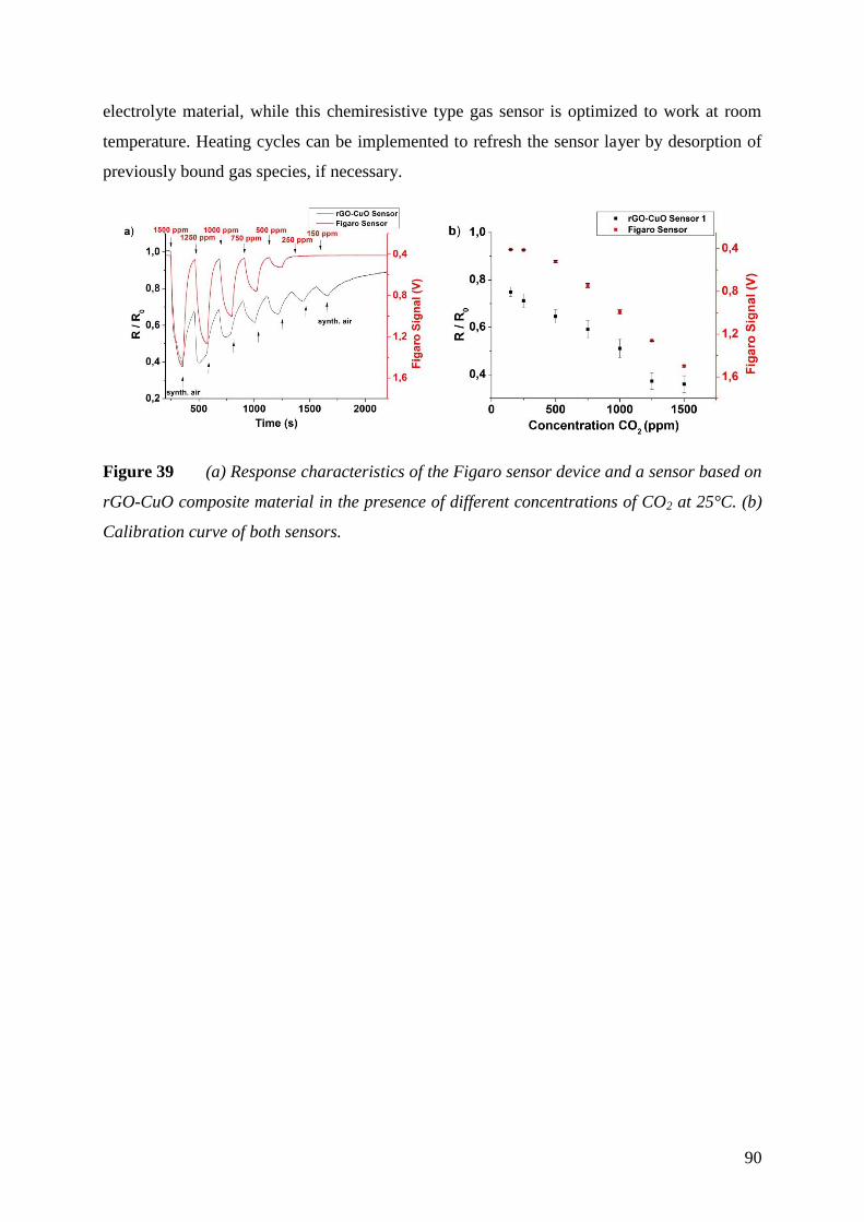
90
electrolyte material, while this chemiresistive type gas sensor is optimized to work at room
temperature. Heating cycles can be implemented to refresh the sensor layer by desorption of
previously bound gas species, if necessary.
Figure 39 (a) Response characteristics of the Figaro sensor device and a sensor based on
rGO-CuO composite material in the presence of different concentrations of CO2 at 25°C. (b)
Calibration curve of both sensors.
91
Conclusion 5.3.5
It was demonstrated that decoration of rGO with CuO nanoparticles leads to an improved
sensitivity of the material towards CO2 in a chemiresistive setup. The sensor material was
optimized in terms of level of doping with metal oxide. Complete coverage of rGO with a
ratio of GO/CuO ratio of 1:2 turned out to have highest sensitivity (3.2·10-4
ppm-1
) even at
room temperature and showed a linear behavior from 150 to 1250 ppm CO2. The sensor
performance was compared to a commercial sensor under identical experimental conditions
and showed great potential in this stage of development. The cross sensitivity of such a device
was rather low towards H2, CH4 and CO, but high towards NO2. Combination with an
unmodified rGO based sensor showed a clear recognition pattern utilizing PCA. An array of
these two electrodes is expected capable for a distinct detection of CO2 and NO2 at room
temperature.
92
6 Summary
The variability of graphene with its exceptional properties gives rise to improve material
chemistry in various fields of applications. The development of graphene is still in the
beginning and up to now only a few niche products have reached the market and are
highlighted as well as the aim of this work in chapter 1. The goal of this thesis was the
investigation of graphene in electrochemical sensor applications. It holds great promise in
terms of miniaturization, improving sensitivity and developing new sensor concepts. Many
approaches are already described for biosensor applications, often utilizing graphene in an
amperometric detection scheme. Therefore, the impact of preparation technique on the
detection of the model analyte H2O2 was investigated, applying different graphene materials
as electrode material. Further, graphene was studied as tunable sensor material in gas sensing
applications and how it can be customized for the room temperature detection of CO2.
Chapter 2 summarizes the author’s publications and patents developed in the frame of this
work.
The perspectives of graphene in electrochemical sensors were investigated by the means of
research performed in this field and are described in chapter 3. The most prominent
preparation techniques and the application in biosensor and gas sensor technologies are
discussed. Every method provides graphene materials of different characteristics, scalability
and further usability. It was shown that defects in the ideal sp2 carbon lattice decide on the
sensor performance, but also on device fabrication and appropriate functionalization. A higher
quality of graphene can lead to more sensitivity and reliable device production in
electrochemical biosensor technologies. In contrast, a defective structure can enhance the
sensitivity and applicability in gas sensing applications, providing additional adsorption sites.
In chapter 4, the experimental work, performed during this work, is described in detail.
Chapter 5 comprises the results on graphene and graphene composite materials applied in the
electrochemical detection of H2O2 and as tunable recognition element in gas sensors. In a first
part, graphene materials derived by different preparation techniques were studied as electrode
material. The electrochemical behavior of the different materials has been investigated as well
as the feasibility in device fabrication was compared. It was shown, that the quality of the
graphene has an enormous impact on the reductive amperometric detection of H2O2. A
93
defective structure like in reduced graphene oxide leads to almost no significant improvement
in signal enhancement compared to a standard carbon disc electrode. In contrast, fewer
defects like in graphene prepared by chemical vapor deposition, resulted in a higher
sensitivity, which is 50 times better compared to reduced graphene oxide. This technique was
found to be most suitable for the production of highly sensitive electrodes to be further used
in amperometric detection and development of biosensors.
Whereas, a material of high quality is desired in electrochemical biosensor applications,
defects are beneficial using graphene as transducer in a chemiresistive setup for the detection
of gaseous analytes. In the next part of the work, reduced graphene oxide was demonstrated to
be an applicable candidate for gas detection at moderate (85 °C) or even room temperature.
Analyte gases like NO2, CH4 and H2 were detected due to fast changes in the electrical
resistance at of 85 °C. To overcome the poor selectivity, the material was further altered with
octadecylamine, metal nanoparticles such as Pd and Pt, and metal oxides such as MnO2, and
TiO2. This changed the sensor response towards the studied gases and the different response
patterns for six different materials allowed a clear discrimination of all test gases by pattern
recognition based on principal component analysis.
Based on the feasibility of this concept, a graphene-based sensor for the room temperature
detection of CO2 was developed. Decoration of reduced graphene oxide with CuO
nanoparticles led to an improved sensing performance for the target analyte. Different levels
of metal oxide doping were applied by wet chemical and electrochemical preparation methods
and the resulting composite materials were characterized. It was shown that a complete
coverage obtained by wet chemical functionalization leads to highest sensitivity, comparable
to a commercial CO2 sensor, which was also tested in the frame of this work. An array
consisting of reduced graphene oxide and the composite with CuO nanoparticles was capable
to differentiate CO2 from NO2, CO, H2 and CH4. This sensor material can lead to the
development of miniaturized chemical sensors comprising high and adjustable sensitivity,
which can be applied for monitoring air quality and ventilation management. The presented
sensor concept based on customized graphene materials can be tailored for the versatile use in
appropriate applications.
One of the main challenges remains the reproducible large-scale production of graphene and
functionalized graphene combined with reliable transfer techniques in terms of an industrial
application. This problem has not been solved completely up to now. But the extensive
research going on in this field will lead to a solution in near future and will help graphene to

94
find its way to be integrated into many electrochemical sensor devices. Further studies should
preferably aim for large scale production of the material and devices. The use of high quality
graphene with a distinct introduction of defects and a better controlled way of
functionalization may be a route to tune the material properties in favorable directions.
95
7 Zusammenfassung
Graphen birgt großes Potential in den unterschiedlichsten Anwendungsgebieten, aufgrund der
bemerkenswerten Eigenschaften, die es in einem Material vereint. Bis jetzt kann es jedoch nur
in einigen wenigen kommerziellen Nischenprodukte wiedergefunden werden. Das Ziel dieser
Arbeit war es, die Anwendung von Graphen in elektrochemischen Sensoren zu untersuchen.
In diesem Zusammenhang kann es dazu beitragen, Bauelemente zu miniaturisieren, die
Empfindlichkeiten von Sensoren zu verbessern und neue Messkonzepte zu entwickeln. Viele
Studien auf dem Gebiet der Biosensorik befassen sich mit der Anwendung von Graphen und
basieren meist auf einem amperometrischen Detektionsprinzip. Hierzu wurden
unterschiedlich hergestellte Graphenmaterialien als Elektrodenmaterial zum Nachweis eines
Modelanalyten (H2O2) eingesetzt und der Einfluss der Materialqualität untersucht.
Desweiteren wurde gezeigt, dass sich chemisch hergestelltes Graphen als Sensormaterial zur
Detektion von Gasen eignet und wie dessen Ansprech-verhalten zur selektiven Detektion von
CO2 angepasst werden kann.
Kapitel 2 gibt einen Überblick über die Publikationen und Patente des Autors, die im Rahmen
dieser Arbeit entstanden sind.
Kapitel 3 beschreibt die Perspektiven von Graphen und dessen Einsatz in elektrochemischen
Sensoren anhand der Ergebnisse aktueller Forschung. Zunächst wird auf die bedeutendsten
Methoden zu dessen Herstellung eingegangen und deren Anwendbarkeit in Gas- und Bio-
sensortechnologien diskutiert. Dabei liefert jede Herstellungsmethode Graphenematerialien
mit unterschiedlichen Eigenschaften und in verschiedenen Dimensionen und Mengen, was die
weitere Verarbeitung stark beeinflusst. Es zeigte sich, dass Defekte in der sp2 Struktur der
Kohlenstoffschichten maßgeblich die Leistungsfähigkeit der Sensoren, als auch deren
Herstellung und entsprechende Funktionalisierung beeinflussen. Graphen von hoher Qualität
zeigt erhöhte Sensitivität und zuverlässigere Produktion in elektrochemischen Biosensoren.
Im Gegensatz dazu profitieren Anwendungen in der Gassensorik von Defekten im Material,
da diese zusätzliche Bindungstellen für Analyten aufweisen.
In Kapitel 4 werden die experimentellen Einzelheiten dieser Arbeit beschrieben. Die dabei
entstandenen Ergebnisse zu Graphen und Kompositmaterialien, angewandt zur elektro-
chemischen Detektion von H2O2 und als variables Sensorelement zur Detektion von Gasen,
96
sind in Kapitel 5 dargestellt. Im ersten Teil wurden die elektrochemischen Eigenschaften von
Graphenelektroden, welche auf unterschiedliche Weise hergestellt wurden, verglichen. Es
wurde gezeigt, dass die Qualität des Graphens einen erheblichen Einfluss auf die reduktive
amperometrische Erfassung von H2O2 hat. Eine defektreiche Struktur, wie in reduziertem
Graphenoxid, zeigte dabei nur geringfügige Signalverstärkung verglichen mit einer
herkömmlichen Kohlenstoffelektrode. Graphen, welches durch chemische Gasphasen-
abscheidung hergestellt wurde und eine geringere Anzahl an Defekten aufweist, zeigte im
Gegensatz dazu eine um den Faktor 50 höhere Sensitivität als reduziertes Graphenoxid. Daher
eignet sich gerade diese Methode der Graphensynthese zur Herstellung von hochsensitiven
Elektroden und Entwicklung amperometrischer Biosensoren.
In elektrochemischen Biosensoren ist eine hohe Materialqualität wünschenswert. Bei der
Detektion von Gasen können sich jedoch Defekte im Material vorteilhaft auswirken. Im
nächsten Teil der Arbeit wurde reduziertes Graphenoxid als signalgebendes Element in
chemischen Leitfähigkeitssensoren eingesetzt. Diese Sensoren konnten bei niedrigen
Temperaturen von 85 °C oder sogar Raumtemperatur betrieben werden. Eine schnelle
Änderung des elektrischen Widerstandes, bedingt durch die Adsorption von Gasmolekülen,
ermöglichte eine Erfassung von NO2, CH4 und H2, jedoch mit schlechter Selektivität. Um
diese zu verbessern, wurde das Material mit Octadecylamin, Metallnanopartikeln (Pd, Pt)
sowie Metalloxiden (TiO2, MnO2) modifiziert und ein verändertes Ansprechverhalten
gegenüber der getesteten Gase festgestellt. Das Erkennungsmuster von sechs verschiedenen
Sensormaterialien kombiniert mit einer Datenverarbeitung mittels Hauptkomponentenanalyse
ermöglichte schließlich eine klare Unterscheidung der Gase.
Basierend auf diesem Konzept, wurde im nächsten Teil der Arbeit ein Sensor zur Detektion
von CO2 bei Raumtemperatur entwickelt. Das Ansprechverhalten von reduziertem
Graphenoxid konnte durch Abscheidung von CuO Nanopartikeln erheblich verbessert
werden. Reduziertes Graphenoxid wurde mittels nasschemischer und elektrochemischer
Methoden in unterschiedlichen Verhältnissen mit dem Metalloxid beladen und die
entsprechenden Kompositmaterialien charakterisiert. Eine vollständige Bedeckung mit
Metalloxidpartikeln führte dabei zur höchsten Sensitivität gegenüber CO2. Im Rahmen dieses
Projekts wurde ein solcher Sensor mit einem kommerziell erhältlichen CO2-Messgerät
verglichen und lieferte durchaus gleichwertige Resultate. Zusätzlich wurde gezeigt, dass ein
Verbund aus zwei Sensoren, basierend auf reduziertem Graphenoxid und dem
Kompositmaterial mit CuO Nanopartikeln, CO2 gegenüber NO2, CO, H2 und CH4 selektiv
97
unterscheiden kann. Das Kompositmaterial kann zur Entwicklung von miniaturisierten
chemischen Sensoren mit hoher und einstellbarer Sensitivität beitragen und könnte in der
Überwachung von Luftqualität und automatischer Belüftungssteuerung Anwendung finden.
Das hier vorgestellte Sensorkonzept, bestehend aus individuell modifizierten Graphen-
materialien, bietet ein vielseitiges Spektrum an Einsatzmöglichkeiten.
Die reproduzierbare Herstellung von Graphen und funktionalisierten Materialien im
Zusammenspiel mit geeigneten Transfertechniken ist bis heute der limitierende Faktor, um es
letztendlich für industrielle Anwendungen verfügbar zu machen. Dieses Problem ist noch
nicht vollständig gelöst. Jedoch lässt der Fortschritt auf diesem Gebiet auf Lösungsansätze in
naher Zukunft hoffen. Graphen wird damit voraussichtlich den Weg in viele elektrochemische
Sensoren finden. Weiterführende Studien sollten sich daher vor allem mit der groß angelegten
Produktion von Materialen und Sensoren befassen. Eine Möglichkeit ist hierbei die
Verwendung von Graphenmaterialien von definierter Qualität, in die auf kontrollierter Weise
Defekte und entsprechende Funktionalisierungen eingebracht werden.
98
8 References
1. Novoselov KS, Geim AK, Morozov SV, Jiang D, Zhang Y, Dubonos SV, Grigorieva
IV, Firsov AA (2004) Electric Field Effect in Atomically Thin Carbon Films. Science
306:666–669
2. (2013) Graphene in the heart of Europe. Nat Nanotechnol 8:221–221
3. Liu F, Ming P, Li J (2007) Ab initio calculation of ideal strength and phonon instability
of graphene under tension. Phys Rev B 76:064120
4. Geim AK (2009) Graphene: Status and Prospects. Science 324:1530–1534
5. Nair RR, Blake P, Grigorenko AN, Novoselov KS, Booth TJ, Stauber T, Peres NMR,
Geim AK (2008) Fine structure constant defines visual transparency of graphene.
Science 320:1308
6. Chae HK, Siberio-Pérez DY, Kim J, Go Y, Eddaoudi M, Matzger AJ, O’Keeffe M,
Yaghi OM (2004) A route to high surface area, porosity and inclusion of large
molecules in crystals. Nature 427:523–527
7. http://store.vorbeck.com/collections/all, 13.06.2015.
8. http://www.head.com/tennis/technologies/?region=eu&id=706, 13.06.2015.
9. http://www.vittoria.com/news/the-revolutionary-g-material/, 13.06.2015.
10. http://www.catlike.es/global/en/innovation/helmets-technologies/graphene/,
13.06.2015.
11. Li Y, Chopra N (2014) Progress in Large-Scale Production of Graphene. Part 2: Vapor
Methods. JOM 67:44–52
12. https://www.zapandgocharger.com/pre-order.html, 12.06.2015.
13. Yuan W, Shi G (2013) Graphene-based gas sensors. J Mater Chem A 1:10078–10091
14. Samet JM (2007) Traffic, Air Pollution, and Health. Inhal Toxicol 19:1021–1027
15. Moseley PT (1997) Solid state gas sensors. Meas Sci Technol 8:223
16. Korotcenkov G (2007) Metal oxides for solid-state gas sensors: What determines our
choice? Mater Sci Eng B 139:1–23
17. Tricoli A, Righettoni M, Teleki A (2010) Semiconductor Gas Sensors: Dry Synthesis
and Application. Angew Chem Int Ed 49:7632–7659
18. Robinson JT, Perkins FK, Snow ES, Wei Z, Sheehan PE (2008) Reduced graphene
oxide molecular sensors. Nano Lett 8:3137–3140
99
19. Leenaerts O, Partoens B, Peeters FM (2008) Adsorption of H2O, NH3, CO, NO2, and
NO on graphene: A first-principles study. Phys Rev B 77:125416
20. Schedin F, Geim AK, Morozov SV, Hill EW, Blake P, Katsnelson MI, Novoselov KS
(2007) Detection of individual gas molecules adsorbed on graphene. Nat Mater 6:652–
655
21. Stoller MD, Park S, Zhu Y, An J, Ruoff RS (2008) Graphene-Based Ultracapacitors.
Nano Lett 8:3498–3502
22. Rao CNR, Sood AK, Subrahmanyam KS, Govindaraj A (2009) Graphene: The New
Two-Dimensional Nanomaterial. Angew Chem Int Ed 48:7752–7777
23. Hummers WS, Offeman RE (1958) Preparation of Graphitic Oxide. J Am Chem Soc
80:1339–1339
24. Li D, Müller MB, Gilje S, Kaner RB, Wallace GG (2008) Processable aqueous
dispersions of graphene nanosheets. Nat Nanotechnol 3:101–105
25. Gómez-Navarro C, Weitz RT, Bittner AM, Scolari M, Mews A, Burghard M, Kern K
(2007) Electronic transport properties of individual chemically reduced graphene oxide
sheets. Nano Lett 7:3499–3503
26. Gilje S, Han S, Wang M, Wang KL, Kaner RB (2007) A chemical route to graphene
for device applications. Nano Lett 7:3394–3398
27. Zhang Y-H, Chen Y-B, Zhou K-G, Liu C-H, Zeng J, Zhang H-L, Peng Y (2009)
Improving gas sensing properties of graphene by introducing dopants and defects: a
first-principles study. Nanotechnology 20:185504
28. Eda G, Fanchini G, Chhowalla M (2008) Large-area ultrathin films of reduced
graphene oxide as a transparent and flexible electronic material. Nat Nanotechnol
3:270–274
29. Xu Y, Shi G (2011) Assembly of chemically modified graphene: methods and
applications. J Mater Chem 21:3311–3323
30. Persily AK (1997) Evaluating building IAQ and ventilation with indoor carbon dioxide.
Trans-Am Soc Heat Refrig AIR Cond Eng 103:193–204
31. Scheff PA, Paulius VK, Huang SW, Conroy LM (2000) Indoor Air Quality in a Middle
School, Part I: Use of CO2 as a Tracer for Effective Ventilation. Appl Occup Environ
Hyg 15:824–834
32. Pérez-López B, Merkoçi A (2012) Carbon nanotubes and graphene in analytical
sciences. Microchim Acta 179:1–16
100
33. Zhou M, Zhai Y, Dong S (2009) Electrochemical Sensing and Biosensing Platform
Based on Chemically Reduced Graphene Oxide. Anal Chem 81:5603–5613
34. Kochmann S, Hirsch T, Wolfbeis OS (2012) Graphenes in chemical sensors and
biosensors. TrAC Trends Anal Chem 39:87–113
35. Marcano DC, Kosynkin DV, Berlin JM, Sinitskii A, Sun Z, Slesarev A, Alemany LB,
Lu W, Tour JM (2010) Improved Synthesis of Graphene Oxide. ACS Nano 4:4806–
4814
36. Kim KS, Zhao Y, Jang H, Lee SY, Kim JM, Kim KS, Ahn J-H, Kim P, Choi J-Y, Hong
BH (2009) Large-scale pattern growth of graphene films for stretchable transparent
electrodes. Nature 457:706–710
37. Shang NG, Papakonstantinou P, McMullan M, Chu M, Stamboulis A, Potenza A,
Dhesi SS, Marchetto H (2008) Catalyst-Free Efficient Growth, Orientation and
Biosensing Properties of Multilayer Graphene Nanoflake Films with Sharp Edge
Planes. Adv Funct Mater 18:3506–3514
38. Novoselov KS, Fal′ko VI, Colombo L, Gellert PR, Schwab MG, Kim K (2012) A
roadmap for graphene. Nature 490:192–200
39. Park S, Ruoff RS (2009) Chemical methods for the production of graphenes. Nat
Nanotechnol 4:217–224
40. Tang L, Wang Y, Li Y, Feng H, Lu J, Li J (2009) Preparation, Structure, and
Electrochemical Properties of Reduced Graphene Sheet Films. Adv Funct Mater
19:2782–2789
41. Ambrosi A, Chua CK, Bonanni A, Pumera M (2014) Electrochemistry of Graphene and
Related Materials. Chem Rev 114:7150–7188
42. Lu L-M, Qiu X-L, Zhang X-B, Shen G-L, Tan W, Yu R-Q (2013) Supramolecular
assembly of enzyme on functionalized graphene for electrochemical biosensing.
Biosens Bioelectron 45:102–107
43. Chen S, Yuan R, Chai Y, Hu F (2012) Electrochemical sensing of hydrogen peroxide
using metal nanoparticles: a review. Microchim Acta 180:15–32
44. Cui X, Wu S, Li Y, Wan G (Gary) (2014) Sensing hydrogen peroxide using a glassy
carbon electrode modified with in-situ electrodeposited platinum-gold bimetallic
nanoclusters on a graphene surface. Microchim Acta 182:265–272
45. Li S-J, Du J-M, Zhang J-P, Zhang M-J, Chen J (2014) A glassy carbon electrode
modified with a film composed of cobalt oxide nanoparticles and graphene for
electrochemical sensing of H2O2. Microchim Acta 181:631–638
101
46. Chen X, Wu G, Cai Z, Oyama M, Chen X (2013) Advances in enzyme-free
electrochemical sensors for hydrogen peroxide, glucose, and uric acid. Microchim Acta
181:689–705
47. Yuping Lin XC (2015) Non-enzymatic sensing of hydrogen peroxide using a glassy
carbon electrode modified with a nanocomposite made from carbon nanotubes and
molybdenum disulfide. Microchim Acta. doi: 10.1007/s00604-015-1517-5
48. Kang J, Shin D, Bae S, Hong BH (2012) Graphene transfer: key for applications.
Nanoscale 4:5527–5537
49. Roddaro S, Pingue P, Piazza V, Pellegrini V, Beltram F (2007) The Optical Visibility
of Graphene: Interference Colors of Ultrathin Graphite on SiO2. Nano Lett 7:2707–
2710
50. Morozov SV, Novoselov KS, Katsnelson MI, Schedin F, Elias DC, Jaszczak JA, Geim
AK (2008) Giant Intrinsic Carrier Mobilities in Graphene and Its Bilayer. Phys Rev
Lett 100:016602
51. Emtsev KV, Bostwick A, Horn K, et al (2009) Towards wafer-size graphene layers by
atmospheric pressure graphitization of silicon carbide. Nat Mater 8:203–207
52. Moon JS, Curtis D, Hu M, et al (2009) Epitaxial-Graphene RF Field-Effect Transistors
on Si-Face 6H-SiC Substrates. IEEE Electron Device Lett 30:650–652
53. Lin Y-M, Dimitrakopoulos C, Jenkins KA, Farmer DB, Chiu H-Y, Grill A, Avouris P
(2010) 100-GHz transistors from wafer-scale epitaxial graphene. Science 327:662
54. Li X, Cai W, Colombo L, Ruoff RS (2009) Evolution of Graphene Growth on Ni and
Cu by Carbon Isotope Labeling. Nano Lett 9:4268–4272
55. Ago H, Ogawa Y, Tsuji M, Mizuno S, Hibino H (2012) Catalytic Growth of Graphene:
Toward Large-Area Single-Crystalline Graphene. J Phys Chem Lett 3:2228–2236
56. Mattevi C, Kim H, Chhowalla M (2011) A review of chemical vapour deposition of
graphene on copper. J Mater Chem 21:3324–3334
57. Bae S, Kim H, Lee Y, et al (2010) Roll-to-roll production of 30-inch graphene films for
transparent electrodes. Nat Nanotechnol 5:574–578
58. Ryu J, Kim Y, Won D, et al (2014) Fast Synthesis of High-Performance Graphene
Films by Hydrogen-Free Rapid Thermal Chemical Vapor Deposition. ACS Nano
8:950–956
59. Stankovich S, Dikin DA, Piner RD, Kohlhaas KA, Kleinhammes A, Jia Y, Wu Y,
Nguyen ST, Ruoff RS (2007) Synthesis of graphene-based nanosheets via chemical
reduction of exfoliated graphite oxide. Carbon 45:1558–1565
102
60. Gao X, Jang J, Nagase S (2010) Hydrazine and Thermal Reduction of Graphene Oxide:
Reaction Mechanisms, Product Structures, and Reaction Design. J Phys Chem C
114:832–842
61. Shao Y, Wang J, Engelhard M, Wang C, Lin Y (2010) Facile and controllable
electrochemical reduction of graphene oxide and its applications. J Mater Chem
20:743–748
62. Dreyer DR, Park S, Bielawski CW, Ruoff RS (2009) The chemistry of graphene oxide.
Chem Soc Rev 39:228–240
63. Pham VH, Cuong TV, Hur SH, Shin EW, Kim JS, Chung JS, Kim EJ (2010) Fast and
simple fabrication of a large transparent chemically-converted graphene film by spray-
coating. Carbon 48:1945–1951
64. Zheng Q, Ip WH, Lin X, Yousefi N, Yeung KK, Li Z, Kim J-K (2011) Transparent
Conductive Films Consisting of Ultralarge Graphene Sheets Produced by Langmuir–
Blodgett Assembly. ACS Nano 5:6039–6051
65. Torrisi F, Hasan T, Wu W, et al (2012) Inkjet-Printed Graphene Electronics. ACS Nano
6:2992–3006
66. Ciesielski A, Samorì P (2013) Graphene via sonication assisted liquid-phase
exfoliation. Chem Soc Rev 43:381–398
67. Hernandez Y, Nicolosi V, Lotya M, et al (2008) High-yield production of graphene by
liquid-phase exfoliation of graphite. Nat Nanotechnol 3:563–568
68. Schlierf A, Yang H, Gebremedhn E, et al (2013) Nanoscale insight into the exfoliation
mechanism of graphene with organic dyes: effect of charge, dipole and molecular
structure. Nanoscale 5:4205–4216
69. Lotya M, King PJ, Khan U, De S, Coleman JN (2010) High-Concentration, Surfactant-
Stabilized Graphene Dispersions. ACS Nano 4:3155–3162
70. Paton KR, Varrla E, Backes C, et al (2014) Scalable production of large quantities of
defect-free few-layer graphene by shear exfoliation in liquids. Nat Mater 13:624–630
71. Narita A, Feng X, Müllen K (2015) Bottom-Up Synthesis of Chemically Precise
Graphene Nanoribbons. Chem Rec 15:295–309
72. Cai J, Ruffieux P, Jaafar R, et al (2010) Atomically precise bottom-up fabrication of
graphene nanoribbons. Nature 466:470–473
73. Dreyer DR, Ruoff RS, Bielawski CW (2010) From Conception to Realization: An
Historial Account of Graphene and Some Perspectives for Its Future. Angew Chem Int
Ed 49:9336–9344

103
74. Ferrari AC, Meyer JC, Scardaci V, et al (2006) Raman Spectrum of Graphene and
Graphene Layers. Phys Rev Lett 97:187401
75. Tille T (2012) Requirements for Gas Sensors in Automotive Air Quality Applications.
In: Fleischer M, Lehmann M (eds) Solid State Gas Sens. - Ind. Appl. Springer Berlin
Heidelberg, pp 13–33
76. Galatsis K, Wlodarski W, Kalantar-Zadeh K, Trinchi A (2002) Investigation of gas
sensors for cabin air quality monitoring. In: 2002 Conf. Optoelectron. Microelectron.
Mater. Devices. pp 229–232
77. Blaschke M, Tille T, Robertson P, Mair S, Weimar U, Ulmer H (2006) MEMS Gas-
Sensor Array for Monitoring the Perceived Car-Cabin Air Quality. IEEE Sens J
6:1298–1308
78. Ampuero S, Bosset JO (2003) The electronic nose applied to dairy products: a review.
Sens Actuators B Chem 94:1–12
79. Becher C, Kaul P, Mitrovics J, Warmer J (2010) The detection of evaporating
hazardous material released from moving sources using a gas sensor network. Sens
Actuators B Chem 146:513–520
80. Wang J (2007) Electrochemical Sensing of Explosives. Electroanalysis 19:415–423
81. Fine GF, Cavanagh LM, Afonja A, Binions R (2010) Metal Oxide Semi-Conductor Gas
Sensors in Environmental Monitoring. Sensors 10:5469–5502
82. Capone S, Forleo A, Francioso L, Rella R, Siciliano P, Spadavecchia J, Presicce DS,
Taurino AM (2003) Solid state gas sensors: state of the art and future activities. J
Optoelectron Adv Mater 5:1335–1348
83. Mao S, Lu G, Chen J (2014) Nanocarbon-based gas sensors: progress and challenges. J
Mater Chem A 2:5573–5579
84. Hill EW, Vijayaragahvan A, Novoselov K (2011) Graphene Sensors. IEEE Sens J
11:3161–3170
85. Zhang T, Mubeen S, Myung NV, Deshusses MA (2008) Recent progress in carbon
nanotube-based gas sensors. Nanotechnology 19:332001
86. Llobet E (2013) Gas sensors using carbon nanomaterials: A review. Sens Actuators B
Chem 179:32–45
87. Dai J, Yuan J, Giannozzi P (2009) Gas adsorption on graphene doped with B, N, Al,
and S: A theoretical study. Appl Phys Lett 95:232105
104
88. Huang B, Li Z, Liu Z, Zhou G, Hao S, Wu J, Gu B-L, Duan W (2008) Adsorption of
Gas Molecules on Graphene Nanoribbons and Its Implication for Nanoscale Molecule
Sensor. J Phys Chem C 112:13442–13446
89. Lu G, Ocola LE, Chen J (2009) Gas detection using low-temperature reduced graphene
oxide sheets. Appl Phys Lett 94:083111
90. Hu N, Yang Z, Wang Y, Zhang L, Wang Y, Huang X, Wei H, Wei L, Zhang Y (2014)
Ultrafast and sensitive room temperature NH3 gas sensors based on chemically reduced
graphene oxide. Nanotechnology 25:025502
91. Lu G, Ocola LE, Chen J (2009) Reduced graphene oxide for room-temperature gas
sensors. Nanotechnology 20:445502
92. Muhammad Hafiz S, Ritikos R, Whitcher TJ, et al (2014) A practical carbon dioxide
gas sensor using room-temperature hydrogen plasma reduced graphene oxide. Sens
Actuators B Chem 193:692–700
93. Zhang H, Feng J, Fei T, Liu S, Zhang T (2014) SnO2 nanoparticles-reduced graphene
oxide nanocomposites for NO2 sensing at low operating temperature. Sens Actuators B
Chem 190:472–478
94. Wang Z, Xiao Y, Cui X, Cheng P, Wang B, Gao Y, Li X, Yang T, Zhang T, Lu G
(2014) Humidity-Sensing Properties of Urchinlike CuO Nanostructures Modified by
Reduced Graphene Oxide. ACS Appl Mater Interfaces 6:3888–3895
95. Ghosh R, Midya A, Santra S, Ray SK, Guha PK (2014) Humidity Sensing by
Chemically Reduced Graphene Oxide. In: Jain VK, Verma A (eds) Phys. Semicond.
Devices. Springer International Publishing, pp 699–701
96. Tran QT, Hoa HTM, Yoo D-H, Cuong TV, Hur SH, Chung JS, Kim EJ, Kohl PA
(2014) Reduced graphene oxide as an over-coating layer on silver nanostructures for
detecting NH3 gas at room temperature. Sens Actuators B Chem 194:45–50
97. Su P-G, Shieh H-C (2014) Flexible NO2 sensors fabricated by layer-by-layer covalent
anchoring and in situ reduction of graphene oxide. Sens Actuators B Chem 190:865–
872
98. Iezhokin I, Offermans P, Brongersma SH, Giesbers AJM, Flipse CFJ (2013) High
sensitive quasi freestanding epitaxial graphene gas sensor on 6H-SiC. Appl Phys Lett
103:053514
99. Yavari F, Castillo E, Gullapalli H, Ajayan PM, Koratkar N (2012) High sensitivity
detection of NO2 and NH3 in air using chemical vapor deposition grown graphene.
Appl Phys Lett 100:203120
105
100. Chen G, Paronyan TM, Harutyunyan AR (2012) Sub-ppt gas detection with pristine
graphene. Appl Phys Lett 101:053119
101. Kumar S, Kaushik S, Pratap R, Raghavan S (2015) Graphene on Paper: A Simple,
Low-Cost Chemical Sensing Platform. ACS Appl Mater Interfaces 7:2189–2194
102. Dan Y, Lu Y, Kybert NJ, Luo Z, Johnson ATC (2009) Intrinsic Response of Graphene
Vapor Sensors. Nano Lett 9:1472–1475
103. Wei X-L, Chen Y-P, Liu W-L, Zhong J-X (2012) Enhanced gas sensor based on
nitrogen-vacancy graphene nanoribbons. Phys Lett A 376:559–562
104. Niu F, Liu J-M, Tao L-M, Wang W, Song W-G (2013) Nitrogen and silica co-doped
graphene nanosheets for NO2 gas sensing. J Mater Chem A 1:6130–6133
105. Cagliani A, Mackenzie DMA, Tschammer LK, Pizzocchero F, Almdal K, Bøggild P
(2014) Large-area nanopatterned graphene for ultrasensitive gas sensing. Nano Res
7:743–754
106. Zhang H, Luo X, Lin X, Lu X, Leng Y, Song H (2013) Density functional theory
calculations on the adsorption of formaldehyde and other harmful gases on pure, Ti-
doped, or N-doped graphene sheets. Appl Surf Sci 283:559–565
107. Tavakol H, Mollaei-Renani A (2014) DFT, AIM, and NBO study of the interaction of
simple and sulfur-doped graphenes with molecular halogens, CH3OH, CH3SH, H2O,
and H2S. Struct Chem 25:1659–1667
108. Chung MG, Kim DH, Lee HM, Kim T, Choi JH, Seo D kyun, Yoo J-B, Hong S-H,
Kang TJ, Kim YH (2012) Highly sensitive NO2 gas sensor based on ozone treated
graphene. Sens Actuators B Chem 166–167:172–176
109. Peng Y, Li J (2013) Ammonia adsorption on graphene and graphene oxide: a first-
principles study. Front Environ Sci Eng 7:403–411
110. Tang S, Cao Z (2011) Adsorption of nitrogen oxides on graphene and graphene oxides:
Insights from density functional calculations. J Chem Phys 134:044710
111. Fowler JD, Allen MJ, Tung VC, Yang Y, Kaner RB, Weiller BH (2009) Practical
Chemical Sensors from Chemically Derived Graphene. ACS Nano 3:301–306
112. Lee H-K, Lee J, Choi N-J, Moon SE, Lee H, Yang WS (2011) Efficient Reducing
Method of Graphene Oxide for Gas Sensor Applications. Procedia Eng 25:892–895
113. Lipatov A, Varezhnikov A, Wilson P, Sysoev V, Kolmakov A, Sinitskii A (2013)
Highly selective gas sensor arrays based on thermally reduced graphene oxide.
Nanoscale 5:5426–5434
106
114. Lu G, Park S, Yu K, Ruoff RS, Ocola LE, Rosenmann D, Chen J (2011) Toward
Practical Gas Sensing with Highly Reduced Graphene Oxide: A New Signal Processing
Method To Circumvent Run-to-Run and Device-to-Device Variations. ACS Nano
5:1154–1164
115. Varghese SS, Lonkar S, Singh KK, Swaminathan S, Abdala A (2015) Recent advances
in graphene based gas sensors. Sens Actuators B Chem 218:160–183
116. Li X, Xie D, Dai R, Xu J, Jiang Y (2014) Formadelyde-sensing properties of reduced
graphene oxide by layer-by-layer self-assemble method. Int. Conf. Electron Devices
Solid-State Circuits EDSSC. 1–2
117. Zhou Y, Jiang Y, Xie T, Tai H, Xie G (2014) A novel sensing mechanism for resistive
gas sensors based on layered reduced graphene oxide thin films at room temperature.
Sens Actuators B Chem 203:135–142
118. Prezioso S, Perrozzi F, Giancaterini L, Cantalini C, Treossi E, Palermo V, Nardone M,
Santucci S, Ottaviano L (2013) Graphene Oxide as a Practical Solution to High
Sensitivity Gas Sensing. J Phys Chem C 117:10683–10690
119. Ma C, Shao X, Cao D (2014) Nitrogen-doped graphene as an excellent candidate for
selective gas sensing. Sci China Chem 57:911–917
120. Hussain T, Panigrahi P, Ahuja R (2014) Enriching physisorption of H2S and NH3
gases on a graphane sheet by doping with Li adatoms. Phys Chem Chem Phys
16:8100–8105
121. Niu F, Tao L-M, Deng Y-C, Wang Q-H, Song W-G (2014) Phosphorus doped
graphene nanosheets for room temperature NH3 sensing. New J Chem 38:2269–2272
122. Cui S, Pu H, Mattson EC, Wen Z, Chang J, Hou Y, Hirschmugl CJ, Chen J (2014)
Ultrasensitive Chemical Sensing through Facile Tuning Defects and Functional Groups
in Reduced Graphene Oxide. Anal Chem 86:7516–7522
123. Pak Y, Kim S-M, Jeong H, et al (2014) Palladium-Decorated Hydrogen-Gas Sensors
Using Periodically Aligned Graphene Nanoribbons. ACS Appl Mater Interfaces
6:13293–13298
124. Cui S, Mao S, Wen Z, Chang J, Zhang Y, Chen J (2013) Controllable synthesis of
silver nanoparticle-decorated reduced graphene oxide hybrids for ammonia detection.
Analyst 138:2877–2882
125. Li W, Geng X, Guo Y, et al (2011) Reduced Graphene Oxide Electrically Contacted
Graphene Sensor for Highly Sensitive Nitric Oxide Detection. ACS Nano 5:6955–6961
107
126. Gutés A, Hsia B, Sussman A, Mickelson W, Zettl A, Carraro C, Maboudian R (2012)
Graphene decoration with metal nanoparticles: Towards easy integration for sensing
applications. Nanoscale 4:438–440
127. Huang L, Wang Z, Zhang J, Pu J, Lin Y, Xu S, Shen L, Chen Q, Shi W (2014) Fully
Printed, Rapid-Response Sensors Based on Chemically Modified Graphene for
Detecting NO2 at Room Temperature. ACS Appl Mater Interfaces 6:7426–7433
128. Liu S, Yu B, Zhang H, Fei T, Zhang T (2014) Enhancing NO2 gas sensing
performances at room temperature based on reduced graphene oxide-ZnO nanoparticles
hybrids. Sens Actuators B Chem 202:272–278
129. Mao S, Cui S, Lu G, Yu K, Wen Z, Chen J (2012) Tuning gas-sensing properties of
reduced graphene oxide using tin oxide nanocrystals. J Mater Chem 22:11009–11013
130. Uddin ASMI, Chung G-S (2014) Synthesis of highly dispersed ZnO nanoparticles on
graphene surface and their acetylene sensing properties. Sens Actuators B Chem
205:338–344
131. Meng H, Yang W, Ding K, Feng L, Guan Y (2014) Cu2O nanorods modified by
reduced graphene oxide for NH3 sensing at room temperature. J Mater Chem A
3:1174–1181
132. Su P-G, Peng S-L (2015) Fabrication and NO2 gas-sensing properties of reduced
graphene oxide/WO3 nanocomposite films. Talanta 132:398–405
133. Zhang L, Li C, Liu A, Shi G (2012) Electrosynthesis of graphene oxide/polypyrene
composite films and their applications for sensing organic vapors. J Mater Chem
22:8438–8443
134. Yang Y, Yang X, Yang W, Li S, Xu J, Jiang Y (2014) Porous conducting polymer and
reduced graphene oxide nanocomposites for room temperature gas detection. RSC Adv
4:42546–42553
135. Wang Y, Zhang L, Hu N, Wang Y, Zhang Y, Zhou Z, Liu Y, Shen S, Peng C (2014)
Ammonia gas sensors based on chemically reduced graphene oxide sheets self-
assembled on Au electrodes. Nanoscale Res Lett 9:251
136. Xie T, Xie G, Zhou Y, Huang J, Wu M, Jiang Y, Tai H (2014) Thin film transistors gas
sensors based on reduced graphene oxide poly(3-hexylthiophene) bilayer film for
nitrogen dioxide detection. Chem Phys Lett 614:275–281
137. Clark LC, Lyons C (1962) Electrode systems for continuous monitoring in
cardiovascular surgery. Ann N Y Acad Sci 102:29–45
108
138. Wang J (2001) Glucose Biosensors: 40 Years of Advances and Challenges.
Electroanalysis 13:983–988
139. Hua L, Wu X, Wang R (2012) Glucose sensor based on an electrochemical reduced
graphene oxide-poly(L-lysine) composite film modified GC electrode. Analyst
137:5716–5719
140. Zheng D, Vashist SK, Al-Rubeaan K, Luong JHT, Sheu F-S (2012) Mediatorless
amperometric glucose biosensing using 3-aminopropyltriethoxysilane-functionalized
graphene. Talanta 99:22–28
141. Mani V, Devadas B, Chen S-M (2013) Direct electrochemistry of glucose oxidase at
electrochemically reduced graphene oxide-multiwalled carbon nanotubes hybrid
material modified electrode for glucose biosensor. Biosens Bioelectron 41:309–315
142. Cai C-J, Xu M-W, Bao S-J, Lei C, Jia D-Z (2012) A facile route for constructing a
graphene-chitosan-ZrO2 composite for direct electron transfer and glucose sensing.
RSC Adv 2:8172–8178
143. Jiang Y, Zhang Q, Li F, Niu L, Jiang Y, Zhang Q, Li F, Niu L (2012) Glucose oxidase
and graphene bionanocomposite bridged by ionic liquid unit for glucose biosensing
application. Sens Actuators B Chem 161:728–733
144. Zhang Q, Wu S, Zhang L, Lu J, Verproot F, Liu Y, Xing Z, Li J, Song X-M (2011)
Fabrication of polymeric ionic liquid/graphene nanocomposite for glucose oxidase
immobilization and direct electrochemistry. Biosens Bioelectron 26:2632–2637
145. Yang MH, Choi BG, Park H, Hong WH, Lee SY, Park TJ (2010) Development of a
Glucose Biosensor Using Advanced Electrode Modified by Nanohybrid Composing
Chemically Modified Graphene and Ionic Liquid. Electroanalysis 22:1223–1228
146. Yang J, Deng S, Lei J, Ju H, Gunasekaran S (2011) Electrochemical synthesis of
reduced graphene sheet–AuPd alloy nanoparticle composites for enzymatic biosensing.
Biosens Bioelectron 29:159–166
147. Wang K, Liu Q, Guan Q-M, Wu J, Li H-N, Yan J-J (2011) Enhanced direct
electrochemistry of glucose oxidase and biosensing for glucose via synergy effect of
graphene and CdS nanocrystals. Biosens Bioelectron 26:2252–2257
148. Unnikrishnan B, Palanisamy S, Chen S-M (2013) A simple electrochemical approach
to fabricate a glucose biosensor based on graphene–glucose oxidase biocomposite.
Biosens Bioelectron 39:70–75
109
149. Wu P, Shao Q, Hu Y, Jin J, Yin Y, Zhang H, Cai C (2010) Direct electrochemistry of
glucose oxidase assembled on graphene and application to glucose detection.
Electrochimica Acta 55:8606–8614
150. Kang X, Wang J, Wu H, Aksay IA, Liu J, Lin Y (2009) Glucose Oxidase–graphene–
chitosan modified electrode for direct electrochemistry and glucose sensing. Biosens
Bioelectron 25:901–905
151. Qiu J-D, Huang J, Liang R-P (2011) Nanocomposite film based on graphene oxide for
high performance flexible glucose biosensor. Sens Actuators B Chem 160:287–294
152. Chen Y, Li Y, Sun D, Tian D, Zhang J, Zhu J-J (2011) Fabrication of gold
nanoparticles on bilayer graphene for glucose electrochemical biosensing. J Mater
Chem 21:7604–7611
153. Liu S, Tian J, Wang L, Luo Y, Lu W, Sun X (2011) Self-assembled graphene platelet–
glucose oxidase nanostructures for glucose biosensing. Biosens Bioelectron 26:4491–
4496
154. Tavares AJ, Noor MO, Vannoy CH, Algar WR, Krull UJ (2012) On-Chip Transduction
of Nucleic Acid Hybridization Using Spatial Profiles of Immobilized Quantum Dots
and Fluorescence Resonance Energy Transfer. Anal Chem 84:312–319
155. Zeng Q, Cheng J-S, Liu X-F, Bai H-T, Jiang J-H (2011) Palladium
nanoparticle/chitosan-grafted graphene nanocomposites for construction of a glucose
biosensor. Biosens Bioelectron 26:3456–3463
156. Claussen JC, Kumar A, Jaroch DB, Khawaja MH, Hibbard AB, Porterfield DM, Fisher
TS (2012) Nanostructuring Platinum Nanoparticles on Multilayered Graphene Petal
Nanosheets for Electrochemical Biosensing. Adv Funct Mater 22:3399–3405
157. Guo Y, Han Y, Shuang S, Dong C (2012) Rational synthesis of graphene–metal
coordination polymer composite nanosheet as enhanced materials for electrochemical
biosensing. J Mater Chem 22:13166–13173
158. Baby TT, Aravind SSJ, Arockiadoss T, Rakhi RB, Ramaprabhu S (2010) Metal
decorated graphene nanosheets as immobilization matrix for amperometric glucose
biosensor. Sens Actuators B Chem 145:71–77
159. Zhiguo G, Shuping Y, Zaijun L, Xiulan S, Guangli W, Yinjun F, Junkang L (2011) An
ultrasensitive electrochemical biosensor for glucose using CdTe-CdS core–shell
quantum dot as ultrafast electron transfer relay between graphene-gold nanocomposite
and gold nanoparticle. Electrochimica Acta 56:9162–9167
110
160. Kavitha T, Gopalan AI, Lee K-P, Park S-Y (2012) Glucose sensing, photocatalytic and
antibacterial properties of graphene–ZnO nanoparticle hybrids. Carbon 50:2994–3000
161. Palanisamy S, Vilian AE, Chen S-M (2012) Direct electrochemistry of glucose oxidase
at reduced graphene oxide/zinc oxide composite modified electrode for glucose sensor.
Int J Electrochem Sci 7:2153–2163
162. Norouzi P, Ganjali H, Larijani B, Ganjali MR, Faridbod F, Zamani HA (2011) A
glucose biosensor based on nanographene and ZnO nanoparticles using FFT continuous
cyclic voltammetry. Int J Electrochem Sci 6:5189–5195
163. Gu H, Yu Y, Liu X, Ni B, Zhou T, Shi G (2012) Layer-by-layer self-assembly of
functionalized graphene nanoplates for glucose sensing in vivo integrated with on-line
microdialysis system. Biosens Bioelectron 32:118–126
164. Ping J, Wang Y, Fan K, Wu J, Ying Y (2011) Direct electrochemical reduction of
graphene oxide on ionic liquid doped screen-printed electrode and its electrochemical
biosensing application. Biosens Bioelectron 28:204–209
165. Alwarappan S, Liu C, Kumar A, Li C-Z (2010) Enzyme-doped graphene nanosheets for
enhanced glucose biosensing. J Phys Chem C 114:12920–12924
166. Shi J, Zhang H, Snyder A, Wang M, Xie J, Porterfield DM, Stanciu LA (2012) An
aqueous media based approach for the preparation of a biosensor platform composed of
graphene oxide and Pt-black. Biosens Bioelectron 38:314–320
167. Wu H, Wang J, Kang X, Wang C, Wang D, Liu J, Aksay IA, Lin Y (2009) Glucose
biosensor based on immobilization of glucose oxidase in platinum
nanoparticles/graphene/chitosan nanocomposite film. Talanta 80:403–406
168. Shan C, Yang H, Han D, Zhang Q, Ivaska A, Niu L (2010) Graphene/AuNPs/chitosan
nanocomposites film for glucose biosensing. Biosens Bioelectron 25:1070–1074
169. Zhang Y, Liu S, Wang L, Qin X, Tian J, Lu W, Chang G, Sun X (2012) One-pot green
synthesis of Ag nanoparticles-graphene nanocomposites and their applications in
SERS, H 2 O 2, and glucose sensing. Rsc Adv 2:538–545
170. Wang Y, Shao Y, Matson DW, Li J, Lin Y (2010) Nitrogen-doped graphene and its
application in electrochemical biosensing. ACS Nano 4:1790–1798
171. Alwarappan S, Boyapalle S, Kumar A, Li C-Z, Mohapatra S (2012) Comparative study
of single-, few-, and multilayered graphene toward enzyme conjugation and
electrochemical response. J Phys Chem C 116:6556–6559
172. Rao CNR, Sood AK, Subrahmanyam KS, Govindaraj A (2009) Graphen, das neue
zweidimensionale Nanomaterial. Angew Chem 121:7890–7916
111
173. Lu C-H, Yang H-H, Zhu C-L, Chen X, Chen G-N (2009) A graphene platform for
sensing biomolecules. Angew Chem Int Ed Engl 48:4785–4787
174. Manohar S, Mantz AR, Bancroft KE, Hui C-Y, Jagota A, Vezenov DV (2008) Peeling
single-stranded DNA from graphite surface to determine oligonucleotide binding
energy by force spectroscopy. Nano Lett 8:4365–4372
175. Varghese N, Mogera U, Govindaraj A, Das A, Maiti PK, Sood AK, Rao CNR (2009)
Binding of DNA nucleobases and nucleosides with graphene. ChemPhysChem 10:206–
210
176. Liu Z, Robinson JT, Sun X, Dai H (2008) PEGylated nanographene oxide for delivery
of water-insoluble cancer drugs. J Am Chem Soc 130:10876–10877
177. Antony J, Grimme S (2008) Structures and interaction energies of stacked graphene–
nucleobase complexes. Phys Chem Chem Phys 10:2722–2729
178. Gowtham S, Scheicher RH, Ahuja R, Pandey R, Karna SP (2007) Physisorption of
nucleobases on graphene: Density-functional calculations. Phys Rev B 76:033401
179. Ortmann F, Schmidt WG, Bechstedt F (2005) Attracted by long-range electron
correlation: adenine on graphite. Phys Rev Lett 95:186101
180. Zhu W, Zhao Z, Li Z, Jiang J, Shen G, Yu R (2012) A graphene oxide platform for the
assay of DNA 3′-phosphatases and their inhibitors based on hairpin primer and
polymerase elongation. J Mater Chem B 1:361–367
181. Yang L, Liu C, Ren W, Li Z (2012) Graphene surface-anchored fluorescence sensor for
sensitive detection of microRNA coupled with enzyme-free signal amplification of
hybridization chain reaction. ACS Appl Mater Interfaces 4:6450–6453
182. Zhou D-M, Xi Q, Liang M-F, Chen C-H, Tang L-J, Jiang J-H (2013) Graphene oxide-
hairpin probe nanocomposite as a homogeneous assay platform for DNA base excision
repair screening. Biosens Bioelectron 41:359–365
183. Tan X, Chen T, Xiong X, Mao Y, Zhu G, Yasun E, Li C, Zhu Z, Tan W (2012)
Semiquantification of ATP in live cells using nonspecific desorption of DNA from
graphene oxide as the internal reference. Anal Chem 84:8622–8627
184. Zhang C, Xu J, Zhang S, Ji X, He Z (2012) One‐Pot Synthesized DNA–CdTe Quantum
Dots Applied in a Biosensor for the Detection of Sequence‐Specific Oligonucleotides.
Chem- Eur J 18:8296–8300
185. Xing X-J, Liu X-G, Luo Q-Y, Tang H-W, Pang D-W (2012) Graphene oxide based
fluorescent aptasensor for adenosine deaminase detection using adenosine as the
substrate. Biosens Bioelectron 37:61–67
112
186. Tao Y, Lin Y, Huang Z, Ren J, Qu X (2012) DNA-templated silver nanoclusters–
graphene oxide nanohybrid materials: a platform for label-free and sensitive
fluorescence turn-on detection of multiple nucleic acid targets. Analyst 137:2588–2592
187. Mei Q, Zhang Z (2012) Photoluminescent graphene oxide ink to print sensors onto
microporous membranes for versatile visualization bioassays. Angew Chem Int Ed
51:5602–5606
188. Liu X, Aizen R, Freeman R, Yehezkeli O, Willner I (2012) Multiplexed aptasensors
and amplified DNA sensors using functionalized graphene oxide: application for logic
gate operations. ACS Nano 6:3553–3563
189. Jang H, Kim Y-K, Kwon H-M, Yeo W-S, Kim D-E, Min D-H (2010) A Graphene‐
Based Platform for the Assay of Duplex‐DNA Unwinding by Helicase. Angew Chem
122:5839–5843
190. He S, Song B, Li D, Zhu C, Qi W, Wen Y, Wang L, Song S, Fang H, Fan C (2010) A
graphene nanoprobe for rapid, sensitive, and multicolor fluorescent DNA analysis. Adv
Funct Mater 20:453–459
191. Drummond TG, Hill MG, Barton JK (2003) Electrochemical DNA sensors. Nat
Biotechnol 21:1192–1199
192. Ba X, Luo L, Ding Y, Zhang Z, Chu Y, Wang B, Ouyang X (2012) Poly (alizarin
red)/graphene modified glassy carbon electrode for simultaneous determination of
purine and pyrimidine. Anal Chim Acta 752:94–100
193. Du M, Yang T, Jiao K (2010) Immobilization-free direct electrochemical detection for
DNA specific sequences based on electrochemically converted gold
nanoparticles/graphene composite film. J Mater Chem 20:9253–9260
194. Merchant CA, Healy K, Wanunu M, Ray V, Peterman N, Bartel J, Fischbein MD,
Venta K, Luo Z, Johnson AC (2010) DNA translocation through graphene nanopores.
Nano Lett 10:2915–2921
195. Yang T, Guan Q, Guo X, Meng L, Du M, Jiao K (2013) Direct and Freely Switchable
Detection of Target Genes Engineered by Reduced Graphene Oxide-Poly(m-
Aminobenzenesulfonic Acid) Nanocomposite via Synchronous Pulse Electrosynthesis.
Anal Chem 85:1358–1366
196. Sun W, Qi X, Zhang Y, Yang H, Gao H, Chen Y, Sun Z (2012) Electrochemical DNA
biosensor for the detection of Listeria monocytogenes with dendritic nanogold and
electrochemical reduced graphene modified carbon ionic liquid electrode.
Electrochimica Acta 85:145–151
113
197. Dong H, Zhu Z, Ju H, Yan F (2012) Triplex signal amplification for electrochemical
DNA biosensing by coupling probe-gold nanoparticles–graphene modified electrode
with enzyme functionalized carbon sphere as tracer. Biosens Bioelectron 33:228–232
198. Du M, Yang T, Li X, Jiao K (2012) Fabrication of DNA/graphene/polyaniline
nanocomplex for label-free voltammetric detection of DNA hybridization. Talanta
88:439–444
199. Dong X, Shi Y, Huang W, Chen P, Li L-J (2010) Electrical Detection of DNA
Hybridization with Single‐Base Specificity Using Transistors Based on CVD‐Grown
Graphene Sheets. Adv Mater 22:1649–1653
200. Du Y, Guo S, Dong S, Wang E (2011) An integrated sensing system for detection of
DNA using new parallel-motif DNA triplex system and graphene–mesoporous silica–
gold nanoparticle hybrids. Biomaterials 32:8584–8592
201. Dong X, Huang W, Chen P (2010) In Situ Synthesis of Reduced Graphene Oxide and
Gold Nanocomposites for Nanoelectronics and Biosensing. Nanoscale Res Lett 6:60
202. Tang Z, Wu H, Cort JR, Buchko GW, Zhang Y, Shao Y, Aksay IA, Liu J, Lin Y (2010)
Constraint of DNA on functionalized graphene improves its biostability and specificity.
Small 6:1205–1209
203. Dubuisson E, Yang Z, Loh KP (2011) Optimizing label-free DNA electrical detection
on graphene platform. Anal Chem 83:2452–2460
204. Bonanni A, Pumera M (2011) Graphene platform for hairpin-DNA-based impedimetric
genosensing. ACS Nano 5:2356–2361
205. Bo Y, Yang H, Hu Y, Yao T, Huang S (2011) A novel electrochemical DNA biosensor
based on graphene and polyaniline nanowires. Electrochimica Acta 56:2676–2681
206. Dikin DA, Stankovich S, Zimney EJ, Piner RD, Dommett GHB, Evmenenko G,
Nguyen ST, Ruoff RS (2007) Preparation and characterization of graphene oxide paper.
Nature 448:457–460
207. Liang Y, Wu D, Feng X, Müllen K (2009) Dispersion of graphene sheets in organic
solvent supported by ionic interactions. Adv Mater 21:1679–1683
208. Xu Y, Bai H, Lu G, Li C, Shi G (2008) Flexible graphene films via the filtration of
water-soluble noncovalent functionalized graphene sheets. J Am Chem Soc 130:5856–
5857
209. Dong H, Gao W, Yan F, Ji H, Ju H (2010) Fluorescence resonance energy transfer
between quantum dots and graphene oxide for sensing biomolecules. Anal Chem
82:5511–5517
114
210. Liu C, Wang Z, Jia H, Li Z (2011) Efficient fluorescence resonance energy transfer
between upconversion nanophosphors and graphene oxide: a highly sensitive
biosensing platform. Chem Commun 47:4661–4663
211. Liu F, Piao Y, Choi KS, Seo TS (2012) Fabrication of free-standing graphene
composite films as electrochemical biosensors. Carbon 50:123–133
212. Lin L, Liu Y, Tang L, Li J (2011) Electrochemical DNA sensor by the assembly of
graphene and DNA-conjugated gold nanoparticles with silver enhancement strategy.
Analyst 136:4732–4737
213. Zhang Y, Jiang W (2012) Decorating graphene sheets with gold nanoparticles for the
detection of sequence-specific DNA. Electrochimica Acta 71:239–245
214. Niu S, Sun J, Nan C, Lin J (2013) Sensitive DNA biosensor improved by 1,10-
phenanthroline cobalt complex as indicator based on the electrode modified by gold
nanoparticles and graphene. Sens Actuators B Chem 176:58–63
215. Yin H, Zhou Y, Zhang H, Meng X, Ai S (2012) Electrochemical determination of
microRNA-21 based on graphene, LNA integrated molecular beacon, AuNPs and
biotin multifunctional bio bar codes and enzymatic assay system. Biosens Bioelectron
33:247–253
216. Zhu W, Zhao Z, Li Z, Jiang J, Shen G, Yu R (2012) A ligation-triggered highly
sensitive fluorescent assay of adenosine triphosphate based on graphene oxide. Analyst
137:5506–5509
217. Wang L, Xu M, Han L, Zhou M, Zhu C, Dong S (2012) Graphene Enhanced Electron
Transfer at Aptamer Modified Electrode and Its Application in Biosensing. Anal Chem
84:7301–7307
218. Wang L, Zhu J, Han L, Jin L, Zhu C, Wang E, Dong S (2012) Graphene-Based
Aptamer Logic Gates and Their Application to Multiplex Detection. ACS Nano
6:6659–6666
219. Huang P-JJ, Kempaiah R, Liu J (2011) Synergistic pH effect for reversible shuttling
aptamer-based biosensors between graphene oxide and target molecules. J Mater Chem
21:8991–8993
220. Luo M, Chen X, Zhou G, Xiang X, Chen L, Ji X, He Z (2012) Chemiluminescence
biosensors for DNA detection using graphene oxide and a horseradish peroxidase-
mimicking DNAzyme. Chem Commun 48:1126–1128
115
221. Wu C, Zhou Y, Miao X, Ling L (2011) A novel fluorescent biosensor for sequence-
specific recognition of double-stranded DNA with the platform of graphene oxide.
Analyst 136:2106–2110
222. Hu Y, Hua S, Li F, Jiang Y, Bai X, Li D, Niu L (2011) Green-synthesized gold
nanoparticles decorated graphene sheets for label-free electrochemical impedance DNA
hybridization biosensing. Biosens Bioelectron 26:4355–4361
223. Hu Y, Li F, Bai X, Li D, Hua S, Wang K, Niu L (2011) Label-free electrochemical
impedance sensing of DNA hybridization based on functionalized graphene sheets.
Chem Commun 47:1743–1745
224. Gong Q, Yang H, Dong Y, Zhang W (2015) A sensitive impedimetric DNA biosensor
for the determination of the HIV gene based on electrochemically reduced graphene
oxide. Anal Methods 7:2554–2562
225. Kitano H (2002) Systems biology: a brief overview. Science 295:1662–1664
226. Srinivas PR, Kramer BS, Srivastava S (2001) Trends in biomarker research for cancer
detection. Lancet Oncol 2:698–704
227. Zeng L, Li Q, Tang D, Chen G, Wei M (2012) Metal platinum-wrapped mesoporous
carbon for sensitive electrochemical immunosensing based on cyclodextrin
functionalized graphene nanosheets. Electrochimica Acta 68:158–165
228. Guo Z, Hao T, Duan J, Wang S, Wei D (2012) Electrochemiluminescence
immunosensor based on graphene–CdS quantum dots–agarose composite for the
ultrasensitive detection of alpha fetoprotein. Talanta 89:27–32
229. Tang J, Tang D, Su B, Li Q, Qiu B, Chen G (2011) Silver nanowire–graphene hybrid
nanocomposites as label for sensitive electrochemical immunoassay of alpha-
fetoprotein. Electrochimica Acta 56:8168–8175
230. Huang K-J, Niu D-J, Sun J-Y, Zhu J-J (2011) An electrochemical amperometric
immunobiosensor for label-free detection of α-fetoprotein based on amine-
functionalized graphene and gold nanoparticles modified carbon ionic liquid electrode.
J Electroanal Chem 656:72–77
231. Kim D-J, Sohn IY, Jung J-H, Yoon OJ, Lee N-E, Park J-S (2013) Reduced graphene
oxide field-effect transistor for label-free femtomolar protein detection. Biosens
Bioelectron 41:621–626
232. Han J, Zhuo Y, Chai Y, Yuan R, Zhang W, Zhu Q (2012) Simultaneous
electrochemical detection of multiple tumor markers based on dual catalysis
116
amplification of multi-functionalized onion-like mesoporous graphene sheets. Anal
Chim Acta 746:70–76
233. Xu S, Liu Y, Wang T, Li J (2011) Positive Potential Operation of a Cathodic
Electrogenerated Chemiluminescence Immunosensor Based on Luminol and Graphene
for Cancer Biomarker Detection. Anal Chem 83:3817–3823
234. Feng L, Wu L, Wang J, Ren J, Miyoshi D, Sugimoto N, Qu X (2012) Detection of a
Prognostic Indicator in Early-Stage Cancer Using Functionalized Graphene-Based
Peptide Sensors. Adv Mater 24:125–131
235. Wang X, Wang C, Qu K, Song Y, Ren J, Miyoshi D, Sugimoto N, Qu X (2010)
Ultrasensitive and Selective Detection of a Prognostic Indicator in Early-Stage Cancer
Using Graphene Oxide and Carbon Nanotubes. Adv Funct Mater 20:3967–3971
236. Xie Y, Chen A, Du D, Lin Y (2011) Graphene-based immunosensor for
electrochemical quantification of phosphorylated p53 (S15). Anal Chim Acta 699:44–
48
237. Du D, Wang L, Shao Y, Wang J, Engelhard MH, Lin Y (2011) Functionalized
Graphene Oxide as a Nanocarrier in a Multienzyme Labeling Amplification Strategy
for Ultrasensitive Electrochemical Immunoassay of Phosphorylated p53 (S392). Anal
Chem 83:746–752
238. Tu W, Wang W, Lei J, Deng S, Ju H (2012) Chemiluminescence excited
photoelectrochemistry using graphene–quantum dots nanocomposite for biosensing.
Chem Commun 48:6535–6537
239. Li Y, Yang W-K, Fan M-Q, Liu A (2011) A Sensitive Label-free Amperometric CEA
Immunosensor Based on Graphene-Nafion Nanocomposite Film as an Enhanced
Sensing Platform. Anal Sci 27:727–727
240. Wang Z, Huang P, Bhirde A, Jin A, Ma Y, Niu G, Neamati N, Chen X (2012) A
nanoscale graphene oxide–peptide biosensor for real-time specific biomarker detection
on the cell surface. Chem Commun 48:9768–9770
241. Zhuo Y, Chai Y-Q, Yuan R, Mao L, Yuan Y-L, Han J (2011) Glucose oxidase and
ferrocene labels immobilized at Au/TiO2 nanocomposites with high load amount and
activity for sensitive immunoelectrochemical measurement of ProGRP biomarker.
Biosens Bioelectron 26:3838–3844
242. Yang M, Javadi A, Li H, Gong S (2010) Ultrasensitive immunosensor for the detection
of cancer biomarker based on graphene sheet. Biosens Bioelectron 26:560–565
117
243. Du D, Zou Z, Shin Y, Wang J, Wu H, Engelhard MH, Liu J, Aksay IA, Lin Y (2010)
Sensitive Immunosensor for Cancer Biomarker Based on Dual Signal Amplification
Strategy of Graphene Sheets and Multienzyme Functionalized Carbon Nanospheres.
Anal Chem 82:2989–2995
244. Zhang B, Cui T (2011) An ultrasensitive and low-cost graphene sensor based on layer-
by-layer nano self-assembly. Appl Phys Lett 98:073116
245. Nierodzik ML, Karpatkin S (2006) Thrombin induces tumor growth, metastasis, and
angiogenesis: Evidence for a thrombin-regulated dormant tumor phenotype. Cancer
Cell 10:355–362
246. Xiao Y, Piorek BD, Plaxco KW, Heeger AJ (2005) A Reagentless Signal-On
Architecture for Electronic, Aptamer-Based Sensors via Target-Induced Strand
Displacement. J Am Chem Soc 127:17990–17991
247. Zhao Q, Lu X, Yuan C-G, Li X-F, Le XC (2009) Aptamer-Linked Assay for Thrombin
Using Gold Nanoparticle Amplification and Inductively Coupled Plasma−Mass
Spectrometry Detection. Anal Chem 81:7484–7489
248. Chang H, Tang L, Wang Y, Jiang J, Li J (2010) Graphene Fluorescence Resonance
Energy Transfer Aptasensor for the Thrombin Detection. Anal Chem 82:2341–2346
249. Zhang M, Yin B-C, Tan W, Ye B-C (2011) A versatile graphene-based fluorescence
“on/off” switch for multiplex detection of various targets. Biosens Bioelectron
26:3260–3265
250. Zhang M, Yin B-C, Wang X-F, Ye B-C (2011) Interaction of peptides with graphene
oxide and its application for real-time monitoring of protease activity. Chem Commun
47:2399–2401
251. Wang Y, Xiao Y, Ma X, Li N, Yang X (2011) Label-free and sensitive thrombin
sensing on a molecularly grafted aptamer on graphene. Chem Commun 48:738–740
252. Bonanni A, Loo AH, Pumera M (2012) Graphene for impedimetric biosensing. TrAC
Trends Anal Chem 37:12–21
253. Bai L, Yuan R, Chai Y, Zhuo Y, Yuan Y, Wang Y (2012) Simultaneous
electrochemical detection of multiple analytes based on dual signal amplification of
single-walled carbon nanotubes and multi-labeled graphene sheets. Biomaterials
33:1090–1096
254. Sun T, Wang L, Li N, Gan X (2011) Label-free electrochemical aptasensor for
thrombin detection based on the nafion@graphene as platform. Bioprocess Biosyst Eng
34:1081–1085
118
255. Gao Y, Li Y, Zou X, Huang H, Su X (2012) Highly sensitive and selective detection of
biothiols using graphene oxide-based “molecular beacon”-like fluorescent probe. Anal
Chim Acta 731:68–74
256. Mei Q, Zhang Z (2012) Photoluminescent Graphene Oxide Ink to Print Sensors onto
Microporous Membranes for Versatile Visualization Bioassays. Angew Chem Int Ed
51:5602–5606
257. Liang J, Chen Z, Guo L, Li L (2011) Electrochemical sensing of L-histidine based on
structure-switching DNAzymes and gold nanoparticle–graphene nanosheet composites.
Chem Commun 47:5476–5478
258. Wei J, Qiu J, Li L, Ren L, Zhang X, Chaudhuri J, Wang S (2012) A reduced graphene
oxide based electrochemical biosensor for tyrosine detection. Nanotechnology
23:335707
259. Shi Y, Wu J, Sun Y, Zhang Y, Wen Z, Dai H, Wang H, Li Z (2012) A graphene oxide
based biosensor for microcystins detection by fluorescence resonance energy transfer.
Biosens Bioelectron 38:31–36
260. Xie WY, Huang WT, Li NB, Luo HQ (2011) Design of a dual-output fluorescent DNA
logic gate and detection of silver ions and cysteine based on graphene oxide. Chem
Commun 48:82–84
261. Lin Y, Tao Y, Pu F, Ren J, Qu X (2011) Combination of Graphene Oxide and Thiol-
Activated DNA Metallization for Sensitive Fluorescence Turn-On Detection of
Cysteine and Their Use for Logic Gate Operations. Adv Funct Mater 21:4565–4572
262. Song Y, Chen Y, Feng L, Ren J, Qu X (2011) Selective and quantitative cancer cell
detection using target-directed functionalized graphene and its synergetic peroxidase-
like activity. Chem Commun 47:4436–4438
263. Staal FJT, Ela SW, Roederer M, Anderson MT, Herzenberg LA (1992) Glutathione
deficiency and human immunodeficiency virus infection. The Lancet 339:909–912
264. Liu B, Tang D, Tang J, Su B, Li Q, Chen G (2011) A graphene-based Au(111) platform
for electrochemical biosensing based catalytic recycling of products on gold
nanoflowers. Analyst 136:2218–2220
265. Liu K, Zhang J-J, Wang C, Zhu J-J (2011) Graphene-assisted dual amplification
strategy for the fabrication of sensitive amperometric immunosensor. Biosens
Bioelectron 26:3627–3632
119
266. Wang G, Huang H, Zhang G, Zhang X, Fang B, Wang L (2010) Gold nanoparticles/L-
cysteine/graphene composite based immobilization strategy for an electrochemical
immunosensor. Anal Methods 2:1692–1697
267. Ohno Y, Maehashi K, Inoue K, Matsumoto K (2011) Label-Free Aptamer-Based
Immunoglobulin Sensors Using Graphene Field-Effect Transistors. Jpn J Appl Phys
50:070120
268. Ohno Y, Maehashi K, Matsumoto K (2010) Label-Free Biosensors Based on Aptamer-
Modified Graphene Field-Effect Transistors. J Am Chem Soc 132:18012–18013
269. Mao S, Lu G, Yu K, Bo Z, Chen J (2010) Specific Protein Detection Using Thermally
Reduced Graphene Oxide Sheet Decorated with Gold Nanoparticle-Antibody
Conjugates. Adv Mater 22:3521–3526
270. Shih C-J, Paulus GLC, Wang QH, Jin Z, Blankschtein D, Strano MS (2012)
Understanding Surfactant/Graphene Interactions Using a Graphene Field Effect
Transistor: Relating Molecular Structure to Hysteresis and Carrier Mobility. Langmuir
28:8579–8586
271. Ohno Y, Maehashi K, Yamashiro Y, Matsumoto K (2009) Electrolyte-Gated Graphene
Field-Effect Transistors for Detecting pH and Protein Adsorption. Nano Lett 9:3318–
3322
272. Pei H, Li J, Lv M, Wang J, Gao J, Lu J, Li Y, Huang Q, Hu J, Fan C (2012) A
Graphene-Based Sensor Array for High-Precision and Adaptive Target Identification
with Ensemble Aptamers. J Am Chem Soc 134:13843–13849
273. Kasry A, Afzali AA, Oida S, Han S-J, Menges B, Tulevski GS (2011) Detection of
Biomolecules via Benign Surface Modification of Graphene. Chem Mater 23:4879–
4881
274. Wang J, Zhao W-W, Li X-R, Xu J-J, Chen H-Y (2012) Potassium-doped graphene
enhanced electrochemiluminescence of SiO2@CdS nanocomposites for sensitive
detection of TATA-binding protein. Chem Commun 48:6429–6431
275. Qu F, Lu H, Yang M, Deng C (2011) Electrochemical immunosensor based on electron
transfer mediated by graphene oxide initiated silver enhancement. Biosens Bioelectron
26:4810–4814
276. Chen Y, Vedala H, Kotchey GP, Audfray A, Cecioni S, Imberty A, Vidal S, Star A
(2012) Electronic Detection of Lectins Using Carbohydrate-Functionalized
Nanostructures: Graphene versus Carbon Nanotubes. ACS Nano 6:760–770
120
277. Shi Y, Huang WT, Luo HQ, Li NB (2011) A label-free DNA reduced graphene oxide-
based fluorescent sensor for highly sensitive and selective detection of hemin. Chem
Commun 47:4676–4678
278. Qin H, Liu J, Chen C, Wang J, Wang E (2012) An electrochemical aptasensor for chiral
peptide detection using layer-by-layer assembly of polyelectrolyte-methylene
blue/polyelectrolyte-graphene multilayer. Anal Chim Acta 712:127–131
279. Du Y, Guo S, Qin H, Dong S, Wang E (2011) Target-induced conjunction of split
aptamer as new chiral selector for oligopeptide on graphene–mesoporous silica–gold
nanoparticle hybrids modified sensing platform. Chem Commun 48:799–801
280. Liu S-J, Wen Q, Tang L-J, Jiang J-H (2012) Phospholipid–Graphene Nanoassembly as
a Fluorescence Biosensor for Sensitive Detection of Phospholipase D Activity. Anal
Chem 84:5944–5950
281. Wang H, Zhang Q, Chu X, Chen T, Ge J, Yu R (2011) Graphene Oxide–Peptide
Conjugate as an Intracellular Protease Sensor for Caspase-3 Activation Imaging in Live
Cells. Angew Chem Int Ed 50:7065–7069
282. Feng D, Zhang Y, Feng T, Shi W, Li X, Ma H (2011) A graphene oxide–peptide
fluorescence sensor tailor-made for simple and sensitive detection of matrix
metalloproteinase 2. Chem Commun 47:10680–10682
283. Chen C, Zhao J, Jiang J, Yu R (2012) A novel exonuclease III-aided amplification
assay for lysozyme based on graphene oxide platform. Talanta 101:357–361
284. Arya SK, Datta M, Malhotra BD (2008) Recent advances in cholesterol biosensor.
Biosens Bioelectron 23:1083–1100
285. Li J, Peng T, Peng Y (2003) A Cholesterol Biosensor Based on Entrapment of
Cholesterol Oxidase in a Silicic Sol-Gel Matrix at a Prussian Blue Modified Electrode.
Electroanalysis 15:1031–1037
286. Jiang H, Ju H (2007) Enzyme–quantum dots architecture for highly sensitive
electrochemiluminescence biosensing of oxidase substrates. Chem Commun 404–406
287. Ekanayake EMIM, Preethichandra DMG, Kaneto K (2007) Polypyrrole nanotube array
sensor for enhanced adsorption of glucose oxidase in glucose biosensors. Biosens
Bioelectron 23:107–113
288. Dey RS, Raj CR (2010) Development of an Amperometric Cholesterol Biosensor
Based on Graphene−Pt Nanoparticle Hybrid Material. J Phys Chem C 114:21427–
21433
121
289. Saini D, Chauhan R, Solanki PR, Basu T (2012) Gold-Nanoparticle Decorated
Graphene-Nanostructured Polyaniline Nanocomposite-Based Bienzymatic Platform for
Cholesterol Sensing. ISRN Nanotechnol. 2012:
290. Aravind SSJ, Baby TT, Arockiadoss T, Rakhi RB, Ramaprabhu S (2011) A cholesterol
biosensor based on gold nanoparticles decorated functionalized graphene nanoplatelets.
Thin Solid Films 519:5667–5672
291. Manjunatha R, Shivappa Suresh G, Savio Melo J, D’Souza SF, Venkatarangaiah
Venkatesha T (2012) An amperometric bienzymatic cholesterol biosensor based on
functionalized graphene modified electrode and its electrocatalytic activity towards
total cholesterol determination. Talanta 99:302–309
292. Zhang M, Yuan R, Chai Y, Chen S, Zhong X, Zhong H, Wang C (2012) A cathodic
electrogenerated chemiluminescence biosensor based on luminol and hemin-graphene
nanosheets for cholesterol detection. RSC Adv 2:4639–4641
293. Banks CE, Crossley A, Salter C, Wilkins SJ, Compton RG (2006) Carbon Nanotubes
Contain Metal Impurities Which Are Responsible for the “Electrocatalysis” Seen at
Some Nanotube-Modified Electrodes. Angew Chem Int Ed 45:2533–2537
294. Zhiguo G, Shuping Y, Zaijun L, Xiulan S, Guangli W, Yinjun F, Junkang L (2011) An
ultrasensitive hydrogen peroxide biosensor based on electrocatalytic synergy of
graphene–gold nanocomposite, CdTe–CdS core–shell quantum dots and gold
nanoparticles. Anal Chim Acta 701:75–80
295. Zhang Q, Qiao Y, Hao F, Zhang L, Wu S, Li Y, Li J, Song X-M (2010) Fabrication of
a Biocompatible and Conductive Platform Based on a Single-Stranded DNA/Graphene
Nanocomposite for Direct Electrochemistry and Electrocatalysis. Chem – Eur J
16:8133–8139
296. Li M, Xu S, Tang M, Liu L, Gao F, Wang Y (2011) Direct electrochemistry of
horseradish peroxidase on graphene-modified electrode for electrocatalytic reduction
towards H2O2. Electrochimica Acta 56:1144–1149
297. Shan C, Yang H, Han D, Zhang Q, Ivaska A, Niu L (2009) Water-Soluble Graphene
Covalently Functionalized by Biocompatible Poly-l-lysine. Langmuir 25:12030–12033
298. Sheng Q, Wang M, Zheng J (2011) A novel hydrogen peroxide biosensor based on
enzymatically induced deposition of polyaniline on the functionalized graphene–carbon
nanotube hybrid materials. Sens Actuators B Chem 160:1070–1077
122
299. Cui Y, Zhang B, Liu B, Chen H, Chen G, Tang D (2011) Sensitive detection of
hydrogen peroxide in foodstuff using an organic–inorganic hybrid multilayer-
functionalized graphene biosensing platform. Microchim Acta 174:137–144
300. Lu Q, Dong X, Li L-J, Hu X (2010) Direct electrochemistry-based hydrogen peroxide
biosensor formed from single-layer graphene nanoplatelet–enzyme composite film.
Talanta 82:1344–1348
301. Feng X-M, Li R-M, Ma Y-W, Chen R-F, Shi N-E, Fan Q-L, Huang W (2011) One-Step
Electrochemical Synthesis of Graphene/Polyaniline Composite Film and Its
Applications. Adv Funct Mater 21:2989–2996
302. Zhang L, Cheng H, Zhang H, Qu L (2012) Direct electrochemistry and electrocatalysis
of horseradish peroxidase immobilized in graphene oxide–Nafion nanocomposite film.
Electrochimica Acta 65:122–126
303. Zeng Q, Cheng J, Tang L, Liu X, Liu Y, Li J, Jiang J (2010) Self-Assembled
Graphene–Enzyme Hierarchical Nanostructures for Electrochemical Biosensing. Adv
Funct Mater 20:3366–3372
304. Zhang Q, Qiao Y, Zhang L, Wu S, Zhou H, Xu J, Song X-M (2011) Direct
Electrochemistry and Electrocatalysis of Horseradish Peroxidase Immobilized on
Water Soluble Sulfonated Graphene Film via Self-assembly. Electroanalysis 23:900–
906
305. Zhou K, Zhu Y, Yang X, Li C (2011) Preparation and Application of Mediator-Free
H2O2 Biosensors of Graphene-Fe3O4 Composites. Electroanalysis 23:862–869
306. Zhou K, Zhu Y, Yang X, Luo J, Li C, Luan S (2010) A novel hydrogen peroxide
biosensor based on Au–graphene–HRP–chitosan biocomposites. Electrochimica Acta
55:3055–3060
307. Palanisamy S, Unnikrishnan B, Chen S-M (2012) An amperometric biosensor based on
direct immobilization of horseradish peroxidase on electrochemically reduced graphene
oxide modified screen printed carbon electrode. Int J Electrochem Sci 7:7935–7947
308. Schäferling M, Grögel DBM, Schreml S (2011) Luminescent probes for detection and
imaging of hydrogen peroxide. Microchim Acta 174:1–18
309. Moiroux J, Elving PJ (1978) Effects of adsorption, electrode material, and operational
variables on the oxidation of dihydronicotinamide adenine dinucleotide at carbon
electrodes. Anal Chem 50:1056–1062
123
310. Álvarez-González MI, Saidman SB, Lobo-Castañón MJ, Miranda-Ordieres AJ, Tuñón-
Blanco P (2000) Electrocatalytic Detection of NADH and Glycerol by NAD+-Modified
Carbon Electrodes. Anal Chem 72:520–527
311. Guo K, Qian K, Zhang S, Kong J, Yu C, Liu B (2011) Bio-electrocatalysis of NADH
and ethanol based on graphene sheets modified electrodes. Talanta 85:1174–1179
312. Shan C, Yang H, Han D, Zhang Q, Ivaska A, Niu L (2010) Electrochemical
determination of NADH and ethanol based on ionic liquid-functionalized graphene.
Biosens Bioelectron 25:1504–1508
313. Wang X, Wang J, Cheng H, Yu P, Ye J, Mao L (2011) Graphene as a Spacer to Layer-
by-Layer Assemble Electrochemically Functionalized Nanostructures for Molecular
Bioelectronic Devices. Langmuir 27:11180–11186
314. Kumar SP, Manjunatha R, Nethravathi C, Suresh GS, Rajamathi M, Venkatesha TV
(2011) Electrocatalytic Oxidation of NADH on Functionalized Graphene Modified
Graphite Electrode. Electroanalysis 23:842–849
315. Gao W, Chen Y, Xi J, Lin S, Chen Y, Lin Y, Chen Z (2013) A novel
electrochemiluminescence ethanol biosensor based on tris(2,2’-bipyridine) ruthenium
(II) and alcohol dehydrogenase immobilized in graphene/bovine serum albumin
composite film. Biosens Bioelectron 41:776–782
316. Lim CX, Hoh HY, Ang PK, Loh KP (2010) Direct Voltammetric Detection of DNA
and pH Sensing on Epitaxial Graphene: An Insight into the Role of Oxygenated
Defects. Anal Chem 82:7387–7393
317. Sun Z, Fu H, Deng L, Wang J (2013) Redox-active thionine–graphene oxide hybrid
nanosheet: One-pot, rapid synthesis, and application as a sensing platform for uric acid.
Anal Chim Acta 761:84–91
318. Cheng R, Liu Y, Ou S, Pan Y, Zhang S, Chen H, Dai L, Qu J (2012) Optical Turn-On
Sensor Based on Graphene Oxide for Selective Detection of d-Glucosamine. Anal
Chem 84:5641–5644
319. Deng S, Lei J, Cheng L, Zhang Y, Ju H (2011) Amplified electrochemiluminescence of
quantum dots by electrochemically reduced graphene oxide for nanobiosensing of
acetylcholine. Biosens Bioelectron 26:4552–4558
320. Wu X, Hu Y, Jin J, Zhou N, Wu P, Zhang H, Cai C (2010) Electrochemical Approach
for Detection of Extracellular Oxygen Released from Erythrocytes Based on Graphene
Film Integrated with Laccase and 2,2-Azino-bis(3-ethylbenzothiazoline-6-sulfonic
acid). Anal Chem 82:3588–3596
124
321. Bai J, Lai Y, Jiang D, Zeng Y, Xian Y, Xiao F, Zhang N, Hou J, Jin L (2012)
Ultrasensitive electrochemical immunoassay based on graphene oxide–Ag composites
for rapid determination of clenbuterol. Analyst 137:4349–4355
322. Huang Y, Dong X, Liu Y, Li L-J, Chen P (2011) Graphene-based biosensors for
detection of bacteria and their metabolic activities. J Mater Chem 21:12358–12362
323. Wan Y, Wang Y, Wu J, Zhang D (2011) Graphene Oxide Sheet-Mediated Silver
Enhancement for Application to Electrochemical Biosensors. Anal Chem 83:648–653
324. Feng L, Chen Y, Ren J, Qu X (2011) A graphene functionalized electrochemical
aptasensor for selective label-free detection of cancer cells. Biomaterials 32:2930–2937
325. Jung JH, Cheon DS, Liu F, Lee KB, Seo TS (2010) A Graphene Oxide Based Immuno-
biosensor for Pathogen Detection. Angew Chem Int Ed 49:5708–5711
326. Ajjampur SSR, Rajendran P, Ramani S, et al (2008) Closing the diarrhoea diagnostic
gap in Indian children by the application of molecular techniques. J Med Microbiol
57:1364–1368
327. Cao L, Cheng L, Zhang Z, Wang Y, Zhang X, Chen H, Liu B, Zhang S, Kong J (2012)
Visual and high-throughput detection of cancer cells using a graphene oxide-based
FRET aptasensing microfluidic chip. Lab Chip 12:4864–4869
328. Xu C, Wang X, Zhu J (2008) Graphene−Metal Particle Nanocomposites. J Phys Chem
C 112:19841–19845
329. Chen S, Zhu J, Wu X, Han Q, Wang X (2010) Graphene Oxide−MnO2
Nanocomposites for Supercapacitors. ACS Nano 4:2822–2830
330. Li X, Niu J, Zhang J, Li H, Liu Z (2003) Labeling the Defects of Single-Walled Carbon
Nanotubes Using Titanium Dioxide Nanoparticles. J Phys Chem B 107:2453–2458
331. Wang S, Chia P-J, Chua L-L, et al (2008) Band-like Transport in Surface-
Functionalized Highly Solution-Processable Graphene Nanosheets. Adv Mater
20:3440–3446
332. Zhu J, Zeng G, Nie F, Xu X, Chen S, Han Q, Wang X (2010) Decorating graphene
oxide with CuO nanoparticles in a water–isopropanol system. Nanoscale 2:988–994
333. Kudin KN, Ozbas B, Schniepp HC, Prud’homme RK, Aksay IA, Car R (2008) Raman
Spectra of Graphite Oxide and Functionalized Graphene Sheets. Nano Lett 8:36–41
334. Ferrari AC, Robertson J (2000) Interpretation of Raman spectra of disordered and
amorphous carbon. Phys Rev B 61:14095–14107
335. Tuinstra F, Koenig JL (1970) Raman Spectrum of Graphite. J Chem Phys 53:1126–
1130
125
336. Tapas Kuila SB (2012) Chemical functionalization of graphene and its applications.
Prog Mater Sci 57:1061–1105
337. Heydrich S, Hirmer M, Preis C, Korn T, Eroms J, Weiss D, Schüller C (2010) Scanning
Raman spectroscopy of graphene antidot lattices: Evidence for systematic p-type
doping. Appl Phys Lett 97:043113
338. Jin E, Lu X, Cui L, Chao D, Wang C (2010) Fabrication of graphene/prussian blue
composite nanosheets and their electrocatalytic reduction of H2O2. Electrochimica
Acta 55:7230–7234
339. Li L, Du Z, Liu S, Hao Q, Wang Y, Li Q, Wang T (2010) A novel nonenzymatic
hydrogen peroxide sensor based on MnO2/graphene oxide nanocomposite. Talanta
82:1637–1641
340. Liu S, Tian J, Wang L, Li H, Zhang Y, Sun X (2010) Stable Aqueous Dispersion of
Graphene Nanosheets: Noncovalent Functionalization by a Polymeric Reducing Agent
and Their Subsequent Decoration with Ag Nanoparticles for Enzymeless Hydrogen
Peroxide Detection. Macromolecules 43:10078–10083
341. Fu L, Lai G, Jia B, Yu A (2014) Preparation and Electrocatalytic Properties of
Polydopamine Functionalized Reduced Graphene Oxide-Silver Nanocomposites.
Electrocatalysis 6:72–76
342. Maji SK, Sreejith S, Mandal AK, Ma X, Zhao Y (2014) Immobilizing Gold
Nanoparticles in Mesoporous Silica Covered Reduced Graphene Oxide: A Hybrid
Material for Cancer Cell Detection through Hydrogen Peroxide Sensing. ACS Appl
Mater Interfaces 6:13648–13656
343. Jung I, Field DA, Clark NJ, et al (2009) Reduction Kinetics of Graphene Oxide
Determined by Electrical Transport Measurements and Temperature Programmed
Desorption. J Phys Chem C 113:18480–18486
344. Zhi M, Xiang C, Li J, Li M, Wu N (2012) Nanostructured carbon–metal oxide
composite electrodes for supercapacitors: a review. Nanoscale 5:72–88
345. Gautam M, Jayatissa AH (2011) Gas sensing properties of graphene synthesized by
chemical vapor deposition. Mater Sci Eng C 31:1405–1411
346. Yuan W, Liu A, Huang L, Li C, Shi G (2013) High-Performance NO2 Sensors Based
on Chemically Modified Graphene. Adv Mater 25:766–771
347. Yoon HJ, Jun DH, Yang JH, Zhou Z, Yang SS, Cheng MM-C (2011) Carbon dioxide
gas sensor using a graphene sheet. Sens Actuators B Chem 157:310–313
126
348. Ghosh A, Subrahmanyam KS, Krishna KS, Datta S, Govindaraj A, Pati SK, Rao CNR
(2008) Uptake of H2 and CO2 by Graphene. J Phys Chem C 112:15704–15707
349. Wang C, Yin L, Zhang L, Xiang D, Gao R (2010) Metal Oxide Gas Sensors:
Sensitivity and Influencing Factors. Sensors 10:2088–2106
350. Kim H-J, Lee J-H (2014) Highly sensitive and selective gas sensors using p-type oxide
semiconductors: Overview. Sens Actuators B Chem 192:607–627
351. Miller DR, Akbar SA, Morris PA (2014) Nanoscale metal oxide-based heterojunctions
for gas sensing: A review. Sens Actuators B Chem 204:250–272
352. Debbichi L, Marco de Lucas MC, Pierson JF, Krüger P (2012) Vibrational Properties
of CuO and Cu4O3 from First-Principles Calculations, and Raman and Infrared
Spectroscopy. J Phys Chem C 116:10232–10237
353. Kohl C-D, Wagner T (eds) (2014) Gas Sensing Fundamentals. Springer Berlin
Heidelberg, Berlin, Heidelberg
354. Roy BN, Wright T (1996) Electrical Conductivity in Polycrystalline Copper Oxide
Thin Films. Cryst Res Technol 31:1039–1044
355. (2008) Gesundheitliche Bewertung von Kohlendioxid in der Innenraumluft.
Bundesgesundheitsblatt - Gesundheitsforschung - Gesundheitsschutz 51:1358–1369
356. http://download.maritex.com.pl/pdfs/se/FIGCDM4161.pdf Technical Information for
Figaro CDM4161, 01.06.2015.

127
Eidesstattliche Erklärung
Ich erkläre hiermit an Eides statt, dass ich die vorliegende Arbeit ohne unzulässige Hilfe
Dritter und ohne Benutzung anderer als der angegebenen Hilfsmittel angefertigt habe; die aus
anderen Quellen direkt oder indirekt übernommenen Daten und Konzepte sind unter Angabe
des Literaturzitats gekennzeichnet.
Weitere Personen waren an der inhaltlich-materiellen Herstellung der vorliegenden Arbeit
nicht beteiligt. Insbesondere habe ich hierfür nicht die entgeltliche Hilfe eines
Promotionsberaters oder anderer Personen in Anspruch genommen. Niemand hat von mir
weder unmittelbar noch mittelbar geldwerte Leistungen für Arbeiten erhalten, die im
Zusammenhang mit dem Inhalt der vorgelegten Dissertation stehen.
Die Arbeit wurde bisher weder im In- noch im Ausland in gleicher oder ähnlicher Form einer
anderen Prüfungsbehörde vorgelegt.
___________________________________________________________________________
Ort, Datum Unterschrift