carcinoma in a braf-mutant mouse model of papillary thyroid cancer · 2014-04-18 · pathway...
TRANSCRIPT

p53 constrains progression to anaplastic thyroidcarcinoma in a Braf-mutant mouse model ofpapillary thyroid cancerDavid G. McFaddena,b, Amanda Vernona, Philip M. Santiagoa, Raul Martinez-McFalinea, Arjun Bhutkara,Denise M. Crowleya, Martin McMahonc, Peter M. Sadowd, and Tyler Jacksa,1
aKoch Institute for Integrative Cancer Research and Department of Biology, Massachusetts Institute of Technology, Cambridge, MA 02142; bEndocrine/ThyroidUnit and Department of Medicine, and dDepartment of Pathology, Massachusetts General Hospital, Harvard Medical School, Boston, MA 02114; andcDepartment of Cell and Molecular Pharmacology, Helen Diller Family Comprehensive Cancer Center, University of California, San Francisco, CA 94122
Contributed by Tyler Jacks, March 18, 2014 (sent for review December 12, 2013)
Anaplastic thyroid carcinoma (ATC) has among the worst progno-ses of any solid malignancy. The low incidence of the disease hasin part precluded systematic clinical trials and tissue collection, andthere has been little progress in developing effective therapies.v-raf murine sarcoma viral oncogene homolog B (BRAF) and tumorprotein p53 (TP53) mutations cooccur in a high proportion of ATCs,particularly those associated with a precursor papillary thyroidcarcinoma (PTC). To develop an adult-onset model of BRAF-mutantATC, we generated a thyroid-specific CreER transgenic mouse. Weused a Cre-regulated BrafV600E mouse and a conditional Trp53 al-lelic series to demonstrate that p53 constrains progression fromPTC to ATC. Gene expression and immunohistochemical analysesof murine tumors identified the cardinal features of human ATCincluding loss of differentiation, local invasion, distant metastasis,and rapid lethality. We used small-animal ultrasound imaging tomonitor autochthonous tumors and showed that treatment withthe selective BRAF inhibitor PLX4720 improved survival but did notlead to tumor regression or suppress signaling through the MAPKpathway. The combination of PLX4720 and the mapk/Erk kinase(MEK) inhibitor PD0325901 more completely suppressed MAPKpathway activation in mouse and human ATC cell lines and im-proved the structural response and survival of ATC-bearing animals.This model expands the limited repertoire of autochthonous mod-els of clinically aggressive thyroid cancer, and these data suggestthat small-molecule MAPK pathway inhibitors hold clinical prom-ise in the treatment of advanced thyroid carcinoma.
vemurafenib | anaplastic thyroid cancer | MEK inhibitor |genetically-engineered mouse model
Mutations in the v-raf murine sarcoma viral oncogene homo-log B (BRAF) kinase occur in ∼60% of papillary thyroid
carcinomas (PTCs) (www.cbioportal.org/public-portal/data_sets.jsp).PTC generally exhibits an excellent prognosis with conventionaltherapy, including surgery and selective use of radioiodine (1).PTC may progress to clinically aggressive forms of thyroid can-cer, including poorly differentiated thyroid carcinoma (PDTC),which exhibits more rapid growth and poorer clinical outcome.Less commonly, PTC progresses to undifferentiated (anaplastic)thyroid carcinoma (ATC) that is associated with a grim prognosiswith a median survival of 5 mo and a 1-y survival of only 20% (2).Focused sequencing of clinically aggressive subsets of thyroid
cancers including PDTC and ATC suggests acquired cooperatingmutations drive thyroid cancer progression (3, 4). Mutations intumor protein p53 (TP53) occur with increasing frequency inmore aggressive forms of thyroid cancer, culminating in ATC,which harbors the highest frequency of TP53 mutations (5–7).ATC may progress from well-differentiated thyroid carcinomasand is also believed to arise spontaneously, possibly from clini-cally undetectable microscopic well-differentiated thyroid tumors.In the former scenario, ATCs frequently harbor mutations inBRAF, and these mutations are concordant between the anaplastic
and papillary components. This implicates BRAF mutation as aninitiating somatic genetic event and supports the hypothesis thatloss of p53 function is important for progression to ATC (3, 8).Mouse models of thyroid cancer have supported the model of
acquired mutations driving tumor progression. Although each studyhas technical limitations, including embryonic oncogene expressionand/or elevated circulating thyroid-stimulating hormone (TSH)levels, this work generally supports the notion that BRAFT1799A issufficient to initiate PTC (9–12). In addition, deletion of p53 en-abled tumor progression to high-grade thyroid carcinomas in atransgenic mouse model of translocations targeting the ret proto-oncogene (RET/PTC) driven PTC, and a model of follicular thyroidcarcinoma initiated by tissue-specific phosphatase and tensin ho-molog (Pten) deletion (13, 14). These studies provide functionalevidence of an important tumor suppressive role for p53 duringthyroid carcinoma progression, although to date this has not beentested in models of BRAF-mutant PTC.Given the high frequency of BRAF and RAS mutations in
thyroid carcinomas and the success of targeted therapy trials foradvanced thyroid cancers, the potential utility of small-moleculeinhibitors of the MAPK pathway has garnered much recent at-tention (15). These drugs have also been studied in models ofBRAF-mutant thyroid carcinoma. Initial observations using athyroid-specific doxycycline-inducible BRAFT1799A allele suggestedthat BRAF or mapk/Erk kinase (MEK) inhibition induced thyroid
Significance
We generated a thyroid-specific CreER transgenic mouse andused this strain to model progression of v-raf murine sarcomaviral oncogene homolog B (BRAF)-mutant papillary thyroidcancer to anaplastic thyroid cancer (ATC). These murine tumorsrecapitulated the temporal progression and molecular hall-marks of human ATC. We demonstrated that combinedmapk/Erk kinase (MEK) and BRAF inhibition resulted in enhancedantitumor activity vs. single-agent BRAF inhibitors in this pre-clinical model. This model represents a previously lacking mousemodel of BRAF-mutant ATC and adds to the experimental arma-mentarium of a highly lethal disease in need of scientificadvances. These data also suggest that potent inhibition ofthe MAPK pathway may improve outcomes in advanced thy-roid cancers.
Author contributions: D.G.M. and T.J. designed research; D.G.M., A.V., P. M. Santiago,R.M.-M., and D.M.C. performed research; M.M. contributed new reagents/analytictools; D.G.M., A.B., and P. M. Sadow analyzed data; and D.G.M. wrote the paper.
The authors declare no conflict of interest.
Data deposition: The data reported in this paper have been deposited in the Gene Ex-pression Omnibus (GEO) database, www.ncbi.nlm.nih.gov/geo (accession no. GSE55933).1To whom correspondence should be addressed. E-mail: [email protected].
This article contains supporting information online at www.pnas.org/lookup/suppl/doi:10.1073/pnas.1404357111/-/DCSupplemental.
E1600–E1609 | PNAS | Published online April 7, 2014 www.pnas.org/cgi/doi/10.1073/pnas.1404357111
Dow
nloa
ded
by g
uest
on
Feb
ruar
y 10
, 202
0

carcinoma regression and differentiation (9). However, a recentstudy from the same laboratory showed a mitigated responseto BRAF (PLX4032, vemurafenib) inhibition in human pap-illary and ATC cell lines and in an endogenous BrafV600E-drivenPTC mouse model. In response to PLX4032/vemurafenib,feedback inhibition of the human epidermal growth factor re-ceptor 3 (HER3) receptor tyrosine kinase was abrogated, leadingto reactivation of MAPK signaling (16). In addition, responses inpatients treated with the BRAF inhibitor vemurafenib haveexhibited modest activity (17).To develop an adult-onset autochthonous model of clinically
aggressive thyroid carcinoma, we generate a thyroid-specificCreER transgenic mouse and use conditional BrafT1799A andTrp53 alleles. We demonstrate that expression of BRAFV600E issufficient to initiate tumorigenesis in adult animals, and p53 lossenables progression to bona fide ATC recapitulating the cardinalfeatures of the human disease including intrinsic resistance toBRAF inhibitors.
ResultsBrafV600E Initiates PTC in the Adult Murine Thyroid. To model adult-onset thyroid cancer with Cre-regulated alleles in geneticallyengineered mice, we first generated and characterized a thyro-cyte-specific CreER transgenic mouse using a well-characterizedthyroid specific promoter construct (18). We generated two in-dependent transgenic TPOCreER lines, each of which behavedsimilarly with respect to tamoxifen dependence (Fig. S1 B–E).TPOCreER animals were crossed to a Cre-inducible oncogenicBrafV600E allele, BrafCA (Fig. S1A) (19). Several weeks afteradministration of tamoxifen, TPOCreER; BrafCA/+ (referred to asTB) animals developed PTC in a tamoxifen-dependent manner(Fig. 1A and Fig. S1 D and E; 12 wk postinduction). This issimilar to a previously described TgCreER allele, although theTPOCreER allele appears to exhibit less tamoxifen independence(10). TB tumors displayed both papillary growth morphology andnuclear features of PTC and exhibited increased phospho-Erkstaining by immunohistochemistry (IHC) (Fig. 1A and Fig. S1E–G). Even with very long latency (>12–16 mo), extrathyroidalinvasion was not observed, and cervical lymph node metastaseswere not identified in PTC-bearing animals, suggesting that ei-ther additional acquired events are necessary in addition to Brafmutation, or micrometastatic nodal disease exists below thesensitivity of our detection. A single tamoxifen-treated TB ani-mal (of over 50 animals) developed an invasive carcinoma withspindle cell pattern, consistent with ATC, and another animal(with tall cell and columnar cell features in the primary tumor)had detectable lung metastases upon necropsy. Tumor-bearingTB animals exhibited decreased survival relative to controls.However, given the long survival of PTC-bearing animals thatapproached the wild-type murine lifespan, this was not statisti-cally significant (Fig. 1G; P = 0.2600). In addition, these animalsgenerally succumbed to respiratory compromise as a result oflarge noninvasive tumors causing extrinsic tracheal compression.We independently confirmed the sufficiency of BrafV600E to
initiate PTC in the adult murine thyroid by surgical delivery ofCre-expressing adenovirus to the adult murine thyroid gland.BrafCA/+animals at 8–12 wk of age underwent surgical delivery ofa single unilateral injection 1uL of Ad5-CMV-Cre into the thy-roid parenchyma. Four of six animals harbored PTCs severalmonths following tumor initiation, consistent with results fromthe TB animals (Fig. S1H).
p53 Loss Enables Progression to ATC. Because TP53 mutations aremore common in aggressive subtypes of human thyroid carcinoma,we crossed TB mice to conditional Trp53 alleles to acceleratedisease progression (Fig. S1A). Tamoxifen-treated TPOCreER;BrafCA/+; Trp53LSL-R270H/+ developed large PTCs that exhibitedhistologic features associated with clinically aggressive behavior,
including solid growth, tall cell morphology, focal necrosis,and hobnail features (Fig. 1B). In addition, ∼50% of these ani-mals acquired PDTC or ATC and exhibited significantly short-ened survival relative to TPOCreER; BrafCA/+ animals (Fig. 1G;P < 0.0001).Homozygous deletion of p53 (TPOCreER; BrafCA/+;
Trp53Δex2-10/ Δex2-10, referred to as TBP) or expression of pointmutant p53 with conditional loss of the wild-type allele(TPOCreER; BrafCA/+; Trp53LSL-R270H/ Δex2-10) further acceler-ated disease progression to PDTC and overt ATC (Fig. 1 C–E)with a median survival of ∼6 mo following tumor induction (Fig.1G; P < 0.0001; TBP vs. TB animals). These animals quicklydeteriorated after progression to ATC, with rapidly growing neckmasses and development of audible respiratory stridor (referredto as mATC for murine ATC). Histologic analysis confirmed thepresence of undifferentiated (anaplastic) thyroid carcinoma withhighly pleomorphic, atypical cells with evidence of necrosis,tracheal invasion, and extrathyroidal extension (Fig. 1 D and E).We did not discern differences in the histopathology of p53 null(Trp53Δex2-10/ Δex2-10) vs. p53 mutant (Trp53LSL-R270H/ Δex2-10)tumors. Although mutant p53 caused a slight acceleration ofprogression (mean survival 181 d vs. 236 d; P = 0.0121), theseexperiments were not specifically designed to control for tamoxi-fen dose or genetic background; therefore, additional cohorts oflittermate animals would be necessary to confirm accelerationof the model by mutant p53. The predominant histologic patternof ATC in both genotypes was spindle cell, with pleomorphicgiant cell pattern also being frequently observed (Fig. 2A).We did not detect regional lymph node metastases, and mac-
roscopic lung metastases were identified in a minority of TBPanimals. Because lung metastases are common in human ATC, weexamined histological sections from the lungs of 26 mATC-bear-ing animals and identified microscopic metastases in 5/26 animals(19%) (Fig. 1F). This may suggest that hematogenous dissemi-nation occurs following transformation from papillary to ana-plastic carcinoma, although additional studies will be necessary toestablish the timing of metastatic seeding. It is likely that thenatural history of untreated mATC leads to high mortality fromlocal disease progression and precludes the development ofmacroscopic pulmonary disease in most animals.
mATCs Exhibit the Molecular Hallmarks and Explosive Growth ofHuman ATC. We performed immunohistochemical studies withmarkers of thyroid differentiation to confirm thyroid epithelialorigin. Low-level expression of the thyroid master regulatorNkx2-1/TTF-1 and cytokeratin 8 (CK8) expression confirmedthyroid epithelial features (Fig. 2 D and E). In contrast, markersof medullary thyroid carcinoma (chromogranin A; Fig. 2B), andrhabdomyosarcoma (including desmin; Fig. 2C) were absent inthese tumors. These results are consistent with thyroid epithelialorigin of mATC. In the p53 mutant model (Trp53LSL-R270H/ Δex2-10),p53 protein was stabilized only in the ATC compartment, sug-gesting that the cellular signals impinging on p53 are activateduniquely in ATC cells (Fig. 2F).We also used small-animal ultrasound to monitor the tem-
poral dynamics of tumor progression in the TBP model. Tumorsexhibited very slow initial growth upon tumor induction; how-ever, serial ultrasound monitoring of tumors showed rapid tumorgrowth in TBP animals after a variable latency, and histologyconfirmed ATC in these lesions (Fig. 2 G–I). mATC and mPTCcomponents were easily discriminated by ultrasound characteristics:mATC exhibited a more uniform, hypoechoic appearance, whereasmPTC were heterogeneous and often partially cystic lesions. Serialmeasurement of tumor area in these animals demonstrated thesudden conversion and rapid growth of ATC in the TBP model,mimicking the explosive growth of human ATC (Fig. 2J).
McFadden et al. PNAS | Published online April 7, 2014 | E1601
MED
ICALSC
IENCE
SPN
ASPL
US
Dow
nloa
ded
by g
uest
on
Feb
ruar
y 10
, 202
0

Fig. 1. p53 loss and mutation accelerate progression to PDTC and undifferentiated thyroid carcinoma. (A–F) H&E histological sections of tamoxifen-treatedTPOCreER; BrafCA animals crossed to Trp53 alleles. (A) TPOCreER; BrafCA/+ animal acquires PTC demonstrating papillary growth (Inset). (B) TPOCreER; BrafCA/+;Trp53LSL-R270H/+ thyroid exhibiting PTC with focal necrosis. (C) TPOCreER; BrafCA/+; Trp53F/F animal with PTC transitioning to PDTC. (D) Low-power viewTPOCreER; BrafCA/+; Trp53F/F animal showing large ATC invading through normal thyroid and abutting the trachea. (E) Extrathyroidal muscle invasion inTPOCreER; BrafCA/+; Trp53F/F animal (arrowhead). (F) Multiple microscopic lung metastases in TPOCreER; BrafCA/+; Trp53F/F animal (arrows). (G) Kaplan–Meiercurves of TBP allelic series animals including TB, TPOCreER; BrafCA/+; Trp53F/F (TBP), TPOCreER; BrafCA/+; Trp53LSL-R270H/+ (TB270/+), and TPOCreER; BrafCA/+;Trp53LSL-R270H/FL (TB270/FL). m, muscle; n, necrosis; NL, normal thyroid; t, thyroid; Tr, trachea.
E1602 | www.pnas.org/cgi/doi/10.1073/pnas.1404357111 McFadden et al.
Dow
nloa
ded
by g
uest
on
Feb
ruar
y 10
, 202
0

Progression to ATC Is Independent of Circulating TSH. CirculatingTSH secreted by the anterior pituitary is a known growth factorfor thyrocytes and thyroid carcinoma cells, and activation of theMAPK pathway has been shown to impair thyroid hormonebiogenesis leading to compensatory TSH elevation and acceler-ation of disease progression in mouse PTC models (10–12, 20).Therefore, we measured TSH following induction of BrafV600E
in these models. In contrast to previously published models inwhich TSH was elevated ∼1000 fold, we detected a less than10-fold elevation of TSH in tumor-bearing animals (Fig. 3A). Thismost likely results from the mosaic activity of the TPOCreERtransgene in the follicular epithelium (Fig. S1C). To conclusivelyexclude a requirement for supraphysiologic TSH signaling indisease progression, we added supraphysiologic levels of L-thy-roxine to the water of a cohort of TBP animals immediatelyfollowing tamoxifen administration and completely suppressedTSH (Fig. 3A). These animals also developed ATCs with a similarhistology and latency as untreated controls (Fig. 3B; P = 0.0896),confirming that TSH stimulation is not a necessary component ofdisease progression to ATC in this model.
Murine ATCs Exhibit a Gene Expression Profile of High-Grade,Undifferentiated Carcinomas. To begin to define the molecu-lar events that discriminate murine PTC and ATC, we per-formed gene expression profiling with Affymetrix Gene 1.0 STarrays on five TB tumors and four TBP tumors (Dataset S1,Table S1).Gene-set enrichment analysis (GSEA) was used to compare
global gene expression patterns in mPTC and mATC (21). Usinga false-discovery rate of less than 25% and P < 0.01, we identified411 differentially up-regulated and 178 down-regulated gene setsin mATC compared with mPTC. Differentially expressed genesets included signatures of cellular proliferation, consistent withthe rapid growth observed by ultrasound monitoring. In addition,signatures of PDTC and ATC were up-regulated in mATC (Fig.4A and Dataset S1, Table S3). Epithelial gene expression sig-natures were down-regulated in TBP tumors, consistent withepithelial-to-mesenchymal transition (EMT) during progressionfrom PTC to ATC. Genes down-regulated in metastatic vs. pri-mary melanomas were also down-regulated mATC vs. mPTC,consistent with the increase in tumor grade and frequency of
Fig. 2. TBP mATC exhibits histological features and growth dynamics of human ATC. (A–F) Histological analysis of mATC. (A) H&E staining of TBP mATCshowing pleomorphic giant cell subtype. (B) Anti-chromogranin A (CgA) staining shows positive staining in a parathyroid gland and negative staining inmATC cells. (C) Anti-desmin IHC demonstrating focal positive staining in muscle and myofibroblasts. (D) Positive anti-CK8 IHC in mATC cells. (E) Anti-Nkx2-1/TTF1 staining showing positive staining in normal thyroid follicular epithelial cells and weak staining in mATC cells. (F) Anti-p53 IHC in TB; Trp53LSLR270H/F
tumor demonstrating mutant p53 stabilization in ATC compartment. (G–I) Serial small-animal ultrasound images showing explosive growth of ATC after longinitial latency. Tumor area, shown at lower left, was calculated based on cross sectional measurements. (J) Chart of tumor area of three tamoxifen-treated TBPanimals by serial ultrasound imaging highlighting the rapid onset of ATC with explosive growth. p, parathyroid; t, normal thyroid.
McFadden et al. PNAS | Published online April 7, 2014 | E1603
MED
ICALSC
IENCE
SPN
ASPL
US
Dow
nloa
ded
by g
uest
on
Feb
ruar
y 10
, 202
0
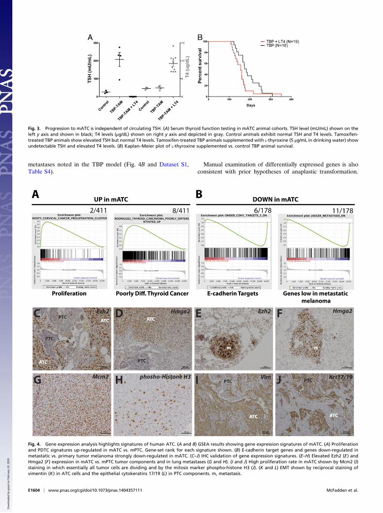
metastases noted in the TBP model (Fig. 4B and Dataset S1,Table S4).
Manual examination of differentially expressed genes is alsoconsistent with prior hypotheses of anaplastic transformation.
Fig. 3. Progression to mATC is independent of circulating TSH. (A) Serum thyroid function testing in mATC animal cohorts. TSH level (mU/mL) shown on theleft y axis and shown in black; T4 levels (μg/dL) shown on right y axis and depicted in gray. Control animals exhibit normal TSH and T4 levels. Tamoxifen-treated TBP animals show elevated TSH but normal T4 levels. Tamoxifen-treated TBP animals supplemented with L-thyroxine (5 μg/mL in drinking water) showundetectable TSH and elevated T4 levels. (B) Kaplan–Meier plot of L-thyroxine supplemented vs. control TBP animal survival.
Fig. 4. Gene expression analysis highlights signatures of human ATC. (A and B) GSEA results showing gene expression signatures of mATC. (A) Proliferationand PDTC signatures up-regulated in mATC vs. mPTC. Gene-set rank for each signature shown. (B) E-cadherin target genes and genes down-regulated inmetastatic vs. primary tumor melanoma strongly down-regulated in mATC. (C–J) IHC validation of gene expression signatures. (E–H) Elevated Ezh2 (E) andHmga2 (F) expression in mATC vs. mPTC tumor components and in lung metastases (G and H). (I and J) High proliferation rate in mATC shown by Mcm2 (I)staining in which essentially all tumor cells are dividing and by the mitosis marker phospho-histone H3 (J). (K and L) EMT shown by reciprocal staining ofvimentin (K) in ATC cells and the epithelial cytokeratins 17/19 (L) in PTC components. m, metastasis.
E1604 | www.pnas.org/cgi/doi/10.1073/pnas.1404357111 McFadden et al.
Dow
nloa
ded
by g
uest
on
Feb
ruar
y 10
, 202
0

Markers of thyroid differentiation and epithelial fate were sig-nificantly down-regulated in mATC (Dataset S1, Table S1).Conversely, markers of mesenchymal fate were up-regulatedalong with embryonic markers, consistent with EMT and markedcellular dedifferentiation evident in human ATC (22, 23). Al-though EMT and embryonic markers have been associated withstem cell-like properties, we do not believe these data argue foror against a cancer stem cell model in mATC, and additionalwork would be necessary to address this possibility.We validated the observed gene expression patterns by IHC
using specific antibodies (Fig. 4 C–J). Consistent with activationof an embryonic transcriptional signature, the transcriptionalregulators enhancer of zeste homolog 2 (Ezh2) and high mobilitygroup AT-hook 2 (Hmga2) were up-regulated in mATC primarytumors and metastases compared with PTC components withinthe same tumors (Fig. 4 C–F). In addition, staining for markersof cell proliferation, minichromosome maintenance complex com-ponent 2 (Mcm2) and phospho-histone H3, revealed an extremelyhigh rate of cell division in mATC (Fig. 4 G and H). Finally, we
observed reciprocal expression of the mesenchymal markervimentin (high in ATC, low in PTC) and the epithelial cytoker-atins 17/19 (low in ATC, high in PTC) within tumors harboringboth ATC and PTC regions (Fig. 4 I and J).We also assessed cellular signaling in mATC. We did not
detect increased phospho-Erk staining in ATC components rel-ative to PTC regions in the TBP tumors (Fig. 5A). In contrast,examination of PI3K and mammalian target of rapamycin (mTOR)signaling by phospho-specific antibodies demonstrated increasedphospho-Akt (S473), and phospho-S6 (S235/236) in ATC com-partments (Fig. 5 B and C). This suggests that these pathwaysare activated during progression to ATC. Therefore, we se-quenced the Pten and phosphatidylinositol 4,5 bisphosphate 3kinase catalytic subunit alpha (Pik3ca) genes in six mATCtumors for hotspot mutations (Pik3ca codons 541–551 and1041–1051; Pten codons 125–135). We detected no mutationsat these sites. Epigenetic silencing of Pten has been observedin human ATC and other cancer types (24). Therefore, weperformed IHC to determine whether Pten might be silenced
Fig. 5. Incomplete mATC response to BRAF inhibition. (A–F ) IHC assessment of signaling in control (A–C ) vs. PLX4720-treated (D–F ) mATC. Persistentphospho-Erk (Thr202/Tyr204) (A and D), phospho-Akt (S473) (B and E ), and phospho-S6 (S235/236) in the ATC component but not control normalthyroid tissue in both control and PLX4720-treated tumors. (G) Kaplan–Meier plot of control chow vs. PLX4720-treated TBP ATC-bearing animals. (H)Relative cross-sectional area of PTC vs. ATC components in three PLX4720-treated ATC-bearing animals. (I and J) Serial ultrasound images of PLX4720-treated TBP animal. Note the PTC-bearing left thyroid gland exhibits a partial response to treatment whereas the ATC component increases in sizeduring ten days of treatment. NL, normal thyroid.
McFadden et al. PNAS | Published online April 7, 2014 | E1605
MED
ICALSC
IENCE
SPN
ASPL
US
Dow
nloa
ded
by g
uest
on
Feb
ruar
y 10
, 202
0

in TBP mATC. We detected strong expression of Pten in ta-moxifen-treated TBP mATC but not control TPOCreER;BrafCA/+; PtenΔ/Δ tumors (Fig. S2 A and B). In addition, weconfirmed that Pten is expressed in murine ATC tumor cell linesgenerated from these animals (Fig. S2E). Dissociated mATCtumor cells rapidly proliferated upon placement in culture, andare immortalized as all lines showed no evidence of senescencewith serial passaging. Consistent with IHC analysis of TBPmATC tumors in vivo, these lines weakly express Nkx2-1 (Fig.S2C). PCR-genotyping of the engineered BrafCA and Trp53alleles additionally established tumor origin and suggested hightumor purity after 3–7 passages (Fig. S2D). These data suggestedthat activation of PI3K and/or the mTOR pathways occurred byas yet unknown mechanisms in TBP mATC cells. Together,these data suggest that BrafV600E expression in thyroid epithelialcells is sufficient for initiation of PTC, and that loss of p53function enables progression to highly malignant, lethal ATCthat exhibits the cardinal features of human ATC including anembryonic transcriptional program and activation of PI3K andmTOR signaling.
Murine ATCs Respond Incompletely to BRAF Inhibition. A singleclinical case report has described a dramatic clinical response ina patient with metastatic ATC to the BRAF inhibitor vemur-afenib/PLX4032 (25). However, the recently published experi-ence of a major thyroid cancer referral center with vemurafenibsuggests a more modest response (17, 25). Therefore, we de-veloped a preclinical treatment protocol that accounted for thevariability in the timing of anaplastic conversion in tumor-bear-ing TBP animals (Fig. S3A). Following tumor induction, TBPcohorts were monitored for a decline in body condition score. Atthe first sign of a decline in body condition, small-animal ultra-sound was performed to assess for the presence of ATC. mATC-bearing animals were then treated with control or PLX4720-compounded chow (PLX4720 is a sister compound to PLX4032/vemurafenib with increased bioavailability in mice). Animalswith rapid disease progression and decline in body conditionwithin 48 h were euthanized and excluded from the analysis toensure adequate absorption of PLX4720. Ultrasound was per-formed at day zero and every 48 h during treatment to monitorthe structural response, and survival was assessed.Animals receiving PLX4720-compounded chow exhibited a
statistically significant survival benefit (Fig. 5G; P = 0.0168);however, we identified no decrease in the ATC cross sectionalarea in response to PLX4720. In fact, most tumors grew largerduring treatment with PLX4720 (Fig. 5 I and J). We per-formed immunohistochemical studies to assess cellular pro-liferation and apoptosis, and although PLX4720-treatedtumors did not exhibit an increase in apoptosis, a slight butstatistically significant decrease (P = 0.02) in proliferation wasidentified by phospho-histone H3 staining (Fig. S4A). Al-though this likely contributed to improved survival, wesearched for additional factors that might explain the clear im-provement in survival of treated animals. Careful review of theimaging results revealed a decrease in contralateral mPTCspresent in TBP mice (Fig. 5 H–J). This suggests that early stageBraf-mutant mPTC may be more sensitive to BRAF inhibition,consistent with prior publications in a doxycycline-regulatedtransgenic BRAFV600E mouse model (9). Because respiratorydistress caused by tracheal invasion and compression is theapparent cause of death in tumor-bearing TBP animals, wehypothesized that the decrement in contralateral low-gradePTC components in response to PLX4720 improved the acuteupper airway obstruction and therefore survival.We assessed cellular signaling in PLX4720-treated vs. control
mATC by IHC in dissected tumor specimens. Both treated andcontrol tumors exhibited positive phospho-Erk staining, sug-gesting that MAPK pathway inhibition was incomplete in re-
sponse to PLX4720 in mATC (Fig. 5 A and D). In addition,phospho-Akt and phospho-S6 staining was maintained inPLX4720-treated tumors (Fig. 5 B, C, E, and F). This is consis-tent with recently proposed reactivation of upstream receptor-tyrosine kinase that occurred in response to BRAF inhibition inthyroid carcinoma cell lines (16). However, activation of pAktand pS6 also occurred in untreated tumors. We were technicallyunable to reliably detect phosphorylated HER3 in treated anduntreated tumors, and are therefore unable to determine ifHER3 signaling is consistently activated in untreated mATC orin response to PLX4720 treatment. We also performed Westernblotting using tumor protein extracts to more accurately quantifyMAPK and PI3K signaling; however, we detected no consistentdifference between treated and untreated tumors (Fig. S4B).This may reflect heterogeneity in the tumor response to PLX4720,or variability in the duration of treatment because animals wereeuthanized based on body condition rather than at defined timepoints. Therefore, at this time we cannot determine whethersignaling through these pathways is consistently increased ordecreased in response to long-term PLX4720 in vivo.
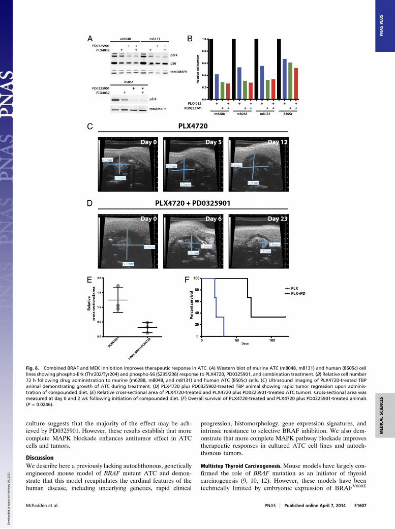
Combined MEK/BRAF Inhibition Enhances Efficacy of MAPK PathwayTargeting in mATC. Treatment of mATC-bearing animals withPLX4720 incompletely suppressed phospho-Erk activity in mATCtumors. The use of combined MEK and BRAF inhibitors inBRAF-mutant melanoma has resulted in improved clinical out-comes, suggesting that more potent pathway inhibition may im-prove efficacy in MAPK pathway mutant cancer cells (26). Inaddition, the proposed mechanism of BRAF-inhibitor resistancein thyroid carcinoma cells involving the reactivation of upstreamreceptor tyrosine kinase-driven signaling predicts that down-stream MEK inhibition should result in more complete MAPKpathway blockade (16). Therefore, we examined this hypothesisin the mATC model.Treatment of mATC cells with PLX4720 resulted in incomplete
suppression of phospho-Erk activity, consistent with autochtho-nous TBP mATC tumors (Fig. 6A). In contrast, the selective MEKinhibitor PD0325901 more completely suppressed pErk in mATCcells and in the human ATC cell line 8505c. The combination ofPD0325901 and PLX4032 resulted in similar pErk levels toPD0325901 alone. In addition, the decrement in pS6 levelsroughly correlated with the degree of pErk suppression, sug-gesting that BrafV600E-mediated signaling impinges upon themTOR pathway as has been previously suggested (27, 28). Weadditionally assessed cellular growth of mATC and 8505c cellsin response to PLX4032, PD0325901, and the combination ofdrugs. Consistent with the phospho-Erk response, PD0325901and combination PD0325901 plus PLX4720 resulted in morecomplete inhibition of cellular growth in mATC cells vs.PLX4720 alone (Fig. 6B).We next treated a cohort of mATC-bearing animals with
PLX4720-compounded and combined PD0325901/PLX4720-compounded chow. To capture mATC outgrowth before significantanimal morbidity, tamoxifen-treated TBP animals were monitoredwith weekly ultrasound imaging (Fig. S3B). Upon detection of ATC,animals were randomly assigned to receive PLX4720-compoundedor PLX4720 plus PD0325901-compounded chow. Consistent withresults obtained in cultured cells, we observed a dramatic responseto combined PLX4720/PD0325901 administration compared withPLX4720 monotherapy (Fig. 6 C and D). Combination-treatedanimals exhibited an enhanced tumor response, including completeregression in three of four mATC-bearing animals, and significantlyprolonged survival (P = 0.0246) (Fig. 6 E and F). We have notassessed the response of mATC to single agent MEK inhibitors invivo; therefore, we cannot determine whether the antitumoreffect in combination treated animals was due to enhanced ef-ficacy of downstream MEK inhibition or to the combined impactof these drugs. The response of mATC and 8505c cells in
E1606 | www.pnas.org/cgi/doi/10.1073/pnas.1404357111 McFadden et al.
Dow
nloa
ded
by g
uest
on
Feb
ruar
y 10
, 202
0
culture suggests that the majority of the effect may be ach-ieved by PD0325901. However, these results establish that morecomplete MAPK blockade enhances antitumor effect in ATCcells and tumors.
DiscussionWe describe here a previously lacking autochthonous, geneticallyengineered mouse model of BRAF mutant ATC and demon-strate that this model recapitulates the cardinal features of thehuman disease, including underlying genetics, rapid clinical
progression, histomorphology, gene expression signatures, andintrinsic resistance to selective BRAF inhibition. We also dem-onstrate that more complete MAPK pathway blockade improvestherapeutic responses in cultured ATC cell lines and autoch-thonous tumors.
Multistep Thyroid Carcinogenesis. Mouse models have largely con-firmed the role of BRAF mutation as an initiator of thyroidcarcinogenesis (9, 10, 12). However, these models have beentechnically limited by embryonic expression of BRAFV600E
Fig. 6. Combined BRAF and MEK inhibition improves therapeutic response in ATC. (A) Western blot of murine ATC (m8048, m8131) and human (8505c) celllines showing phospho-Erk (Thr202/Tyr204) and phospho-S6 (S235/236) response to PLX4720, PD0325901, and combination treatment. (B) Relative cell number72 h following drug administration to murine (m6288, m8048, and m8131) and human ATC (8505c) cells. (C) Ultrasound imaging of PLX4720-treated TBPanimal demonstrating growth of ATC during treatment. (D) PLX4720 plus PD0325902-treated TBP animal showing rapid tumor regression upon adminis-tration of compounded diet. (E) Relative cross-sectional area of PLX4720-treated and PLX4720 plus PD0325901-treated ATC tumors. Cross-sectional area wasmeasured at day 0 and 2 wk following initiation of compounded diet. (F) Overall survival of PLX4720-treated and PLX4720 plus PD0325901-treated animals(P = 0.0246).
McFadden et al. PNAS | Published online April 7, 2014 | E1607
MED
ICALSC
IENCE
SPN
ASPL
US
Dow
nloa
ded
by g
uest
on
Feb
ruar
y 10
, 202
0

and/or supraphysiologic circulating TSH levels that can promotethyroid cellular growth. In addition, there has recently beencontroversy regarding whether BRAFV600E is sufficient to inducePTC in the adult murine thyroid gland and whether BRAFT17799A
mutation is a clonal event in human PTC (29, 30). Although webelieve the preponderance of experimental data supports therole of BRAF as an initiating genetic event, our results are thestrongest to date that mosaic BRAFV600E expression in the adultmurine thyroid is sufficient to initiate PTC. The fact that PTCis not observed in corn oil-treated TB animals 12 wk after in-duction, and all tamoxifen-treated TB animals harbor PTC,strongly argues for the sufficiency of BRAFV600E to initiate PTCin the adult. In addition, in contrast to Shimamura et al. (30), wehave successfully induced PTC in BrafCA/+ animals by surgicaldelivery of Cre-expressing adenovirus.Analyses of human ATC specimens have consistently impli-
cated p53 as an important tumor suppressor during thyroidcancer progression. TP53 mutations are detected at high fre-quency in ATCs, at lower frequency in PDTC, and uncommonlyin PTCs (5, 6). In addition, prior work from a mouse model ofPTC driven by transgenic overexpression of a RET/PTC fusionhas implicated p53 loss in progression from papillary to PDTCand possibly ATC (13). Our data demonstrate that combinedBRAF mutation and loss of p53 cooperate in vivo to facilitateprogression to ATC. Interestingly, although loss of p53 appearsto be required for progression to ATC, our data suggest thatadditional events may be required for ATC conversion. Firstly,the ∼6-mo latency after induction of BrafV600E expression andp53 loss suggests this combination of events is insufficient todirect the complete ATC phenotype. In addition, the slow tem-poral progression of TBP tumors followed by sudden ATC out-growth as assessed by serial ultrasound imaging is consistent withacquired somatic genetic or epigenetic alterations driving tumorprogression and conversion to the anaplastic phenotype. Themouse model described here provides an ideal autochthonousplatform to begin to dissect the molecular mechanisms ofthis transition.Recently, a genetically engineered mouse model of follicular
thyroid carcinoma progression to ATC was generated by com-pound deletion of p53 and Pten in the murine thyroid follicularepithelium (14). The TPOCre; PtenΔ/Δ; p53Δ/Δ animals developinvasive, undifferentiated carcinoma with sarcomatoid features.Although this model exhibits a longer ATC latency, the histo-logic phenotype is indeed quite similar to the features we detectin the TBP model. This may support the notion that each ofthese models acquires convergent events such that the TBPmodel activates the PI3K pathway, and TPOCre; PtenΔ/Δ; p53Δ/Δ
animals acquire MAPK pathway alterations. However, geno-typing for Ras family hotspot mutations was negative in TPOCre;PtenΔ/Δ; p53Δ/Δ tumors (14). Conversely, we detected no Pten orPik3ca hotspot mutations in TBP mATC tumors, and thesetumors maintained expression of Pten, as assessed by IHC. Thissuggests that neither MAPK nor PIK3CA alterations are essentialfor anaplastic conversion. However, other acquired mechanisms ofactivating the PI3K pathway in the TBP mATC model have notbeen excluded. Together, these models provide strong in vivoevidence for the important role of p53 loss and PI3K pathwayactivation during progression to ATC.
Response of ATC to MAPK Inhibition. Previous studies using MAPKpathway inhibitors in ATC had relied upon highly passaged hu-man ATC cells in culture or transplanted into immunocompro-mised mice (16, 31, 32). Our results demonstrating an incompleteresponse of mATCs to PLX4720 are consistent with the reducedsensitivity of a variety of human thyroid carcinoma cell lines toMAPK pathway inhibition in cultured conditions. Relief ofnegative feedback to the HER3 receptor tyrosine kinase signal-ing pathway has been implicated as a mechanism of intrinsic
resistance of ATC cells in culture (16). This mechanism predictsthat downstream MEK inhibition may blunt reactivation ofMAPK signaling. Upstream RTK activation may also impingeupon the PI3K/Akt pathway, suggesting that MAPK inhibitionmay be insufficient to suppress growth and/or survival of BRAF-mutant thyroid carcinoma cells.Our data both in vitro and in vivo, however, suggest that po-
tent MAPK blockade may be sufficient for a robust initial anti-tumor response. mATC tumors also exhibit increased signalingthrough the PI3K/Akt pathway, and although the response tocombination PLX4720/PD0325901 was robust in our study,mATC tumors eventually recurred. Human ATCs also harborfrequent alterations in the PI3K pathway (4). Therefore, it willbe important to determine whether combined MAPK/PI3K in-hibition will improve outcomes in this model system, as has beensuggested from studies in several cancer types (33). It will also beinteresting to interrogate the mechanisms underlying tumor re-currence following combined BRAF and MEK inhibition in thismodel. The data presented here, however, strongly argue fora central role of MAPK pathway inhibitors in treatment regi-mens for patients with advanced forms of thyroid cancer andsuggest that more potent pathway blockade may result in im-proved responses.
Mouse Models for Uncommon Aggressive Tumors.A lack of availabletissue and participants in clinical trials has hampered scientificprogress and therapeutic improvements for many uncommontumors, particularly those exhibiting highly aggressive clinical be-havior. Using the somatic genetics of human ATC, we describea valid preclinical mouse model recapitulating the temporal pro-gression, gene expression programs, and resistance to targetedBRAF inhibitors seen in human thyroid carcinoma. We proposethis model as a suitable experimental platform from which tobegin to genetically dissect the molecular determinants of ATCprogression and resistance to therapies.
MethodsCloning and Generation of TPOCreER Mice. The TPOCre vector was provided byShioko Kimura (National Cancer Institute, Bethesda, MD) (18). A partial SalIplus MluI digest was performed to remove the Cre coding sequence. This wasreplaced by a PCR-generated CreER (T2) ORF flanked by SalI and MluI re-striction sites. Restriction mapping and DNA sequencing confirmed allcloning junctions. TPOCreER expression construct was excised with KpnI andinjected into fertilized oocytes using standard techniques. Two independenttransgenic animals were generated. One founder strain (1139) was used forall experiments described in this paper.
Animal Studies. All studies were performed under an Institutional AnimalCare and Use Committee- and Massachusetts Institute of TechnologyCommittee on Animal Care-approved animal protocol. TPOCreER ani-mals were crossed to the previously described mouse strains availablefrom The Jackson Laboratory: Braftm1Mmcm/J (stock no. 017837), Gt(ROSA)26Sortm9(CAG_tdTomato)Hze/J (stock no. 007909), Trp53tm1Brn/J (stockno. 008462), and Trp53tm3.1Tyj/J (stock no. 008182). All mice were main-tained on a mixed C57BL/6; 129SvJae background. Genotyping primers andprotocols are available upon request. Tumor induction was performed by i.p.administration of 0.1–0.2 mg/g tamoxifen dissolved in corn oil (Sigma) forone or two administrations at age 8–12 wk. Ad5-CMV-Cre was obtainedfrom the University of Iowa Gene Transfer Vector Core Facility. PLX4720 andPD0325901 were administered in compounded chow from Research Diets.PLX4720 was administered at 400 ppm in the initial monotherapy trial (Fig.S2A) and at 200 ppm in the combination trial (Fig. S2B). PD0325901 wasadministered at 7 ppm in diet. A Vevo 770 small-animal ultrasound witha 704 probe was used to image thyroid tumors for treatment studies. Cross-sectional area was calculated by measuring maximal anterior-posterior andleft-right dimension using the standard area for an ellipse. GraphPad Prism5.0 or 6.0 was used for all statistical analyses. All survival P values werecalculated using a log-rank (Mantel–Cox) test.
Hormone Measurements. Blood was collected from male and female mice byretro-orbital bleeding and allowed to clot at room temperature for 1 h and
E1608 | www.pnas.org/cgi/doi/10.1073/pnas.1404357111 McFadden et al.
Dow
nloa
ded
by g
uest
on
Feb
ruar
y 10
, 202
0

then centrifuged. Serum was stored at −80 °C until assays were performed.Samuel Refetoff (University of Chicago, Chicago, IL) performed TSH and T4measurement, as previously described, on a fee-for-service agreement (34).
Gene Expression Analyses. Total RNA was harvested from five snap-frozenTB/+ (PTC) and TBP tumors after histological confirmation of papillary oranaplastic phenotype. Affymetrix Mouse Exon 1.0 ST arrays were used aspreviously described. Raw hybridization intensities from Affymetrix MouseGene 1.0 ST arrays were normalized and summarized using the open-sourceAffymetrix Power Tools software package (Affymetrix) to derive gene-levelexpression estimates. Multiple probe sets targeting the same gene werecollapsed using their median value in order to eliminate duplicates from thedataset. Differential gene expression analysis was performed using thelimma package in the R environment for statistical computing (www.r-pro-ject.org and ref. 35). GSEA was performed with GSEA software and themolecular signature database (MSigDB) (21). Gene expression datasets havebeen deposited in the National Center for Biotechnology Information GeneExpression Omnibus database (accession no. GSE55933).
IHC. See Dataset S1, Table S2 for a list of antibodies used in this study. IHCwas performed using standard protocols on a ThermoFisher Lab VisionAutostainer 360.
Cell Line Studies. mATC tumors were dissociated by mincing freshly dissectedtumors and digestion in HBSS-free with 100 μL of trypsin (0.25%), 200 μL ofDispase (BD), and 100 μL of collagenase (10 mg/mL; Worthington) for 30 minto 1 h. Following incubation and filtering through a 40- to 100-μm cell
strainer, dissociated cells were plated on to tissue culture dishes in DMEMsupplemented with 10% FBS and pen/strep. Cells were passaged and frozenusing standard techniques. Human 8505c cells were obtained from thelaboratory of Sareh Parangi (Massachusetts General Hospital). PD0325901was added to media to a final concentration of 50 nM, and PLX4720 wassupplemented to 2 μM in all experiments. Protein extracts were harvestedafter 24 h of incubation in drug vs. vehicle (DMSO) media using radioim-munoassay immunoprecipitation assay buffer supplemented with proteaseand phosphatase inhibitors (Roche). See Dataset S1, Table S2 for a list ofantibodies used in this study. Cell number was assessed 72 h following ad-dition of drug or DMSO using the Celltiter-Glo kit (Promega).
Note Added in Proof. Separately, Charles et al. demonstrated that combinedexpression of BRAF(V600E) with silencing of PTEN also elicits anaplasticthyroid cancer in a related mouse model (36).
ACKNOWLEDGMENTS. We thank Plexxikon for providing PLX4720 andPD0325901 for experiments, Dr. Shioko Kimura [National Cancer Institute(NCI)] for providing the TPOCre plasmid, Dr. Samuel Refetoff (University ofChicago) for mouse TSH and T4 assays, Dr. Sareh Parangi (MassachusettsGeneral Hospital) for sharing 8505c human ATC cells, and Kaitlyn Sanders fortechnical assistance. We thank Thales Papagiannakopoulos, TuomasTammela, Eric Snyder, and Gilbert H. Daniels for critical review of themanuscript. This work was supported by the Howard Hughes Medical Institute(T.J.), National Institutes of Health R01CA131261 (to M.M.), and an AmericanThyroid Association research award (to D.M.). D.M. is supported by NCI CareerDevelopment Award K08CA160658.
1. Cooper DS, et al.; American Thyroid Association (ATA) Guidelines Taskforce on Thy-roid Nodules and Differentiated Thyroid Cancer (2009) Revised American ThyroidAssociation management guidelines for patients with thyroid nodules and differen-tiated thyroid cancer. Thyroid 19(11):1167–1214.
2. Smallridge RC, et al.; American Thyroid Association Anaplastic Thyroid CancerGuidelines Taskforce (2012) American Thyroid Association guidelines for manage-ment of patients with anaplastic thyroid cancer. Thyroid 22(11):1104–1139.
3. Ricarte-Filho JC, et al. (2009) Mutational profile of advanced primary and metastaticradioactive iodine-refractory thyroid cancers reveals distinct pathogenetic roles forBRAF, PIK3CA, and AKT1. Cancer Res 69(11):4885–4893.
4. Santarpia L, El-Naggar AK, Cote GJ, Myers JN, Sherman SI (2008) Phosphatidylinositol3-kinase/akt and ras/raf-mitogen-activated protein kinase pathway mutations in an-aplastic thyroid cancer. J Clin Endocrinol Metab 93(1):278–284.
5. Fagin JA, et al. (1993) High prevalence of mutations of the p53 gene in poorly dif-ferentiated human thyroid carcinomas. J Clin Invest 91(1):179–184.
6. Ito T, et al. (1992) Unique association of p53 mutations with undifferentiated but notwith differentiated carcinomas of the thyroid gland. Cancer Res 52(5):1369–1371.
7. Nikiforov YE, Nikiforova MN (2011) Molecular genetics and diagnosis of thyroidcancer. Nat Rev Endocrinol 7(10):569–580.
8. Gauchotte G, et al. (2011) BRAF, p53 and SOX2 in anaplastic thyroid carcinoma: Evi-dence for multistep carcinogenesis. Pathology 43(5):447–452.
9. Chakravarty D, et al. (2011) Small-molecule MAPK inhibitors restore radioiodine in-corporation in mouse thyroid cancers with conditional BRAF activation. J Clin Invest121(12):4700–4711.
10. Charles RP, Iezza G, Amendola E, Dankort D, McMahon M (2011) Mutationally ac-tivated BRAF(V600E) elicits papillary thyroid cancer in the adult mouse. Cancer Res71(11):3863–3871.
11. Franco AT, et al. (2011) Thyrotrophin receptor signaling dependence of Braf-inducedthyroid tumor initiation in mice. Proc Natl Acad Sci USA 108(4):1615–1620.
12. Knauf JA, et al. (2005) Targeted expression of BRAFV600E in thyroid cells of trans-genic mice results in papillary thyroid cancers that undergo dedifferentiation. CancerRes 65(10):4238–4245.
13. La Perle KM, Jhiang SM, Capen CC (2000) Loss of p53 promotes anaplasia and localinvasion in ret/PTC1-induced thyroid carcinomas. Am J Pathol 157(2):671–677.
14. Antico Arciuch VG, et al. (2011) Thyrocyte-specific inactivation of p53 and Pten resultsin anaplastic thyroid carcinomas faithfully recapitulating human tumors. Oncotarget2(12):1109–1126.
15. Sherman SI (2011) Targeted therapies for thyroid tumors. Mod Pathol 24(Suppl 2):S44–S52.
16. Montero-Conde C, et al. (2013) Relief of feedback inhibition of HER3 transcription byRAF and MEK inhibitors attenuates their antitumor effects in BRAF-mutant thyroidcarcinomas. Cancer Discov 3(5):520–533.
17. Kim KB, et al. (2013) Clinical responses to vemurafenib in patients with metastaticpapillary thyroid cancer harboring BRAF(V600E) mutation. Thyroid 23(10):1277–1283.
18. Kusakabe T, Kawaguchi A, Kawaguchi R, Feigenbaum L, Kimura S (2004) Thyrocyte-specific expression of Cre recombinase in transgenic mice. Genesis 39(3):212–216.
19. Dankort D, et al. (2007) A new mouse model to explore the initiation, progression,and therapy of BRAFV600E-induced lung tumors. Genes Dev 21(4):379–384.
20. Orim F, et al. (2014) Thyrotropin signaling confers more aggressive features withhigher genomic instability on BRAF(V600E)-induced thyroid tumors in a mouse model.Thyroid 24(3):502–510.
21. Subramanian A, et al. (2005) Gene set enrichment analysis: A knowledge-basedapproach for interpreting genome-wide expression profiles. Proc Natl Acad Sci USA102(43):15545–15550.
22. Knauf JA, et al. (2011) Progression of BRAF-induced thyroid cancer is associated withepithelial-mesenchymal transition requiring concomitant MAP kinase and TGFβ sig-naling. Oncogene 30(28):3153–3162.
23. Wenig BM (1993) Atlas of Head and Neck Pathology (Saunders, Philadelphia).24. Hou P, Ji M, Xing M (2008) Association of PTEN gene methylation with genetic al-
terations in the phosphatidylinositol 3-kinase/AKT signaling pathway in thyroid tu-mors. Cancer 113(9):2440–2447.
25. Rosove MH, Peddi PF, Glaspy JA (2013) BRAF V600E inhibition in anaplastic thyroidcancer. N Engl J Med 368(7):684–685.
26. Flaherty KT, et al. (2012) Combined BRAF and MEK inhibition in melanoma with BRAFV600 mutations. N Engl J Med 367(18):1694–1703.
27. Faustino A, et al. (2012) mTOR pathway overactivation in BRAF mutated papillarythyroid carcinoma. J Clin Endocrinol Metab 97(7):E1139–E1149.
28. Jin N, Jiang T, Rosen DM, Nelkin BD, Ball DW (2011) Synergistic action of a RAF in-hibitor and a dual PI3K/mTOR inhibitor in thyroid cancer. Clin Cancer Res 17(20):6482–6489.
29. Guerra A, et al. (2012) The primary occurrence of BRAF(V600E) is a rare clonal event inpapillary thyroid carcinoma. J Clin Endocrinol Metab 97(2):517–524.
30. Shimamura M, et al. (2013) Postnatal expression of BRAFV600E does not inducethyroid cancer in mouse models of thyroid papillary carcinoma. Endocrinology154(11):4423–4430.
31. Nehs MA, et al. (2012) Late intervention with anti-BRAF(V600E) therapy induces tu-mor regression in an orthotopic mouse model of human anaplastic thyroid cancer.Endocrinology 153(2):985–994.
32. Nucera C, et al. (2011) Targeting BRAFV600E with PLX4720 displays potent anti-migratory and anti-invasive activity in preclinical models of human thyroid cancer.Oncologist 16(3):296–309.
33. Wong KK, Engelman JA, Cantley LC (2010) Targeting the PI3K signaling pathway incancer. Curr Opin Genet Dev 20(1):87–90.
34. Pohlenz J, et al. (1999) Improved radioimmunoassay for measurement of mousethyrotropin in serum: Strain differences in thyrotropin concentration and thyrotrophsensitivity to thyroid hormone. Thyroid 9(12):1265–1271.
35. Gentleman RC, et al. (2004) Bioconductor: Open software development for compu-tational biology and bioinformatics. Genome Biol 5(10):R80.
36. Charles RP, Silva J, Iezza G, Phillips W, McMahon M (2014) BRAF(V600E) cooperateswith PIK3CA(H1074R) to promote anaplastic thyroid carcinogenesis. Mol Cancer Res,in press.
McFadden et al. PNAS | Published online April 7, 2014 | E1609
MED
ICALSC
IENCE
SPN
ASPL
US
Dow
nloa
ded
by g
uest
on
Feb
ruar
y 10
, 202
0